Abstract
Background and objective: Left atrium (LA) is an important biomarker of adverse cardiovas-cular outcomes and cerebrovascular events. This study aimed to evaluate LA myocardial deformation using cardiac magnetic resonance feature tracking (CMR-FT) in patients with acute ST-segment elevation myocardial infarction (STEMI) and secondary mitral regurgitation (MR). Additionally, to assess interobserver and intraobserver variability of the technique. Materials and methods: Twenty patients with STEMI underwent CMR with a 1.5 Tesla MRI.scanner. According to the presence of MR patients were divided into two groups: MR(+) and MR(−). Total LA strain (εs), passive LA strain (εe), and active LA strain (εa) were obtained. Additionally, total, passive and active strain rates (SRs, SRe, and SRa) were calculated. To assess interobserver agreement data analysis was performed by second independent observer. Results: LA volumetric and functional parameters were similar in both groups. All LA strain values were significantly higher in patients with MR: εs (27.67 ± 10.25 for MR(−) vs. 32.80 ± 6.95 for MR(+); P = 0.01), εe (15.29 ± 7.30 for MR(−) vs. 19.22 ± 6.04 for MR(+); P = 0.01) and εa (12.38 ± 4.23 for MR(−) vs. 14.44 ± 5.19 for MR(+); P = 0.03). Only SRe significantly increased in patients with MR (−0.57 ± 0.24 for MR(−) vs. 0.70 ± 0.20 for MR(+); P = 0.01). All LA deformation parameters demonstrated high interobserver and intraobserver agreement. Conclusions: Conventional volumetric and functional LA parameters do not detect early changes in LA performance in patients with STEMI and secondary MR. In contrast, LA reservoir, passive and active strain are significantly higher in patients with MR. Only peak early negative strain rate substantially increases during secondary MR. LA deformation parameters derived from conventional cine images using CMR-FT technique are highly reproducible.
1. Introduction
The prognosis of individuals who survived acute myocardial infarction (AMI) has substantially improved over the last decades. However, more precise and preferably individual risk stratification is necessary to further reduce cardiovascular burden in this population. Almost half of patients presenting with AMI experience acute mitral regurgitation (MR) which carries additional risk of heart failure and premature death [1].
Current evidence suggests that left atrial (LA) size and function are important markers of adverse cardiovascular outcomes and cerebrovascular events [2,3]. LA function during AMI is affected by acute ischemia of atrial myocardium. Meanwhile, MR stimulates cardiac remodeling and is associated with LA failure, atrial fibrillation and cardiac death [4].
LA size and function can be assessed using a number of noninvasive cardiac imaging modalities such as echocardiography, computed tomography or cardiac magnetic resonance (CMR). Advanced cardiac imaging for the assessment of myocardial mechanics provides additional information about atrial performance, furthermore, it allows detection of early functional changes and predicts future events [5].
Assessment of myocardial deformation using CMR imaging became possible after the introduction of myocardial tagging technique. However, the need for additional image acquisition and time consuming postprocessing make this technique less attractive. CMR feature tracking (CMR-FT) algorithm focuses on tracking of endocardial and epicardial contours. Myocardial strain and strain rate can be derived from conventional balanced steady state free precession (bSSFP) cine images and used to quantify myocardial function.
We performed this study to assess LA myocardial performance during acute ischemia and subsequent volume overload due to MR. Additionally, we evaluated interobserver and intraobserver reproducibility of CMR-FT derived LA strain and strain rate measurements.
2. Materials and methods
2.1. Study population
Patients were consecutively enrolled into the study if they presented with first ST-segment elevation myocardial infarction (STEMI) and were treated with primary coronary intervention (PCI). The diagnosis of STEMI was based on typical symptoms, specific electrocardiographic (ECG) changes (ST-segment elevation greater than 1 mm in two contiguous limb leads or more than 2 mm in precordial leads or new left bundle branch block), elevated troponin levels and detection of occluded coronary artery during conventional coronary angiography.
Transthoracic two-dimensional echocardiography was performed within 72 h from admission and primary PCI. The severity of MR was assessed according to the European Association of Cardiovascular Imaging (EACVI) recommendations by proximal isovelocity surface area (PISA) method or semi-quantitative color flow Doppler when quantitative assessment of MR was not feasible (unmeasurable PISA or continuous Doppler trace) [6]. Previously has been proved that only mild secondary MR has impact on LA longitudinal deformation therefore we selected STEMI patients with mild-to-moderate MR [7]. Ischemic MR is highly load dependent. There was no significant difference on hemodynamic conditions measured by echocardiography. According to our findings patients were divided into two following groups: patients without MR (MR(−)) or with functional MR (MR(+)). Patients with trace MR were considered as MR(−). The final study population consisted of 20 STEMI patients: 10 without and 10 with secondary MR.
The exclusion criteria were as follows: medical history of ischemic heart disease (known coronary artery disease, previous MI, PCI, coronary artery bypass grafting), structural cardiac valve disease (including previous valvular surgery), previously known MR. We excluded subjects with anterior STEMI in order to avoid distinct ischemic effect on LA myocardial performance (Table S1). Patients with absolute contraindications for CMR (ferromagnetic implants, vascular aneurysm clips or claustrophobia) and those with irregular heart rhythm (multiple premature beats or atrial fibrillation) were also excluded. The study complies with the Declaration of Helsinki and was approved by the local Ethics Committee. All patients gave written informed consent before entering the study.
2.2. Cardiac magnetic resonance
All CMR images were acquired using a 1.5 Tesla scanner (Siemens Magnetom Aera, Siemens AG Healthcare Sector, Erlangen, Germany) with an 18-channel phased array coil in a supine position. CMR was performed within 72 h from echocardiography. The study protocol included an initial survey to define imaging planes. Cine images were acquired using retrospectively gated bSSFP sequence with short periods of breath-holding in three left ventricular (LV) long-axis (two-chamber, three-chamber and four-chamber) planes. The ventricular two-chamber and four-chamber planes were used to plan contiguous stack of short-axis slices covering entire LV. The in-plane resolution of cine images was 0.9 mm × 0.9 mm, slice thickness of 8 mm with a 2-mm interslice gap and 25 phases per cardiac cycle.
2.3. Volumetric analysis
Volumetric analysis was performed using vendor dedicated software (Syngo.via, Siemens AG Healthcare Sector, Erlangen, Germany). LV end-diastolic (LVEDV) and end-systolic volumes (LVESV) were quantified using manual planimetry of the endocardial and epicardial surface from short-axis stack as previously described and LV ejection fraction (LVEF) was calculated. Papillary muscles were considered as being part of the blood pool. Ventricular volumes were adjusted to body surface area. To assess phasic atrial function by the volumetric method, LA volumes were estimated using the previously validated biplane-area method according to the formula [8]:
LA volume (ml) = 0.85 × LA area2ch × LA area4ch/LA length
LA length was defined as the longest distance measured between posterior atrial wall and mid-portion of mitral annulus plane in either the two-chamber or four-chamber view. LA volumes were estimated at three different time points of the cardiac cycle: LA maximal volume was measured during ventricular end-systole at the phase of mitral valve opening, LA minimal volume was obtained at late ventricular end-diastole after LA contraction and mitral valve closure and LA volume at the onset of atrial systole was considered at left ventricular diastole immediately before LA contraction [9]. Phasic LA function was assessed with the calculation of LA emptying volumes and fractions: LA total emptying volume and fraction (LAEVtotal and LAEFtotal, corresponding to LA reservoir function), LA passive emptying volume and fraction (LAEVpassive and LAEFpassive, corresponding to LA conduit function) and LA active emptying volume and fraction (LAEVactive and LAEFactive, corresponding to LA contractile booster pump function) and were derived using equations shown in Table 1. Atrial emptying volumes were indexed to body surface area.

Table 1.
Equations used for calculation of emptying volumes and fractions to assess phasic functions of LA.
2.4. Feature tracking
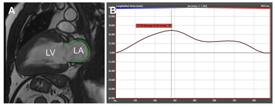
CMR-FT was processed using commercially available tissue tracking software (TomTec Imaging Systems, 2D CPA MR, Cardiac Performance Analysis, Unterschleissheim, Germany) allowing semi-automated analysis of cardiac mechanics using conventional cine images. LA endocardial and epicardial surface was manually contoured by a point-and-click approach in both two-chamber and four-chamber views at LV end-systolic phase (Fig. 1A). After application of tissue tracking algorithm endocardial borders were detected through all cardiac phases. Strain and strain rate curves were generated for six atrial segments. Segments with inadequate contouring quality due to pulmonary vein or LA appendage were excluded from the analysis. In these patients strain and strain rate values were calculated by averaging measurements obtained from the remaining segments. The global strain and strain rate profiles were derived and used for further analysis (Fig. 1B). All parameters were derived twice in both four-chamber and two-chamber views. LA global strain and strain rate values were calculated by averaging the strain curves of both four-chamber and two-chamber long-axis views. Similarly to previous descriptions from echocardiographic speckle tracking and CMR-FT studies three LA longitudinal deformation parameters were assessed: total LA strain (εs, corresponding to LA reservoir function), passive LA strain (εe, corresponding to LA conduit function) and active LA strain (εa, corresponding to LA contractile function) [10,11]. Additionally, total, passive and active LA strain rate (SRs, SRe, and SRa, corresponding to all three atrial functional phases) were obtained. For the assessment of inter-observer reproducibility, all patients were analyzed by two independent observers. Moreover, to assess intraobserver agreement data analysis of 10 patients was repeated four weeks after first assessment.

Fig. 1.
The end-diastolic frame (A) of two-chamber view with contouring of LA myocardium after application of tracking algorithm. Example of LA strain curve (B) corresponding to reservoir, conduit and contractile booster pump function. LA, left atrium.
2.5. Statistical analysis
Data analysis was performed using Microsoft Excel and IBM SPSS Statistics version 22.0 software (SPSS Inc., Chicago, IL, USA) for Macintosh. The Kolmogorov–Smirnov test was used to determine whether the data was normally distributed. Continuous variables are expressed as mean standard deviation (SD) and qualitative data as count and percentage. Continuous variables of two groups were compared by the paired samples t test if the data were normally distributed, whereas Wilcoxon signed ranks test was used to compare nonparametric data. A P value of <0.05 was considered significant. Interobserver and intraobserver reproducibility was quantified by using intraclass correlation coefficient (ICC) and Bland–Altman analysis [12]. Agreement was considered excellent for ICC > 0.74, good for ICC 0.60–0.74, fair for ICC 0.40–0.59, and poor for ICC < 0.40 [13].
3. Results
In total, 20 patients were included into our study. Table 2 shows the demographic characteristics, atrial and ventricular volumetric parameters of the study population. There was no significant difference in age, gender, and BMI between the two groups. Patients with MR demonstrated significantly greater LVEDV, LVESV and LVSV. There was no significant difference in LVEF between both groups (62.34 ± 2.94 for MR(−) group vs. 59.69 ± 3.20 for MR(+) group; P = 0.07). LA emptying volumes and fractions were similar in both groups.

Table 2.
Characteristics of the study population.
All LA strain values were significantly higher in patients with MR (Table 3): εs (27.67 ± 10.25 for MR(−) vs. 32.80 ± 6.95 for MR(+); P = 0.01), εe (15.29 ± 7.30 for MR(−) vs. 19.22 6.04 for MR (+); P = 0.01) and εa (12.38 ± 4.23 for MR(−) vs. 14.44 5.19 for MR(+); P = 0.03). SRs and SRa were similar in both groups. Only SRe corresponding to atrial conduit function was significantly increased in patients with MR (−0.57 0.24 for MR(−) vs. 0.70 ± 0.20 for MR(+); P = 0.01).

Table 3.
Comparison of CMR-FT obtained LA strain and strain rate measures between two groups.
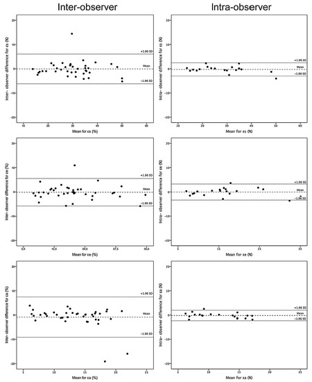
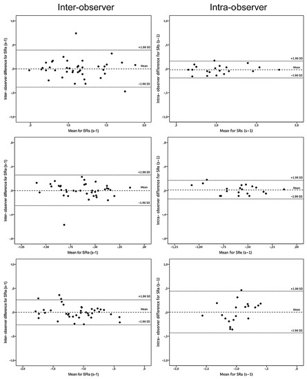
Excellent interobserver reproducibility was found in all measurements. Only εa demonstrated higher variability: ICC 0.76 (95% CI, 0.54–0.87). The intraobserver agreement was better for all strain parameters and total and passive LA strain rate. The least reproducible measure for intra-observer level was SRa: ICC 0.79 (95% CI, 0.49–0.92). Table 4 and Table 5 summarize the mean ± standard deviation, limits of agreement, and ICC for both levels. Bland–Altman plots demonstrate interobserver and intraobserver reproducibility of all strain and strain rate parameters and are depicted in Fig. 2 and Fig. 3.

Table 4.
Interobserver reproducibility for LA strain and strain rate measurements.

Table 5.
Intraobserver reproducibility for LA strain and strain rate measurements.

Fig. 2.
Bland–Altman plots with limits of agreement (1.96 standard deviations) demonstrates interobserver and intraobserver reproducibility of CMR-FT for LA strain parameters. The middle dotted line is the mean of difference of measurements. The upper and lower dotted lines are ±1.96 standard deviation. LA, left atrium; εs, LA total strain; εe, LA passive strain; εa, LA active strain.

Fig. 3.
Bland–Altman plots with limits of agreement (1.96 standard deviations) demonstrates interobserver and intraobserver reproducibility of CMR-FT for LA strain rate parameters. The middle dotted line is the mean of difference of measurements. The upper and lower dotted lines are ±1.96 standard deviation. LA, left atrium; SRs, LA total strain rate; SRe, LA passive strain rate; SRa, LA active strain rate.
4. Discussion
The current study was designed to assess LA deformation in patients presenting with STEMI and newly developed MR as well as interobserver and intraobserver reproducibility using CMR-FT. Our study indicates several important findings:
- LV volumes are significantly larger in patients with MR, whereas LV ejection fraction is similar when compared with those without MR.
- LA volumes and emptying fractions are similar in both groups and do not show early changes in LA myocardial deformation in patients with acute STEMI and secondary MR.
- LA reservoir, passive and active strain are significantly higher in patients with MR, but only peak early negative strain rate exhibits substantial augmentation.
- Study demonstrates excellent interobserver and intraobserver reproducibility for all LA strain and strain rate values.
The MR after AMI is associated with increased risk of heart failure and death [14]. Even mild MR reduces patient's survival rate [15]. Approximately 50% of patients with AMI experience MR which is silent, therefore frequently clinically unrecognized. LA function in patients with AMI and MR is affected by acute ischemia of atrial myocardium and hemodynamic changes due to developed mitral insufficiency.
The LA myocardium is supplied mainly by branches of the proximal left circumflex artery (LCx) with less contribution from the right coronary artery (RCA). The left anterior descending (LAD) coronary artery does not supply LA myocardium [16]. In experimental animal model of acute LV ischemia Bauer et al. demonstrated dramatic deterioration of LA function during LCx occlusion compared with ischemia induced by obstruction of LAD [17]. Since we excluded patients with LAD occlusion and demonstrated increased LA strain and strain rate in subjects with MR, one might infer that LA performance is influenced not only by acute ischemia. However, we did not have the ability to compare our data with patients experienced anterior STEMI.
Most importantly, our study shows that atrial volumetric and functional parameters such as emptying volumes and fractions do not depict early changes in LA myocardial performance in patients with acute STEMI and secondary MR.
Hemodynamically, the LA actively modulates ventricular filling through its functional components. There are well recognized three phases of the LA function: the reservoir phase in which the atrium fills rapidly from the pulmonary veins during early LV systole, the conduit phase when blood from the atrium is sucked into the ventricle, and contractile or booster pump phase which completes LV filling in late diastole [9,18].
LA myocardial motion can be assessed using advanced tissue tracking techniques as speckle tracking echocardiography (STE) or CMR-FT. The feasibility of measuring LA deformation parameters during all three functional phases was demonstrated in previous studies [11]. However, the technical challenges as insufficient image quality still limit its use in patient with obesity or poor acoustic window. CMR-FT as a novel tissue tracking technique became available in 2009 [19]. Due to lack of intramyocardial landmarks and excellent contrast between blood pool and myocardium, CMR-FT relies on identification of endocardial and epicardial contours [20].
Peak longitudinal strain allows quantification of the LA reservoir function which is essential for LV filling and closely related to atrial compliance [21]. Cameli et al. reported negative correlation between LA peak longitudinal strain and MR degree. Furthermore, impaired LA reservoir function was associated with higher incidence of paroxysmal atrial fibrillation [22]. Borg et al. investigated LA deformation indices and reported that reservoir strain and strain rate increase during chronic MR [23]. In our study we also found that LA reservoir function was enhanced in patients with acute MR.
During MR, the early diastolic ventricular filling increases as a response to suddenly developed volume overload. The latter induces LA dilatation and is the main cause of atrial arrhythmias and thromboembolic events. The LA conduit function in patients with MR is also augmented due to deteriorated LV compliance and increased LA passive recoil [24]. Our study demonstrated higher LA passive strain and strain rate in subjects with MR.
However, it remains unclear whether these changes are present during acute phase of MR or persist over time. Also it is not known whether LA strain and strain rate decrease if MR persists or disappears. To answer this question serial imaging over several years after STEMI would be necessary.
Higher reproducibility enables smaller changes to be detected with greater reliability. Recent studies have reported excellent reproducibility for all strain and strain rate parameters at interobserver and intraobserver levels [10]. The reservoir function is the most reproducible, followed by the conduit and booster pump function [25]. Our study also demonstrated excellent interobserver and intraobserver agreement for all LA deformation parameters.
4.1. Limitations
Our study is limited due to the small number of participants and larger population could increase the strength of the investigation. Contouring of the LA myocardium, especially at the insertion points of pulmonary veins and atrial appendage may influence calculation of LA volumes and emptying fractions and become a potential source of an error. Conventionally acquired long-axis cine images not always display actual size of LA. Therefore, complete stack for two- and four-chamber cine images covering entire LA should be obtained. Additionally, CMR-FT was performed on two-dimensional cine images. Three-dimensional acquisition techniques would eliminate though-plane motion effect and allow tracking of myocardial tissue simultaneously in all directions.
5. Conclusions
LA volumetric and functional parameters such as emptying volumes and fractions do not detect early changes in LA performance in patients with acute STEMI and secondary MR. LA reservoir, passive and active strains are significantly higher in patients with MR. Only peak early negative strain rate exhibits substantial augmentation in subjects with STEMI and secondary MR. LA deformation parameters derived from conventional cine images using CMR-FT technique are highly reproducible.
Conflict of interest
The authors state no conflict of interest.
Appendix A. Supplementary data
Supplementary data associated with this article can be found, in the online version, at doi:10.1016/j.medici.2017.02.001.
REFERENCES
- Bursi, F.; Enriquez-Sarano, M.; Nkomo, V.T.; Jacobsen, S.J.; Weston, S.A.; Meverden, R.A.; et al. Heart failure and death after myocardial infarction in the community: the emerging role of mitral regurgitation. Circulation 2005, 111, 295–301. [Google Scholar] [CrossRef] [PubMed]
- Benjamin, E.J.; D'Agostino, R.B.; Belanger, A.J.; Wolf, P.A.; Levy, D. Left atrial size and the risk of stroke and death. The Framingham Heart Study. Circulation 1995, 92, 835–41. [Google Scholar] [CrossRef] [PubMed]
- Hoit, B.D. Left atrial size and function: role in prognosis. J Am Coll Cardiol 2014, 63, 493–505. [Google Scholar] [CrossRef] [PubMed]
- Grigioni, F.; Avierinos, J.F.; Ling, L.H.; Scott, C.G.; Bailey, K.R.; Tajik, A.J.; et al. Atrial fibrillation complicating the course of degenerative mitral regurgitation: determinants and long-term outcome. J Am Coll Cardiol 2002, 40, 84–92. [Google Scholar] [CrossRef]
- Kaminski, M.; Steel, K.; Jerosch-Herold, M.; Khin, M.; Tsang, S.; Hauser, T.; et al. Strong cardiovascular prognostic implication of quantitative left atrial contractile function assessed by cardiac magnetic resonance imaging in patients with chronic hypertension. J Cardiovasc Magn Reson 2011, 13, 42. [Google Scholar] [CrossRef] [PubMed]
- Lancellotti, P.; Tribouilloy, C.; Hagendorff, A.; Popescu, B.A.; Edvardsen, T.; Pierard, L.A.; et al. Scientific Document Committee of the European Association of Cardiovascular Imaging. Recommendations for the echocardiographic assessment of native valvular regurgitation: an executive summary from the European Association of Cardiovascular Imaging. Eur Heart J Cardiovasc Imaging 2013, 14, 611–44. [Google Scholar] [CrossRef] [PubMed]
- Lapinskas, T.; Urbonaite, L.; Bucius, P.; Fedaravicius, A.P.; Stabinskaite, A.; Ejsmont, M.; et al. Evaluation of left atrial myocardial deformation in patients with acute MR after STEMI using CMR feature tracking. J Cardiovasc Magn Reson 2016, 18 (Suppl. 1), P64. [Google Scholar] [CrossRef]
- Sievers, B.; Kirchberg, S.; Addo, M.; Bakan, A.; Brandts, B.; Trappe, H.J. Assessment of left atrial volumes in sinus rhythm and atrial fibrillation using biplane area-length method and cardiovascular magnetic resonance imaging with TrueFISP. J Cardiovasc Magn Reson 2004, 6, 855–63. [Google Scholar] [CrossRef] [PubMed]
- Blume, G.G.; Mcleod, C.J.; Barnes, M.E.; Seward, J.B.; Pellikka, P.A.; Bastiansen, P.M.; et al. Left atrial function: physiology, assessment and clinical implications. Eur J Echocardiogr 2011, 12, 421–30. [Google Scholar] [CrossRef] [PubMed]
- Kowallick, J.T.; Kutty, S.; Edemann, F.; Chiribiri, A.; Villa, A.; Steinmetz, M.; et al. Quantification of left atrial strain and strain rate using cardiovascular magnetic resonance myocardial feature tracking: a feasibility study. J Cardiovasc Magn Reson 2014, 16, 60. [Google Scholar] [CrossRef] [PubMed]
- Cameli, M.; Caputo, M.; Mondillo, S.; Ballo, P.; Palmerini, E.; Lisi, M.; et al. Feasibility and reference values of left atrial longitudinal strain imaging by two-dimensional speckle tracking. Cardiovasc Ultrasound 2009, 7, 6. [Google Scholar] [CrossRef] [PubMed]
- Bland, J.M.; Altman, D.G. Statistical methods for assessing agreement between two methods of clinical measurement. Lancet 1996, 8476, 307–10. [Google Scholar] [CrossRef]
- Oppo, K.; Leen, E.; Angerson, W.J.; Cooke, T.G.; McArdle, C.S. Doppler perfusion index: an interobserver and intraobserver reproducibility study. Radiology 1998, 208, 453–7. [Google Scholar] [CrossRef] [PubMed]
- Lamas, G.A.; Mitchell, G.F.; Flaker, G.C.; Smith, S.C., Jr.; Gersh, B.J.; Bast; et al. Clinical significance of mitral regurgiation after acute myocardial infarction. Survival and ventricular enlargement investigators. Circulation 1997, 96, 827–33. [Google Scholar] [CrossRef] [PubMed]
- Grigioni, F.; Enriquez-Sarano, M.; Zehr, K.J.; Bailey, K.R.; Tajik, A.J. Ischemic mitral regurgitation: long-term outcome and prognostic implications with quantitative Doppler assessment. Circulation 2001, 103, 1759–64. [Google Scholar] [CrossRef] [PubMed]
- James, T.N.; Burch, G.E. Blood supply of the human interventricular septum. Circulation 1958, 17, 391–6. [Google Scholar] [CrossRef] [PubMed]
- Bauer, F.; Jones, M.; Qin, J.X.; Castro, P.; Asada, J.; Sitges, M.; et al. Quantitative analysis of left atrial function during left ventricular ischemia with and without left atrial ischemia: a real-time 3-dimensional echocardiographic study. J Am Soc Echocardiogr 2005, 18, 795–801. [Google Scholar] [CrossRef] [PubMed]
- Payne, R.M.; Stone, H.L.; Engelken, E.J. Atrial function during volume loading. J Appl Physiol 1971, 31, 326–31. [Google Scholar] [CrossRef] [PubMed]
- Maret, E.; Todt, T.; Brudin, L.; Nylander, E.; Swahn, E.; Ohlsson, J.; et al. Functional measurements based on feature tracking of cine magnetic resonance images identify left ventricular segments with myocardial scar. Cardiovasc Ultrasound 2009, 7, 53. [Google Scholar] [CrossRef] [PubMed]
- Schuster, A.; Hor, K.N.; Kowallick, J.T.; Beerbaum, P.; Kutty, S. Cardiovascular magnetic resonance myocardial feature tracking: concepts and clinical applications. Circ Cardiovasc Imaging 2016, 9, e004077. [Google Scholar] [CrossRef] [PubMed]
- Grant, C.; Bunnell, I.L.; Greene, D.G. The reservoir function of the left atrium during ventricular systole. An angiocardiographic study of atrial stroke volume and work. Am J Med 1964, 37, 36–43. [Google Scholar] [CrossRef]
- Cameli, M.; Lisi, M.; Righini, F.M.; Focardi, M.; Alfieri, O.; Mondillo, S. Left atrial speckle tracking analysis in patients with mitral insufficiency and history of paroxysmal atrial fibrillation. Int J Cardiovasc Imaging 2012, 28, 1663–70. [Google Scholar]
- Borg, A.N.; Pearce, K.A.; Williams, S.G.; Ray, S.G. Left atrial function and deformation in chronic primary mitral regurgitation. Eur J Echocardiogr 2009, 10, 833–40. [Google Scholar] [CrossRef] [PubMed][Green Version]
- Katayama, K.; Tajimi, T.; Guth, B.D.; Matsuzaki, M.; Lee, J.D.; Seitelberger, R.; et al. Early diastolic filling dynamics during experimental mitral regurgitation in the conscious dog. Circulation 1988, 78, 390–400. [Google Scholar] [CrossRef] [PubMed]
- Kowallick, J.T.; Morton, G.; Lamata, P.; Jogiya, R.; Kutty, S.; Hasenfuß, G.; et al. Quantification of atrial dynamics using cardiovascular magnetic resonance: inter-study reproducibility. J Cardiovasc Magn Reson 2015, 17, 36. [Google Scholar] [CrossRef] [PubMed]
© 2017 The Lithuanian University of Health Sciences. Production and hosting by Elsevier Sp. z o.o. This is an open access article under the CC BY-NC-ND license (http://creativecommons.org/licenses/by-nc-nd/4.0/).