
Host–Pathogen Interaction in Leishmaniasis: Immune Response and Vaccination Strategies


Abstract
1. Introduction
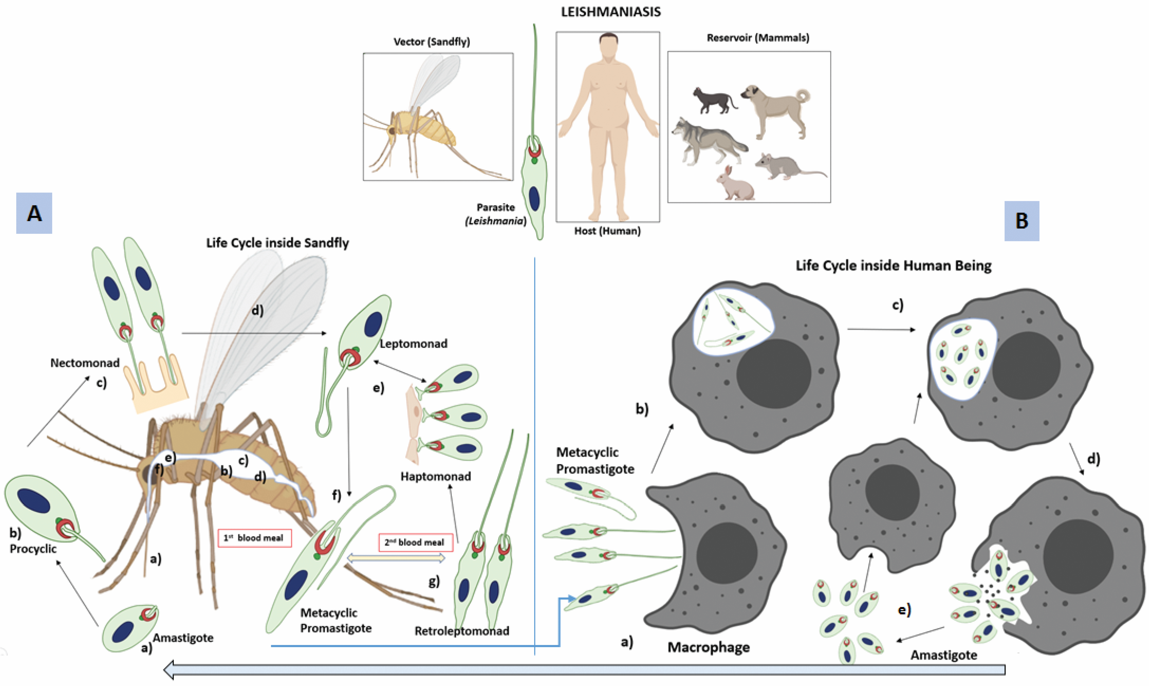
2. Life Cycle of Leishmania
2.1. Sandfly-Specific Stages of the Parasite
2.2. Stages Inside the Mammalian Phagocytes
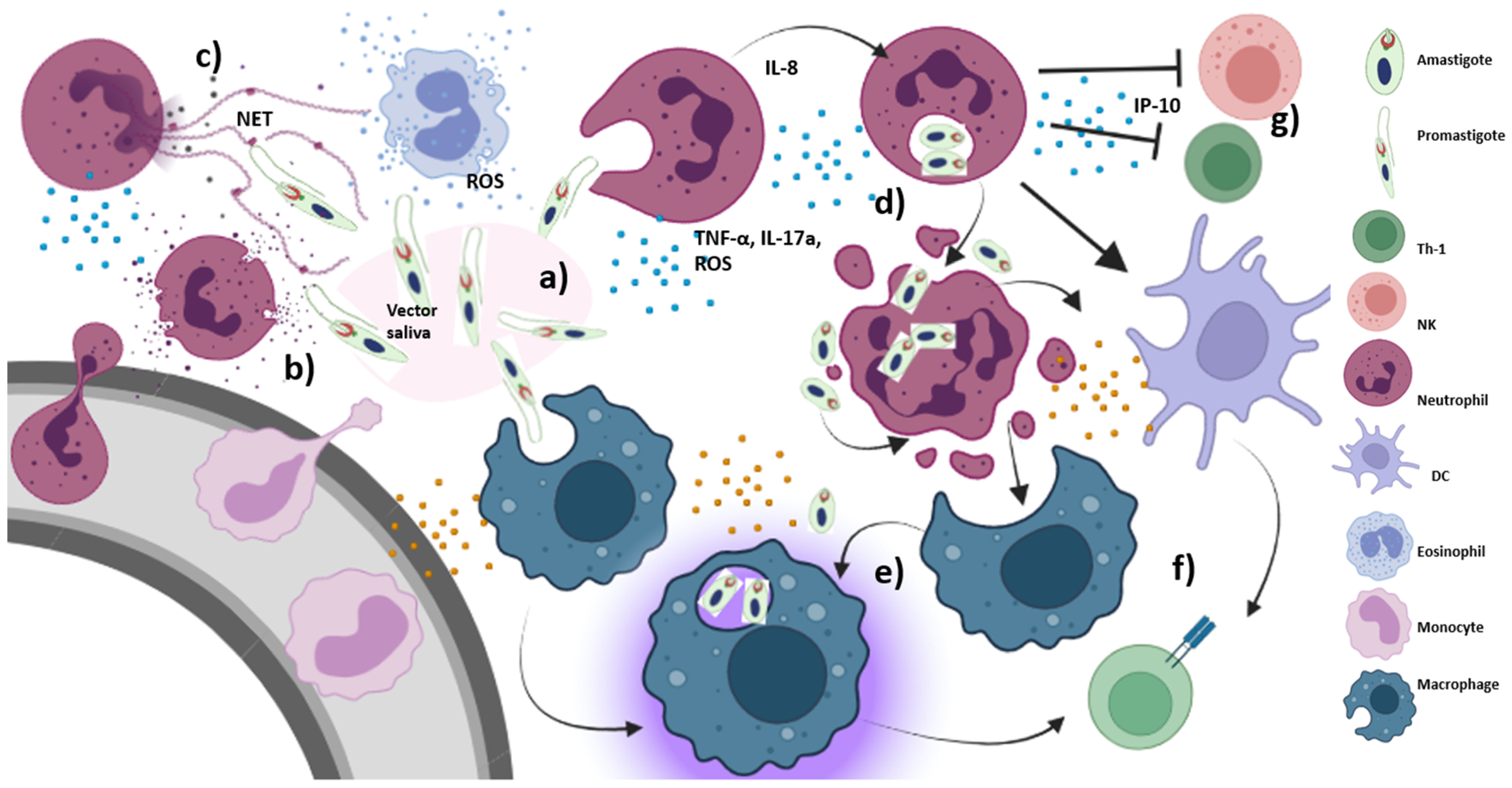
3. Host Immune Response
3.1. Innate Immune Response
3.1.1. Role of Neutrophils and Salivary Components of Sandfly during Early Immune Response
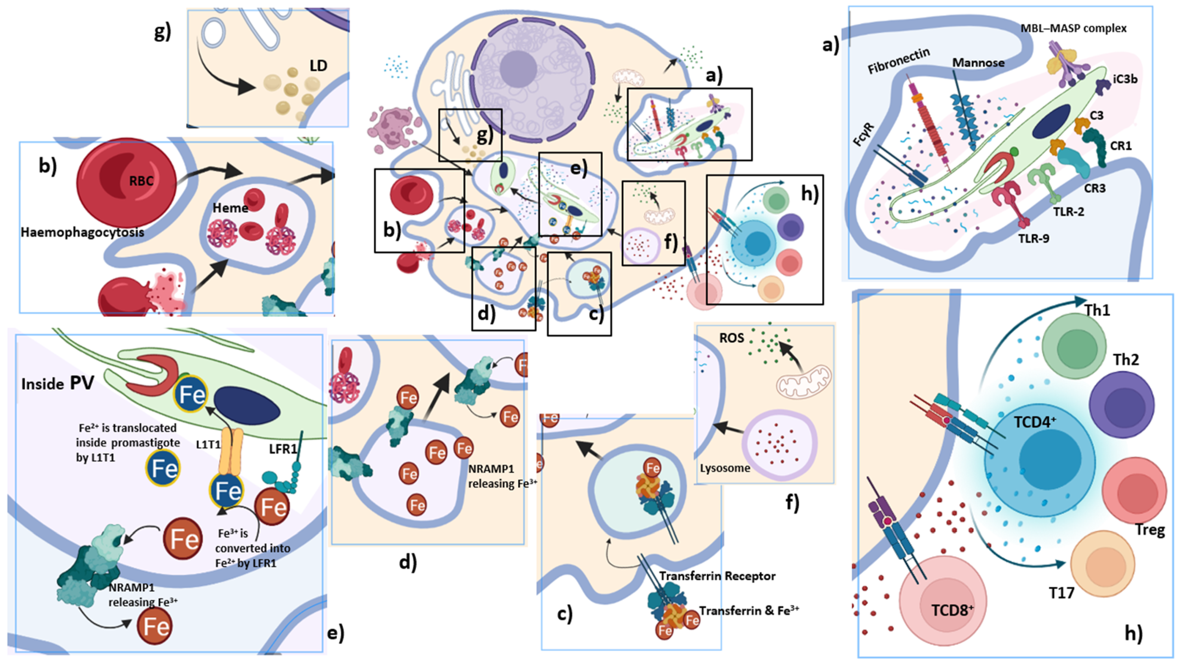
3.1.2. Macrophage as a Cellular Host of Leishmania
Parasitophorous Vacuoles (PV) as a Safe Haven for Leishmania
Leishmania-Induced Hemophagocytosis and Iron Uptake
Lipid Uptake by Leishmania
Cytokine Induced Macrophage Activation
3.1.3. Dual Role of Complement
3.1.4. Role of Dendritic Cell
3.2. Adaptive Immune Response
3.2.1. Immunomodulation by T Helper Subtypes
Th1 and Th2 Polarization
T Regulatory and T17 Cells
3.2.2. CD8 T Cells
3.2.3. Defects in Antigen Presentation to T Cells
4. Vaccine Strategies
4.1. First-Generation Vaccine
4.1.1. Live Attenuated Vaccine
4.1.2. Killed Parasite Vaccines
4.2. 2nd Generation Vaccination
4.2.1. gp63
4.2.2. Leishmania Homolog for Receptors of Activated C Kinase (LACK)
4.2.3. Kinetoplastid Membrane Protein-11 (KMP-11)
4.2.4. Fucose Mannose Ligand (FML) and Monophosphoryl Lipid A (MPL-A)
4.2.5. Hypothetical, In Silico Protein Constructs and Other Proteins
4.2.6. Vaccines against Canine Leishmaniasis
4.3. Third-Generation Vaccine
5. Conclusions
Author Contributions
Funding
Institutional Review Board Statement
Informed Consent Statement
Data Availability Statement
Acknowledgments
Conflicts of Interest
References
- Ashford, R.W. Leishmaniasis reservoirs and their significance in control. Clin. Dermatol. 1996, 14, 523–532. [Google Scholar] [CrossRef]
- Weekly Epidemiological Report. 2020. Available online: http://www.who.int/wer (accessed on 10 January 2022).
- Herwaltd, B.L. Leishmaniasis. Lancet 1999, 354, 1191–1199. [Google Scholar] [CrossRef]
- Bailey, M.S.; Lockwood, D.N. Cutaneous leishmaniasis. Clin. Dermatol. 2007, 25, 203–211. [Google Scholar] [CrossRef] [PubMed]
- Weigle, K.; Saravia, N.G. Natural history, clinical evolution, and the host-parasite interaction in New World cutaneous Leishmania-sis. Clin. Dermatol. 1996, 14, 433–450. [Google Scholar] [CrossRef]
- Burza, S.; Croft, S.L.; Boelaert, M. Leishmaniasis. Lancet 2018, 392, 951–970. [Google Scholar] [CrossRef]
- Murray, H.W.; Berman, J.D.; Davies, C.R.; Saravia, N.G. Advances in leishmaniasis. Lancet 2005, 366, 1561–1577. [Google Scholar] [CrossRef]
- Gregory, D.J.; Sladek, R.; Olivier, M.; Matlashewski, G. Comparison of the effects of Leishmania major or Leishmania do-novani infection on macrophage gene expression. Infect. Immun. 2008, 76, 1186–1192. [Google Scholar] [CrossRef]
- Cruz, I.; Morales, M.A.; Noguer, I.; Rodríguez, A.; Alvar, J. Leishmania in discarded syringes from intravenous drug users. Lancet 2002, 359, 1124–1125. [Google Scholar] [CrossRef]
- Pagliano, P.; Carannante, N.; Rossi, M.; Gramiccia, M.; Gradoni, L.; Faella, F.S.; Gaeta, G.B. Visceral leishmaniasis in pregnancy: A case series and a systematic review of the literature. J. Antimicrob. Chemother. 2005, 55, 229–233. [Google Scholar] [CrossRef]
- Costa, C.H.N.; Gomes, R.B.B.; Silva, M.R.B.; Garcez, L.M.; Ramos, P.K.; Santos, R.S.; Shaw, J.J.; David, J.R.; Maguire, J.H. Competence of the Human Host as a Reservoir forLeishmania chagasi. J. Infect. Dis. 2000, 182, 997–1000. [Google Scholar] [CrossRef]
- Chitimia, L.; Muñoz-García, C.; Sánchez-Velasco, D.; Lizana, V.; del Río, L.; Murcia, L.; Fisa, R.; Riera, C.; Giménez-Font, P.; Jiménez-Montalbán, P.; et al. Cryptic Leishmaniosis by Leishmania infantum, a feature of canines only? A study of natural infection in wild rabbits, humans and dogs in southeastern Spain. Vet. Parasitol. 2011, 181, 12–16. [Google Scholar] [CrossRef] [PubMed]
- Dipineto, L.; Manna, L.; Baiano, A.; Gala, M.; Fioretti, A.; Gravino, A.E.; Menna, L.F. Presence of Leishmania infantum in Red Foxes (Vulpes vulpes) in Southern Italy. J. Wildl. Dis. 2007, 43, 518–520. [Google Scholar] [CrossRef] [PubMed]
- Chatzis, M.K.; Andreadou, M.; Leontides, L.; Kasabalis, D.; Mylonakis, M.; Koutinas, A.F.; Rallis, T.; Ikonomopoulos, J.; Saridomichelakis, M.N. Cytological and molecular detection of Leishmania infantum in different tissues of clinically normal and sick cats. Vet. Parasitol. 2014, 202, 217–225. [Google Scholar] [CrossRef] [PubMed]
- Pereira, A.A.S.; Ferreira, E.; Lima, A.C.V.M.d.R.; Tonelli, G.B.; Rêgo, F.D.; Paglia, A.P.; Andrade-Filho, J.D.; Paz, G.F.; Gontijo, C.M.F. Detection of Leishmania spp in silvatic mammals and isolation of Leishmania (Viannia) braziliensis from Rattus rattus in an endemic area for leishmaniasis in Minas Gerais State, Brazil. PLoS ONE 2017, 12, e0187704. [Google Scholar] [CrossRef]
- Singh, N.; Mishra, J.; Singh, R.; Singh, S. Animal Reservoirs of Visceral Leishmaniasis in India. J. Parasitol. 2013, 99, 64–67. [Google Scholar] [CrossRef]
- Oryan, A.; Akbari, M. Worldwide risk factors in leishmaniasis. Asian Pac. J. Trop. Med. 2016, 9, 925–932. [Google Scholar] [CrossRef]
- Cloots, K.; Marino, P.; Burza, S.; Gill, N.; Boelaert, M.; Hasker, E. Visceral Leishmaniasis-HIV Coinfection as a Predictor of Increased Leishmania Transmission at the Village Level in Bihar, India. Front. Cell. Infect. Microbiol. 2021, 11, 604117. [Google Scholar] [CrossRef]
- Aschale, Y.; Ayehu, A.; Worku, L.; Tesfa, H.; Birhanie, M.; Lemma, W. Malaria-visceral leishmaniasis co-infection and associated factors among migrant laborers in West Armachiho district, North West Ethiopia: Community based cross-sectional study. BMC Infect. Dis. 2019, 19, 239. [Google Scholar] [CrossRef]
- Shen, S.-S.; Qu, X.-Y.; Zhang, W.-Z.; Li, J.; Lv, Z.-Y. Infection against infection: Parasite antagonism against parasites, viruses and bacteria. Infect. Dis. Poverty 2019, 8, 49. [Google Scholar] [CrossRef]
- Bates, P.A. Transmission of Leishmania metacyclic promastigotes by phlebotomine sand flies. Int. J. Parasitol. 2007, 37, 1097–1106. [Google Scholar] [CrossRef]
- Rogers, M.E.; Ilg, T.; Nikolaev, A.V.; Ferguson, M.; Bates, P.A. Transmission of cutaneous leishmaniasis by sand flies is enhanced by regurgitation of fPPG. Nature 2004, 430, 463–467. [Google Scholar] [CrossRef] [PubMed]
- Bates, P.; Rogers, M. New Insights into the Developmental Biology and Transmission Mechanisms of Leishmania. Curr. Mol. Med. 2004, 4, 601–609. [Google Scholar] [CrossRef] [PubMed]
- Gossage, S.M.; Rogers, M.E.; Bates, P.A. Two separate growth phases during the development of Leishmania in sand flies: Implications for understanding the life cycle. Int. J. Parasitol. 2003, 33, 1027–1034. [Google Scholar] [CrossRef]
- Killick-Kendrick, R.; Molyneux, D.H.; Ashford, R.W. Leishmania in phlebotomid sandflies. I. Modifications of the flagellum associated with attachment to the mid-gut and oesophageal valve of the sandfly. Proc. R Soc. Lond. B Biol. Sci. 1974, 187, 409–419. [Google Scholar]
- Giraud, E.; Martin, O.; Yakob, L.; Rogers, M. Quantifying Leishmania Metacyclic Promastigotes from Individual Sandfly Bites Reveals the Efficiency of Vector Transmission. Commun. Biol. 2019, 2, 84. [Google Scholar] [CrossRef]
- Bates, P.A. Revising Leishmania’s life cycle. Nat. Microbiol. 2018, 3, 529–530. [Google Scholar] [CrossRef]
- Rogers, M.E.; Chance, M.L.; Bates, P.A. The role of promastigote secretory gel in the origin and transmission of the infective stage of Leishmania mexicana by the sandfly Lutzomyia longipalpis. Parasitology 2002, 124, 495–507. [Google Scholar] [CrossRef]
- Sunter, J.; Gull, K. Shape, form, function and Leishmania pathogenicity: From textbook descriptions to biological understanding. Open Biol. 2017, 7, 170165. [Google Scholar] [CrossRef]
- Rittig, M.G.; Schröppel, K.; Seack, K.H.; Sander, U.; N’Diaye, E.N.; Maridonneau-Parini, I.; Solbach, W.; Bogdan, C. Coiling phag-ocytosis of trypanosomatids and fungal cells. Infect. Immun. 1998, 66, 4331–4339. [Google Scholar] [CrossRef]
- Petropolis, D.B.; Rodrigues, J.C.; Viana, N.B.; Pontes, B.; Pereira, C.F.; Silva-Filho, F.C. Leishmania amazonensis promastigotes in 3D Collagen I culture: An in vitro physiological environment for the study of extracellular matrix and host cell interactions. PeerJ 2014, 2, e317. [Google Scholar] [CrossRef]
- Dedet, J.P.; Pratlong, F.; Lanotte, G.; Ravel, C. Cutaneous leishmaniasis. The parasite. Clin. Dermatol. 1999, 17, 261–268. [Google Scholar] [CrossRef]
- Yao, C.; Donelson, J.E.; Wilson, M.E. The major surface protease (MSP or GP63) of Leishmania sp. Biosynthesis, regulation of expression, and function. Mol. Biochem. Parasitol. 2003, 132, 1–16. [Google Scholar] [CrossRef]
- Naderer, T.; McConville, M.J. The Leishmania-macrophage interaction: A metabolic perspective. Cell. Microbiol. 2008, 10, 301–308. [Google Scholar] [CrossRef] [PubMed]
- Lo, S.K.; Bovis, L.; Matura, R.; Zhu, B.; He, S.; Lum, H.; Turco, S.J.; Ho, J.L. Leishmania lipophosphoglycan reduces monocyte transendothelial migration: Modulation of cell adhesion molecules, intercellular junctional proteins, and chemoattractants. J. Immunol. 1998, 160, 1857–1865. [Google Scholar] [PubMed]
- Bogdan, C. Macrophages as host, effector and immunoregulatory cells in leishmaniasis: Impact of tissue micro-environment and metabolism. Cytokine X 2020, 2, 100041. [Google Scholar] [CrossRef]
- Liu, D.; Uzonna, J.E. The early interaction of Leishmania with macrophages and dendritic cells and its influence on the host immune response. Front. Cell. Infect. Microbiol. 2012, 2, 83. [Google Scholar] [CrossRef]
- Novais, F.O.; Santiago, R.C.; Báfica, A.; Khouri, R.; Afonso, L.; Borges, V.; Brodskyn, C.; Netto, M.B.; Barral, A.; De Oliveira, C.I. Neutrophils and Macrophages Cooperate in Host Resistance against Leishmania braziliensis Infection. J. Immunol. 2009, 183, 8088–8098. [Google Scholar] [CrossRef]
- de Souza Carmo, E.V.; Katz, S.; Barbiéri, C.L. Neutrophils reduce the parasite burden in Leishmania (Leishmania) amazonensis-infected macrophages. PLoS ONE 2010, 3, e13815. [Google Scholar]
- Pearson, R.D.; Steigbigel, R.T. Phagocytosis and killing of the protozoan Leishmania donovani by human polymorphonuclear leukocytes. J. Immunol. 1981, 127, 1438–1443. [Google Scholar]
- Guimaraes-Costa, A.B.; Nascimento, M.T.C.; Froment, G.S.; Soares, R.P.P.; Morgado, F.N.; Conceicao-Silva, F.; Saraiva, E.M. Leishmania amazonensis promastigotes induce and are killed by neutrophil extracellular traps. Proc. Natl. Acad. Sci. USA 2009, 106, 6748–6753. [Google Scholar] [CrossRef]
- Titus, R.G.; Ribeiro, J.M. Salivary gland lysates from the sand fly Lutzomyia longipalpis enhance Leishmania infectivity. Science 1988, 239, 1306–1308. [Google Scholar] [CrossRef]
- Dey, R.; Joshi, A.B.; Oliveira, F.; Pereira, L.; Guimarães-Costa, A.B.; Serafim, T.D.; de Castro, W.; Coutinho-Abreu, I.V.; Bhattacharya, P.; Townsend, S.; et al. Gut Microbes Egested during Bites of Infected Sand Flies Augment Severity of Leishmaniasis via Inflammasome-Derived IL-1β. Cell Host Microbe 2018, 23, 134.e6–143.e6. [Google Scholar] [CrossRef] [PubMed]
- Chagas, A.C.; Oliveira, F.; Debrabant, A.; Valenzuela, J.G.; Ribeiro, J.M.; Calvo, E. Lundep, a sand fly salivary endonuclease in-creases Leishmania parasite survival in neutrophils and inhibits XIIa contact activation in human plasma. PLoS Pathog. 2014, 10, e1003923. [Google Scholar] [CrossRef] [PubMed]
- Guimaraes-Costa, A.B.; Shannon, J.P.; Waclawiak, I.; Oliveira, J.; Meneses, C.; de Castro, W.; Wen, X.; Brzostowski, J.; Serafim, T.D.; Andersen, J.F.; et al. A sand fly salivary protein acts as a neutrophil chemo-attractant. Nat. Commun. 2021, 12, 3213. [Google Scholar] [CrossRef] [PubMed]
- Sangare, M.; Coulibaly, Y.I.; Huda, N.; Vidal, S.; Tariq, S.; Coulibaly, M.E.; Coulibaly, S.Y.; Soumaoro, L.; Dicko, I.; Traore, B.; et al. Individuals co-exposed to sand fly saliva and filarial parasites exhibit altered monocyte function. PLoS Negl. Trop. Dis. 2021, 15, e0009448. [Google Scholar] [CrossRef]
- Charmoy, M.; Megnekou, R.; Allenbach, C.; Zweifel, C.; Perez, C.; Monnat, K.; Breton, M.; Ronet, C.; Launois, P.; Tacchini-Cottier, F. Leishmania major induces distinct neutrophil phenotypes in mice that are resistant or susceptible to infection. J. Leukoc. Biol. 2007, 82, 288–299. [Google Scholar] [CrossRef]
- Ribeiro-Gomes, F.L.; Moniz-de-Souza, M.C.; Alexandre-Moreira, M.S.; Dias, W.B.; Lopes, M.F.; Nunes, M.P.; Lungarella, G.; DosReis, G.A. Neutrophils activate macrophages for intracellular killing of Leishmania major through recruitment of TLR4 by neutrophil elastase. J. Immunol. 2007, 179, 3988–3994. [Google Scholar] [CrossRef]
- Ribeiro-Gomes, F.L.; Sacks, D. The influence of early neutrophil-Leishmania interactions on the host immune response to infec-tion. Front. Cell. Infect. Microbiol. 2012, 2, 59. [Google Scholar] [CrossRef]
- Singh, T.P.; Carvalho, A.M.; Sacramento, L.A.; Grice, E.A.; Scott, P. Microbiota instruct IL-17A-producing innate lymphoid cells to promote skin inflammation in cutaneous leishmaniasis. PLOS Pathog. 2021, 17, e1009693. [Google Scholar] [CrossRef]
- van Zandbergen, G.; Hermann, N.; Laufs, H.; Solbach, M.D.W.; Laskay, T. Leishmania Promastigotes Release a Granulocyte Chemotactic Factor and Induce Interleukin-8 Release but Inhibit Gamma Interferon-Inducible Protein 10 Production by Neutrophil Granulocytes. Infect. Immun. 2002, 70, 4177–4184. [Google Scholar] [CrossRef]
- Kostka, S.L.; Dinges, S.; Griewank, K.; Iwakura, Y.; Udey, M.C.; Von Stebut, E. IL-17 Promotes Progression of Cutaneous Leishmaniasis in Susceptible Mice. J. Immunol. 2009, 182, 3039–3046. [Google Scholar] [CrossRef] [PubMed]
- Boaventura, V.S.; Santos, C.S.; Cardoso, C.R.D.B.; De Andrade, J.; Dos-Santos, W.L.; Clarêncio, J.; Silva, J.S.; Borges, V.; Netto, M.B.; Brodskyn, C.I.; et al. Human mucosal leishmaniasis: Neutrophils infiltrate areas of tissue damage that express high levels of Th17-related cytokines. Eur. J. Immunol. 2010, 40, 2830–2836. [Google Scholar] [CrossRef] [PubMed]
- van Zandbergen, G.; Klinger, M.; Mueller, A.; Dannenberg, S.; Gebert, A.; Solbach, W.; Laskay, T. Cutting Edge: Neutrophil Granulocyte Serves as a Vector for Leishmania Entry into Macrophages. J. Immunol. 2004, 173, 6521–6525. [Google Scholar] [CrossRef] [PubMed]
- Gueirard, P.; Laplante, A.; Rondeau, C.; Milon, G.; Desjardins, M. Trafficking of Leishmania donovani promastigotes in non-lytic compartments in neutrophils enables the subsequent transfer of parasites to macrophages. Cell. Microbiol. 2008, 10, 100–111. [Google Scholar] [CrossRef]
- Rossi, M.; Fasel, N. How to master the host immune system? Leishmania parasites have the solutions! Int. Immunol. 2018, 30, 103–111. [Google Scholar] [CrossRef]
- Beverley, S.M.; Turco, S.J. Lipophosphoglycan (LPG) and the identification of virulence genes in the protozoan parasite Leishmania. Trends Microbiol. 1998, 6, 135–140. [Google Scholar] [CrossRef]
- Ferguson, M. The surface glycoconjugates of trypanosomatid parasites. Philos. Trans. R Soc. Lond. B Biol. Sci. 1997, 352, 1295–1302. [Google Scholar] [CrossRef]
- McConville, M.J.; Ralton, J.E. Developmentally regulated changes in the cell surface architecture of Leishmania parasites. Behring Inst. Mitt. 1997, 99, 34–43. [Google Scholar]
- Ferguson, M.A. The structure, biosynthesis and functions of glycosylphosphatidylinositol anchors, and the contributions of trypanosome research. J. Cell Sci. 1999, 112, 2799–2809. [Google Scholar] [CrossRef]
- Kane, M.M.; Mosser, D.M. Leishmania parasites and their ploys to disrupt macrophage activation. Curr. Opin. Hematol. 2000, 7, 26–31. [Google Scholar] [CrossRef]
- Joshi, P.B.; Kelly, B.L.; Kamhawi, S.; Sacks, D.L.; McMaster, W.R. Targeted gene deletion in Leishmania major identifies leishmanolysin (GP63) as a virulence factor. Mol. Biochem. Parasitol. 2002, 120, 33–40. [Google Scholar] [CrossRef]
- Pelletier, I.; Hashidate, T.; Urashima, T.; Nishi, N.; Nakamura, T.; Futai, M.; Arata, Y.; Kasai, K.; Hirashima, M.; Hirabayashi, J.; et al. Specific recognition of Leishmania major poly-beta-galactosyl epitopes by galectin-9: Possible implication of galectin-9 in interaction between L. major and host cells. J. Biol. Chem. 2003, 278, 22223–22230. [Google Scholar] [CrossRef] [PubMed]
- Kamhawi, S.; Ramalho-Ortigao, M.; Pham, V.M.; Kumar, S.; Lawyer, P.G.; Turco, S.J.; Barillas-Mury, C.; Sacks, D.L.; Valenzuela, J.G. A Role for Insect Galectins in Parasite Survival. Cell 2004, 119, 329–341. [Google Scholar] [CrossRef] [PubMed]
- Soulat, D.; Bogdan, C. Function of Macrophage and Parasite Phosphatases in Leishmaniasis. Front. Immunol. 2017, 8, 1838. [Google Scholar] [CrossRef] [PubMed]
- Srivastava, S.; Pandey, S.P.; Jha, M.K.; Chandel, H.S.; Saha, B. Leishmania expressed lipophosphoglycan interacts with Toll-like receptor (TLR)-2 to decrease TLR-9 expression and reduce anti-leishmanial responses. Clin. Exp. Immunol. 2013, 172, 403–409. [Google Scholar] [CrossRef]
- Frankenburg, S.; Leibovici, V.; Mansbach, N.; Turco, S.J.; Rosen, G. Effect of glycolipids of Leishmania parasites on human monocyte activity. Inhibition by lipophosphoglycan. J. Immunol. 1990, 145, 4284–4289. [Google Scholar]
- Ibraim, I.C.; de Assis, R.R.; Pessoa, N.L.; Campos, M.A.; Melo, M.N.; Turco, S.J.; Soares, R.P. Two biochemically distinct lipophosphoglycans from Leishmania braziliensis and Leishmania infantum trigger different innate immune responses in murine macrophages. Parasites Vectors 2013, 6, 54. [Google Scholar] [CrossRef]
- Barral-Netto, M.; Barral, A.; Brownell, C.E.; Skeiky, Y.A.; Ellingsworth, L.R.; Twardzik, D.R.; Reed, S.G. Transforming growth factor-beta in leishmanial infection: A parasite escape mechanism. Science 1992, 257, 545–548. [Google Scholar] [CrossRef]
- Chang, K.; Fong, D. Cell Biology of Host-Parasite Membrane Interactions in Leishmaniasis. Ciba Found. Symp. 1983, 99, 113–137. [Google Scholar] [CrossRef]
- Forestier, C.-L.; Machu, C.; Loussert, C.; Pescher, P.; Späth, G.F. Imaging Host Cell-Leishmania Interaction Dynamics Implicates Parasite Motility, Lysosome Recruitment, and Host Cell Wounding in the Infection Process. Cell Host Microbe 2011, 9, 319–330. [Google Scholar] [CrossRef]
- Gluenz, E.; Ginger, M.L.; McKean, P.G. Flagellum assembly and function during the Leishmania life cycle. Curr. Opin. Microbiol. 2010, 13, 473–479. [Google Scholar] [CrossRef] [PubMed]
- Gluenz, E.; Höög, J.L.; Smith, A.E.; Dawe, H.R.; Shaw, M.K.; Gull, K. Beyond 9+0: Noncanonical axoneme structures characterize sensory cilia from protists to humans. FASEB J. 2010, 24, 3117–3121. [Google Scholar] [CrossRef] [PubMed]
- Burchmore, R.J.; Barrett, M. Life in vacuoles—Nutrient acquisition by Leishmania amastigotes. Int. J. Parasitol. 2001, 31, 1311–1320. [Google Scholar] [CrossRef]
- Dermine, J.-F.; Goyette, G.; Houde, M.; Turco, S.J.; Desjardins, M. Leishmania donovani lipophosphoglycan disrupts phagosome microdomains in J774 macrophages. Cell. Microbiol. 2005, 7, 1263–1270. [Google Scholar] [CrossRef] [PubMed]
- Vinet, A.F.; Fukuda, M.; Turco, S.J.; Descoteaux, A. The Leishmania donovani lipophosphoglycan excludes the vesicular proton-ATPase from phagosomes by impairing the recruitment of synaptotagmin V. PLoS Pathog. 2009, 5, e1000628. [Google Scholar] [CrossRef]
- Chakraborty, P.; Das, P.K. Suppression of macrophage lysosomal enzymes after Leishmania donovani infection. Biochem. Med. Metab. Biol. 1989, 41, 46–55. [Google Scholar] [CrossRef]
- Henriques, C.; Atella, G.C.; Bonilha, V.L.; de Souza, W. Biochemical analysis of proteins and lipids found in parasitophorous vacuoles containing Leishmania amazonensis. Parasitol. Res. 2003, 89, 123–133. [Google Scholar]
- Courret, N.; Fréhel, C.; Gouhier, N.; Pouchelet, M.; Prina, E.; Roux, P.; Antoine, J.C. Biogenesis of Leishmania-harbouring parasitophorous vacuoles following phagocytosis of the metacyclic promastigote or amastigote stages of the parasites. J. Cell Sci. 2002, 115, 2303–2316. [Google Scholar] [CrossRef]
- Körner, U.; Fuss, V.; Steigerwald, J.; Moll, H. Biogenesis of Leishmania major -Harboring Vacuoles in Murine Dendritic Cells. Infect. Immun. 2006, 74, 1305–1312. [Google Scholar] [CrossRef]
- Podinovskaia, M.; Descoteaux, A. Leishmania and the macrophage: A multifaceted interaction. Future Microbiol. 2015, 10, 111–129. [Google Scholar] [CrossRef]
- Mittra, B.; Andrews, N.W. IRONy OF FATE: Role of iron-mediated ROS in Leishmania differentiation. Trends Parasitol. 2013, 29, 489–496. [Google Scholar] [CrossRef] [PubMed]
- Mittra, B.; Cortez, M.; Haydock, A.; Ramasamy, G.; Myler, P.; Andrews, N. Iron uptake controls the generation of Leishmania infective forms through regulation of ROS levels. J. Exp. Med. 2013, 210, 401–416. [Google Scholar] [CrossRef] [PubMed]
- Glaser, T.A.; Baatz, J.E.; Kreishman, G.P.; Mukkada, A.J. pH homeostasis in Leishmania donovani amastigotes and promastigotes. Proc. Natl. Acad. Sci. USA 1988, 85, 7602–7606. [Google Scholar] [CrossRef] [PubMed]
- Schaible, U.E.; Schlesinger, P.H.; Steinberg, T.H.; Mangel, W.F.; Kobayashi, T.; Russell, D.G. Parasitophorous vacuoles of Leishmania mexicana acquire macromolecules from the host cell cytosol via two independent routes. J. Cell Sci. 1999, 112, 681–693. [Google Scholar] [CrossRef]
- Real, F.; Mortara, R.A.; Rabinovitch, M. Fusion between Leishmania amazonensis and Leishmania major Parasitophorous Vacuoles: Live Imaging of Coinfected Macrophages. PLoS Negl. Trop. Dis. 2010, 4, e905. [Google Scholar] [CrossRef]
- Chang, K.-P.; Dwyer, D. Leishmania Donovani. Hamster macrophage interactions in vitro: Cell entry, intracellular survival, and multiplication of amastigotes. J. Exp. Med. 1978, 147, 515–530. [Google Scholar] [CrossRef]
- Saha, S.; Basu, M.; Guin, S.; Gupta, P.; Mitterstiller, A.-M.; Weiss, G.; Jana, K.; Ukil, A. Leishmania donovani Exploits Macrophage Heme Oxygenase-1 To Neutralize Oxidative Burst and TLR Signaling–Dependent Host Defense. J. Immunol. 2018, 202, 827–840. [Google Scholar] [CrossRef]
- Wei, X.-Q.; Charles, I.G.; Smith, A.C.; Ure, J.; Feng, G.-J.; Huang, F.-P.; Xu, D.; Muller, W.; Moncada, S.; Liew, F.Y. Altered immune responses in mice lacking inducible nitric oxide synthase. Nature 1995, 375, 408–411. [Google Scholar] [CrossRef]
- Murray, H.W.; Nathan, C.F. Macrophage Microbicidal Mechanisms In Vivo: Reactive Nitrogen versus Oxygen Intermediates in the Killing of Intracellular Visceral Leishmania donovani. J. Exp. Med. 1999, 189, 741–746. [Google Scholar] [CrossRef]
- Henter, J.-I.; Horne, A.; Aricó, M.; Egeler, R.M.; Filipovich, A.H.; Imashuku, S.; Ladisch, S.; McClain, K.; Webb, D.; Winiarski, J.; et al. HLH-2004: Diagnostic and therapeutic guidelines for hemophagocytic lymphohistiocytosis. Pediatr. Blood Cancer 2007, 48, 124–131. [Google Scholar] [CrossRef]
- Mallory, F.B. A histological study of typhoid fever. J. Exp. Med. 1898, 3, 611–638. [Google Scholar] [CrossRef] [PubMed]
- Balasubramanian, S.; Kaarthigeyan, K.; Aparna, V.; Srinivas, S. Tuberculosis associated hemophagocytic syndrome in infancy. Indian Pediatr. 2008, 45, 593–595. [Google Scholar] [PubMed]
- Slovut, D.P.; Benedetti, E.; Matas, A.J. Babesiosis and hemophagocytic syndrome in an asplenic renal transplant recipient. Transplantation 1996, 62, 537–539. [Google Scholar] [CrossRef] [PubMed]
- Granert, C.; Elinder, G.; Ost, A.; Henter, J.I. Kala-azar in a one-year-old Swedish child. Diagnostic difficulties because of active hemophagocytosis. Acta Paediatr. 1993, 82, 794–796. [Google Scholar] [CrossRef]
- Fathalla, M.; Hashim, J.; Alkindy, H.; Wali, Y. Cerebrospinal fluid involvement in a case of visceral leishmaniasis associated with hemophagocytic lymphohistiocytosis. Sultan Qaboos Univ. Med. J. 2007, 7, 253–256. [Google Scholar]
- Kilani, B.; Ammari, L.; Kanoun, F.; Ben Chaabane, T.; Abdellatif, S.; Chaker, E. Hemophagocytic syndrome associated with visceral leishmaniasis. Int. J. Infect. Dis. 2006, 10, 85–86. [Google Scholar] [CrossRef][Green Version]
- Ozyurek, E.; Özçay, F.; Yilmaz, B.; Özbek, N. Hemophagocytic lymphohistiocytosis associated with visceral Leishmaniasis: A Case Report. Pediatr. Hematol. Oncol. 2005, 22, 409–414. [Google Scholar] [CrossRef]
- Gagnaire, M.-H.; Galambrun, C.; Stéphan, J.L. Hemophagocytic Syndrome: A Misleading Complication of Visceral Leishmaniasis in Children—A Series of 12 Cases. Pediatrics 2000, 106, e58. [Google Scholar] [CrossRef]
- Marom, D.; Offer, I.; Tamary, H.; Jaffe, C.L.; Garty, B.Z. Hemophagocytic Lymphohistocytosis associated with Visceral Leishmaniasis. J. Pediatr. Hematol. Oncol. 2001, 18, 65–70. [Google Scholar] [CrossRef]
- Bode, S.; Bogdan, C.; Beutel, K.; Behnisch, W.; Greiner, J.; Henning, S.; Jorch, N.; Jankofsky, M.; Jakob, M.; Schmid, I.; et al. Hemophagocytic Lymphohistiocytosis in Imported Pediatric Visceral Leishmaniasis in a Nonendemic Area. J. Pediatr. 2014, 165, 147.e1–153.e1. [Google Scholar] [CrossRef]
- Pilonieta, M.C.; Moreland, S.M.; English, C.N.; Detweiler, C.S. Salmonella enterica infection stimulates macrophages to hemophagocytose. mBio 2014, 5, e02211. [Google Scholar] [CrossRef] [PubMed]
- Brown, D.E.; McCoy, M.W.; Pilonieta, M.C.; Nix, R.N.; Detweiler, C.S. Chronic Murine Typhoid Fever Is a Natural Model of Secondary Hemophagocytic Lymphohistiocytosis. PLoS ONE 2010, 5, e9441. [Google Scholar] [CrossRef] [PubMed]
- Jordan, M.B.; Hildeman, D.; Kappler, J.; Marrack, P. An animal model of hemophagocytic lymphohistiocytosis (HLH): CD8+ T cells and interferon gamma are essential for the disorder. Blood 2004, 104, 735–743. [Google Scholar] [CrossRef] [PubMed]
- Brisse, E.; Imbrechts, M.; Put, K.; Avau, A.; Mitera, T.; Berghmans, N.; Rutgeerts, O.; Waer, M.; Ninivaggi, M.; Kelchtermans, H.; et al. Mouse Cytomegalovirus Infection in BALB/c Mice Resembles Virus-Associated Secondary Hemophagocytic Lymphohistiocytosis and Shows a Pathogenesis Distinct from Primary Hemophagocytic Lymphohistiocytosis. J. Immunol. 2016, 196, 3124–3134. [Google Scholar] [CrossRef]
- Cnops, J.; De Trez, C.; Stijlemans, B.; Keirsse, J.; Kauffmann, F.; Barkhuizen, M.; Keeton, R.; Boon, L.; Brombacher, F.; Magez, S. NK-, NKT- and CD8-Derived IFNγ Drives Myeloid Cell Activation and Erythrophagocytosis, Resulting in Trypanosomosis-Associated Acute Anemia. PLoS Pathog. 2015, 11, e1004964. [Google Scholar] [CrossRef]
- Morimoto, A.; Omachi, S.; Osada, Y.; Chambers, J.K.; Uchida, K.; Sanjoba, C.; Matsumoto, Y.; Goto, Y. Hemophagocytosis in Experimental Visceral Leishmaniasis by Leishmania donovani. PLoS Negl. Trop. Dis. 2016, 10, e0004505. [Google Scholar] [CrossRef]
- Marquis, J.F.; Gros, P. Intracellular Leishmania: Your iron or mine? Trends Microbiol. 2007, 15, 93–95. [Google Scholar] [CrossRef]
- Blackwell, J.M.; Goswami, T.; Evans, C.A.; Sibthorpe, D.; Papo, N.; White, J.K.; Searle, S.; Miller, E.N.; Peacock, C.S.; Mohammed, H.; et al. SLC11A1 (formerly NRAMP1) and disease resistance. Cell Microbiol. 2001, 3, 773–784. [Google Scholar] [CrossRef]
- Banerjee, S.; Datta, R. Leishmania infection triggers hepcidin-mediated proteasomal degradation of Nramp1 to increase phagolysosomal iron availability. Cell Microbiol. 2020, 22, e13253. [Google Scholar] [CrossRef]
- Das, N.K.; Biswas, S.; Solanki, S.; Mukhopadhyay, C.K. Leishmania donovani depletes labile iron pool to exploit iron uptake capacity of macrophage for its intracellular growth. Cell Microbiol. 2009, 11, 83–94. [Google Scholar] [CrossRef]
- Borges, V.M.; Vannier-Santos, M.A.; De Souza, W. Subverted transferrin trafficking in Leishmania-infected macrophages. Parasitol Res. 1998, 84, 811–822. [Google Scholar] [CrossRef] [PubMed]
- Srivastav, S.; Ball, W.B.; Gupta, P.; Giri, J.; Ukil, A.; Das, P.K. Leishmania donovani Prevents Oxidative Burst-mediated Apoptosis of Host Macrophages through Selective Induction of Suppressors of Cytokine Signaling (SOCS) Proteins. J. Biol. Chem. 2014, 289, 1092–1105. [Google Scholar] [CrossRef] [PubMed]
- Ghosh, J.; Guha, R.; Das, S.; Roy, S. Liposomal cholesterol delivery activates the macrophage innate immune arm to facilitate intracellular Leishmania donovani killing. Infect. Immun. 2014, 82, 607–617. [Google Scholar] [CrossRef] [PubMed]
- Ghosh, J.; Lal, C.S.; Pandey, K.; Das, V.N.R.; Das, P.; Roychoudhury, K.; Roy, S. Human visceral leishmaniasis: Decrease in serum cholesterol as a function of splenic parasite load. Ann. Trop. Med. Parasitol. 2011, 105, 267–271. [Google Scholar] [CrossRef] [PubMed]
- Messaoud, H.B.-B.; Guichard, M.; Lawton, P.; Delton, I.; Azzouz-Maache, S. Changes in Lipid and Fatty Acid Composition During Intramacrophagic Transformation of Leishmania donovani Complex Promastigotes into Amastigotes. Lipids 2017, 52, 433–441. [Google Scholar] [CrossRef]
- Zhang, K.; Beverley, S.M. Phospholipid and sphingolipid metabolism in Leishmania. Mol. Biochem. Parasitol. 2010, 170, 55–64. [Google Scholar] [CrossRef]
- Zhang, O.; Wilson, M.C.; Xu, W.; Hsu, F.F.; Turk, J.; Kuhlmann, F.M.; Wang, Y.; Soong, L.; Key, P.; Beverley, S.M.; et al. Deg-radation of host sphingomyelin is essential for Leishmania virulence. PLoS Pathog. 2009, 5, e1000692. [Google Scholar] [CrossRef]
- Vallochi, A.L.; Teixeira, L.; Oliveira, K.D.S.; Maya-Monteiro, C.M.; Bozza, P.T. Lipid Droplet, a Key Player in Host-Parasite In-teractions. Front. Immunol. 2018, 9, 1022. [Google Scholar] [CrossRef]
- De Cicco, N.N.; Pereira, M.G.; Corrêa, J.R.; Andrade-Neto, V.V.; Saraiva, F.B.; Chagas-Lima, A.C.; Gondim, K.C.; Torres-Santos, E.C.; Folly, E.; Saraiva, E.M.; et al. LDL uptake by Leishmania amazonensis: Involvement of membrane lipid microdomains. Exp. Parasitol. 2012, 130, 330–340. [Google Scholar] [CrossRef]
- Das, P.; De, T.; Chakraborti, T. Leishmania donovani secretory serine protease alters macrophage inflammatory response via COX-2 mediated PGE-2 production. Indian J. Biochem. Biophys. 2014, 51, 5542–5551. [Google Scholar]
- Carregaro, V.; Valenzuela, J.G.; Cunha, T.M.; Verri, W.A., Jr.; Grespan, R.; Matsumura, G.; Ribeiro, J.M.; Elnaiem, D.E.; Silva, J.S.; Cunha, F.Q. Phlebotomine salivas inhibit immune inflammation-induced neutrophil migration via an autocrine DC-derived PGE 2/IL-10 sequential pathway. J. Leukoc. Biol. 2008, 84, 104–114. [Google Scholar] [CrossRef] [PubMed]
- Araújo-Santos, T.; Prates, D.B.; Andrade, B.B.; Nascimento, D.O.; Clarêncio, J.; Entringer, P.F.; Carneiro, A.B.; Silva-Neto, M.A.C.; Miranda, J.C.; Brodskyn, C.I.; et al. Lutzomyia longipalpis Saliva Triggers Lipid Body Formation and Prostaglandin E2 Production in Murine Macrophages. PLoS Negl. Trop. Dis. 2010, 4, e873. [Google Scholar] [CrossRef] [PubMed]
- Rabhi, S.; Rabhi, I.; Trentin, B.; Piquemal, D.; Regnault, B.; Goyard, S.; Lang, T.; Descoteaux, A.; Enninga, J.; Guizani-Tabbane, L. Lipid droplet formation, their localization and dynamics during Leishmania major macrophage infection. PLoS ONE 2016, 11, 1–19. [Google Scholar] [CrossRef] [PubMed]
- Aoki, J.I.; Muxel, S.M.; Zampieri, R.A.; Müller, K.E.; Nerland, A.H.; Floeter-Winter, L.M. Differential immune response modulation in early Leishmania amazonensis infection of BALB/c and C57BL/6 macrophages based on transcriptome profiles. Sci. Rep. 2019, 9, 19841. [Google Scholar] [CrossRef] [PubMed]
- Tomiotto-Pellissier, F.; Bortoleti, B.; Assolini, J.P.; Gonçalves, M.D.; Carloto, A.C.M.; Miranda-Sapla, M.M.; Conchon-Costa, I.; Bordignon, J.; Pavanelli, W.R. Macrophage Polarization in Leishmaniasis: Broadening Horizons. Front. Immunol. 2018, 9, 2529. [Google Scholar] [CrossRef]
- Kropf, P.; Fuentes, J.M.; Fähnrich, E.; Arpa, L.; Herath, S.; Weber, V.; Soler, G.; Celada, A.; Modolell, M.; Müller, I. Arginase and polyamine synthesis are key factors in the regulation of experimental leishmaniasis in vivo. FASEB J. 2005, 19, 1000–1002. [Google Scholar] [CrossRef]
- Kumar, A.; Das, S.; Mandal, A.; Verma, S.; Abhishek, K.; Kumar, A.; Kumar, V.; Ghosh, A.K.; Das, P. Leishmania infection activates host mTOR for its survival by M2 macrophage polarization. Parasite Immunol. 2018, 40, e12586. [Google Scholar] [CrossRef]
- Parmar, N.; Chandrakar, P.; Kar, S. Leishmania donovani Subverts Host Immune Response by Epigenetic Reprogramming of Macrophage M(Lipopolysaccharides + IFN-γ)/M(IL-10) Polarization. J. Immunol. 2020, 204, 2762–2778. [Google Scholar] [CrossRef]
- Descoteaux, A.; Matlashewski, G. c-fos and TNF gene expression in Leishmania donovani–infected macrophages. Mol. Cell. Biol. 1989, 9, 5223–5227. [Google Scholar]
- Reiner, N.E.; Ng, W.; McMaster, W.R. Parasite-accessory cell-interactions in murine leishmaniasis 2. Leishmania donovani suppresses macrophage expression of class-I and class-II major histocompatibility complex gene-products. J. Immunol. 1987, 138, 1926–1932. [Google Scholar]
- Olivier, M.; Tanner, C.E. Effect of cyclosporin A in murine leishmaniasis. Trop. Med. Parasitol. 1989, 40, 32–38. [Google Scholar] [PubMed]
- Hatzigeorgiou, D.E.; Geng, J.; Zhu, B.; Zhang, Y.; Liu, K.; Rom, W.N.; Fenton, M.J.; Turco, S.J.; Ho, J.L. Lipophosphoglycan from Leishmania suppresses agonist-induced interleukin 1 beta gene expression in human monocytes via a unique promoter se-quence. Proc. Natl. Acad. Sci. USA 1996, 93, 14708–14713. [Google Scholar] [CrossRef] [PubMed]
- Cillari, E.; Dieli, M.; Maltese, E.; Milano, S.; Salerno, A.; Liew, F.Y. Enhancement of macrophage IL-1 production by Leishmania major infection in vitro and its inhibition by IFN-g. J. Immunol. 1989, 143, 2001–2005. [Google Scholar] [PubMed]
- Matte, C.; Olivier, M. Leishmania-Induced Cellular Recruitment during the Early Inflammatory Response: Modulation of Proinflammatory Mediators. J. Infect. Dis. 2002, 185, 673–681. [Google Scholar] [CrossRef]
- Ghalib, H.W.; Piuvezam, M.R.; Skeiky, Y.A.; Siddig, M.; Hashim, F.A.; El-Hassan, A.M.; Russo, D.M.; Reed, S.G. Interleukin 10 production correlates with pathology in human Leishmania donovani infections. J. Clin. Investig. 1993, 92, 324–329. [Google Scholar] [CrossRef]
- Nylen, S.R.; Maurya, L.; Eidsmo, K.D.; Manandhar, S.S.; Sacks, D. Splenic accumulation of IL-10 mRNA in T cells distinct from CD4+CD25+ (Foxp3) regulatory T cells in human visceral leishmaniasis. J. Exp. Med. 2007, 204, 805–817. [Google Scholar] [CrossRef]
- Panaro, M.; Acquafredda, A.; Lisi, S.; Lofrumento, D.; Mitolo, V.; Sisto, M.; Fasanella, A.; Trotta, T.; Bertani, F.; Consenti, B.; et al. Nitric oxide production by macrophages of dogs vaccinated with killed Leishmania infantum promastigotes. Comp. Immunol. Microbiol. Infect. Dis. 2001, 24, 187–195. [Google Scholar] [CrossRef]
- Pinelli, E.; Gebhard, D.; Mommaas, A.; van Hoeij, M.; Langermans, J.A.; Ruitenberg, E.; Rutten, V.P. Infection of a canine macrophage cell line with Leishmania infantum: Determination of nitric oxide production and anti-leishmanial activity. Vet. Parasitol. 2000, 92, 181–189. [Google Scholar] [CrossRef]
- Ghalib, H.W.; Whittle, J.A.; Kubin, M.; Hashim, F.A.; El-Hassan, A.M.; Grabstein, K.H.; Trinchieri, G.; Reed, S.G. IL-12 enhances Th1-type responses in human Leishmania donovani infections. J. Immunol. 1995, 154, 4623–4629. [Google Scholar]
- Sacks, D.L.; Lal, S.L.; Shrivastava, S.N.; Blackwell, J.; Neva, F.A. An analysis of T cell responsiveness in Indian kala-azar. J. Immunol. 1987, 138, 908–913. [Google Scholar]
- Mosser, D.M.; Edelson, P.J. Activation of the alternative complement pathway by Leishmania promastigotes: Parasite lysisand attachment to macrophages. J. Immunol. 1984, 132, 1501–1505. [Google Scholar] [PubMed]
- Mosser, D.M.; Burke, S.K.; Coutavas, E.E.; Wedgwood, J.F.; Edelson, P.J. Leishmania species: Mechanisms of complement activation by five strains of promastigotes. Exp. Parasitol. 1986, 62, 394–404. [Google Scholar] [CrossRef]
- Laurenti, M.D.; Corbett, C.; Sotto, M.; Sinhorini, I.; Goto, H. The role of complement in the acute inflammatory process in the skin and in host–parasite interaction in hamsters inoculated with Leishmania (Leishmania) chagasi. Int. J. Exp. Pathol. 1996, 77, 15–24. [Google Scholar] [CrossRef] [PubMed]
- Brittingham, A.; Mosser, D. Exploitation of the complement system by Leishmania promastigotes. Parasitol. Today 1996, 12, 444–447. [Google Scholar] [CrossRef]
- Rosenthal, L.A.; Sutterwala, F.S.; Kehrli, M.E.; Mosser, D.M. Leishmania major-human macrophage interactions: Cooperation between Mac-1 (CD11b/CD18) and complement receptor type 1 (CD35) in promastigote adhesion. Infect. Immun. 1996, 64, 2206–2215. [Google Scholar] [CrossRef] [PubMed]
- Blackwell, J.M. Role of macrophage complement and lectin-like receptors in binding Leishmania parasites to host macrophages. Immunol. Lett. 1985, 11, 227–232. [Google Scholar] [CrossRef]
- Ueno, N.; Wilson, M.E. Receptor-mediated phagocytosis of Leishmania: Implications for intracellular survival. Trends Parasitol. 2012, 28, 335–344. [Google Scholar] [CrossRef] [PubMed]
- Filho, A.A.P.; Nascimento, A.A.d.S.; Saab, N.A.A.; Fugiwara, R.T.; Pessoa, G.C.D.; Koerich, L.B.; Pereira, M.H.; Araújo, R.N.; Sant’Anna, M.R.V.; Gontijo, N.F. Evasion of the complement system by Leishmania through the uptake of factor H, a complement regulatory protein. Acta Trop. 2021, 224, 106152. [Google Scholar] [CrossRef]
- Ambrosio, A.R.; Bavia, L.; Hiraiwa, P.M.; Tirado, T.C.; Figueiredo, F.B.; de Messias-Reason, I.J. The lectin pathway of complement and the initial recognition of Leishmania infantum promastigotes. Life Sci. 2021, 282, 119793. [Google Scholar] [CrossRef]
- Vargas-Inchaustegui, D.A.; Tai, W.; Xin, L.; Hogg, A.E.; Corry, D.B.; Soong, L. Distinct roles for MyD88 and toll-like receptor 2 during Leishmania braziliensis infection in mice. Infect. Immun. 2009, 77, 2948–2956. [Google Scholar] [CrossRef]
- Argueta-Donohué, J.; Wilkins-Rodríguez, A.A.; Aguirre-García, M.; Gutiérrez-Kobeh, L. Differential phagocytosis of Leishmania mexicana promastigotes and amastigotes by monocyte-derived dendritic cells. Microbiol. Immunol. 2016, 60, 369–381. [Google Scholar] [CrossRef] [PubMed]
- Sacramento, L.; Trevelin, S.C.; Nascimento, M.S.; Lima-Jùnior, D.S.; Costa, D.L.; Almeida, R.P.; Cunha, F.Q.; Silva, J.S.; Carregaro, V. Toll-like receptor 9 signaling in dendritic cells regulates neutrophil recruitment to inflammatory foci following Leishmania infantum infection. Infect. Immun. 2015, 83, 4604–4616. [Google Scholar] [CrossRef] [PubMed]
- Liese, J.; Schleicher, U.; Bogdan, C. TLR9 signaling is essential for the innate NK cell response in murine cutaneous leishmaniasis. Eur. J. Immunol. 2007, 37, 3424–3434. [Google Scholar] [CrossRef] [PubMed]
- Revest, M.; Donaghy, L.; Cabillic, F.; Guiguen, C.; Gangneux, J.-P. Comparison of the immunomodulatory effects of L. donovani and L. major excreted–secreted antigens, particulate and soluble extracts and viable parasites on human dendritic cells. Vaccine 2008, 26, 6119–6123. [Google Scholar] [CrossRef]
- Ato, M.; Maroof, A.; Zubairi, S.; Nakano, H.; Kakiuchi, T.; Kaye, P.M. Loss of Dendritic Cell Migration and Impaired Resistance to Leishmania donovani Infection in Mice Deficient in CCL19 and CCL21. J. Immunol. 2006, 176, 5486–5493. [Google Scholar] [CrossRef]
- Sato, N.; Ahuja, S.K.; Quinones, M.; Kostecki, V.; Reddick, R.L.; Melby, P.C.; Kuziel, W.A.; Ahuja, S.S. CC chemokine receptor (CCR)2 is required for langerhans cell migration and localization of T helper cell type 1 (Th1)-inducing dendritic cells. absence of CCR2 shifts the Leishmania major-resistant phenotype to a susceptible state dominated by Th2 cytokin, B Cell Outgrowth, and Sustained Neutrophilic Inflammation. J. Exp. Med. 2000, 192, 205–218. [Google Scholar]
- von Stebut, E.; Belkaid, Y.; Jakob, T.; Sacks, D.L.; Udey, M.C. Uptake of Leishmania major amastigotes results in activation and in-terleukin 12 release from murine skin–derived dendritic cells: Implications for the initiation of Anti-Leishmania immunity. J. Exp. Med. 1998, 188, 1547–1552. [Google Scholar] [CrossRef]
- Favali, C.; Tavares, N.; Clarencio, J.; Barral, A.; Barral-Netto, M.; Brodskyn, C. Leishmania amazonensis infection impairs differentiation and function of human dendritic cells. J. Leukoc. Biol. 2007, 82, 1401–1406. [Google Scholar] [CrossRef][Green Version]
- Resende, M.; Moreira, D.; Augusto, J.; Cunha, J.; Neves, B.; Cruz, M.T.; Estaquier, J.; Cordeiro-da-Silva, A.; Silvestre, R. Leishmania-infected MHC Class II high dendritic cells polarize CD4+ T cells toward a nonprotective T-bet+ IFN- + IL-10+ phenotype. J. Immunol. 2013, 191, 262–273. [Google Scholar] [CrossRef]
- Amaral, V.F.; Ransatto, V.A.; Conceicão-Silva, F.; Molinaro, E.; Ferreira, V.; Coutinho, S.G.; McMahon-Pratt, D.; Grimaldi, G. Leishmania amazonensis: The Asian rhesus macaques (Macaca mulatta) as an experimental model for study of cutaneous leishmaniasis. Exp. Parasitol. 1996, 82, 34–44. [Google Scholar] [CrossRef]
- Gonçalves-de-Albuquerque, S.D.C.; Pessoa-E-Silva, R.; Trajano-Silva, L.A.M.; de Goes, T.C.; de Morais, R.C.S.; da C. Oliveira, C.N.; de Lorena, V.M.B.; de Paiva-Cavalcanti, M. The Equivocal Role of Th17 Cells and Neutrophils on Immunopathogenesis of Leishmaniasis. Front. Immunol. 2017, 8, 1437. [Google Scholar] [CrossRef] [PubMed]
- Alexander, J.; Brombacher, F. T helper 1/T helper 2 cells and resistance/susceptibility to Leishmania infection: Is this paradigm still relevant? Front. Immunol. 2012, 2012. 3, 80. [Google Scholar]
- Castes, M.; Cabrera, M.; Trujillo, D.; Convit, J. T-cell subpopulations, expression of interleukin-2 receptor, and production of inter-leukin-2 and gamma interferon in human American cutaneous leishmaniasis. J. Clin. Microbiol. 1988, 26, 1207–1213. [Google Scholar] [CrossRef] [PubMed]
- Frankenburg, S.; Kofsky, Y.; Gross, A. In vitro secretion of cytokines by human mononuclear cells of individuals during and after cutaneous leishmaniasis infection. Parasite Immunol. 1993, 15, 509–512. [Google Scholar] [CrossRef] [PubMed]
- Holaday, B.J.; Pompeu, M.M.D.L.; Evans, T.; Braga, D.N.D.M.; Texeira, M.J.; Sousa, A.D.Q.; Sadick, M.D.; Vasconcelos, A.W.; Abrams, J.S.; Pearson, R.D.; et al. Correlates of Leishmania-Specific Immunity in the Clinical Spectrum of Infection with Leishmania chagasi. J. Infect. Dis. 1993, 167, 411–417. [Google Scholar] [CrossRef] [PubMed]
- Amit, A.; Dikhit, M.R.; Singh, A.; Kumar, V.; Suman, S.S.; Singh, A.; Kumar, A.; Thakur, A.K.; Das, V.R.; Das, P.; et al. Immunization with Leishmania donovani protein disulfide isomerase DNA construct induces Th1 and Th17 dependent immune response and protection against experimental visceral leishmaniasis in Balb/c mice. Mol. Immunol. 2017, 82, 104–113. [Google Scholar] [CrossRef] [PubMed]
- McMahon-Pratt, D.; Alexander, J. Does the Leishmania major paradigm of pathogenesis and protection hold for New World cutaneous leishmaniases or the visceral disease? Immunol. Rev. 2004, 201, 206–224. [Google Scholar] [CrossRef]
- Atri, C.; Guerfali, F.Z.; Laouini, D. Role of Human Macrophage Polarization in Inflammation during Infectious Diseases. Int. J. Mol. Sci. 2018, 19, 1801. [Google Scholar] [CrossRef]
- Jaiswal, A.K.; Khare, P.; Joshi, S.; Kushawaha, P.K.; Sundar, S.; Dube, A. Th1 stimulatory proteins of Leishmania donovani: Comparative cellular and protective responses of rTriose phosphate isomerase, rProtein disulfide isomerase and rElongation factor-2 in combination with rHSP70 against visceral leishmaniasis. PLoS ONE 2014, 9, e108556. [Google Scholar] [CrossRef]
- Rodrigues, L.S.; Barreto, A.S.; Bomfim, L.G.S.; Gomes, M.C.; Ferreira, N.L.C.; da Cruz, G.S.; Magalhães, L.S.; de Jesus, A.R.; Palatnik-De-Sousa, C.B.; Corrêa, C.B.; et al. Multifunctional, TNF-α and IFN-γ-Secreting CD4 and CD8 T Cells and CD8High T Cells Are Associated with the Cure of Human Visceral Leishmaniasis. Front. Immunol. 2021, 12, 773983. [Google Scholar] [CrossRef]
- Hejazi, S.; Hoseini, S.; Javanmard, S.; Zarkesh, S.; Khamesipour, A. Interleukin-10 and Transforming Growth Factor-β in Early and Late Lesions of Patients with Leishmania major induced Cutaneous Leishmaniasis. Iran. J. Parasitol. 2012, 7, 53–60. [Google Scholar] [PubMed]
- Bhowmick, S.; Ravindran, R.; Ali, N. IL-4 contributes to failure, and colludes with IL-10 to exacerbate Leishmania donovani infection following administration of a subcutaneous leishmanial antigen vaccine. BMC Microbiol. 2014, 14, 8. [Google Scholar] [CrossRef] [PubMed]
- Muxel, S.M.; Aoki, J.I.; Fernandes, J.C.R.; Laranjeira-Silva, M.F.; Zampieri, R.A.; Acuña, S.M.; Müller, K.E.; Vanderlinde, R.H.; Floeter-Winter, L.M. Arginine and polyamines fate in Leishmania infection. Front. Microbiol. 2018, 15, 2682. [Google Scholar] [CrossRef] [PubMed]
- Li, S.N.; Wang, W.; Fu, S.P.; Wang, J.F.; Liu, H.M.; Xie, S.S.; Liu, B.R.; Li, Y.; Lv, Q.K.; Li, Z.Q.; et al. IL-21 modulates release of proinflammatory cytokines in LPS-stimulated macrophages through distinct signaling path-ways. Mediat. Inflamm. 2013, 2013, 548073. [Google Scholar] [CrossRef]
- Poudel, B.; Yorek, M.S.; Mazgaeen, L.; Brown, S.A.; Kanneganti, T.-D.; Gurung, P. Acute IL-4 Governs Pathogenic T Cell Responses during Leishmania major Infection. ImmunoHorizons 2020, 4, 546–560. [Google Scholar] [CrossRef]
- Belkaid, Y.; Hoffmann, K.F.; Mendez, S.; Kamhawi, S.; Udey, M.C.; Wynn, T.; Sacks, D.L. The Role of Interleukin (IL)-10 in the Persistence of Leishmania major in the Skin after Healing and the Therapeutic Potential of Anti–IL-10 Receptor Antibody for Sterile Cure. J. Exp. Med. 2001, 194, 1497–1506. [Google Scholar] [CrossRef]
- Belkaid, Y.; Piccirillo, C.A.; Mendez, S. CD4+ CD25+ regulatory T cells control Leishmania major persistence and immunity. Nature 2002, 420, 633–637. [Google Scholar] [CrossRef]
- Anderson, C.F.; Oukka, M.; Kuchroo, V.J.; Sacks, D. CD4+CD25-Foxp3- Th1 cells are source of IL-10-mediated immune suppression in chronic cutaneous leishmaniasis. J. Exp. Med. 2007, 204, 285–297. [Google Scholar] [CrossRef]
- Kane, M.M.; Mosser, D.M. The Role of IL-10 in Promoting Disease Progression in Leishmaniasis. J. Immunol. 2001, 166, 1141–1147. [Google Scholar] [CrossRef]
- Gautam, S.; Kumar, R.; Maurya, R.; Nylén, S.; Ansari, N.; Rai, M.; Sundar, S.; Sacks, D. IL-10 Neutralization Promotes Parasite Clearance in Splenic Aspirate Cells from Patients with Visceral Leishmaniasis. J. Infect. Dis. 2011, 204, 1134–1137. [Google Scholar] [CrossRef]
- Schwarz, T.; Remer, K.A.; Nahrendorf, W.; Masic, A.; Siewe, L.; Muller, W.; Roers, A.; Moll, H. T Cell-Derived IL-10 Determines Leishmaniasis Disease Outcome and Is Suppressed by a Dendritic Cell Based Vaccine. PLoS Pathog. 2013, 9, e1003476. [Google Scholar] [CrossRef] [PubMed]
- Gupta, G.; Majumdar, S.; Adhikari, A.; Bhattacharya, P.; Mukherjee, A.K.; Majumdar, S.B.; Majumdar, S. Treatment with IP-10 induces host-protective immune response by regulating the T regulatory cell functioning in Leishmania donovani-infected mice. Med. Microbiol. Immunol. 2011, 200, 241–253. [Google Scholar] [CrossRef] [PubMed]
- Mendez, S.; Reckling, S.K.; Piccirillo, C.A.; Sacks, D.L.; Belkaid, Y. Role for CD4+ CD25+ Regulatory T Cells in Reactivation of Persistent Leishmaniasis and Control of Concomitant Immunity. J. Exp. Med. 2004, 200, 201–210. [Google Scholar] [CrossRef] [PubMed]
- Ji, J.; Masterson, J.; Sun, J.; Soong, L. CD4+CD25+ regulatory T cells restrain pathogenic responses during Leishmania amazonen-sis infection. J. Immunol. 2005, 174, 7147–7153. [Google Scholar] [CrossRef]
- Rai, A.K.; Thakur, C.P.; Singh, A.; Seth, T.; Srivastava, S.K.; Singh, P.; Mitra, D.K. Regulatory T Cells Suppress T Cell Activation at the Pathologic Site of Human Visceral Leishmaniasis. PLoS ONE 2012, 7, e31551. [Google Scholar] [CrossRef]
- Rodriguez-Pinto, D.; Navas, A.; Blanco, V.M.; Ramírez, L.; Garcerant, D.; Cruz, A.; Craft, N.; Saravia, N.G. Regulatory T Cells in the Pathogenesis and Healing of Chronic Human Dermal Leishmaniasis Caused by Leishmania (Viannia) Species. PLoS Negl. Trop. Dis. 2012, 6, e1627. [Google Scholar] [CrossRef]
- Rodriguez-Pinto, D.; Saravia, N.G.; McMahon-Pratt, D. CD4 T cell activation by B cells in human Leishmania (Viannia)infection. BMC Infect. Dis. 2014, 14, 108. [Google Scholar] [CrossRef]
- Ehrlich, A.; Castilho, T.M.; Goldsmith-Pestana, K.; Chae, W.J.; Bothwell, A.L.; Sparwasser, T.; McMahon-Pratt, D. The immunotherapeutic role of regulatory T cells in Leishmania (Viannia) panamensis infection. J. Immunol. 2014, 193, 2961–2970. [Google Scholar] [CrossRef]
- Anderson, C.F.; Stumhofer, J.S.; Hunter, C.A.; Sacks, D. IL-27 regulates IL-10 and IL-17 from CD4+ cells in non-healing Leishmania major infection. J. Immunol. 2009, 183, 4619–4627. [Google Scholar] [CrossRef]
- Castilho, T.M.; Goldsmith-pestana, K.; Lozano, C.; Valderrama, L.; Saravia, N.G.; Mcmahon-pratt, D. Murine model of chronic L. (Viannia) panamensis infection: Role of IL-13 in disease. Eur. J. Immunol. 2010, 40, 2816–2829. [Google Scholar] [CrossRef]
- Katara, G.K.; Raj, A.; Kumar, R.; Avishek, K.; Kaushal, H.; Ansari, N.A.; Bumb, R.A.; Salotra, P. Analysis of localized immune responses reveals presence of Th17 and Treg cells in cutaneous leishmaniasis due to Leishmania tropica. BMC Immunol. 2013, 14, 52. [Google Scholar] [CrossRef] [PubMed]
- Espir, T.T.; Figueira, L.D.P.; Naiff, M.D.F.; Costa, A.; Ramalho-Ortigao, M.; Malheiro, A.; Franco, A.M.R. The Role of Inflammatory, Anti-Inflammatory, and Regulatory Cytokines in Patients Infected with Cutaneous Leishmaniasis in Amazonas State, Brazil. J. Immunol. Res. 2014, 2014, 481750. [Google Scholar] [CrossRef] [PubMed]
- Asad, M.; Sabur, A.; Kamran, M.; Shadab, M.; Das, S.; Ali, N. Effector functions of Th17 cells are regulated by IL-35 and TGF-β in visceral leishmaniasis. FASEB J. 2021, 35, e21755. [Google Scholar] [CrossRef] [PubMed]
- Khatonier, R.; Ahmed, G.; Sarmah, P.; Narain, K.; Khan, A.M. Immunomodulatory role of Th17 pathway in experimental visceral leishmaniasis. Immunobiology 2021, 226, 152148. [Google Scholar] [CrossRef]
- Nateghi, R.M.; Keshavarz, V.H.; Eskandari, S.E.; Miramin, M.A.; Shahrestani, S.T.; Sarraf-Nejad, A.; Khamesipour, A. Differential in vitro CD4+/CD8+ T-cell response to live vs. killed Leishmania major. Parasite Immunol. 2010, 32, 101–110. [Google Scholar] [CrossRef]
- Santos, C.D.; Boaventura, V.; Ribeiro Cardoso, C.; Tavares, N.; Lordelo, M.J.; Noronha, A.; Costa, J.; Borges, V.M.; de Oliveira, C.I.; Van Weyenbergh, J.; et al. CD8(+) granzyme B(+)-mediated tissue injury vs. CD4(+)IFNgamma(+)-mediated parasite killing in human cutaneous leishmaniasis. J. Investig. Dermatol. 2013, 133, 1533–1540. [Google Scholar] [CrossRef]
- Joshi, T.; Rodriguez, S.; Perovic, V.; Cockburn, I.; Stäger, S. B7-H1 Blockade Increases Survival of Dysfunctional CD8+ T Cells and Confers Protection against Leishmania donovani Infections. PLoS Pathog. 2009, 5, e1000431. [Google Scholar] [CrossRef]
- Gautam, S.; Kumar, R.; Singh, N.; Singh, A.K.; Rai, M.; Sacks, D.; Sundar, S.; Nylén, S. CD8 T cell exhaustion in human visceral leishmaniasis. J. Infect. Dis. 2014, 209, 290–299. [Google Scholar] [CrossRef]
- Clarencio, J.; de Oliveira, C.I.; Favali, C.; Medina, O.; Caldas, A.; Costa, C.H.; Costa, D.L.; Brodskyn, C.; Barral, A.; Barral-Netto, M. Could the lower frequency of CD8+CD18+CD45RO+ lymphocytes be biomarkers of human VL? Int. Immunol. 2009, 21, 137–144. [Google Scholar] [CrossRef][Green Version]
- Bourreau, E.; Ronet, C.; Couppié, P.; Sainte-Marie, D.; Tacchini-Cottier, F.; Launois, P. IL-10 producing CD8+ T cells in human infection with Leishmania guyanensis. Microbes Infect. 2007, 9, 1034–1041. [Google Scholar] [CrossRef]
- Saha, S.; Mondal, S.; Ravindran, R.; Bhowmick, S.; Modak, D.; Mallick, S.; Rahman, M.; Kar, S.; Goswami, R.; Guha, S.K.; et al. IL-10- and TGF-beta-mediated susceptibility in kala-azar and post-kala-azar dermal leishmaniasis: The significance of amphotericin B in the control of Leishmania donovani infection in India. J. Immunol. 2007, 179, 5592–5603. [Google Scholar] [CrossRef] [PubMed]
- Katara, G.K.; Ansari, N.A.; Verma, S.; Ramesh, V.; Salotra, P. Foxp3 and IL-10 Expression Correlates with Parasite Burden in Lesional Tissues of Post Kala Azar Dermal Leishmaniasis (PKDL) Patients. PLoS Negl. Trop. Dis. 2011, 5, e1171. [Google Scholar] [CrossRef] [PubMed]
- Dong, H.; Zhu, G.; Tamada, K.; Chen, L. B7-H1, a third member of the B7 family, co-stimulates T-cell proliferation and interleukin-10 secretion. Nat. Med. 1999, 5, 1365–1369. [Google Scholar] [CrossRef] [PubMed]
- Murphy, M.L.; Cotterell, S.E.; Gorak, P.M.; Engwerda, C.R.; Kaye, P.M. Blockade of CTLA-4 enhances host resistance to the intra-cellular pathogen, Leishmania donovani. J. Immunol. 1998, 161, 4153–4160. [Google Scholar] [PubMed]
- Zubairi, S.; Sanos, S.L.; Hill, S.; Kaye, P.M. Immunotherapy with OX40L-Fc or anti-CTLA-4 enhances local tissue responses and killing ofLeishmania donovani. Eur. J. Immunol. 2004, 34, 1433–1440. [Google Scholar] [CrossRef] [PubMed]
- Prina, E.; Jouanne, C.; Lão, S.D.S.; Szabo, A.; Guillet, J.G.; Antoine, J.C. Antigen presentation capacity of murine macrophages infected with Leishmania amazonensis amastigotes. J. Immunol. 1993, 151, 2050–2061. [Google Scholar]
- Lang, T.; de Chastellier, C.; Frehel, C.; Hellio, R.; Metezeau, P.; Leao, S.; Antoine, J.C. Distribution of MHC class I and of MHC class II molecules in macrophages infected with Leishmania amazonensis. J. Cell. Sci. 1994, 107, 69–82. [Google Scholar] [CrossRef]
- Fruth, U.; Solioz, N.; Louis, J.A. Leishmania major interferes with antigen presentation by infected macrophages. J. Immunol. 1993, 150, 1857–1864. [Google Scholar]
- De Almeida, M.; Cardoso, S.; Barral-Netto, M. Leishmania (Leishmania) chagasi infection alters the expression of cell adhesion and costimulatory molecules on human monocyte and macrophage. Int. J. Parasitol. 2003, 33, 153–162. [Google Scholar] [CrossRef]
- Kaye, M.; Rogers, N.J.; Curry, A.J.; Scott, J.C. Deficient expression of co-stimulatory molecules on Leishmania infected macrophages. Eur. J. Immunol. 1994, 24, 2850–2854. [Google Scholar] [CrossRef]
- Saha, B.; Das, G.; Vohra, H.; Ganguly, N.K.; Mishra, G.C. Macrophage-T cell interaction in experimental visceral leishmaniasis: Failure to express costimulatory molecules on Leishmania infected macrophages and its implication in the suppression of cell-mediated immunity. Eur. J. Immunol. 1995, 25, 2492–2498. [Google Scholar] [CrossRef] [PubMed]
- Soong, L.; Xu, J.-C.; Grewal, I.; Kima, P.; Sun, J.; Longley, B.; Ruddle, N.H.; McMahon-Pratt, D.; Flavell, R.A. Disruption of CD40–CD40 Ligand Interactions Results in an Enhanced Susceptibility to Leishmania amazonensis Infection. Immunity 1996, 4, 263–273. [Google Scholar] [CrossRef]
- Kamanaka, M.; Yu, P.; Yasui, T.; Oshida, K.; Kawabe, T.; Horii, T.; Kishimoto, T.; Kikutani, H. Protective role of CD40 in Leishmania major infection at two distinct phases of cell mediated immunity. Immunity 1996, 4, 275–281. [Google Scholar] [CrossRef]
- Heinzel, F.P.; Rerko, R.M.; Hujer, A.M. Underproduction of Interleukin-12 in Susceptible Mice during Progressive Leishmaniasis Is Due to Decreased CD40 Activity. Cell. Immunol. 1998, 184, 129–142. [Google Scholar] [CrossRef]
- Awasthi, A.; Mathur, R.; Khan, A.; Joshi, B.N.; Jain, N.; Sawant, S.; Boppana, R.; Mitra, D.; Saha, B. CD40 signaling is impaired in L. major-infected macrophages and is rescued by a p38MAPK activator establishing a host-protective memory T cell response. J. Exp. Med. 2003, 197, 1037–1043. [Google Scholar] [CrossRef]
- Nyambura, L.W.; Jarmalavicius, S.; Walden, P. Impact of Leishmania donovani infection on the HLA I self peptide repertoire of human macrophages. PLoS ONE 2018, 13, e0200297. [Google Scholar] [CrossRef]
- Rezvan, H.; Khodadadi, A.; Ali, S.A. CTL responses to DCs stimulated with Leishmania antigens detected by DCs expressing Leishmania gp63. Iran. J. Immunol. 2014, 11, 65–73. [Google Scholar]
- Imbert, S.; Palous, M.; Meyer, I.; Dannaoui, E.; Mazier, D.; Datry, A.; Fekkar, A. In VitroCombination of Voriconazole and Miltefosine against Clinically Relevant Molds. Antimicrob. Agents Chemother. 2014, 58, 6996–6998. [Google Scholar] [CrossRef]
- Ashwin, H.; Sadlova, J.; Vojtkova, B.; Becvar, T.; Lypaczewski, P.; Schwartz, E.; Greensted, E.; Van Bocxlaer, K.; Pasin, M.; Lipinski, K.S.; et al. Characterization of a new Leishmania major strain for use in a controlled human infection model. Nat. Commun. 2021, 12, 215. [Google Scholar] [CrossRef]
- Gavron, D.; Saul, A. Pioneer of Tropical Medicine; Balaban Publishers: Rehovot, Israel, 1997; p. 92. ISBN 0-86689-045-9. [Google Scholar]
- Nadim, A.; Javadian, E.; Mohebali, M. The experience of leishmanization in the Islamic Republic of Iran. EMHJ East. Mediter-Ranean Health J. 1997, 3, 284–289. [Google Scholar] [CrossRef]
- Koufman, Z.; Egoz, N.; Greenblatt, C.L.; Handman, E.; Montilio, B.; Even-Paz, Z. Observations on immunization against cutaneous leish-maniasis in Israel. Isr. J. Med. Sci. 1978, 14, 218–222. [Google Scholar] [PubMed]
- Nadim, A.; Javadian, E.; Tahvildar-Bidruni, G.; Ghorbani, M. Effectiveness of leishmanization in the control of cutaneous leishman-iasis. Bull. Soc. Pathol. Exot. Filiales. 1983, 76, 4377–4383. [Google Scholar]
- Seyed, N.; Peters, N.C.; Rafati, S. Translating Observations from Leishmanization into Non-Living Vaccines: The Potential of Dendritic Cell-Based Vaccination Strategies Against Leishmania. Front. Immunol. 2018, 9, 1227. [Google Scholar] [CrossRef] [PubMed]
- Sacks, D.L. Vaccines against tropical parasitic diseases: A persisting answer to a persisting problem. Nat. Immunol. 2014, 15, 403–405. [Google Scholar] [CrossRef]
- Zabala-Peñafiel, A.; Todd, D.; Daneshvar, H.; Burchmore, R. The potential of live attenuated vaccines against Cutaneous Leishmaniasis. Exp. Parasitol. 2020, 210, 107849. [Google Scholar] [CrossRef]
- Noazin, S.; Modabber, F.; Khamesipour, A.; Smith, P.G.; Moulton, L.H.; Nasseri, K.; Sharifi, I.; Khalil, E.A.; Bernal, I.D.V.; Antunes, C.M.; et al. First generation leishmaniasis vaccines: A review of field efficacy trials. Vaccine 2008, 26, 6759–6767. [Google Scholar] [CrossRef]
- Daneshvar, H.; Coombs, G.H.; Hagan, P.; Phillips, R.S. Leishmania mexicana and Leishmania major: Attenuation of Wild? Type Parasites and Vaccination with the Attenuated Lines. J. Infect. Dis. 2003, 187, 1662–1668. [Google Scholar] [CrossRef]
- Nagill, R.; Kaur, S. Vaccine candidates for leishmaniasis: A review. Int. Immunopharmacol. 2011, 11, 1464–1488. [Google Scholar] [CrossRef]
- De Oliveira, B.C.; Duthie, M.S.; Alves, P.V.R. Vaccines for leishmaniasis and the implications of their development for American tegumentary leishmaniasis. Hum. Vaccin. Immunother. 2020, 16, 919–930. [Google Scholar] [CrossRef]
- Pandey, S.C.; Kumar, A.; Samant, M. Genetically modified live attenuated vaccine: A potential strategy to combat visceral leishmaniasis. Parasite Immunol. 2020, 42, e12732. [Google Scholar] [CrossRef]
- Volpedo, G.; Huston, R.H.; Holcomb, E.A.; Pacheco-Fernandez, T.; Gannavaram, S.; Bhattacharya, P.; Nakhasi, H.L.; Satoskar, A.R. From infection to vaccination: Reviewing the global burden, history of vaccine development, and recurring challenges in global leishmaniasis protection. Expert Rev. Vaccines 2021, 20, 1431–1446. [Google Scholar] [CrossRef] [PubMed]
- Titus, R.G.; Gueiros-Filho, F.J.; de Freitas, L.A.; Beverley, S.M. Development of a safe live Leishmania vaccine line by gene replacement. Proc. Natl. Acad. Sci. USA 1995, 92, 10267–11071. [Google Scholar] [CrossRef] [PubMed]
- Veras, P.; Brodskyn, C.; Balestieri, F.; De Freitas, L.; Ramos, A.; Queiroz, A.; Barral, A.; Beverley, S.; Barral-Netto, M. A dhfr-ts- Leishmania major Knockout Mutant Cross-protects against Leishmania amazonensis. Mem. Inst. Oswaldo Cruz. 1999, 94, 491–496. [Google Scholar] [CrossRef] [PubMed]
- Amaral, V.F.; Teva, A.; Oliveira-Neto, M.P.; Silva, A.J.; Pereira, M.S.; Cupolillo, E.; Porrozzi, R.; Coutinho, S.G.; Pirmez, C.; Beverley, S.M.; et al. Study of the safety, immunogenicity and efficacy of attenuated and killed Leishmania major vaccines in a rhesus monkey (Macaca mulatta) model of the human disease. Mem. Inst. Oswaldo Cruz. 2002, 97, 1041–1048. [Google Scholar] [CrossRef]
- Alexander, J.; Coombs, G.H.; Mottram, J. Leishmania mexicana cysteine proteinase-deficient mutants have attenuated virulence for mice and potentiate a Th1 response. J. Immunol. 1998, 161, 6794–6801. [Google Scholar]
- Saravia, N.G.; Escorcia, B.; Osorio, Y.; Valderrama, L.; Brooks, D.; Arteaga, L.; Coombs, G.; Mottram, J.; Travi, B.L. Pathogenicity and protective immunogenicity of cysteine proteinase-deficient mutants of Leishmania mexicana in non-murine models. Vaccine 2006, 24, 4247–4259. [Google Scholar] [CrossRef]
- Selvapandiyan, A.; Debrabant, A.; Duncan, R.; Muller, J.; Salotra, P.; Sreenivas, G.; Salisbury, J.; Nakhasi, H.L. Centrin Gene Disruption Impairs Stage-specific Basal Body Duplication and Cell Cycle Progression in Leishmania. J. Biol. Chem. 2004, 279, 25703–25710. [Google Scholar] [CrossRef]
- Fiuza, J.A.; Santiago, H.; Selvapandiyan, A.; Gannavaram, S.; Ricci, D.; Bueno, L.L.; Bartholomeu, D.C.; Correa-Oliveira, R.; Nakhasi, H.L.; Fujiwara, R.T. Induction of immunogenicity by live attenuated L. donovani centrin parasite in dogs. Vaccine 2013, 31, 1785–1792. [Google Scholar] [CrossRef][Green Version]
- Fiuza, J.A.; Gannavaram, S.; Santiago, H.D.C.; Selvapandiyan, A.; Souza, D.M.; Passos, L.S.A.; de Mendonça, L.Z.; Lemos-Giunchetti, D.D.S.; Ricci, N.D.; Bartholomeu, D.C.; et al. Vaccination using live attenuated Leishmania donovani centrin deleted parasites induces protection in dogs against Leishmania infantum. Vaccine 2015, 33, 280–288. [Google Scholar] [CrossRef]
- Viana, K.F.; Fiuza, J.A.; Gannavaam, S.; Dey, R.; Selvapandiyan, A.; Bartholomeu, D.C.; da Silveira-Lemos, D.; Bueno, L.L.; Dutra, W.O.; Fujiwara, R.T.; et al. Application of rapid in vitro co-culture system of macrophages and T-cell subsets to assess the immunogenicity of dogs vaccinated with live attenuated Leishmania donovani centrin deleted parasites (LdCen-/-). Parasites Vectors 2016, 9, 250. [Google Scholar] [CrossRef]
- Bhattacharya, P.; Dey, R.; Dagur, P.K.; Joshi, A.B.; Ismail, N.; Gannavaram, S.; Debrabant, A.; Akue, A.D.; Kukuruga, M.A.; Selvapandiyan, A.; et al. Live Attenuated Leishmania donovani Centrin Knock Out Parasites Generate Non-inferior Protective Immune Response in Aged Mice against Visceral Leishmaniasis. PLoS Negl. Trop. Dis. 2016, 10, e0004963. [Google Scholar] [CrossRef] [PubMed]
- Ismail, N.; Kaul, A.; Bhattacharya, P.; Gannavaram, S.; Nakhasi, H.L. Immunization with Live Attenuated Leishmania donovani Centrin-/- Parasites Is Efficacious in Asymptomatic Infection. Front. Immunol. 2017, 12, 1788. [Google Scholar] [CrossRef] [PubMed]
- Fiuza, J.A.; Dey, R.; Davenport, D.; Abdeladhim, M.; Meneses, C.; Oliveira, F.; Kamhawi, S.; Valenzuela, J.G.; Gannavaram, S.; Nakhasi, H.L. Intradermal Immunization of Leishmania donovani Centrin Knock-Out Parasites in Combination with Salivary Protein LJM19 from Sand Fly Vector Induces a Durable Protective Immune Response in Hamsters. PLoS Negl. Trop. Dis. 2016, 10, e0004322. [Google Scholar] [CrossRef] [PubMed]
- Banerjee, A.; Bhattacharya, P.; Dagur, P.K.; Karmakar, S.; Ismail, N.; Joshi, A.B.; Akue, A.D.; KuKuruga, M.; McCoy, J.P., Jr.; Dey, R.; et al. Live Attenuated Leishmania donovani Centrin Gene–Deleted Parasites Induce IL-23–Dependent IL-17–Protective Immune Response against Visceral Leishmaniasis in a Murine Model. J. Immunol. 2018, 200, 163–176. [Google Scholar] [CrossRef]
- Afshar, M.J.A.; Elikaee, S.; Saberi, R.; Mohtasebi, S.; Mohebali, M. Expression analysis of centrin gene in promastigote and amastigote forms of Leishmania infantum iranian isolates: A promising target for live attenuated vaccine development against canine leishmaniasis. BMC Vet. Res. 2021, 17, 162. [Google Scholar] [CrossRef]
- Dey, R.; Dagur, P.K.; Selvapandiyan, A.; McCoy, J.P.; Salotra, P.; Duncan, R.; Nakhasi, H.L. Live attenuated Leishmania donovani p27 gene knockout parasites are nonpathogenic and elicit long-term protective immunity in BALB/c mice. J. Immunol. 2013, 190, 2138–2149. [Google Scholar] [CrossRef]
- Elikaee, S.; Mohebali, M.; Rezaei, S.; Eslami, H.; Khamesipour, A.; Keshavarz, H.; Eshraghian, M.R. Development of a new live attenuated Leishmania major p27 gene knockout: Safety and immunogenicity evaluation in BALB/c mice. Cell. Immunol. 2018, 332, 24–31. [Google Scholar] [CrossRef]
- Elikaee, S.; Mohebali, M.; Rezaei, S.; Eslami, H.; Khamesipour, A.; Keshavarz, H.; Eshraghian, M.R. Leishmania major p27 gene knockout as a novel live attenuated vaccine candidate: Protective immunity and efficacy evaluation against cutaneous and visceral leishmaniasis in BALB/c mice. Vaccine 2019, 37, 3221–3228. [Google Scholar] [CrossRef]
- Solana, J.C.; Ramírez, L.; Cook, E.C.; Hernández-García, E.; Sacristán, S.; Martín, M.E.; Manuel González, V.; Reguera, R.M.; Balaña-Fouce, R.; Fresno, M.; et al. Subcutaneous Immunization of Leishmania HSP70-II Null Mutant Line Reduces the Severity of the Experimental Visceral Leishmaniasis in BALB/c Mice. Vaccines 2020, 8, 141. [Google Scholar] [CrossRef]
- Soto, M.; Ramírez, L.; Solana, J.C.; Cook, E.; Hernández-García, E.; Requena, J.M.; Iborra, S. Inoculation of the Leishmania infantum HSP70-II Null Mutant Induces Long-Term Protection against L. amazonensis Infection in BALB/c Mice. Microorganisms 2021, 9, 363. [Google Scholar] [CrossRef]
- Didwania, N.; Shadab, M.; Sabur, A.; Ali, N. Alternative to Chemotherapy—The Unmet Demand against Leishmaniasis. Front. Immunol. 2017, 8, 1779. [Google Scholar] [CrossRef] [PubMed]
- Noazin, S.; Khamesipour, A.; Moulton, L.H.; Tanner, M.; Nasseri, K.; Modabber, F.; Sharifi, I.; Khalil, E.; Bernal, I.D.V.; Antunes, C.M.; et al. Efficacy of killed whole-parasite vaccines in the prevention of leishmaniasis—A meta-analysis. Vaccine 2009, 27, 4747–4753. [Google Scholar] [CrossRef] [PubMed]
- Bruhn, K.W.; Birnbaum, R.; Haskell, J.; Vanchinathan, V.; Greger, S.; Narayan, R.; Chang, P.-L.; Tran, T.A.; Hickerson, S.M.; Beverley, S.M.; et al. Killed but Metabolically Active Leishmania infantum as a Novel Whole-Cell Vaccine for Visceral Leishmaniasis. Clin. Vaccine Immunol. 2012, 19, 490–498. [Google Scholar] [CrossRef] [PubMed]
- Rostamian, M.; Bahrami, F.; Niknam, H.M. Vaccination with whole-cell killed or recombinant leishmanial protein and toll-like receptor agonists against Leishmania tropica in BALB/c mice. PLoS ONE 2018, 13, e0204491. [Google Scholar] [CrossRef] [PubMed]
- Maarouf, M.; Abdlwahab, A.A. Investigating the Existence of Ribosomal Protein L5 Gene in Syrian Strain of Leishmania tropica Genome: Sequencing It and Evaluating Its Immune Response as DNA Vaccine. J. Parasitol. Res. 2021, 2021, 6617270. [Google Scholar] [CrossRef]
- Mutiso, J.M.; Macharia, J.C.; Kiio, M.N.; Ichagichu, J.M.; Rikoi, H.; Gicheru, M.M. Development of Leishmania vaccines: Predicting the future from past and present experience. J. Biomed. Res. 2013, 27, 85–102. [Google Scholar] [CrossRef]
- Sinha, S.; Sundaram, S.; Singh, A.P.; Tripathi, A. A gp63 based vaccine candidate against Visceral Leishmaniasis. Bioinformation 2011, 5, 320–325. [Google Scholar] [CrossRef]
- Bhowmick, S.; Ravindran, R.; Ali, N. gp63 in Stable Cationic Liposomes Confers Sustained Vaccine Immunity to Susceptible BALB/c Mice Infected with Leishmania donovani. Infect. Immun. 2008, 76, 1003–1015. [Google Scholar] [CrossRef]
- Mazumder, S.; Maji, M.; Ali, N. Potentiating Effects of MPL on DSPC Bearing Cationic Liposomes Promote Recombinant GP63 Vaccine Efficacy: High Immunogenicity and Protection. PLoS Negl. Trop. Dis. 2011, 5, e1429. [Google Scholar] [CrossRef]
- Rezvan, H.; Rees, R.; Ali, S. Immunogenicity of MHC Class I Peptides Derived from Leishmania mexicana Gp63 in HLA-A2.1 Transgenic (HHDII) and BALB/C Mouse Models. Iran. J. Parasitol. 2012, 7, 27–40. [Google Scholar]
- Rezvan, H. Immunogenicity of HLA-DR1 Restricted Peptides Derived from Leishmania major gp63 Using FVB/N-DR1 Transgenic Mouse Model. Iran. J. Parasitol. 2013, 8, 273–279. [Google Scholar] [PubMed]
- Silva, L.P.; Paciello, M.O.; Teixeira, W.P.A.; Rivas, A.V.; Agular, R.W.S.; Cangussu, A.S.R.; Barbosa, L.C.B.; Marchetto, R.; Giunchetti, R.C.; Viana, K.F. Immunogenicity of HLA-DR1 and HLA-A2 peptides derived from Leishmania major Gp63 in golden hamsters. Parasite Immunol. 2020, 42, e12780. [Google Scholar] [CrossRef] [PubMed]
- Zhang, J.; He, J.; Li, J.; Zhou, Q.; Chen, H.; Zheng, Z.; Chen, Q.; Chen, D.; Chen, J. The immunogenicity and protective immunity of multi-epitopes DNA prime-protein boost vaccines encoding Amastin-Kmp-11, Kmp11-Gp63 and Amastin-Gp63 against visceral leishmaniasis. PLoS ONE 2020, 15, e0230381. [Google Scholar] [CrossRef] [PubMed]
- Motamedpour, L.; Dalimi, A.; Pirestani, M.; Ghafarifar, F. In silico analysis and expression of a new chimeric antigen as a vaccine candidate against cutaneous leishmaniasis. Iran. J. Basic Med. Sci. 2020, 23, 1409–1418. [Google Scholar] [CrossRef] [PubMed]
- Mougneau, E.; Altare, F.; Wakil, A.E.; Zheng, S.; Coppola, T.; Wang, Z.-E.; Waldmann, R.; Locksley, R.M.; Glaichenhaus, N. Expression Cloning of a Protective Leishmania Antigen. Science 1995, 268, 563–566. [Google Scholar] [CrossRef]
- Hugentobler, F.; Yam, K.K.; Gillard, J.; Mahbuba, R.; Olivier, M.; Cousineau, B. Immunization against Leishmania major Infection Using LACK- and IL-12-Expressing Lactococcus lactis Induces Delay in Footpad Swelling. PLoS ONE 2012, 7, e30945. [Google Scholar] [CrossRef]
- Hugentobler, F.; Di Roberto, R.B.; Gillard, J.; Cousineau, B. Oral immunization using live Lactococcus lactis co-expressing LACK and IL-12 protects BALB/c mice against Leishmania major infection. Vaccine 2012, 30, 5726–5732. [Google Scholar] [CrossRef]
- Salari, S.; Sharifi, I.; Keyhani, A.R.; Almani, P.G.N. Evaluation of a new live recombinant vaccine against cutaneous leishmaniasis in BALB/c mice. Parasites Vectors 2020, 13, 415. [Google Scholar] [CrossRef]
- Lakhal-Naouar, I.; Koles, N.; Rao, M.; Morrison, E.B.; Childs, J.M.; Alving, C.R.; Aronson, N.E. Transcutaneous immunization using SLA or rLACK skews the immune response towards a Th1 profile but fails to protect BALB/c mice against a Leishmania major challenge. Vaccine 2019, 37, 516–523. [Google Scholar] [CrossRef]
- Naseri, H.; Eskandari, F.; Jaafari, M.R.; Khamesipour, A.; Abbasi, A.; Badiee, A. PEGylation of cationic liposomes encapsulating soluble Leishmania antigens reduces the adjuvant efficacy of liposomes in murine model. Parasite Immunol. 2017, 39, e12492. [Google Scholar] [CrossRef]
- Hezarjaribi, H.Z.; Soosaraei, M.; Fakhar, M.; Akhtari, J.; Rafiei, A.; Jorjani, O.N. Preparation and characterization of a nanoliposomal vaccine of pcLACK candidate against cutaneous leishmaniasis. Infect. Disord. Drug Targets 2020, 21, 527–533. [Google Scholar] [CrossRef] [PubMed]
- Basu, R.; Bhaumik, S.; Haldar, A.K.; Naskar, K.; De, T.; Dana, S.K.; Walden, P.; Roy, S. Hybrid Cell Vaccination Resolves Leishmania donovani Infection by Eliciting a Strong CD8 + Cytotoxic T-Lymphocyte Response with Concomitant Suppression of Interleukin-10 (IL-10) but Not IL-4 or IL-13. Infect. Immun. 2007, 75, 5956–5966. [Google Scholar] [CrossRef]
- Agallou, M.; Margaroni, M.; Karagouni, E. Cellular vaccination with bone marrow-derived dendritic cells pulsed with a peptide of Leishmania infantum KMP-11 and CpG oligonucleotides induces protection in a murine model of visceral leishmaniasis. Vaccine 2011, 29, 5053–5064. [Google Scholar] [CrossRef] [PubMed]
- Chaudhary, R.; Amit, A.; Kumar, A.; Dikhit, M.; Pandey, K.; Das, P.; Bimal, S. A New Vaccine Strategy of Dendritic Cell Presented Kinetoplastid Membrane (KMP-11) as Immunogen for Control against Experimental Visceral Leishmaniasis. Modern Res. Inflamm. 2017, 6, 15–28. [Google Scholar] [CrossRef][Green Version]
- Santos, D.M.; Carneiro, M.W.; de Moura, T.R.; Soto, M.; Luz, N.F.; Prates, D.B.; Irache, J.M.; Brodskyn, C.; Barral, A.; Netto, M.B.; et al. PLGA nanoparticles loaded with KMP-11 stimulate innate immunity and induce the killing of Leishmania. Nanomed. Nanotechnol. Biol. Med. 2013, 9, 985–995. [Google Scholar] [CrossRef]
- Salari, S.; Sharifi, I.; Bamorovat, M.; Almani, P.G.N. The immunity of the recombinant prokaryotic and eukaryotic subunit vaccines against cutaneous leishmaniasis. Microb. Pathog. 2021, 153, 104807. [Google Scholar] [CrossRef]
- Saraiva, E.M.; Barbosa, A.d.F.; Santos, F.N.; Borja-Cabrera, G.P.; Nico, D.; Souza, L.O.P.; Mendes-Aguiar, C.d.O.; de Souza, E.P.; Fampa, P.; Parra, L.E.; et al. The FML-vaccine (Leishmune®) against canine visceral leishmaniasis: A transmission blocking vaccine. Vaccine 2006, 24, 2423–2431. [Google Scholar] [CrossRef]
- Foroughi-Parvar, F.; Hatam, G.-R.; Sarkari, B.; Kamali-Sarvestani, E. Leishmania infantum FML pulsed-dendritic cells induce a protective immune response in murine visceral leishmaniasis. Immunotherapy 2015, 7, 3–12. [Google Scholar] [CrossRef]
- Ahmadabad, H.N.; Shafiei, R.; Hatam, G.R.; Emameh, R.Z.; Aspatwar, A. Cytokine profile and nitric oxide levels in peritoneal macrophages of BALB/c mice exposed to the fucose-mannose ligand of Leishmania infantum combined with glycyrrhizin. Parasites Vectors 2020, 13, 363. [Google Scholar] [CrossRef]
- Brito, R.; Ruiz, J.; Cardoso, J.; Ostolin, T.; Reis, L.; Mathias, F.; Aguiar-Soares, R.; Roatt, B.; Corrêa-Oliveira, R.; Resende, D.; et al. Chimeric Vaccines Designed by Immunoinformatics-Activated Polyfunctional and Memory T Cells That Trigger Protection against Experimental Visceral Leishmaniasis. Vaccines 2020, 8, 252. [Google Scholar] [CrossRef]
- Ostolin, T.L.V.D.P.; Gusmão, M.R.; Mathias, F.A.S.; Cardoso, J.M.D.O.; Roatt, B.M.; Aguiar-Soares, R.D.D.O.; Ruiz, J.C.; Resende, D.D.M.; de Brito, R.C.F.; Reis, A.B. A chimeric vaccine combined with adjuvant system induces immunogenicity and protection against visceral leishmaniasis in BALB/c mice. Vaccine 2021, 39, 2755–2763. [Google Scholar] [CrossRef] [PubMed]
- Duarte, M.C.; Lage, D.P.; Martins, V.T.; Costa, L.E.; Carvalho, A.M.R.S.; Ludolf, F.; Santos, T.T.D.O.; Vale, D.L.; Roatt, B.; Menezes-Souza, D.; et al. A vaccine composed of a hypothetical protein and the eukaryotic initiation factor 5a from Leishmania braziliensis cross-protection against Leishmania amazonensis infection. Immunobiology 2017, 222, 251–260. [Google Scholar] [CrossRef]
- Martins, V.T.; Lage, D.P.; Duarte, M.C.; Costa, L.E.; Garde, E.; Rodrigues, M.R.; Chávez-Fumagalli, M.A.; Menezes-Souza, D.; Roatt, B.M.; Tavares, C.A.P.; et al. A new Leishmania-specific hypothetical protein, LiHyT, used as a vaccine antigen against visceral leishmaniasis. Acta Trop. 2016, 154, 73–81. [Google Scholar] [CrossRef]
- Santos, T.T.; Martins, V.T.; Lage, D.P.; Costa, L.E.; Salles, B.C.; Carvalho, A.M.; Dias, D.S.; Ribeiro, P.A.; Chávez-Fumagalli, M.A.; de Ávila, R.A.M.; et al. Probing the efficacy of a heterologous Leishmania/L. viannia braziliensis recombinant enolase as a candidate vaccine to restrict the development of L. infantum in BALB/c mice. Acta Trop. 2017, 171, 8–16. [Google Scholar] [CrossRef] [PubMed]
- Yadav, S.; Prakash, J.; Shukla, H.; Das, K.C.; Tripathi, T.; Dubey, V.K. Design of a multi-epitope subunit vaccine for immune-protection against Leishmania parasite. Pathog. Glob. Heal. 2020, 114, 471–481. [Google Scholar] [CrossRef] [PubMed]
- Atapour, A.; Ghalamfarsa, F.; Naderi, S.; Hatam, G. Designing of a Novel Fusion Protein Vaccine Candidate Against Human Visceral Leishmaniasis (VL) Using Immunoinformatics and Structural Approaches. Int. J. Pept. Res. Ther. 2021, 27, 1885–1898. [Google Scholar] [CrossRef]
- Reis, A.B.; Giunchetti, R.C.; Carrillo, E.; Martins-Filho, O.A.; Moreno, J. Immunity to Leishmania and the rational search for vaccines against canine leishmaniasis. Trends Parasitol. 2010, 26, 341–349. [Google Scholar] [CrossRef]
- Velez, R.; Gállego, M. Commercially approved vaccines for canine leishmaniosis: A review of available data on their safety and efficacy. Trop. Med. Int. Health 2020, 25, 540–557. [Google Scholar] [CrossRef]
- Parra, L.; Borja-Cabrera, G.P.; Santos, F.; Souza, L.; Palatnik-De-Sousa, C.; Menz, I. Safety trial using the Leishmune® vaccine against canine visceral leishmaniasis in Brazil. Vaccine 2007, 25, 2180–2186. [Google Scholar] [CrossRef]
- Aguiar-Soares, R.; Roatt, B.; Mathias, F.; Reis, L.; Cardoso, J.; Brito, R.; Ker, H.; Corrêa-Oliveira, R.; Giunchetti, R.; Reis, A. Phase I and II Clinical Trial Comparing the LBSap, Leishmune®, and Leish-Tec® Vaccines against Canine Visceral Leishmaniasis. Vaccines 2020, 8, 690. [Google Scholar] [CrossRef]
- Ministerio da Agricultura, Pecuaria e Abastecimento (MAPA). Suspensao da Licence de Fabricacao e Comercialixacao do Produto Leishmune®. Nota Tecnia n 038/2014/DFIP/SDA. Brasila. 2014. Available online: http://www.agricultura.gov.br/assuntos/politica-agricola/arquivos/nota-tecnica-dfip-38-14-leishmune.pdf/view (accessed on 11 November 2021).
- Bongiorno, G.; Paparcone, R.; Manzillo, V.F.; Oliva, G.; Cuisinier, A.-M.; Gradoni, L. Vaccination with LiESP/QA-21 (CaniLeish®) reduces the intensity of infection in Phlebotomus perniciosus fed on Leishmania infantum infected dogs—A preliminary xenodiagnosis study. Veter-Parasitol. 2013, 197, 691–695. [Google Scholar] [CrossRef] [PubMed]
- Fernandes, A.P.; Costa, M.M.S.; Coelho, E.A.F.; Michalick, M.S.M.; de Freitas, E.; Melo, M.N.; Tafuri, W.L.; Resende, D.; Hermont, V.; Abrantes, C.D.F.; et al. Protective immunity against challenge with Leishmania (Leishmania) chagasi in beagle dogs vaccinated with recombinant A2 protein. Vaccine 2008, 26, 5888–5895. [Google Scholar] [CrossRef] [PubMed]
- Toepp, A.J.; Petersen, C.A. The balancing act: Immunology of leishmaniosis. Res. Veter. Sci. 2020, 130, 19–25. [Google Scholar] [CrossRef] [PubMed]
- Ikeogu, N.M.; Akaluka, G.N.; Edechi, C.A.; Salako, E.S.; Onyilagha, C.; Barazandeh, A.F.; Uzonna, J.E. Leishmania Immunity: Advancing Immunotherapy and Vaccine Development. Microorganisms 2020, 8, 1201. [Google Scholar] [CrossRef]
- Grimaldi, G., Jr.; Teva, A.; dos-Santos, C.B.; Santos, F.N.; Pinto, I.-S.; Fux, B.; Leite, G.R.; Falqueto, A. Field trial of efficacy of the Leish-tec® vaccine against canine leishmaniasis caused by Leishmania infantum in an endemic area with high transmission rates. PLoS ONE 2017, 12, e0185438. [Google Scholar] [CrossRef]
- Moreno, J.; Vouldoukis, I.; Martin, V.; McGahie, D.; Cuisinier, A.-M.; Gueguen, S. Use of a LiESP/QA-21 Vaccine (CaniLeish) Stimulates an Appropriate Th1-Dominated Cell-Mediated Immune Response in Dogs. PLoS Negl. Trop. Dis. 2012, 6, e1683. [Google Scholar] [CrossRef]
- European Medicines Agency. CaniLeish: Summary of Product Characteristics. 2011. Available online: http://www.ema.europa.eu/docs/en_GB/document_library/EPAR_Summary_for_the_public/veterinary/002232/WC500104955.pdf (accessed on 20 January 2022).
- Velez, R.; Domenech, E.; Rodríguez-Cortés, A.; Barrios, D.; Tebar, S.; Fernández-Arévalo, A.; Aguilar, R.; Dobaño, C.; Alberola, J.; Cairó, J.; et al. Evaluation of canine leishmaniosis vaccine CaniLeish® under field conditions in native dog populations from an endemic Mediterranean area–A randomized controlled trial. Acta Trop. 2020, 205, 105387. [Google Scholar] [CrossRef]
- European Medicines Agency. Lentifend: Summary of Product Characteristics. Available online: https://www.ema.europa.eu/en/documents/product-information/letifend-epar-product-information_en.pdf (accessed on 20 January 2022).
- Cotrina, J.F.; Iniesta, V.; Monroy, I.; Baz, V.; Hugnet, C.; Marañon, F.; Fabra, M.; Gómez-Nieto, L.C.; Alonso, C. A large-scale field randomized trial demonstrates safety and efficacy of the vaccine LetiFend® against canine leishmanio-sis. Vaccine 2018, 36, 1972–1982. [Google Scholar] [CrossRef]
- Cacheiro-Llaguno, C.; Parody, N.; Renshaw-Calderón, A.; Osuna, C.; Alonso, C.; Carnés, J. Vaccination with LetiFend® reduces circulating immune complexes in dogs experimentally infected with L. infantum. Vaccine 2020, 38, 890–896. [Google Scholar] [CrossRef]
- Giunchetti, R.C.; Corrêa-Oliveira, R.; Martins-Filho, O.A.; Teixeira-Carvalho, A.; Roatt, B.M.; Aguiar-Soares, R.D.D.O.; Coura-Vital, W.; de Abreu, R.T.; Malaquias, L.C.C.; Gontijo, N.F.; et al. A killed Leishmania vaccine with sand fly saliva extract and saponin adjuvant displays immunogenicity in dogs. Vaccine 2008, 26, 623–638. [Google Scholar] [CrossRef]
- Roatt, B.M.; Aguiar-Soares, R.D.; Vitoriano-Souza, J.; Coura-Vital, W.; Braga, S.L.; Corrêa-Oliveira, R.; Martins-Filho, O.A.; Teixeira-Carvalho, A.; de Lana, M.; Figueiredo Gontijo, N.; et al. Performance of LBSap Vaccine after Intradermal Challenge with L. infantum and Saliva of Lu. longipalpis: Immunogenicity and Parasi-tological Evaluation. PLoS ONE 2012, 7, e49780. [Google Scholar] [CrossRef]
- Lemesre, J.L.; Holzmuller, P.; Cavaleyra, M.; Gonçalves, R.B.; Hottin, G.; Papierok, G. Protection against experimental visceral leishmaniasis infection in dogs immunized with purified excreted secreted antigens of Leishmania infantum promastigotes. Vaccine 2005, 23, 2825–2840. [Google Scholar] [CrossRef] [PubMed]
- Lemesre, J.L.; Holzmuller, P.; Gonçalves, R.B.; Bourdoiseau, G.; Hugnet, C.; Cavaleyra, M.; Papierok, G. Long-lasting protection against canine visceral leishmaniasis using the LiESAp-MDP vaccine in endemic areas of France: Double-blind randomised efficacy field trial. Vaccine 2007, 25, 4223–4234. [Google Scholar] [CrossRef] [PubMed]
- Holzmuller, P.; Cavaleyra, M.; Moreaux, J.; Kovacic, R.; Vincendeau, P.; Papierok, G.; Lemesre, J.-L. Lymphocytes of dogs immunised with purified excreted-secreted antigens of Leishmania infantum co-incubated with Leishmania infected macrophages produce IFN gamma resulting in nitric oxide-mediated amastigote apoptosis. Vet. Immunol. Immunopathol. 2005, 106, 247–257. [Google Scholar] [CrossRef]
- Roatt, B.M.; Aguiar-Soares, R.D.D.O.; Reis, L.E.S.; Cardoso, J.M.D.O.; Mathias, F.A.S.; de Brito, R.C.F.; da Silva, S.M.; Gontijo, N.D.F.; Ferreira, S.D.A.; Valenzuela, J.G.; et al. A Vaccine Therapy for Canine Visceral Leishmaniasis Promoted Significant Improvement of Clinical and Immune Status with Reduction in Parasite Burden. Front. Immunol. 2017, 8, 217. [Google Scholar] [CrossRef]
- Shermeh, A.S.; Zahedifard, F.; Habibzadeh, S.; Taheri, T.; Rafati, S.; Seyed, N. Evaluation of protection induced by in vitro maturated BMDCs presenting CD8+ T cell stimulating peptides after a heterologous vaccination regimen in BALB/c model against Leishmania major. Exp. Parasitol. 2021, 223, 108082. [Google Scholar] [CrossRef]
- Das, A.; Ali, N. Vaccine Development Against Leishmania donovani. Front. Immunol. 2012, 3, 99. [Google Scholar] [CrossRef]
- Liu, M.A.; Wahren, B.; Hedestam, G.B.K. DNA vaccines: Recent developments and future possibilities. Hum. Gene Ther. 2006, 17, 1051–1061. [Google Scholar] [CrossRef]
- Alarcon, J.B.; Waine, G.W.; McManus, D.P. DNA Vaccines: Technology and Application as Anti-parasite and Anti-microbial Agents. Adv. Parasitol. 1999, 42, 343–410. [Google Scholar] [CrossRef]
- Dunning, N. Leishmania vaccines: From leishmanization to the era of DNA technology. Biosci. Horiz. 2009, 2, 73–82. [Google Scholar] [CrossRef]
- Tewary, P.; Jain, M.; Sahani, M.H.; Saxena, S.; Madhubala, R. A Heterologous Prime-Boost Vaccination Regimen Using ORFF DNA and Recombinant ORFF Protein Confers Protective Immunity against Experimental Visceral Leishmaniasis. J. Infect. Dis. 2005, 191, 2130–2137. [Google Scholar] [CrossRef] [PubMed]
- Yang, D.M.; Fairweather, N.; Button, L.L.; McMaster, W.R.; Kahl, L.P.; Liew, F.Y. Oral Salmonella typhimurium (AroA-) vaccine expressing a major leishmanial surface protein (gp63) preferentially induces T helper 1 cells and protective immunity against leishmaniasis. J. Immunol. 1990, 145, 2281–2285. [Google Scholar]
- Dumonteil, E.; Jesus, R.S.M.; Javier, E.O.; Del Rosario, G.M.M. DNA vaccines induce partial protection against Leishmania mexicana. Vaccine 2003, 21, 2161–2168. [Google Scholar] [CrossRef]
- Rosado-Vallado, M.; Mut-Martin, M.; García-Miss Mdel, R.; Dumonteil, E. Aluminium phosphate potentiates the efficacy of DNA vaccines against Leishmania mexicana. Vaccine 2005, 23, 5372–5379. [Google Scholar] [CrossRef] [PubMed]
- Tsagozis, P.; Karagouni, E.; Dotsika, E. Dendritic Cells Pulsed with Peptides of GP63 Induce Differential Protection against Experimental Cutaneous Leishmaniasis. Int. J. Immunopathol. Pharmacol. 2004, 17, 343–352. [Google Scholar] [CrossRef]
- Forster, K.M.; Hartwig, D.D.; Oliveira, T.L.; Bacelo, K.L.; Schuch, R.; Amaral, M.G.; Dellagostin, O.A. DNA prime-protein boost based vaccination with a conserved region of leptospiral immunoglobulin-like A and B proteins enhances protection against leptospirosis. Mem. Inst. Oswaldo Cruz 2015, 110, 989–995. [Google Scholar] [CrossRef]
- Aguilar-Be, I.; Zardo, R.D.S.; de Souza, E.P.; Borja-Cabrera, G.P.; Vallado, M.E.R.; Mut-Martin, M.; García-Miss, M.D.R.; Palatnik-De-Sousa, C.B.; Dumonteil, E. Cross-Protective Efficacy of a Prophylactic Leishmania donovani DNA Vaccine against Visceral and Cutaneous Murine Leishmaniasis. Infect. Immun. 2005, 73, 812–819. [Google Scholar] [CrossRef]
- Campos-Neto, A.; Webb, J.R.; Greeson, K.; Coler, R.N.; Skeiky, Y.A.W.; Reed, S.G. Vaccination with Plasmid DNA Encoding TSA/LmSTI1 Leishmanial Fusion Proteins Confers Protection against Leishmania major Infection in Susceptible BALB/c Mice. Infect. Immun. 2002, 70, 2828–2836. [Google Scholar] [CrossRef]
- Méndez, S.; Gurunathan, S.; Kamhawi, S.; Belkaid, Y.; Moga, M.A.; Skeiky, Y.A.W.; Campos-Neto, A.; Reed, S.; Seder, R.A.; Sacks, D. The Potency and Durability of DNA- and Protein-Based Vaccines Against Leishmania major Evaluated Using Low-Dose, Intradermal Challenge. J. Immunol. 2001, 166, 5122–5128. [Google Scholar] [CrossRef]
- Ahmed, S.B.H.; Touihri, L.; Chtourou, Y.; Dellagi, K.; Bahloul, C. DNA based vaccination with a cocktail of plasmids encoding immunodominant Leishmania (Leishmania) major antigens confers full protection in BALB/c mice. Vaccine 2009, 27, 99–106. [Google Scholar] [CrossRef]
- Saldarriaga, O.A.; Travi, B.L.; Park, W.; Perez, L.E.; Melby, P.C. Immunogenicity of a multicomponent DNA vaccine against visceral leishmaniasis in dogs. Vaccine 2006, 24, 1928–1940. [Google Scholar] [CrossRef] [PubMed]
- Iborra, S.; Soto, M.; Carrión, J.; Alonso, C.; Requena, J.M. Vaccination with a plasmid DNA cocktail encoding the nucleosomal histones of Leishmania confers protection against murine cutaneous leishmaniosis. Vaccine 2004, 22, 3865–3876. [Google Scholar] [CrossRef] [PubMed]
- Carrión, J. Mechanisms of immunity to Leishmania major infection in mice: The contribution of DNA vaccines coding for two novel sets of histones (H2A–H2B or H3–H4). Comp. Immunol. Microbiol. Infect. Dis. 2011, 34, 381–386. [Google Scholar] [CrossRef] [PubMed]
- Moreno, E.; Schwartz, J.; Calvo, A.; Blanco, L.; Larrea, E.; Irache, J.M.; Sanmartín, C.; Coulman, S.A.; Soto, M.; Birchall, J.C.; et al. Skin vaccination using microneedles coated with a plasmid DNA cocktail encoding nucleosomal histones of Leishmania spp. Int. J. Pharm. 2017, 533, 236–244. [Google Scholar] [CrossRef] [PubMed]
- Baharia, R.K.; Tandon, R.; Sahasrabuddhe, A.A.; Sundar, S.; Dube, A. Nucleosomal Histone Proteins of L. donovani: A Combination of Recombinant H2A, H2B, H3 and H4 Proteins Were Highly Immunogenic and Offered Optimum Prophylactic Efficacy against Leishmania Challenge in Hamsters. PLoS ONE 2014, 9, e97911. [Google Scholar] [CrossRef]
- Carneiro, M.W.; Santos, D.M.; Fukutani, K.F.; Clarencio, J.; Miranda, J.C.; Brodskyn, C.; Barral, A.; Barral-Netto, M.; Soto, M.; de Oliveira, C.I. Vaccination with L. infantum chagasi Nucleosomal Histones Confers Protection against New World Cutaneous Leishmaniasis Caused by Leishmania braziliensis. PLoS ONE 2012, 7, e52296. [Google Scholar] [CrossRef]
- Mortazavidehkordi, N.; Farjadfar, A.; Khanahmad, H.; Ghayour Najafabadi, Z.; Hashemi, N.; Fallah, A.; Najafi, A.; Kia, V.; Hejazi, S.H. Evaluation of a novel lentiviral vaccine expressing KMP11-HASPB fusion protein against Leishmania infantum in BALB/c mice. Parasite Immunol. 2016, 38, 670–677. [Google Scholar] [CrossRef]
- Mortazavidehkordi, N.; Fallah, A.; Abdollahi, A.; Kia, V.; Khanahmad, H.; Najafabadi, Z.G.; Hashemi, N.; Estiri, B.; Roudbari, Z.; Najafi, A.; et al. A lentiviral vaccine expressing KMP11-HASPB fusion protein increases immune response to Leishmania major in BALB/C. Parasitol. Res. 2018, 117, 2265–2273. [Google Scholar] [CrossRef]
- Dalimi, A.; Nasiri, V.D. Construction and Immunogenicity Assessment of pEGFP-N1-KMP11-GP96 (Fusion) as a DNA Vaccine Candidate against Leishmania major Infection in BALB/c Mice. Iran. J. Parasitol. 2020, 15, 11–21. [Google Scholar] [CrossRef]
- Guha, R.; Gupta, D.; Rastogi, R.; Vikram, R.; Krishnamurthy, G.; Bimal, S.; Roy, S.; Mukhopadhyay, A. Vaccination with Leishmania Hemoglobin Receptor–Encoding DNA Protects Against Visceral Leishmaniasis. Sci. Transl. Med. 2013, 5, 202ra121. [Google Scholar] [CrossRef]
- Das, S.; Freier, A.; Boussoffara, T.; Das, S.; Oswald, D.; Losch, F.O.; Selka, M.; Sacerdoti-Sierra, N.; Schönian, G.; Wiesmüller, K.-H.; et al. Modular Multiantigen T Cell Epitope–Enriched DNA Vaccine Against Human Leishmaniasis. Sci. Transl. Med. 2014, 6, 234ra56. [Google Scholar] [CrossRef] [PubMed]
- Duthie, M.; Van Hoeven, N.; Macmillen, Z.; Picone, A.; Mohamath, R.; Erasmus, J.; Hsu, F.-C.; Stinchcomb, D.T.; Reed, S.G. Heterologous Immunization with Defined RNA and Subunit Vaccines Enhances T Cell Responses That Protect Against Leishmania donovani. Front. Immunol. 2018, 9, 2420. [Google Scholar] [CrossRef] [PubMed]
- Burgos-Reyes, M.A.; Baylón-Pacheco, L.; Espíritu-Gordillo, P.; Galindo-Gómez, S.; Tsutsumi, V.; Rosales-Encina, J.L. Effect of Prophylactic Vaccination with the Membrane-Bound Acid Phosphatase Gene of Leishmania mexicana in the Murine Model of Localized Cutaneous Leishmaniasis. J. Immunol. Res. 2021, 2021, 6624246. [Google Scholar] [CrossRef] [PubMed]
- Gurunathan, S.; Sacks, D.L.; Brown, D.R.; Reiner, S.L.; Charest, H.; Glaichenhaus, N.; Seder, R.A. Vaccination with DNA Encoding the Immunodominant LACK Parasite Antigen Confers Protective Immunity to Mice Infected with Leishmania major. J. Exp. Med. 1997, 186, 1137–1147. [Google Scholar] [CrossRef]
- Gonzalo, R.M.; Rodriguez, J.R.; Rodriguez, D.; Gonzalez-Aseguinolaza, G.; Larraga, V.; Esteban, M. Protective immune response against cutaneous leishmaniasis by prime/booster immunization regimens with vaccinia virus recombinants expressing Leishmania infantum p36/LACK and IL-12 in combination with purified p36. Microbes Infect. 2001, 3, 701–711. [Google Scholar] [CrossRef]
- Ramos, I.; Alonso, A.; Marcen, J.; Peris, A.; Castillo, J.; Colmenares, M.; Larraga, V. Heterologous prime-boost vaccination with a non-replicative vaccinia recombinant vector expressing LACK confers protection against canine visceral leishmaniasis with a predominant Th1-specific immune response. Vaccine 2008, 26, 333–344. [Google Scholar] [CrossRef]
- Ramiro, M.J.; Zárate, J.J.; Hanke, T.; Rodriguez, D.; Rodriguez, J.R.; Esteban, M.; Lucientes, J.; Castillo, J.A.; Larraga, V. Protection in dogs against visceral leishmaniasis caused by Leishmania infantum is achieved by immunization with a heterologous prime-boost regime using DNA and vaccinia recombinant vectors expressing LACK. Vaccine 2003, 21, 2474–2484. [Google Scholar] [CrossRef]
- Ramos, I.; Alonso, A.; Peris, A.; Marcen, J.; Abengozar, M.; Alcolea, P.; Castillo, J.; Larraga, V. Antibiotic resistance free plasmid DNA expressing LACK protein leads towards a protective Th1 response against Leishmania infantum infection. Vaccine 2009, 27, 6695–6703. [Google Scholar] [CrossRef]
- Jorjani, O.; Ghaffarifar, F.; Sharifi, Z.; Dalimi, A.; Ziaei-Hezarjaribi, H.; Talebi, B. LACK Gene’s Immune Response Induced by Cocktail DNA Vaccine with IL-12 Gene Against Cutaneous Leishmaniasis in BALB/c Mice. Avicenna J. Med. Biotechnol. 2018, 10, 134–140. [Google Scholar]
- Ghaffarifar, F.; Jorjani, O.; Sharifi, Z.; Dalimi, A.; Hassan, Z.M.; Tabatabaie, F.; Khoshzaban, F.; Hezarjaribi, H.Z. Enhancement of immune response induced by DNA vaccine cocktail expressing complete LACK and TSA genes against Leishmania major. APMIS 2013, 121, 290–298. [Google Scholar] [CrossRef]
- Maspi, N.; Ghaffarifar, F.; Sharifi, Z.; Dalimi, A.; Dayer, M.S. Comparative Assessment of Induced Immune Responses Following Intramuscular Immunization with Fusion and Cocktail of LeIF, LACK and TSA Genes Against Cutaneous Leishmaniasis in BALB/c Mice. Arch. Immunol. Ther. Exp. 2018, 66, 55–64. [Google Scholar] [CrossRef] [PubMed]
- Salehi-Sangani, G.; Mohebali, M.; Jajarmi, V.; Khamesipour, A.; Bandehpour, M.; Mahmoudi, M.; Zahedi-Zavaram, H. Immunization against Leishmania major infection in BALB/c mice using a subunit-based DNA vaccine derived from TSA, LmSTI1, KMP11, and LACK predominant antigens. Iran. J. Basic Med. Sci. 2019, 22, 1493–1501. [Google Scholar] [PubMed]
- Alcolea, P.J.; Alonso, A.; Esteban, A.; Peris, P.; Cortés, A.; Castillo, J.A.; Larraga, V. IL12 p35 and p40 subunit genes administered as pPAL plasmid constructs do not improve protection of pPAL-LACK vaccine against canine leishmaniasis. PLoS ONE 2019, 14, e0212136. [Google Scholar] [CrossRef]
- Osman, M.; Mistry, A.; Keding, A.; Gabe, R.; Cook, E.; Forrester, S.; Wiggins, R.; Di Marco, S.; Colloca, S.; Siani, L.; et al. A third generation vaccine for human visceral leishmaniasis and post kala azar dermal leishmaniasis: First-in-human trial of ChAd63-KH. PLoS Negl. Trop. Dis. 2017, 11, e0005527. [Google Scholar] [CrossRef]
- Younis, B.M.; Osman, M.; Khalil, E.A.; Santoro, F.; Furini, S.; Wiggins, R.; Keding, A.; Carraro, M.; Musa, A.E.; Abdarahaman, M.A.; et al. Safety and immunogenicity of ChAd63-KH vaccine in post-kala-azar dermal leishmaniasis patients in Sudan. Mol. Ther. 2021, 29, 2366–2377. [Google Scholar] [CrossRef]
- Oliveira-Da-Silva, J.A.; Lage, D.P.; Ramos, F.F.; Machado, A.S.; Tavares, G.S.; Mendonça, D.V.; Pereira, I.A.; Martins, V.T.; Carvalho, L.M.; Ludolf, F.; et al. Leishmania infantum pyridoxal kinase evaluated in a recombinant protein and DNA vaccine to protects against visceral leishmaniasis. Mol. Immunol. 2020, 124, 161–171. [Google Scholar] [CrossRef]
- Martínez-Flórez, A.; Martori, C.; Monteagudo, P.L.; Rodriguez, F.; Alberola, J.; Rodríguez-Cortés, A.; Muntsant, C.M. Sirolimus enhances the protection achieved by a DNA vaccine against Leishmania infantum. Parasites Vectors 2020, 13, 294. [Google Scholar] [CrossRef]


Publisher’s Note: MDPI stays neutral with regard to jurisdictional claims in published maps and institutional affiliations. |
© 2022 by the authors. Licensee MDPI, Basel, Switzerland. This article is an open access article distributed under the terms and conditions of the Creative Commons Attribution (CC BY) license (https://creativecommons.org/licenses/by/4.0/).
Share and Cite
Yasmin, H.; Adhikary, A.; Al-Ahdal, M.N.; Roy, S.; Kishore, U. Host–Pathogen Interaction in Leishmaniasis: Immune Response and Vaccination Strategies. Immuno 2022, 2, 218-254. https://doi.org/10.3390/immuno2010015
Yasmin H, Adhikary A, Al-Ahdal MN, Roy S, Kishore U. Host–Pathogen Interaction in Leishmaniasis: Immune Response and Vaccination Strategies. Immuno. 2022; 2(1):218-254. https://doi.org/10.3390/immuno2010015
Chicago/Turabian StyleYasmin, Hadida, Anureeta Adhikary, Mohammed N. Al-Ahdal, Syamal Roy, and Uday Kishore. 2022. "Host–Pathogen Interaction in Leishmaniasis: Immune Response and Vaccination Strategies" Immuno 2, no. 1: 218-254. https://doi.org/10.3390/immuno2010015
APA StyleYasmin, H., Adhikary, A., Al-Ahdal, M. N., Roy, S., & Kishore, U. (2022). Host–Pathogen Interaction in Leishmaniasis: Immune Response and Vaccination Strategies. Immuno, 2(1), 218-254. https://doi.org/10.3390/immuno2010015