Commentary

The New Anatomy of Neuroimmunology

by
Claudio Solaro
1,2,†,
David Barratt
3 and
Mauro Vaccarezza
3,*,†
1
Department of Rehabilitation, CRRF “Mons. Luigi Novarese”, 13040 Moncrivello, Italy
2
Occupational Therapy, Catholic University of the Sacred Heart, 13040 Moncrivello, Italy
3
Curtin Medical School, Curtin Health Innovation Research Institute, Faculty of Health Sciences, Curtin University, Perth, WA 6102, Australia
*
Author to whom correspondence should be addressed.
These authors contributed equally to this work.
Immuno 2022, 2(1), 255-259; https://doi.org/10.3390/immuno2010016
Submission received: 19 February 2022 / Revised: 10 March 2022 / Accepted: 15 March 2022 / Published: 16 March 2022

Abstract

:
In the past few years, a renowned interest in the interplay between the immune system and central nervous systems (CNS) has sparked a wealth of new experimental studies. Two recent publications in Science shed new light on the “resident” immune cell populations in the CNS and their functions in homeostasis and pathological status, with potential implications in understanding CNS disease mechanisms and in designing new “intelligent” therapies.

1. Introduction

Precincts around the central nervous system (CNS) protect neurons from the variable chemicals and cells of the bloodstream by managing the movement of molecules and cells between blood and CNS. These barriers also ensure that CNS can be kept under surveillance by certain immune cells but restrict the access of blood-derived immune cells and molecules to specific compartments at the CNS border.
Historically, it was a common concept that the brain was excluded from peripheral immune activity and, instead, had a self-capacity regarding defence and repair [1,2]. The classical view of a cell population in the CNS with immune-like properties defined these cells under the term “microglia” [3,4]. Of note, microglial cells share phenotypic characteristics (as well as lineage properties) with bone marrow-derived monocytes/macrophages, and as the resident macrophage cells, they act as the first and main form of active immune defense in the central nervous system (CNS) [3,4]. This simplified view of the CNS immune system has been confuted by recent discoveries concerning the presence of an immunologically relevant lymphatic system servicing brain parenchyma and has led us to reconsider the relationship between the immune and nervous systems in health and disease [5,6,7,8,9,10,11,12].

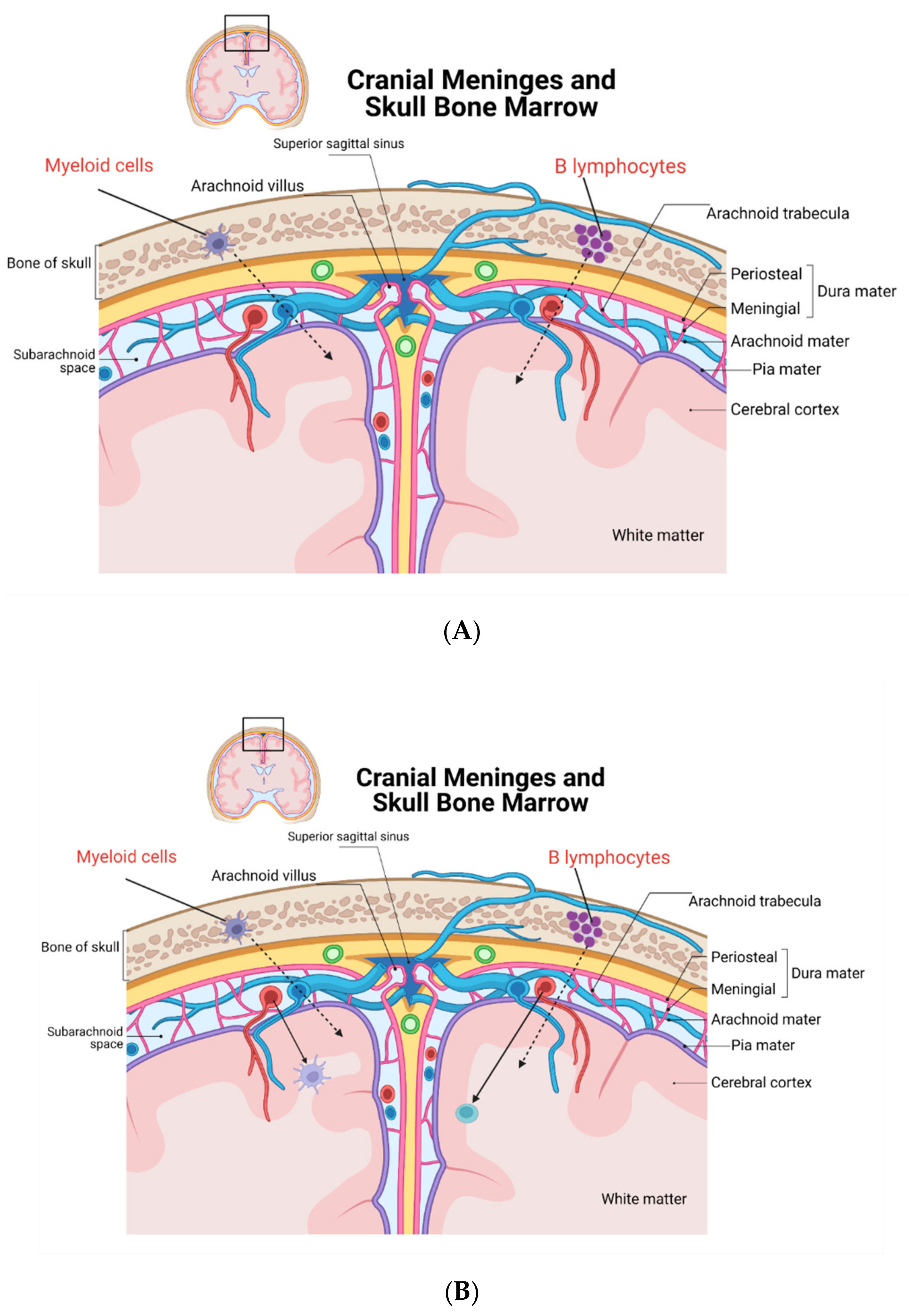
2. New Discoveries on CNS Leukocytes and Their Origin

On top of these new concepts, the latest studies add substantial significance to the peculiar characteristics of the immune-cell-patrolling mammalian CNS. A recent work by Cugurra et al. analysed the heterogeneous and somewhat conflicting function of myeloid cells using parabiosis and several models of CNS injury [13].
Cugurra et al. [13] confirmed previous observations by Herisson et al. describing the existence of exquisite channels connecting skull bone marrow and the dura mater [14]. Noteworthy, Cugurra et al. reported similar channels also connecting the vertebral bone barrow with the spinal dura mater [13]. These channels likely serve as anatomical routes for myeloid cell migration and partly contribute to parenchymal cell infiltrations observed by Cugurra et al. in several CNS disease models, including spinal cord injury, experimental autoimmune encephalomyelitis (EAE), and optic nerve damage [13].
Interestingly, the CNS-infiltrating Ly6C+ monocyte population did not derive from blood (as per weak green fluorescent protein [GFP] expression as reported in the experimental procedures [13]), whereas the major contribution in the inflamed CNS was clearly from blood with respect to neutrophils, CD4+ T cells, and Ly6C− monocytes. Ly6C+ cells in mice identified a subpopulation of bone marrow-derived monocytes endowed with immunosuppressive and immunoregulatory activities [15], Bronte Nat Comm 2016. An immunohistochemistry analysis of CNS tissues in experimental settings confirmed that this pattern was dominated by “resident” LyC6+ cell infiltration [13].
Moreover, results from single-cell RNA sequencing suggest a potential and important non-redundant role between blood-derived and CNS-derived myeloid cell lineages, with blood-originating cells demonstrating a more inflammatory and deleterious behaviour.
CNS border-derived myeloid cells, thus, demonstrate a less inflammatory and more “regulatory” phenotype: this population could act as an “attenuator” of the immune and inflammatory response with important implications for other disease states including CNS viral and bacterial infections and in primary and metastatic tumours in the CNS compartment.
In a companion study, Brioschi et al. studied another compartment of meningeal immunity: meningeal B cells [16]. As with the previous study, parabiosis experiments and single-cell RNA sequencing were utilised together with flow cytometry techniques.
The main discovery described in this study is the peculiar meningeal mouse B cell lineage found in the dura mater (the most external meningeal layer), which showed a phenotype and developmental pattern similar to the classical bone marrow-derived B murine cells. Most of these cells were of the B2 type, whereas innate B cells (B1 cells) were much less represented among lymphocyte populations. The main B cell population was extravascular with no infiltration in the CNS parenchyma, and it was mainly localised close to the sagittal and transverse sinuses and could exit CNS through lymphatics [16]. Furthermore, bone marrow reconstitution experiments showed that meningeal B cells were very similar to bone marrow B cells but not to peripheral blood B cells. These B cells from the peripheral circulation were minimally involved in colonising the mouse meninges under physiological conditions. Resident dura mater B cells migrated along vascular channels from the calvarial bone marrow to the mouse meninges similarly to the myeloid cells described previously [10,11]. Likely, the same vascular channels recently described [14] and reported also by Cugurra et al. form a direct physical interaction between calvaria and the meningeal space [12,13,14] allowing the migration of B cells as demonstrated by Brioschi et al. [16].
Lastly, Brioschi et al. also demonstrate that old mice accumulate age-associated B cells and plasma cells in the dura mater, mainly from the peripheral blood compartment. These cells identified as “age associated B cells” were antigen-experienced and accumulate in the dura with age (of note, these findings were basically reproduced and confirmed recently by Shafflick et al. and Wang et al. [17,18]). Overall, calvaria can indeed supply an early and quick source of B cells that develop in the dura mater, resulting in a negative selection of B cells with a high affinity for local self-epitopes. It would be of interest to further study the various B cell populations in pathological conditions (such as in mouse models of Multiple Sclerosis [MS]), considering the pivotal role of B cells in MS pathogenesis) [19,20].
Taken together, these studies suggest the almost revolutionary concept that cell migration through the blood–brain barrier is not required for meningeal-derived immune cell infiltration and immune surveys of brain tissues. Figure 1A,B show a synopsis of the new discoveries and concepts illustrated in this paragraph.

3. Conclusions

Overall, these new studies shed light on the “special” immunology of CNS. What is the importance of these new anatomical concepts in neuro-immunology?
The obvious and significant impact is on the comprehension of immune privilege and immunity in CNS in special regard to the B cells and myeloid compartment. It would be of course very interesting also to assess the phenotype and behaviour of the T cell compartment in the meningeal space and to assess if T cells are using the same migration patterns by traveling the inner skull vascular channels as well (preliminary data generated by Brioschi et al. in their experimental model found a very low number of T cells in the dura mater area in homeostatic conditions [16]).
Clinical implications are also compelling: as an example, we could mention gliomas (primary brain tumour difficult to treat) that are usually infiltrated by myeloid cells. These cells have an important role in promoting tumorigenesis and resistance to therapy [21]. A potential exploitation of the skull–meninges connection could influence myeloid cell chemotaxis as a new immunotherapy. Another unmet medical need that could be exploited by these new concepts is the field of traumatic brain injury due to the proven effects of myeloid cells on vascular repair after traumatic injury [22].
More experimental procedures in animal models and carefully designed human studies can provide us with more insight on the proposed new anatomy of neuroimmunology.
In conclusion, an exciting new era for better understanding the function of dura mater immune cells at the CNS level in physiological and pathology settings is now emerging, thus paving a way to modulate (boosting or inhibiting) immune responses at CNS levels.

Author Contributions

Conceptualisation, M.V. and C.S.; methodology, M.V.; software, M.V.; literature analysis: M.V., C.S. and D.B.; writing—original draft preparation, C.S., D.B. and M.V.; writing—review and editing, C.S. and M.V.; supervision: M.V. All authors have read and agreed to the published version of the manuscript.

Funding

This research received no external funding.

Institutional Review Board Statement

Not applicable.

Informed Consent Statement

Not applicable.

Data Availability Statement

Not applicable.

Acknowledgments

The authors thank Devahuti R. Chaliha for English proofreading.

Conflicts of Interest

The authors declare no conflict of interest.

References

  1. Abbott, N.J.; Patabendige, A.A.; Dolman, D.E.; Yusof, S.R.; Begley, D.J. Structure and function of the blood-brain barrier. Neurobiol. Dis. 2010, 37, 13–25. [Google Scholar] [CrossRef] [PubMed]
  2. Forrester, J.V.; McMenamin, P.G.; Dando, S.J. CNS infection and immune privilege. Nat. Rev. Neurosci. 2018, 19, 655–871. [Google Scholar] [CrossRef] [PubMed]
  3. Ginhoux, F.; Prinz, M. Origin of microglia: Current concepts and past controversies. Cold Spring Harb. Perspect. Biol. 2015, 7, a020537. [Google Scholar] [CrossRef] [PubMed] [Green Version]
  4. Filiano, A.J.; Gadani, S.P.; Kipnis, J. Interactions of innate and adaptive immunity in brain development and function. Brain Res. 2015, 1617, 18–27. [Google Scholar] [CrossRef] [PubMed] [Green Version]
  5. Tavares, G.A.; Louveau, A. Meningeal Lymphatics: An Immune Gateway for the Central Nervous System. Cells 2021, 10, 3385. [Google Scholar] [CrossRef] [PubMed]
  6. Yankova, G.; Bogomyakova, O.; Tulupov, A. The glymphatic system and meningeal lymphatics of the brain: New understanding of brain clearance. Rev. Neurosci. 2021, 32, 693–705. [Google Scholar] [CrossRef] [PubMed]
  7. Sandrone, S.; Moreno-Zambrano, D.; Kipnis, J.; van Gijn, J. A (delayed) history of the brain lymphatic system. Nat. Med. 2019, 25, 538–540. [Google Scholar] [CrossRef]
  8. Ahn, J.H.; Cho, H.; Kim, J.-H.; Kim, S.H.; Ham, J.-S.; Park, I.; Suh, S.H.; Hong, S.P.; Song, J.-H.; Hong, Y.-K.; et al. Meningeal lymphatic vessels at the skull base drain cerebrospinal fluid. Nature 2019, 572, 62–66. [Google Scholar] [CrossRef]
  9. Absinta, M.; Ha, S.-K.; Nair, G.; Sati, P.; Luciano, N.J.; Palisoc, M.; Louveau, A.; Zaghloul, K.A.; Pittaluga, S.; Kipnis, J.; et al. Human and nonhuman primate meninges harbor lymphatic vessels that can be visualized noninvasively by MRI. Elife 2017, 6, e29738. [Google Scholar] [CrossRef]
  10. Mastorakos, P.; McGavern, D. The anatomy and immunology of vasculature in the central nervous system. Sci. Immunol. 2019, 4, eaav0492. [Google Scholar] [CrossRef]
  11. Mundt, S.; Keller, A.; Greter, M. The dural sinus hub: More than just a brain drain. Cell 2021, 184, 858–860. [Google Scholar] [CrossRef] [PubMed]
  12. Rustenhoven, J.; Drieu, A.; Mamuladze, T.; Alves de Lima, K.; Dykstra, T.; Wall, M.; Papadopoulos, Z.; Kanamori, M.; Salvador, A.F.; Baker, W.; et al. Functional characterization of the dural sinuses as a neuroimmune interface. Cell 2021, 184, 1000–1016. [Google Scholar] [CrossRef] [PubMed]
  13. Cugurra, A.; Mamuladze, T.; Rustenhoven, J.; Dykstra, T.; Beroshvili, G.; Greenberg, Z.J.; Baker, W.; Papadopoulos, Z.; Drieu, A.; Blackburn, S.; et al. Skull and vertebral bone marrow are myeloid cell reservoirs for the meninges and CNS parenchyma. Science 2021, 373, eabf7844. [Google Scholar] [CrossRef] [PubMed]
  14. Herisson, F.; Frodermann, V.; Courties, G.; Rohde, D.; Sun, Y.; Vandoorne, K.; Wojtkiewicz, G.R.; Santos Masson, G.; Vinegoni, C.; Kim, J.; et al. Direct vascular channels connect skull bone marrow and the brain surface enabling myeloid cell migration. Nat. Neurosci. 2018, 21, 1209–1217. [Google Scholar] [CrossRef] [PubMed]
  15. Bronte, V.; Brandau, S.; Chen, S.H.; Colombo, M.P.; Frey, A.B.; Greten, T.F.; Mandruzzato, S.; Murray, P.J.; Ochoa, A.; Ostrand-Rosenberg, S.; et al. Recommendations for myeloid-derived suppressor cell nomenclature and characterization standards. Nat. Commun. 2016, 7, 12150. [Google Scholar] [CrossRef] [Green Version]
  16. Brioschi, S.; Wang, W.-L.; Peng, V.; Wang, M.; Shchukina, I.; Greenberg, Z.J.; Bando, J.K.; Jaeger, N.; Czepielewski, R.S.; Swain, A.; et al. Heterogeneity of meningeal B cells reveals a lymphopoietic niche at the CNS borders. Science 2021, 373, eabf9277. [Google Scholar] [CrossRef]
  17. Schafflick, D.; Wolbert, J.; Heming, M.; Thomas, C.; Hartlehnert, M.; Börsch, A.-L.; Ricci, A.; Martín-Salamanca, S.; Li, X.; Lu, I.-N.; et al. Single-cell profiling of CNS border compartment leukocytes reveals that B cells and their progenitors reside in non-diseased meninges. Nat. Neurosci. 2021, 24, 1225–1234. [Google Scholar] [CrossRef]
  18. Wang, Y.; Chen, D.; Xu, D.; Huang, C.; Xing, R.; He, D.; Xu, H. Early developing B cells undergo negative selection by central nervous system-specific antigens in the meninges. Immunity 2021, 54, 2784–2794. [Google Scholar] [CrossRef]
  19. Arneth, B.M. Impact of B cells to the pathophysiology of multiple sclerosis. J. Neuroinflamm. 2019, 16, 128. [Google Scholar] [CrossRef] [Green Version]
  20. Lanz, T.V.; Brewer, R.C.; Ho, P.P.; Moon, J.S.; Jude, K.M.; Fernandez, D.; Fernandes, R.A.; Gomez, A.M.; Nadj, G.S.; Bartley, C.M.; et al. Clonally Expanded B Cells in Multiple Sclerosis Bind EBV EBNA1 and GlialCAM. Nature 2022, 603, 321–327. [Google Scholar] [CrossRef]
  21. Gutmann, D.H.; Kettenmann, H. Microglia/Brain Macrophages as Central Drivers of Brain Tumor Pathobiology. Neuron 2019, 104, 442–449. [Google Scholar] [CrossRef] [PubMed]
  22. Russo, M.V.; Latour, L.L.; McGavern, D.B. Distinct myeloid cell subsets promote meningeal remodeling and vascular repair after mild traumatic brain injury. Nat. Immunol. 2018, 19, 442–452. [Google Scholar] [CrossRef] [PubMed]
Figure 1. New concepts in the topographical organization of the immune response in CNS. (A) Myeloid cells and B lymphocytes normally patrolling the CNS are derived from the skull bone marrow (red characters) and are able to migrate to meninges and to cerebral tissue by travelling through small skull bone vascular channels (not depicted for figure clarity, represented here by dashed arrow lines). (B) Ageing and CNS pathologies could increase tissue infiltration by myeloid cells and B cells from the general blood circulation (represented by the solid arrow lines), altering the local “milieu” and favouring local immune dysfunction, autoimmunity, and altered repair mechanisms (see main text and related references for more details).
Immuno 02 00016 g001
Publisher’s Note: MDPI stays neutral with regard to jurisdictional claims in published maps and institutional affiliations.

