You are currently viewing a new version of our website. To view the old version click .
Molbank
  • Short Note
  • Open Access

23 April 2007

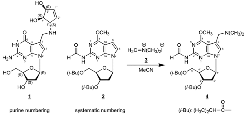
Electrophilic Substitution at C(7) of a Protected 7-Deaza-2’-deoxyguanosine – The 2’-Deoxyribonucleoside Parent Analogue of Queuosine #

and
Organische Chemie I – Bioorganische Chemie, Institut für Chemie, Fachbereich Biologie/Chemie, Universität Osnabrück, Barbarastr. 7, D-49069 Osnabrück, Germany
*
Author to whom correspondence should be addressed.

Abstract

In this manuscript we report on the regioselective electrophilic substitution at C(7) of 7-[2-deoxy-3,5-bis-O-(2-methylpropanoyl)-ß-D-erythro-pentofuranosyl]-2-(formylamino)-4-methoxy-7H-pyrrolo[2,3-d]pyrimidine (2) – a protected precursor of 7-deaza-2’-deoxyguanosine - using N,N-dimethyl-methyleniminium iodide (Eschenmoser’s salt) yielding the Mannich compound 4.
Most naturally occurring 7-deazapurine ribonucleosides [1,2] – both of the adenosine as well as of the guanosine type – carry substituents at the 7-position, and many of them which are found in tRNA represent Mannich bases such as the nucleoside “Q” {2-amino-5-(4,5-cis-dihydroxy-1-cyclopenten-3-yl-trans-aminomethyl-(7-ß-D-ribofuranosyl)-3,7-dihydro-4H-pyrrolo[2,3-d]pyrimidin-4-one, Queuosine, 1}. Many attempts have been made to direct a Mannich side chain regioselectively into position 7. In contrast to 7-deazaadenosine derivatives [3,4] this is not an easy task for 7-deazaguanosine precursors [5] and requires chemical detours [6]. For example, regioselective C(7) Mannich alkylation (morpholine, HCOH/HOAc) can be performed at 3’,5’-bis-O-toluoylated 6-methoxy-2-(methylthio)-7-deazapurine 2’-deoxy-ß-D-ribonucleoside, followed by a three-step aglycone conversion to 7-deazaguanine and deprotection of the glycone [7].
Molbank 2007 m522 i001
It has been reported for 7-deazaguanines that the position of electrophilic substitution strongly depends on the particular substituent pattern of the base as well as of the reaction conditions [8,9,10]. Of decisive importance is the observation that a free 2-amino group directs the electrophilic attack into the undesired 8-position (position 6 using systematic numbering) of the 7-deazaguanine moiety. This is the result of mesomeric stabilization of the σ-complex formed during electrophilic attack at the 8-position. On the other hand, 2-acylamino-7-deazaguanine derivatives form the desired 7-substituted compounds [8]
In this communication we disclose that reaction of the fully protected 7-deaza-2’-deoxy-7-deazaguanosine derivative 2 [8] with N,N-dimethyl-methyleniminium iodide (Eschenmoser’s salt) afforded the C(7) alkylated Mannich compound 4 in moderate yield. This reaction presents a new and alternative route to 7-deazapurine nucleosides with a Mannich side chain in position 7. The structure of 4 was unequivocally assigned by 1H- and 13C-NMR spectroscopy as well as by 1H-NMR NOE difference spectroscopy (see Experimental Procedure). 13C-NMR resonances were assigned applying DEPT-135 and [C,H]HETCOR spectra.
A further transformation of the tertiary amino group of 4 into other Mannich compounds can be accomplished after quarternization. Furthermore, the synthetic or enzymatic incorporation of Mannich bases derived from compounds such as compound 4 allows the introduction of reporter groups in a favourable position of a DNA molecule because the Mannich side chain protrudes into the major groove of a B-DNA double helix.

Experimental Procedure

7-[2-Deoxy-3,5-bis-O-(2-methylpropanoyl)-ß-D-erythro-pentofuranosyl]-5-(dimethylaminomethyl)-2-(formylamino)-4-methoxy-7H-pyrrolo[2,3-d]pyrimidine (4).
Compound 2 (305 mg, 0.7 mmol) was dissolved in acetonitrile (3 mL) and N,N-dimethyl-methyleniminium iodide (3, Eschenmoser’s Salz, 260 mg, 1.4 mmol) was added. After heating to 80°C for 24h, the reaction mixture was evaporated to a small volume, and compound 4 (67 mg, 20 %) was isolated as an amorphous solid by thick-layer chromatography (chloroform/methanol, 9:1, two developments, Rf, 0.4), elution from the silica gel with methanol and centrifugation. 1H-NMR ((D6)DMSO, 500.1 MHz): δ 10.73 (1H, d, HC=O, J = 9.9 Hz); 9.44 (1H, d, NH, J = 9.3 Hz); 7.31 (1H, s, H-C(6)); 6.49 (1H, dd, H-C(1’), J = 6.4, 8.0 Hz); 5.36 (1H, dd, H-C(3’), J = 2.8, 3.6 Hz); 4.28 - 4.17 (3H, m, H-C(4’), H2-C(5’)); 4.03 (3H, s, OCH3); 3.67 (2H, s, CH2); 2.86 (1H, m, iBu-CH); 2.63 – 2.57 (2H, m, H2-C(2’)); 2.47 (1H, m, iBu-CH); 2.28 (6H, s, N(CH3)2); 1.15 and 1.09 (12H, 4 iBu-CH3). 1H-NMR NOE Data ((D6)DMSO): proton irradiated: H-C(1’). NOE observed: H-C(6), 1.5 %; Hα-C(2’), 2.5 %; H-C(4’), 1.5 %. 13C-NMR ((D6)DMSO, 125.8 MHz): δ 175.6, 175.8 (2 C=O, iBu); 163.4 (C-4); 163.3 (C=O, formyl); 152.4 (C-7a); 152.0 (C-2); 121.6 (C-6); 111.4 (C-5); 101.5 (C-4a); 82.9 (C-1’); 81.1 (C-4’); 74.2 (C-3’); 63.6 (C-5’); 53.8 (CH2); 53.7 (OCH3); 44.1 and 33.1 (4 Me, iBu); 35.9 (C-2’); 18.6 (2 Me, N(CH3)2).

Supplementary materials

Supplementary File 1Supplementary File 2Supplementary File 3

References and Notes

  1. Suhadolnik, R.J. Nucleosides As Biological Probes; John Wiley & Sons: New York, 1979; pp. 158–169. [Google Scholar]
  2. Limbach, P.A.; Crain, P.F.; McCloskey, J.A. Nucleic Acids Res 1994, 22, 2183.
  3. Watanabe, S.I.; Ueda, T. Nucleosides Nucleotides 1983, 2, 113.
  4. West, R.A. J. Org. Chem. 1961, 26, 4959. [CrossRef]
  5. Seela, F.; Lüpke, U. Chem. Ber. 1977, 110, 1462. [CrossRef]
  6. Seela, F.; Richter, R. Chem. Ber. 1978, 111, 2925.
  7. Seela, F.; Chen, Y.; Zulauf, M. Synthesis 1997, 1067.
  8. Ramzaeva, N.; Seela, F. Helv. Chim. Acta 1995, 78, 1083. [CrossRef]
  9. Akimoto, H.; Imamiya, E.; Hitaka, T.; Nomura, H.; Nishimura, S. J. Chem. Soc. Perkin Trans 1 1988, 1637.
  10. Benghiat, E.; Crooks, P.A. J. Heterocycl. Chem. 1983, 20, 1023. [CrossRef]
  • #Purine numbering has been used within the General Part of the manuscript, systematic numbering within the Experimental Part.

Article Metrics

Citations

Article Access Statistics

Multiple requests from the same IP address are counted as one view.