In the original publication [1], there was a mistake in Figure 2. The micrograph B representing the sham group was a duplicate of the one representing the control group (micrograph A). The corrected Figure 2 appears below. The authors state that the scientific conclusions are unaffected. This correction was approved by the Academic Editor. The original publication has also been updated.
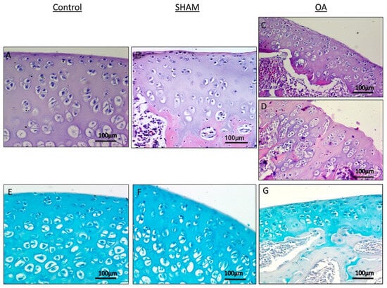
Figure 2.
Histological and histochemical evaluation. (A,B) Histology (H&E staining) demonstrated the absence of structural alterations in control groups (without anterior cruciate ligament transection (ACLT)). In the superficial zone, cells appear flat and small; in the middle and deep zone, cells are organized in columns. Magnification ×20; Scale bars: 100 µm; (C) Histology (H&E staining) demonstrated evidence of structural alterations in cartilage with moderate signs of OA (with ACLT). The structural alterations included a reduction of cartilage thickness in the superficial and the middle zones. The tidemark is no longer intact and the subchondral bone shows fibrillation. Magnification ×20; Scale bars: 100 µm; (D) Histology (H&E staining) demonstrated signs of structural alterations in severe Osteoarthritis (OA) (with ACLT). Severe OA cartilage shows deep surface clefts, disappearance of cells from the superficial zone, cloning, and a lack of cells in the intermediate and deep zone, which are not arranged in columns. The cartilage layers (superficial zone, middle and deep zone) are completely absent. Magnification ×20; Scale bars: 100 µm; (E,F) Histochemistry (toluidine blue staining) showed an absence of structural alterations and preserved GAG, in control groups (without ACLT), as indicated by the intense toluidine blue staining. Magnification ×20; Scale bars: 100 µm; (G) Histochemistry (toluidine blue staining) demonstrated signs of structural alterations in moderate and severe OA cartilage and loss of proteoglycans as evidenced by poor GAG preservation in the OA group (with ACLT), showing reduced toluidine blue staining. Magnification ×20; Scale bars: 100 µm.
Reference
- Giunta, S.; Castorina, A.; Marzagalli, R.; Szychlinska, M.A.; Pichler, K.; Mobasheri, A.; Musumeci, G. Ameliorative Effects of PACAP against Cartilage Degeneration. Morphological, Immunohistochemical and Biochemical Evidence from in Vivo and in Vitro Models of Rat Osteoarthritis. Int. J. Mol. Sci. 2015, 16, 5922–5944. [Google Scholar] [CrossRef] [PubMed]
Disclaimer/Publisher’s Note: The statements, opinions and data contained in all publications are solely those of the individual author(s) and contributor(s) and not of MDPI and/or the editor(s). MDPI and/or the editor(s) disclaim responsibility for any injury to people or property resulting from any ideas, methods, instructions or products referred to in the content. |
© 2024 by the authors. Licensee MDPI, Basel, Switzerland. This article is an open access article distributed under the terms and conditions of the Creative Commons Attribution (CC BY) license (https://creativecommons.org/licenses/by/4.0/).