

Role of Interleukin 6 in Acute Pancreatitis: A Possible Marker for Disease Prognosis

 , , ,
, , , 
Abstract
1. Introduction
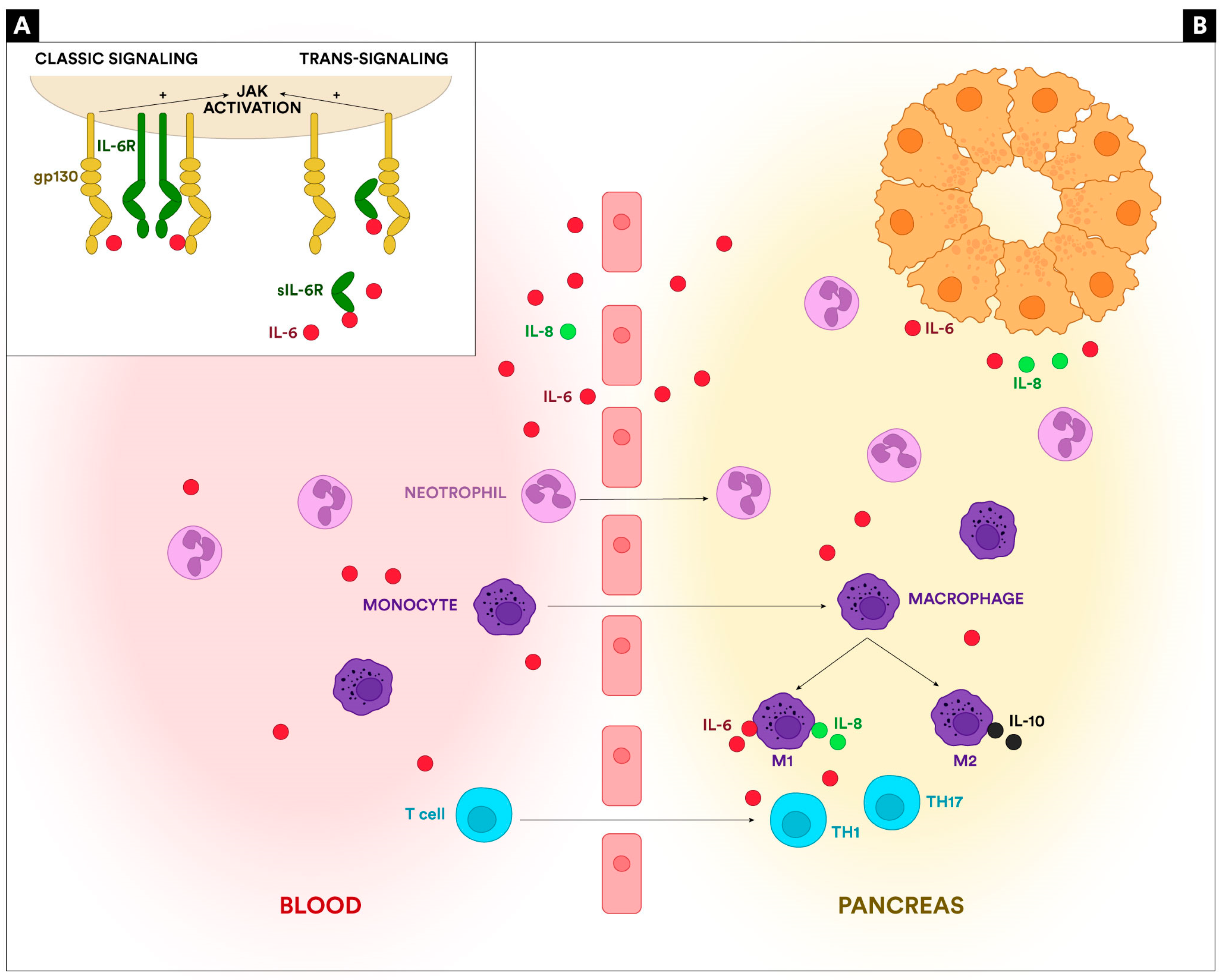
2. IL-6 and IL-6 Receptor
3. IL-6 in the Pathogenesis of Acute Pancreatitis
4. IL-6 as a Prognosis Marker for Acute Pancreatitis
5. Limitations and Future Directions
6. Conclusions
Author Contributions
Funding
Institutional Review Board Statement
Informed Consent Statement
Data Availability Statement
Acknowledgments
Conflicts of Interest
References
- Iannuzzi, J.P.; King, J.A.; Leong, J.H.; Quan, J.; Windsor, J.W.; Tanyingoh, D.; Coward, S.; Forbes, N.; Heitman, S.J.; Shaheen, A.-A.; et al. Global Incidence of Acute Pancreatitis Is Increasing Over Time: A Systematic Review and Meta-Analysis. Gastroenterology 2022, 162, 122–134. [Google Scholar] [CrossRef]
- Morinville, V.D.; Barmada, M.M.; Lowe, M.E. Increasing incidence of acute pancreatitis at an American pediatric tertiary care center: Is greater awareness among physicians responsible? Pancreas 2010, 39, 5–8. [Google Scholar] [CrossRef] [PubMed]
- Banks, P.A.; Bollen, T.L.; Dervenis, C.; Gooszen, H.G.; Johnson, C.D.; Sarr, M.G.; Tsiotos, G.G.; Vege, S.S.; Acute Pancreatitis Classification Working Group. Classification of acute pancreatitis—2012: Revision of the Atlanta classification and definitions by international consensus. Gut 2013, 62, 102–111. [Google Scholar] [CrossRef]
- Wang, G.J.; Gao, C.F.; Wei, D.; Wang, C.; Ding, S.Q. Acute pancreatitis: Etiology and common pathogenesis. World J. Gastroenterol. 2009, 15, 1427–1430. [Google Scholar] [CrossRef]
- Sigounas, D.E.; Tatsioni, A.; Christodoulou, D.K.; Tsianos, E.V.; Ioannidis, J.P. New prognostic markers for outcome of acute pancreatitis: Overview of reporting in 184 studies. Pancreas 2011, 40, 522–532. [Google Scholar] [CrossRef]
- Hunter, C.A.; Jones, S.A. IL-6 as a keystone cytokine in health and disease. Nat. Immunol. 2015, 16, 448–457, Correction in Nat. Immunol. 2017, 18, 1271. [Google Scholar] [CrossRef]
- Hirano, T. IL-6 in inflammation, autoimmunity and cancer. Int. Immunol. 2021, 33, 127–148. [Google Scholar] [CrossRef]
- Colceriu, M.-C.; Aldea, P.L.; Răchişan, A.-L.B.; Bulată, B.; Delean, D.; Grama, A.; Mititelu, A.; Decea, R.M.; Sevastre-Berghian, A.; Clichici, S.; et al. The Utility of Noninvasive Urinary Biomarkers for the Evaluation of Vesicoureteral Reflux in Children. Int. J. Mol. Sci. 2023, 24, 17579. [Google Scholar] [CrossRef] [PubMed]
- Heinrich, P.C.; Castell, J.V.; Andus, T. Interleukin-6 and the acute phase response. Biochem. J. 1990, 265, 621–636. [Google Scholar] [CrossRef] [PubMed]
- Miller, L.L.; Bly, C.G.; Watson, M.L.; Bale, W.F. The dominant role of the liver in plasma protein synthesis; a direct study of the isolated perfused rat liver with the aid of lysine-epsilon-C14. J. Exp. Med. 1951, 94, 431–453. [Google Scholar] [CrossRef]
- Poupart, P.; Vandenabeele, P.; Cayphas, S.; Van Snick, J.; Haegeman, G.; Kruys, V.; Fiers, W.; Content, J. B cell growth modulating and differentiating activity of recombinant human 26-kd protein (BSF-2, HuIFN-beta 2, HPGF). EMBO J. 1987, 6, 1219–1224. [Google Scholar] [CrossRef]
- Castell, J.V.; Gómez-Lechón, M.J.; David, M.; Hirano, T.; Kishimoto, T.; Heinrich, P.C. Recombinant human interleukin-6 (IL-6/BSF-2/HSF) regulates the synthesis of acute phase proteins in human hepatocytes. FEBS Lett. 1988, 232, 347–350. [Google Scholar] [CrossRef] [PubMed]
- Hirano, T.; Yasukawa, K.; Harada, H.; Taga, T.; Watanabe, Y.; Matsuda, T.; Kashiwamura, S.-I.; Nakajima, K.; Koyama, K.; Iwamatsu, A.; et al. Complementary DNA for a novel human interleukin (BSF-2) that induces B lymphocytes to produce immunoglobulin. Nature 1986, 324, 73–76. [Google Scholar] [CrossRef] [PubMed]
- Taga, T.; Kawanishi, Y.; Hardy, R.R.; Hirano, T.; Kishimoto, T. Receptors for B cell stimulatory factor 2. Quantitation, specificity, distribution, and regulation of their expression. J. Exp. Med. 1987, 166, 967–981. [Google Scholar] [CrossRef]
- Wolf, J.; Rose-John, S.; Garbers, C. Interleukin-6 and its receptors: A highly regulated and dynamic system. Cytokine 2014, 70, 11–20. [Google Scholar] [CrossRef]
- Metcalfe, R.D.; Putoczki, T.L.; Griffin, M.D.W. Structural Understanding of Interleukin 6 Family Cytokine Signaling and Targeted Therapies: Focus on Interleukin 11. Front. Immunol. 2020, 11, 1424. [Google Scholar] [CrossRef] [PubMed]
- Hu, Q.; Bian, Q.; Rong, D.; Wang, L.; Song, J.; Huang, H.-S.; Zeng, J.; Mei, J.; Wang, P.-Y. JAK/STAT pathway: Extracellular signals, diseases, immunity, and therapeutic regimens. Front. Bioeng. Biotechnol. 2023, 11, 1110765. [Google Scholar] [CrossRef] [PubMed]
- Hoque, R. Update on innate immunity and perspectives on metabolite regulation in acute pancreatitis. Curr. Opin. Gastroenterol. 2016, 32, 507–512. [Google Scholar] [CrossRef]
- Habtezion, A.; Algul, H. Immune modulation in acute and chronic pancreatitis. Pancreapedia Exocrine Pancreas Knowl. Base 2016. [Google Scholar] [CrossRef]
- Wang, J. Neutrophils in tissue injury and repair. Cell Tissue Res. 2018, 371, 531–539. [Google Scholar] [CrossRef]
- Peng, C.; Li, Z.; Yu, X. The Role of Pancreatic Infiltrating Innate Immune Cells in Acute Pancreatitis. Int. J. Med Sci. 2021, 18, 534–545. [Google Scholar] [CrossRef]
- Hu, F.; Lou, N.; Jiao, J.; Guo, F.; Xiang, H.; Shang, D. Macrophages in pancreatitis: Mechanisms and therapeutic potential. Biomed. Pharmacother. 2020, 131, 110693. [Google Scholar] [CrossRef]
- Tanaka, T.; Narazaki, M.; Kishimoto, T. IL-6 in inflammation, immunity, and disease. Cold Spring Harb. Perspect. Biol. 2014, 6, a016295. [Google Scholar] [CrossRef]
- Zhuang, Q.; Huang, L.; Zeng, Y.; Wu, X.; Qiao, G.; Liu, M.; Wang, L.; Zhou, Y.; Xiong, Y. Dynamic Monitoring of Immunoinflammatory Response Identifies Immunoswitching Characteristics of Severe Acute Pancreatitis in Rats. Front. Immunol. 2022, 13, 876168. [Google Scholar] [CrossRef]
- Lesina, M.; Wörmann, S.M.; Neuhöfer, P.; Song, L.; Algül, H. Interleukin-6 in inflammatory and malignant diseases of the pancreas. Semin. Immunol. 2014, 26, 80–87. [Google Scholar] [CrossRef]
- Pongratz, G.; Hochrinner, H.; Straub, R.H.; Lang, S.; Brünnler, T. B cell activating factor of the tumor necrosis factor family (BAFF) behaves as an acute phase reactant in acute pancreatitis. PLoS ONE. 2013, 8, e54297. [Google Scholar] [CrossRef]
- Suzuki, M.; Sai, J.K.; Shimizu, T. Acute pancreatitis in children and adolescents. World J. Gastrointest Pathophysiol. 2014, 5, 416–426. [Google Scholar] [CrossRef]
- Dambrauskas, Z.; Giese, N.; Gulbinas, A.; Giese, T.; Berberat, P.O.; Pundzius, J.; Barauskas, G.; Friess, H. Different profiles of cytokine expression during mild and severe acute pancreatitis. World J. Gastroenterol. 2010, 16, 1845–1853. [Google Scholar] [CrossRef]
- Ćeranić, D.B.; Zorman, M.; Skok, P. Interleukins and inflammatory markers are useful in predicting the severity of acute pancreatitis. Bosn. J. Basic Med Sci. 2020, 20, 99–105. [Google Scholar] [CrossRef]
- Sternby, H.; Hartman, H.; Thorlacius, H.; Regnér, S. The Initial Course of IL-1β, IL-6, IL-8, IL-10, IL-12, IFN-γ and TNF-α with Regard to Severity Grade in Acute Pancreatitis. Biomolecules 2021, 11, 591. [Google Scholar] [CrossRef]
- Kolber, W.; Dumnicka, P.; Maraj, M.; Kuśnierz-Cabala, B.; Ceranowicz, P.; Pędziwiatr, M.; Maziarz, B.; Mazur-Laskowska, M.; Kuźniewski, M.; Sporek, M.; et al. Does the Automatic Measurement of Interleukin 6 Allow for Prediction of Complications during the First 48 h of Acute Pancreatitis? Int. J. Mol. Sci. 2018, 19, 1820. [Google Scholar] [CrossRef] [PubMed]
- Jain, S.; Midha, S.; Mahapatra, S.J.; Gupta, S.; Sharma, M.K.; Nayak, B.; Jacob, T.G.; Shalimar; Garg, P.K. Interleukin-6 significantly improves predictive value of systemic inflammatory re-sponse syndrome for predicting severe acute pancreatitis. Pancreatology 2018, 18, 500–506. [Google Scholar] [CrossRef] [PubMed]
- Tian, F.; Li, H.; Wang, L.; Li, B.; Aibibula, M.; Zhao, H.; Feng, N.; Lv, J.; Zhang, G.; Ma, X. The diagnostic value of serum C-reactive protein, procalcitonin, interleukin-6 and lactate dehydrogenase in patients with severe acute pancreatitis. Clin. Chim. Acta 2020, 510, 665–670. [Google Scholar] [CrossRef] [PubMed]
- Inagaki, T.; Hoshino, M.; Hayakawa, T.; Ohara, H.; Yamada, T.; Yamada, H.; Iida, M.; Nakazawa, T.; Ogasawara, T.; Uchida, A.; et al. Interleukin-6 is a useful marker for early prediction of the severity of acute pancreatitis. Pancreas 1997, 14, 1–8. [Google Scholar] [CrossRef] [PubMed]
- Pooran, N.; Indaram, A.; Singh, P.; Bank, S. Cytokines (IL-6, IL-8, TNF): Early and reliable predictors of severe acute pancreatitis. J. Clin. Gastroenterol. 2003, 37, 263–266. [Google Scholar] [CrossRef]
- Berney, T.; Gasche, Y.; Robert, J.; Jenny, A.; Mensi, N.; Grau, G.; Vermeulen, B.; Morel, P. Serum profiles of interleukin-6, interleukin-8, and interleukin-10 in patients with severe and mild acute pancreatitis. Pancreas 1999, 18, 371–377. [Google Scholar] [CrossRef] [PubMed]
- Kostic, I.; Spasic, M.; Stojanovic, B.; Jurisevic, M.; Radovanovic, D.; Canović, D.; Stefanovic, S.; Jankovic, S. Early cytokine profile changes in interstitial and necrotic forms of acute pancreatitis. Exp. Clin. Res. 2015, 16, 33–37. [Google Scholar] [CrossRef]
- Viedma, J.A.; Pérez-Mateo, M.; Domínguez, J.E.; Carballo, F. Role of interleukin-6 in acute pancreatitis. Comparison with C-reactive protein and phospholipase A. Gut 1992, 33, 1264–1267. [Google Scholar] [CrossRef]
- Park, J.; Chang, J.H.; Park, S.H.; Lee, H.J.; Lim, Y.S.; Kim, T.H.; Kim, C.W.; Han, S.W. Interleukin-6 is associated with obesity, central fat distribution, and disease severity in patients with acute pancreatitis. Pancreatology 2015, 15, 59–63. [Google Scholar] [CrossRef]
- Gunjaca, I.; Zunic, J.; Gunjaca, M.; Kovac, Z. Circulating cytokine levels in acute pancreatitis-model of SIRS/CARS can help in the clinical assessment of disease severity. Inflammation 2012, 35, 758–763. [Google Scholar] [CrossRef]
- Riché, F.C.; Cholley, B.P.; Laisné, M.-J.C.; Vicaut, E.; Panis, Y.H.; Lajeunie, E.J.; Boudiaf, M.; Valleur, P.D. Inflammatory cytokines, C reactive protein, and procalcitonin as early predictors of necrosis infection in acute necrotizing pancreatitis. Surgery 2003, 133, 257–262. [Google Scholar] [CrossRef] [PubMed]
- Kumar, R.B.; Karim, T.; Jain, A.; Arora, S.; Katiyar, V.K.; Patel, G. Role of Serum Interleukin-6 and C-reactive Protein in Early Prediction of Severe Acute Pancreatitis. West Afr. Coll. Surg. 2022, 12, 20–26. [Google Scholar] [CrossRef]
- Leser, H.-G.; Gross, V.; Scheibenbogen, C.; Heinisch, A.; Salm, R.; Lausen, M.; Rückauer, K.; Andreesen, R.; Farthmann, E.; Schölmerich, J. Elevation of serum interleukin-6 concentration precedes acute-phase response and reflects severity in acute pancreatitis. Gastroenterology 1991, 101, 782–785. [Google Scholar] [CrossRef] [PubMed]
- Khanna, A.K.; Meher, S.; Prakash, S.; Tiwary, S.K.; Singh, U.; Srivastava, A.; Dixit, V.K. Comparison of Ranson, Glasgow, MOSS, SIRS, BISAP, APACHE-II, CTSI Scores, IL-6, CRP, and Procalcitonin in Predicting Severity, Organ Failure, Pancreatic Necrosis, and Mortality in Acute Pancreatitis. HPB Surg. 2013, 2013, 367581. [Google Scholar] [CrossRef] [PubMed]
- Malmstrøm, M.L.; Hansen, M.B.M.; Andersen, A.M.; Ersbøll, A.K.M.; Nielsen, O.H.M.; Jørgensen, L.N.M.; Novovic, S. Cytokines and organ failure in acute pancreatitis: Inflammatory response in acute pancreatitis. Pancreas 2012, 41, 271–277. [Google Scholar] [CrossRef] [PubMed]
- Sathyanarayan, G.; Garg, P.K.; Prasad, H.; Tandon, R.K. Elevated level of interleukin-6 predicts organ failure and severe disease in patients with acute pancreatitis. J. Gastroenterol. Hepatol. 2007, 22, 550–554. [Google Scholar] [CrossRef] [PubMed]
- Yao, J.; Zhang, S.; Zhou, F.; Zhuang, M.; Fei, S. The relationship between inflammatory cytokines and in-hospital complications of acute pancreatitis. Immun. Inflamm. Dis. 2024, 12, e1203. [Google Scholar] [CrossRef] [PubMed]
- De Beaux, A.C.; Goldie, A.S.; Ross, J.A.; Carter, D.C.; Fearon, K.C. Serum concentrations of inflammatory mediators related to organ failure in patients with acute pancreatitis. Br. J. Surg. 1996, 83, 349–353. [Google Scholar] [CrossRef]
- Gregoric, P.; Sijacki, A.; Stankovic, S.; Radenkovic, D.; Ivancevic, N.; Karamarkovic, A.; Popovic, N.; Karadzic, B.; Stijak, L.; Stefanovic, B.; et al. SIRS score on admission and initial concentration of IL-6 as severe acute pancreatitis outcome predictors. Hepatogastroenterology 2010, 57, 349–353, Correction in Hepatogastroenterology 2011, 58, 263. [Google Scholar] [CrossRef]
- Karpavicius, A.; Dambrauskas, Z.; Gradauskas, A.; Samuilis, A.; Zviniene, K.; Kupcinskas, J.; Brimas, G.; Meckovski, A.; Sileikis, A.; Strupas, K. The clinical value of adipokines in predicting the severity and outcome of acute pancreatitis. BMC Gastroenterol. 2016, 16, 99. [Google Scholar] [CrossRef]
- Pezzilli, R.; Billi, P.; Miniero, R.; Fiocchi, M.; Cappelletti, O.; Morselli-Labate, A.M.; Barakat, B.; Sprovieri, G.; Miglioli, M. Serum interleukin-6, interleukin-8, and beta 2-microglobulin in early assessment of severity of acute pancreatitis. Comparison with serum C-reactive protein. Dig. Dis. Sci. 1995, 40, 2341–2348. [Google Scholar] [CrossRef] [PubMed]
- Curley, P.J.; McMahon, M.J.; Lancaster, F.; E Banks, R.; Barclay, G.R.; Shefta, J.; Boylston, A.W.; Whicher, J.T. Reduction in circulating levels of CD4-positive lymphocytes in acute pancreatitis: Relationship to endotoxin, interleukin 6 and disease severity. Br. J. Surg. 1993, 80, 1312–1315. [Google Scholar] [CrossRef] [PubMed]
- Pezzilli, R.; Morselli-Labate, A.M.; Miniero, R.; Barakat, B.; Fiocchi, M.; Cappelletti, O. Simultaneous serum assays of lipase and interleukin-6 for early diagnosis and prognosis of acute pancreatitis. Clin. Chem. 1999, 45, 1762–1767. [Google Scholar] [CrossRef]
- Jiang, C.F.; Shiau, Y.C.; Ng, K.W.; Tan, S.W. Serum interleukin-6, tumor necrosis factor alpha and C-reactive protein in early prediction of severity of acute pancreatitis. J. Chin. Med. Assoc. 2004, 67, 442–446. [Google Scholar]
- Rao, S.A.; Kunte, A.R. Interleukin-6: An Early Predictive Marker for Severity of Acute Pancreatitis. Indian J. Crit. Care Med. 2017, 21, 424–428. [Google Scholar] [CrossRef] [PubMed]
- Mayer, J.; Rau, B.; Gansauge, F.; Beger, H.G. Inflammatory mediators in human acute pancreatitis: Clinical and pathophysiological implications. Gut 2000, 47, 546–552. [Google Scholar] [CrossRef] [PubMed]
- Chen, C.-C.; Wang, S.-S.; Lee, F.-Y.; Chang, F.-Y.; Lee, S.-D. Proinflammatory cytokines in early assessment of the prognosis of acute pancreatitis. Am. J. Gastroenterol. 1999, 94, 213–218. [Google Scholar] [CrossRef] [PubMed]
- Fisic, E.; Poropat, G.; Bilic-Zulle, L.; Licul, V.; Milic, S.; Stimac, D. The Role of IL-6, 8, and 10, sTNFr, CRP, and Pancreatic Elastase in the Prediction of Systemic Complications in Patients with Acute Pancreatitis. Gastroenterol. Res. Pract. 2013, 2013, 282645. [Google Scholar] [CrossRef] [PubMed]
- Bidarkundi, G.K.; Wig, J.D.; Bhatnagar, A.; Majumdar, S. Clinical relevance of intracellular cytokines IL-6 and IL-12 in acute pancreatitis, and correlation with APACHE III score. Br. J. Biomed. Sci. 2002, 59, 85–89. [Google Scholar] [CrossRef]
- Cho, I.R.; Do, M.Y.; Han, S.Y.; Jang, S.I.; Cho, J.H. Comparison of Interleukin-6, C-Reactive Protein, Procalcitonin, and the Computed Tomography Severity Index for Early Prediction of Severity of Acute Pancreatitis. Gut Liver 2023, 17, 629–637. [Google Scholar] [CrossRef] [PubMed]
- Chen, Y.J.; Lin, T.L.; Cai, Z.; Yan, C.H.; Gou, S.R.; Zhuang, Y.D. Assessment of acute pancreatitis severity via determination of serum levels of hsa-miR-126-5p and IL-6. Exp. Ther. Med. 2020, 21, 26. [Google Scholar] [CrossRef] [PubMed]
- Nieminen, A.; Maksimow, M.; Mentula, P.; Kyhälä, L.; Kylänpää, L.; Puolakkainen, P.; Kemppainen, E.; Repo, H.; Salmi, M. Circulating cytokines in predicting development of severe acute pancreatitis. Crit. Care 2014, 18, R104. [Google Scholar] [CrossRef] [PubMed]
- Sternby, H.; Hartman, H.; Johansen, D.; Thorlacius, H.; Regnér, S. IL-6 and CRP are superior in early differentiation between mild and non-mild acute pancreatitis. Pancreatology 2017, 17, 550–554. [Google Scholar] [CrossRef] [PubMed]
- Li, J.; Chen, Z.; Li, L.; Lai, T.; Peng, H.; Gui, L.; He, W. Interleukin-6 is better than C-reactive protein for the prediction of infected pancreatic necrosis and mortality in patients with acute pancreatitis. Front. Cell. Infect. Microbiol. 2022, 12, 933221. [Google Scholar] [CrossRef] [PubMed]
- Farrell, P.R.; Jones, E.K.; Hornung, L.; Thompson, T.; Patel, J.; Lin, T.K.; Nathan, J.D.; Vitale, D.S.; Habtezion, A.; Abu-El-Haija, M. Cytokine Profile Elevations on Admission Can Determine Risks of Severe Acute Pancreatitis in Children. J. Pediatr. 2021, 238, 33–41.e4. [Google Scholar] [CrossRef]

| Authors | Year | Description | Results |
|---|---|---|---|
| Inagaki et al. [34] | 1997 | 12 patients: 7 with mild pancreatitis, 5 with severe pancreatitis IL-6 measured 5, 24, 72, and 120 h after admission | There is a significant difference in IL-6 levels between mild and severe pancreatitis at all moments measured. The peak of IL-6 in the severe group was observed at 5 h after admission, 58.9 ± 18.8 pg/mL. |
| Pooran et al. [35] | 2003 | 36 patients with AP and age-matched controls IL-6 measured 24 h of admission | There is a significant difference in IL-6 levels between controls and mild pancreatitis, controls and severe pancreatitis, and mild and severe pancreatitis. The average serum concentrations of IL-6 were 13.70 ± 1.90 pg/mL, 40.37 ± 4.35 pg/mL, and 82.76 ± 6.98 pg/mL in the control, mild pancreatitis, and severe pancreatitis groups, respectively. The difference in the means between control and mild pancreatitis groups was 26.67 pg/mL, between control and severe pancreatitis was 69.09 pg/mL, and between mild pancreatitis and severe pancreatitis was 42.39 pg/mL. |
| Berney et al. [36] | 1999 | 43 patients with AP: 24 mild AP, 19 severe AP IL-6, IL-8, IL-10, CRP, and PMNE measured at admission and after 1, 2, and 5 days | High levels of IL-6 were detected in both groups upon admission (day 0). Higher levels of IL-6 were detected in patients with severe AP during the first two days (days 1 and 2) compared with those with mild AP. By day 5, IL-6 returned to low concentrations in both groups. The mean values for the mild group were 861 ± 20 pg/mL, 82 ± 28 pg/mL, and 54 ± 13 pg/mL, corresponding to days 0, 1, and 2 of admission. The mean values for the severe group were 33.600 ± 17.600 pg/mL, 486 ± 157 pg/mL, and 579 ± 184 pg/mL on days 0, 1, and 2. A 24 to 48 h delay was observed between peaks of IL-6 and CRP. |
| Kostic et al. [37] | 2015 | 52 patients with AP: 34 mild and 18 severe, necrotizing AP IL-6, IL-8, IL-10, and TNF-α measured on days 1 and 3 after admission | Significantly higher IL-6 concentrations were detected in patients with necrotizing AP compared to those with interstitial AP on both days measured. |
| Viedma et al. [38] | 1992 | 24 patients with AP: 9 mild AP, 15 severe AP IL-6, CRP, and phospholipase A measured at admission and every day until day 7 | Higher IL-6 levels were detected in patients with severe compared with mild AP in the first two days and also in those with necrotizing AP compared with non-necrotizing AP. |
| Park et al. [39] | 2015 | 59 patients with AP: 36 mild AP and 23 moderate AP IL-6, IL-Iβ, IL-1ra, TNF-α, sTNFR-I, and sTNFR-II were measured at admission and after 24 h | Significantly higher levels of IL-6 were detected in moderate compared to mild AP in both days of measurement. In patients with mild AP and with moderate AP, the mean levels of IL-6 on day 2 were 91.1 ± 230 pg/mL and 163 ± 170 pg/mL, respectively. Higher IL-6 levels were also detected in obese patients compared with non-obese. The mean levels of IL-6 on day 2 in overweight and non-overweight patients were 126 ± 124 pg/mL and 115 ± 244 pg/mL, respectively. |
| Gunjaca et al. [40] | 2012 | 20 patients with AP: 13 mild and 7 severe AP IL-6 measured on days 1, 2, 3, 6, and 9 | There is a significant difference in IL-6 levels between mild and severe AP (23.4 pg/mL vs. 539.2 pg/mL, p < 0.0001) |
| Riché et al. [41] | 2003 | 48 patients with necrotizing AP: 15 infected, 33 non-infected IL-6, CRP, PCT, and TNF-α were measured during the first 3 days | Higher IL-6 levels were seen in patients with infected necrosis compared with those without infection IL-6 < 400 pg/mL and PCT < 2 ng/mL combined identified best the patients who were not at risk for necrosis infection |
| Kumar et al. [42] | 2022 | 62 patients with AP: 40 mild, 22 severe forms IL-6 and CRP were measured in the first 24 h of admission and also 48 h after admission | There is a significant difference in IL-6 and CRP mean levels between mild and severe AP. IL-6 at 48 h had the maximum AUC of 0.98. The cut-off values of IL-6 in days 1 and 2 for predicting SAP were 137 pg/mL and 77.3 pg/mL, respectively. IL-6 on day 1 and day 2 and CRP on day 2 were 100% sensitive, but IL-6 on day 1 and day 2 had a maximum specificity of 88.37% among them when compared with a specificity of 81.4% of CRP on day 2. |
| Leser et al. [43] | 1991 | 50 patients with AP: 25 mild AP, 15 severe AP, 10 lethal AP IL-6 and CRP were measured daily for the first 7 days of AP | High IL-6 levels (with a cut-off value of 15 U/mL coresponding to 150 pg/mL) in the first two days of AP predicted (PPV 91%, NPV 82%) a severe or lethal course of the disease more accurately than elevated CRP. IL-6 is a better early parameter for the assessment of the severity of acute pancreatitis than CRP. |
| Khanna et al. [44] | 2013 | 72 patients with AP IL-6, CRP, and procalcitonin measured on the first day of admission | IL-6 (values over 50 pg/mL) has the highest sensitivity for the prediction of SAP (93.1%), organ failure (95.7%), pancreatic necrosis (94.1%), and mortality (100%). The serum concentration of IL-6 on the first day and/or together with serum CRP concentration on the second day of admission is helpful in earlier prediction and assessment of the severity of acute pancreatitis, taking into consideration the disadvantages of multifactorial scoring systems. |
| Malmstrøm et al. [45] | 2012 | 60 patients with severe AP IL-6, IL-8, IL-18, and TNF-α measured at admission and on days 1, 2, and 14 | IL-6 values determined on days 1 and 2 can significantly predict organ failure (renal, respiratory, circulatory, and MOF) in the early stages. The median values of IL-6 in renal, respiratory, and circulatory failure were 840 pg/mL, 185 pg/mL, and 999 pg/mL, respectively, measured on the first day of admission. |
| Sathyanarayan et al. [46] | 2007 | 30 patients with AP: 17 mild AP, 13 severe AP IL-6 measured on days 1, 3, 7, and 14 of admission | IL-6 was significantly higher on day 3 in severe AP than in mild AP (146.29 ± 57.53 pg/mL vs. 91.42 ± 71.65 pg/mL) and was significantly higher in patients who developed organ failure (161.59 ± 53.46 pg/mL vs. 88.16 ± 65.50 pg/mL). At a cut-off value of 122 pg/mL on day 3, IL-6 predicted organ failure and severe pancreatitis with a sensitivity and specificity of 81.8% and 77.7%, respectively. |
| Yao et al. [47] | 2024 | 307 patients with AP: 260 with mild and moderate forms and 47 with SAP IL-6, IL-1β, IL-2, IL-4, IL-5, IL-10, IL-8, IL12p70, IFN-γ, IFN-α, and TNF-α were measured at admission | IL-6 had the best results in discriminating between mild and severe forms compared to the other cytokines studied. At a cut-off of 24.67 pg/mL, IL-6 can predict severe forms with a sensitivity of 87.23% and a specificity of 66.92%. Moreover, IL-6 predicted the occurrence of peripancreatic colections and necrosis. |
| De Beaux et al. [48] | 1996 | 58 patients with AP: 30 mild AP, 28 severe AP IL-6, TNF-α and CRP were measured on the first and second days of admission. | Higher IL-6 levels were associated with severe AP compared to mild forms (p < 0.001) and organ failure development (p < 0.03). |
| Gregoric et al. [49] | 2010 | 234 patients with SAP IL-6 was measured upon admission, and SIRS was calculated | SIRS at admission combined with IL-6 serum levels could be an accurate marker to predict severe AP (performing similarly to APACHE II and Ranson scores). IL-6 levels were significantly higher in the patients with a SIRS score of 3 or higher (72 +/− 67 pg/mL vs. 18 +/− 15 pg/mL). |
| Karpavicius et al. [50] | 2016 | 102 patients with AP: 27 mild, 55 moderate, and 20 severe forms IL-6, adipokines, and CRP were measured at admission and on day 3 of hospitalization | IL-6 was the best predictor for severe forms. IL-6, with a cut-off value of 157 pg/mL, can significantly predict necrosis. |
| Pezzilli et al. [51] | 1995 | 38 patients with AP: 13 mild AP, 15 severe AP IL-6, IL-8, CRP, and β2-microglobulin were measured on admission and daily for 5 days | IL-6 (cut-off 2.7 pg/mL) was the best marker for evaluating the severity of AP (sensitivity 100%, specificity 86%, diagnostic accuracy 91%). |
| Curley et al. [52] | 1993 | 29 patients with AP: 16 mild AP, 13 severe AP IL-1, IL-6, IL-8, and IL-10 were measured at admission and after 48 h | Significant increases of IL-6 (69.5 pg/mL versus <10 pg/mL, p = 0.01) in severe compared with mild AP. |
| Pezzilli et al. [53] | 1999 | 40 patients with AP: 25 mild AP, 15 severe AP Lipase, IL-6, and CRP were measured at admission | IL-6 (cut-off 3.7 μg/L) had a sensitivity of 100%, specificity of 83%, and AUC of 0.91 in discriminating severe from mild AP. C-reactive protein (cut-off values ranging from 6 to 7 mg/L) showed a lower prognostic efficiency than interleukin-6: sensitivity of 87% (13 of 15) and specificity of 46% (11 of 24). |
| Jiang et al. [54] | 2004 | 33 patients with AP: 19 mild AP, 14 severe AP IL-6, CRP, and TNF-α were measured on days 1, 2, 3, and 7 | Significantly higher IL-6 levels were detected in severe compared with mild AP (sensitivity 100%, specificity 89.7%, and accuracy 91% on day 1). |
| Rao et al. [55] | 2017 | 40 patients with severe AP IL-6 measured in the first 24 h of admission | IL-6 (≥28.90 pg/mL) was the most accurate marker (compared with IL-8, IL-10, and CRP) for predicting the progression to severe pancreatitis. |
| Mayer et al. [56] | 2000 | 51 patients with AP: 16 mild AP, 35 severe AP Il-6, IL-1β, IL-8, IL-10, IL-1RA, and sIL-2R were measured daily for 7 days after admission | IL-6 was significantly elevated in patients with distant organ failure and was the best prognostic parameter for pulmonary failure with median values between 989–2071 ng/mL (compared with the other markers investigated). IL-6 reached its highest serum concentration on day 3 after the onset of symptoms |
| Chen et al. [57] | 1999 | 50 patients with AP: 23 mild AP and 18 severe AP TNF-α, IL-1β, IL-6, IL-8, and CRP were measured on days 1, 2, 3, 4, and 7 | Interleukin-6 in day 1 of admission was the most useful prognostic biomarker, compared with the other markers investigated. The sensitivity, specificity and accuracy of predicting a severe attack were 89%, 87%, and 88%, respectively, using IL-6 >400 pg/mL on day 1. |
| Fisic et al. [58] | 2013 | 150 patients with AP: 122 mild AP, 28 severe AP IL-6, IL-8, IL-10, and sTNFr were measured on the first day of admission | IL-6 is the best predictor for severe forms and systemic complications in patients with AP compared with other markers (IL-8, IL-10, and sTNFr). IL-6 greater than 37.9 ng/mL can be considered high-risk in terms of developing systemic complications with a sensitivity of 82% and a specificity of 65%. |
| Bidarkundi et al. [59] | 2002 | 30 patients with AP: 15 mild AP, 15 severe AP, and 15 controls The percentages of PBMCs (peripheral blood mononuclear cells) that contained IL-6 and IL-12 were measured at admission and compared with APACHE III | Higher IL-6 levels were detected in patients with severe compared with mild AP. Values of 25% for IL-6-positive PBMCs and 30 for APACHE III score were the best cut-off values to differentiate mild from severe AP. With IL-6 values >25%, both sensitivity and specificity were 100%, whereas APACHE scores of >30 showed a sensitivity of 80% and specificity of 100%. |
| Cho et al. [60] | 2023 | 103 patients with AP: 42 mild, 53 moderate, and 8 severe AP IL-6, CRP, and procalcitonin were measured at admission, 24 and 48 h after; CTSI was calculated at admission | IL-6 with a cut-off <50 pg/mL can predict mild forms with an accuracy of 83.3% at admission. The accuracy decreases slightly at 24 and 48 h of admission. |
| Chen et al. [61] | 2020 | 90 patients with AP: 30 mild AP, 30 severe AP, 30 recurrent AP IL-6, TNF-α, IL-1, IL-8, IL-10, and miRNAs were measured at admission | Combined determination of serum IL-6 and hsa-miR-126-5p may be useful for the early prediction of AP classification (AUC, 0.991; sensitivity, 93.3%; specificity, 96.7%; cut-off value, 0.802; p < 0.001) |
| Nieminen et al. [62] | 2014 | 163 patients with AP: 103 mild AP, 35 moderate AP, 25 severe AP 48 cytokines, including IL-6 and growth factors, were measured at admission | IL-6 (with a cut-off of 501.6 pg/mL), HGF, and their combination were prognostic biomarkers for severe AP (sensitivities of 56%, 60%, and 72%, respectively, and specificities of 90.6%, 92.8%, and 89.9%, respectively). |
| Sternby et al. [63] | 2017 | 175 patients with AP: 124 mild AP, 41 moderate AP, 10 severe AP 20 biomarkers were measured, including IL-6, at 13–36 h after admission | IL-6 (cut-off 23.6 pg/mL) and CRP (cut-off 23.6 mg/L) combined demonstrate a clinically relevant capacity (AUC 0.803) to differentiate non-mild from mild AP in early stages. |
| Li et al. [64] | 2022 | 67 patients with AP: 10 mild AP, 22 moderate AP, 35 severe AP IL-6 and CRP were measured on the first day after admission | The AUC of IL-6 (cut-off = 121.1 pg/mL) measured within 48 h of onset for the prediction of SAP was 0.69 (95% CI, 0.56–0.82), with a sensitivity of 67.65% and a specificity of 67.74%. IL-6 was more accurate than CRP for predicting mortality (AUC 0.70 vs. 0.75) and infected pancreatic necrosis (AUC 0.65 vs. 0.81) in patients with AP. |
| Farrell et al. [65] | 2021 | 66 children: 36 mild AP, 10 severe AP, and 30 controls IL-6, MCP-1, and CRP were measured at admission | Levels of interleukin-6 (IL-6) and MCP-1 were statistically different between the mild acute pancreatitis and severe acute pancreatitis groups (p = 0.02 for both). IL-6 resulted in a significant ROC curve (AUROC 0.83 [95% CI, 0.67–1.00], p = 0.03) for predicting severe forms. |
Disclaimer/Publisher’s Note: The statements, opinions and data contained in all publications are solely those of the individual author(s) and contributor(s) and not of MDPI and/or the editor(s). MDPI and/or the editor(s) disclaim responsibility for any injury to people or property resulting from any ideas, methods, instructions or products referred to in the content. |
© 2024 by the authors. Licensee MDPI, Basel, Switzerland. This article is an open access article distributed under the terms and conditions of the Creative Commons Attribution (CC BY) license (https://creativecommons.org/licenses/by/4.0/).
Share and Cite
Mititelu, A.; Grama, A.; Colceriu, M.-C.; Benţa, G.; Popoviciu, M.-S.; Pop, T.L. Role of Interleukin 6 in Acute Pancreatitis: A Possible Marker for Disease Prognosis. Int. J. Mol. Sci. 2024, 25, 8283. https://doi.org/10.3390/ijms25158283
Mititelu A, Grama A, Colceriu M-C, Benţa G, Popoviciu M-S, Pop TL. Role of Interleukin 6 in Acute Pancreatitis: A Possible Marker for Disease Prognosis. International Journal of Molecular Sciences. 2024; 25(15):8283. https://doi.org/10.3390/ijms25158283
Chicago/Turabian StyleMititelu, Alexandra, Alina Grama, Marius-Cosmin Colceriu, Gabriel Benţa, Mihaela-Simona Popoviciu, and Tudor Lucian Pop. 2024. "Role of Interleukin 6 in Acute Pancreatitis: A Possible Marker for Disease Prognosis" International Journal of Molecular Sciences 25, no. 15: 8283. https://doi.org/10.3390/ijms25158283
APA StyleMititelu, A., Grama, A., Colceriu, M.-C., Benţa, G., Popoviciu, M.-S., & Pop, T. L. (2024). Role of Interleukin 6 in Acute Pancreatitis: A Possible Marker for Disease Prognosis. International Journal of Molecular Sciences, 25(15), 8283. https://doi.org/10.3390/ijms25158283