Review

The Emerging Role of N-Methyl-D-Aspartate (NMDA) Receptors in the Cardiovascular System: Physiological Implications, Pathological Consequences, and Therapeutic Perspectives

1
Department of Health Sciences, University of Magna Graecia, 88100 Catanzaro, Italy
2
Laboratory of General Physiology, Department of Biology and Biotechnology “L. Spallanzani”, University of Pavia, 27100 Pavia, Italy
3
Department of Biomedicine, School of Medicine, Benemérita Universidad Autónoma de Puebla, Puebla 72410, Mexico
*
Author to whom correspondence should be addressed.
Int. J. Mol. Sci. 2023, 24(4), 3914; https://doi.org/10.3390/ijms24043914
Submission received: 27 January 2023 / Revised: 9 February 2023 / Accepted: 13 February 2023 / Published: 15 February 2023
(This article belongs to the Special Issue Calcium Signaling in Mammalian Cells: From Physiology to Pathology)

Abstract

:
N-methyl-D-aspartate receptors (NMDARs) are ligand-gated ion channels that are activated by the neurotransmitter glutamate, mediate the slow component of excitatory neurotransmission in the central nervous system (CNS), and induce long-term changes in synaptic plasticity. NMDARs are non-selective cation channels that allow the influx of extracellular Na+ and Ca2+ and control cellular activity via both membrane depolarization and an increase in intracellular Ca2+ concentration. The distribution, structure, and role of neuronal NMDARs have been extensively investigated and it is now known that they also regulate crucial functions in the non-neuronal cellular component of the CNS, i.e., astrocytes and cerebrovascular endothelial cells. In addition, NMDARs are expressed in multiple peripheral organs, including heart and systemic and pulmonary circulations. Herein, we survey the most recent information available regarding the distribution and function of NMDARs within the cardiovascular system. We describe the involvement of NMDARs in the modulation of heart rate and cardiac rhythm, in the regulation of arterial blood pressure, in the regulation of cerebral blood flow, and in the blood–brain barrier (BBB) permeability. In parallel, we describe how enhanced NMDAR activity could promote ventricular arrhythmias, heart failure, pulmonary artery hypertension (PAH), and BBB dysfunction. Targeting NMDARs could represent an unexpected pharmacological strategy to reduce the growing burden of several life-threatening cardiovascular disorders.

1. Introduction

Glutamate is the primary excitatory neurotransmitter in the central nervous system (CNS), where it can activate both pre- and post-synaptic ionotropic glutamate receptors (iGluRs) and G-protein coupled metabotropic glutamate receptors (mGluRs) [1]. iGluRs are agonist-operated non-selective cation channels that display different biophysical and pharmacological features and can therefore be subdivided into three structurally distinct classes: α-amino-3-hydroxy-5-methyl-4-isoxasolepropionic acid (AMPA) receptors, kainate receptors, and N-methyl-D-aspartate receptors (NMDARs) [1,2]. While mature AMPA and kainate receptors are only permeable to Na+ and K+, neuronal NMDARs present high permeability to Ca2+ and therefore control many Ca2+-dependent functions [1,2]. These include changes in synaptic strength, learning and memory consolidation during cortical development [1,2,3,4], as well as neuronal migration and synaptogenesis during brain development [5]. Sustained activation of NMDARs can result in cytosolic Ca2+ overload and exert a neurotoxic effect that can be detrimental to the CNS [6]. Mutations in numerous genes encoding for NMDAR subunits may also result in severe neurological disorders, including intellectual disability, autism spectrum disorders, and schizophrenia [6,7]. A large body of evidence has been produced over the last 15 years to show that NMDARs are expressed and fulfil crucial roles also in non-neuronal cells of the CNS, such as astrocytes [8] and cerebrovascular endothelial cells [9], and in non-neuronal peripheral tissues [10,11], in particular the heart and vasculature [12]. Herein, we focus our attention on the growing role played by NMDARs in the cardiovascular system, in which they regulate heart rate and cardiac contraction, arterial blood pressure, endothelial permeability, and neurovascular coupling (NVC). In parallel, we describe how enhanced NMDAR activity could promote ventricular arrhythmias, heart failure, pulmonary artery hypertension (PAH), and blood–brain barrier (BBB) dysfunction in neurological disorders. Acknowledging the multifaceted role played by NMDARs within the heart and blood vessels can pave the way towards novel therapeutic strategies targeting life-threatening cardiovascular disorders. We do not cover the molecular architecture and role of NMDARs in hematopoietic cells, since these issues have been nicely addressed in two recent review articles [13,14].

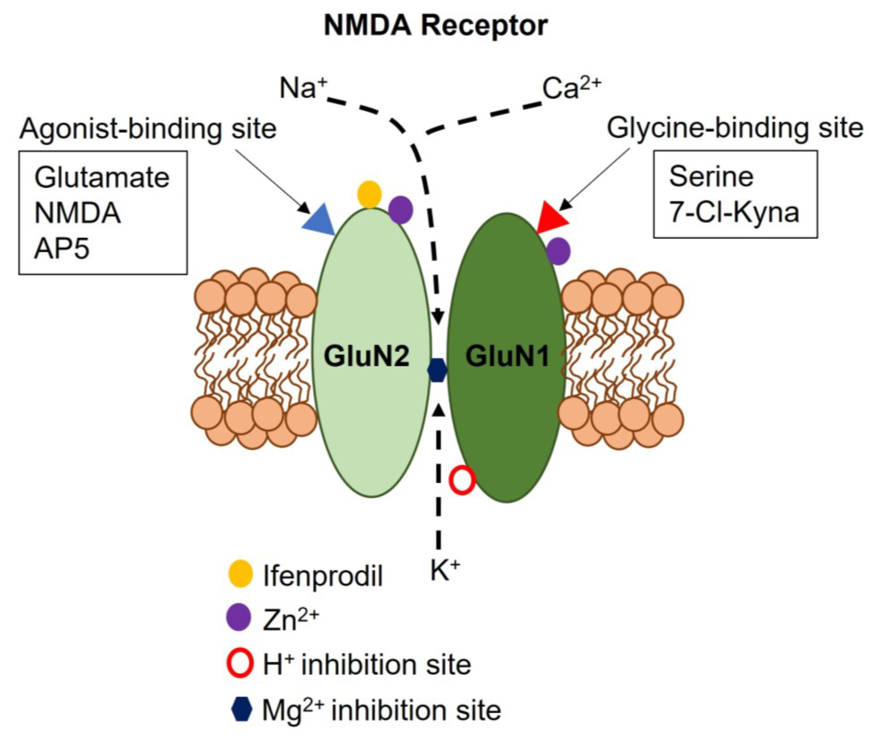
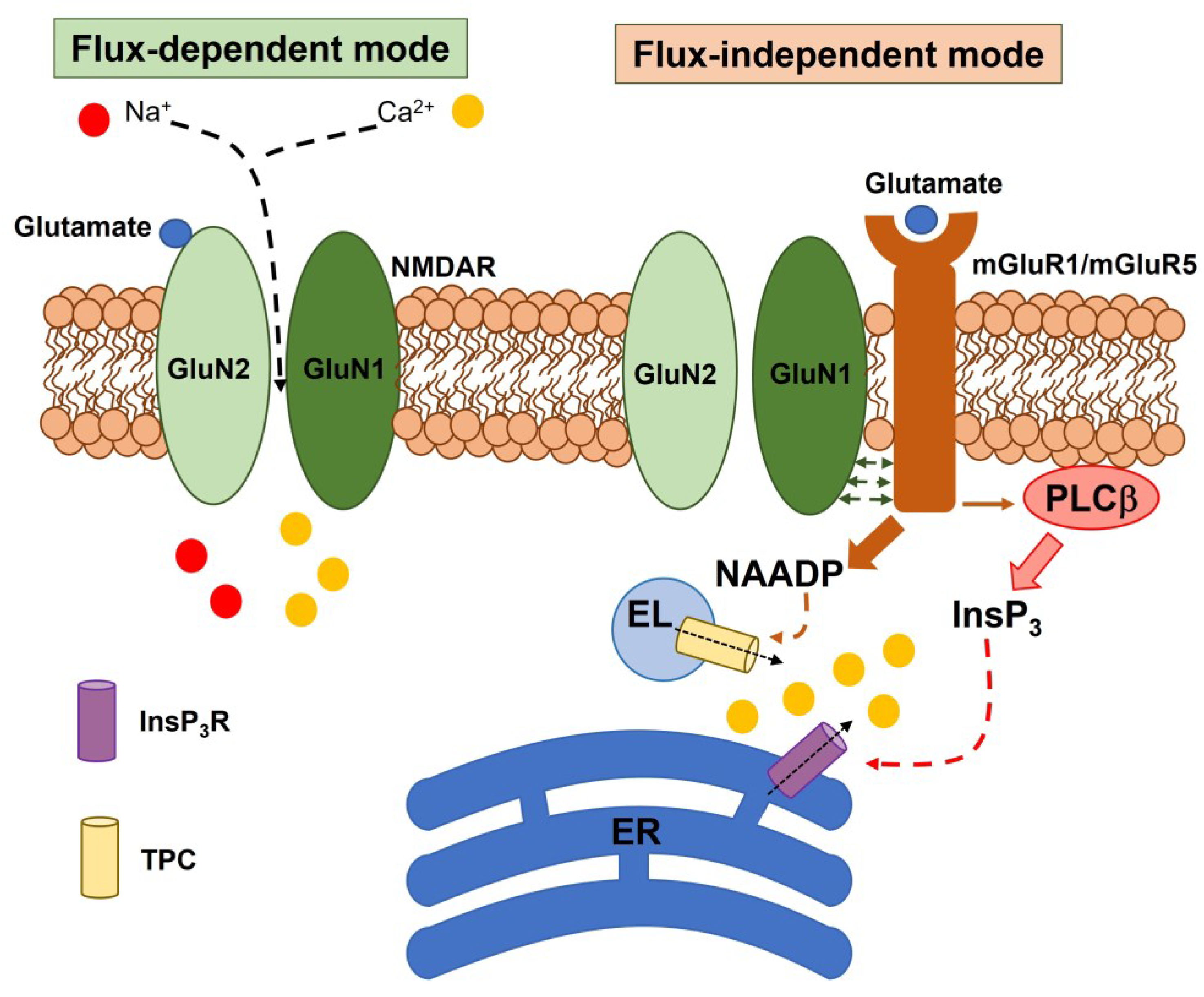
2. Subunit Composition, Regulatory Mechanisms, and Signaling Modes of Neuronal NMDARs

NMDARs assemble as heterotetrameric non-selective cation channels that can be drawn by multiple subunits: GluN1, GluN2A, GluN2B, GluN2C, GluN2D, GluN3A, and GluN3B [1,15]. Neuronal NMDARs comprise two obligatory GluN1 subunits which combine with either two GluN2 subunits or a mixture of GluN2 and GluN3 subunits [9,10]. Furthermore, GluN1 presents eight isoforms (GluN1-1a–GluN1-4a and GluN1-1b–GluN1-4b) due to alternative splicing, but the functional significance of the differential expression of GluN1 isoforms is still unclear [16]. The subunit composition of NMDARs dictates their biophysical and pharmacological features. Canonical NMDARs consist of two glycine-binding GluN1 subunits and two glutamate-binding GluN2 subunits. GluN1 is also required for correct assembly and trafficking of NMDARs to the plasma membrane [1,2]. Neuronal NMDARs differ from AMPA and kainate receptors not only due to their Ca2+ permeability, but also because they undergo a strong inhibition by extracellular Mg2+ at negative membrane potentials and because they require the binding of two co-agonists, i.e., glutamate and glycine (or D-serine), for channel activation [1,2,16]. Therefore, NMDARs serve as coincidence detectors that uniquely respond to presynaptic release of glutamate, which opens the channel pore, and postsynaptic depolarization, which relieves Mg2+-dependent inhibition [1,2]. At central synapses, the rapid activation of AMPA receptors mediates the inward current that depolarizes the postsynaptic neuron and enables Mg2+ unblocking of NMDARs during fast synaptic transmission [1,2]. GluN2A- and GluN2B-containing receptors present high permeability to Ca2+ (permeability ratio of Ca2+ to Cs+ (PCa/PCs) ≈ 7), generate high-conductance single channel openings (50 pS), and are very sensitive to extracellular Mg2+ (IC50 ≈ 15 µM) [16]. The incorporation of GluN2C or GluN2D dramatically reduces the Ca2+ permeability (PCa/PCs ≈ 4.5), single-channel conductance (37 pS), and sensitivity to Mg2+ blockade (IC50 ≈ 80 µM) [16]. The recently discovered glycine-binding GluN3 subunit can further change the biophysical properties of NMDARs [17,18]. The GluN3 subunit can replace one of the two GluN2 subunits in classical NMDARs during a narrow time window of postnatal development and in particular cell types, such as pyramidal neurons in the cortex and hippocampus, cerebellar Purkinje neurons, and multiple types of GABAergic interneurons [17,18]. Compared to classical GluN1/GluN2 receptors, tri-heteromeric GluN1/GluN2/GluN3 receptors display lower Ca2+ permeability (permeability ratio of Ca2+ to the monovalent ions, Na+ and K+ (PCa/PM) of 0.8) and single-channel conductance (27 pS), and are relatively insensitive to Mg2+ inhibition at hyperpolarized membrane potentials [18]. Therefore, GluN3A and GluN3B subunits may serve as dominant-negative regulators of NMDARs [17,18]. Intriguingly, tri-heteromeric GluN1/GluN2/GluN3 receptors do not require membrane depolarization to fully open in response to glutamate release [19]. In addition, omission of the glutamate binding GluN2 subunit can result in the assembly of di-heteromeric GluN1/GluN3A or GluN1/GluN3B receptors, which are insensitive to glutamate, extracellular Mg2+, and other classical NMDAR inhibitors (e.g., MK-801 or memantine, see below), and function as excitatory glycine-gated channels in heterologous expression systems [1,17]. The expression of functional GluN1/GluN3 receptor complexes in native tissues has been confirmed by Ca2+ imaging and electrophysiological recordings of ex vivo mouse optic nerve myelin [20].
The NMDAR complex can be physiologically modulated by multiple endogenous allosteric modulators, including extracellular H+ and Zn2+, which inhibit NMDARs by targeting GluN1 (Figure 1) and lead (Pb2+) cations, which inhibits GluN2A-containing NMDARs [2,11]. Additionally, polyamines, such as spermine and spermidine, exert a voltage-dependent inhibition of NMDARs at high concentrations, whereas they increase the affinity for glycine and promote NMDAR channel opening at micromolar concentrations [11]. A number of synthetic compounds can be exploited to investigate the physiological role of NMDARs with no need for genetic manipulation (Figure 1). These include, of course, the selective agonist NMDA, which recognizes the glutamate-binding site on GluN2, and a number of inhibitors: (1) the competitive antagonist, D-2-amino-5-phosphono-pentanoic acid (AP5), which displaces the agonist from the glutamate binding site; (2) 7-chlorokynurenic acid (7-Cl-Kyna), which is an antagonist for the glycine-binding site on GluN1; (3) MK-801 ((+)-5-methyl-10,11-dihydro-5H-dibenzo[a,d]cyclohepten-5,10-imine maleate), which is an open-channel blocker of NMDAR pores; (4) ifenprodil, which is a selective inhibitor of GluN2B-containing NMDARs; and (5) a variety of substances that directly block NMDARs, including memantine and ketamine [2,11].
Until two decades ago, NMDAR signaling was attributed to its ability to conduct extracellular Ca2+ into active spines during fast synaptic transmission and thereby signal coincidence of synaptic inputs in Hebbian plasticity [21,22]. Unexpectedly, numerous independent studies demonstrated that NMDARs may also signal in a flux-independent mode, i.e., without the need to mediate Ca2+ influx. It has been demonstrated that glutamate binding to GluN2 may induce conformational changes in the cytoplasmic domain of GluN1 that triggers diverse intracellular signaling pathways, e.g., Ca2+ release from the endoplasmic reticulum (ER) and the lysosomal store (Figure 2), protein phosphatase 1 (PP1), and p38 mitogen-activated protein kinase (MAPK) [21,23,24]. Metabotropic NMDAR signaling may be sufficient to induce long-term depression (LTD) and spine shrinkage in the hippocampus [25,26], but has also been reported in rat cortical astrocytes [27,28] and is implicated in synaptic disorders, such as Alzheimer’s disease [29] and schizophrenia [30].

3. NMDAR Expression, Signaling, and Function in the Heart

Cardiac excitability and conduction are finely regulated by the autonomic branch of the peripheral nervous system, which innervates the myocardium with both sympathetic and parasympathetic fibers, respectively, releasing the conventional neurotransmitters norepinephrine and acetylcholine [31]. Intriguingly, recent evidence has indicated that another excitatory neurotransmitter system, based upon glutamate and NMDARs, could modulate heart rate, cardiac conduction, and myocardial contraction [32]. Preliminary investigations revealed that GluN1 was expressed in both monkey [33] and rat [34] hearts, in which they were enriched in the conducting system, in atrial and ventricular cardiomyocytes, in the wall of coronary blood vessels, in coronary cardiac intramural nerve terminals, and in cardiac ganglia [32]. Subsequently, GluN1 expression was confirmed in the same anatomic sites of human myocardium [35], which can also express GluN2C [36] and GluN3 [37]. Finally, a recent transcriptomic analysis showed that all GluN subunits are expressed in rat atria [37]. The cardiac distribution of GluN subunits has been summarized in Table 1. These pieces of evidence fostered the view that NMDARs could play a role in the control of cardiac rhythm and excitation.

3.1. The Role of NMDAR Activation in Ventricular Arrhythmias and Acute Myocardial Infarction (AMI)

An initial report documented that the systemic (intravenous) infusion of NMDA (125, 250, 500, and 1000 μg/kg) induced a dose-independent increase in rat heart rate that persisted after pharmacological ganglion blockade with hexamethonium [48]. A subsequent investigation evaluated the electrophysiological effects of chronic NMDAR activation [41]. Two weeks of NMDA (3 mg/mL/kg) administration caused a significant increase in heart rate and prolonged the duration of ventricular action potential by downregulating the expression of several K+ channels, i.e., Kv4.2, Kv4.3, Kv11.1, and the β-subunit KChIP2, which are involved in cardiac repolarization [41]. In addition, chronic NMDAR activation caused electrical instability, increased the susceptibility to ventricular arrhythmia, and induced myocardial interstitial fibrosis [41]. In accord, Kv4.2 and Kv4.3 associate with KChIP2 to mediate the fast transient outward K+ current (Ito,fast), the reduction of which can prolong ventricular action potential duration [49,50]. Similarly, the loss of function of Kv11.1, also known as human ERG, has long been known to result in QT syndrome [50]. However, these pro-arrhythmic effects were reversed by MK-801 [41]. AMI is consequent to the electrophysiological alterations in myocardial substrates caused by myocardial ischemia and ischemia reperfusion, and leads to ventricular fibrillation and tachycardia [12]. D’Amico et al. demonstrated that bocking NMDARs with MK-801, ketamine, or memantine did not affect the incidence of ventricular arrhythmias and mortality induced by coronary occlusion in a rat model of AMI, but significantly reduced the pro-arrhythmic effects of reperfusion [42]. Similar data were obtained by Sun et al., who confirmed that MK-801 mitigated reperfusion-induced ventricular arrhythmias after occlusion of the left anterior descending coronary artery [43]. These findings suggested that cardiac NMDARs could be hyper-stimulated during reperfusion, probably due to excessive release of glutamate observed under these conditions [43,51]. Accordingly, Liu et al. reported that NMDA infusion significantly increased ischemia-reperfusion-induced Ca2+ overload in isolated rat ventricular cardiomyocytes, which was translated into a significant loss in ventricular function, cardiomyocytes apoptosis, and myocardial damage in an ex vivo model of AMI [44]. These adverse effects were halted by preventing extracellular Ca2+ influx through NMDARs with MK-801 or a Ca2+-free buffer [44]. A recent series of reports confirmed that memantine reduced cardiac arrhythmias and infarct size, alleviated cardiac fibrosis and hypertrophy, and partially rescued cardiac contractility in rat models of isoproterenol-induced AMI [52] and heart failure [53]. Finally, NMDARs could be responsible for the high incidence of lethal arrhythmias that occur in healing myocardium owing to sympathetic nerve sprouting (SNS). SNS can increase the cardiac expression of GluN1 in rats, while the subsequent administration of NMDA induced cardiomyocyte apoptosis and triggered ventricular tachycardia and ventricular fibrillation [45]. These effects were again rescued by blocking NMDARs with AP5 [45], thereby confirming that ventricular NMDARs represent a promising molecular target to treat arrhythmias.

3.2. The Role of Extracellular Ca2+ Entry in NMDA-Induced Ventricular Arrhythmia and Cardiac Remodeling

Multiple mechanisms cooperate to mediate the pro-arrhythmic effects of NMDAR activation in ventricles. An elevation in intracellular Ca2+ concentration ([Ca2+]i) is likely to play a major role in NMDAR-dependent myocardial pathogenesis [41,54]. Glutamate can increase the [Ca2+]i in rat cardiomyocytes by activating NMDARs in the presence of extracellular Mg2+ and in the absence of the co-agonist D-serine (Figure 3A) [46,55]. Therefore, NMDAR activation is able to depolarize ventricular (as well as atrial, see Section 3.3) cardiomyocytes both directly (i.e., by conducting Na+ and Ca2+ into the cytosol) and indirectly (i.e., through the Na+/Ca2+ exchanger (NCX), which can be activated by a subplasmalemmal Ca2+ rise) [56,57]. Thus, a NMDAR-dependent increase in [Ca2+]i could increase the risk of trigger activity and arrhythmias. It is still unclear whether extracellular Ca2+ entry through NMDARs can stimulate Ca2+-induced Ca2+ release (CICR) through type 2 ryanodine receptors (RyR2) (Figure 3B) or increase the sarcoplasmic reticulum (SR) Ca2+ load in a Sarco-Endoplasmic Reticulum Ca2+-ATPase (SERCA) 2a (SERCA2a)-dependent manner, as previously shown for lysosomal two-pore channels [58,59]. In neonatal rat ventricular cardiomyocytes, GluN2B has been detected in the Z-disc, in which it colocalizes with RyR2 on the juxtaposed SR cisternae (Figure 3A) [40]. Moreover, both GluN1 and GluN2B were identified in the outer mitochondrial membrane and inner mitochondrial cristae of rat hearts (Figure 3A) [39]. It is therefore reasonable to speculate that glutamate stimulates RyR2-dependent Ca2+ release and mitochondrial Ca2+ overload (Figure 3B), which would lead to mitochondrial depolarization and opening of the mitochondrial permeability transition pore (mPTP) [60].
In addition, NMDAR-mediated intracellular Ca2+ signals could modulate gene expression and thereby drive the down-regulation of Kv4.2, Kv4.3, Kv11.1, and KChIP2 [41], as well as SERCA2a [43] and proteins (Figure 3B). In agreement with this hypothesis, activation of the Ca2+-dependent transcription factor NF-κB [61,62] can reduce Ito,fast by down-regulating the expression of Kv4.2, Kv4.3, Kv11.1, and KChIP2 in neonatal rat ventricular cardiomyocytes [63]. The reduction in SERCA2a levels in hypertrophic and failing hearts could also involve Ca2+-dependent regulatory pathways [64]. Finally, NMDA-induced myocardial fibrosis, which may predispose ventricles to arrhythmias [57], can also by triggered by intracellular Ca2+ overload. In accord, many reports have demonstrated that NMDAR stimulation can induce mitochondrial depolarization, reactive oxygen species (ROS) production, and caspase-3-mediated apoptosis in rat ventricular cardiomyocytes (Figure 3A) [46,47]. Similarly, NMDARs promoted Ca2+ influx and Ca2+-dependent apoptosis by enhancing p38 MAPK activity in primary human neonatal cardiomyocytes cultured under ischemic conditions [65]. In line with these findings, infusion of MK-801 at the beginning of the reperfusion strongly attenuated oxidative stress in a rat model of AMI [66], whereas co-administration of glutamate and glycine dampened the cardioprotective effects of verapamil by increasing ROS production [67]. Intriguingly, a recent report showed that the anthracycline anticancer drug, pirarubicin, induced Ca2+ overload, oxidative stress, and apoptosis through GluN2D-containing NMDARs in HL-1 (mouse) and H9C2 (rat) cardiomyocytes [68].

3.3. The Role of NMDARs in Atrial Excitability and Conductivity and Their Involvement in Atrial Fibrillation

A recent series of studies have focused on the effect of NMDAR activation in the atria, where they are quite abundant, particularly in the sino-atrial node [35]. In addition, glutamate concentration was found to be enhanced in the left atrial appendages of patients [69]. In agreement with these observations, Xie et al. demonstrated that an intrinsic glutamatergic system is present in rat atrial cardiomyocytes [37]. They found that glutamate-containing vesicles are abundantly located beneath the plasma membrane. Moreover, these cells express glutaminase, which generates glutamate from glutamine; the excitatory amino acid transporter, EAAT1; and many iGluR isoforms, including GluN1 [37]. Accordingly, both glutamate and NMDA elicit transient inward currents and reduce the action potential threshold in rat atrial cardiomyocytes [37]. Furthermore, MK-801 and AP5 attenuate the atrial conduction velocity both ex vivo and in vivo [37]. Finally, an intrinsic glutamatergic system was also detected in human atria, and NMDARs were found to mediate transient inward currents and control also the conduction velocity in cultured human induced pluripotent stem cell (hiPSC)-derived atrial cardiomyocyte monolayers [37].
The evidence described above led to the hypothesis that exaggerated NMDAR activation could also contribute to abnormal excitability and conductivity in atrial fibrillation. In accord, a transcriptomic analysis conducted on a canine model of atrial fibrillation revealed that several genes encoding for glutamate signaling components were targeted by the differentially expressed microRNAs (miRNAs) of the DLK1-DIO3 locus [70]. Furthermore, Shi et al. has previously shown that intraperitoneal injection of NMDA (3 mg/kg) in rats reduced heart rate variability, which is associated with higher adverse outcome of AMI [71], by causing a dramatic sympathovagal imbalance, with a depressed vagal tone and an enhanced sympathetic activity in rats [38]. In addition, NMDA administration caused atrial electrical remodeling and atrial fibrillation by inducing atrial fibrosis and down-regulating connexin 40 through an increase in metalloproteinase 9 (MMP-9) expression [38]. Notably, Xie et al. confirmed that blocking NMDARs with MK-801 and AP5 prevented the occurrence and halted the progression of atrial fibrillation in vivo [37]. Thus, atrial NMDARs could provide a novel molecular target to design novel therapeutic strategies to treat atrial fibrillation, which is the most widespread persistent cardiac arrhythmia [32].

3.4. The Endogenous Agonist of Cardiac NMDARs: Is There A Role for Homocysteine (Hcy)?

Given the evidence that NMDARs are expressed in the heart and control cardiac excitation and conduction, the question as to their physiological agonist is obviously raised. A functional glutamatergic signaling system has been uncovered in atria (see Section 3.3), but not in ventricles. No evidence has been provided in favor of glutamate release from sympathetic nerves, as initially proposed by some authors [46]. It has been suggested that the peripheral organs, including the heart and peripheral vasculature, are exposed to baseline extracellular levels [10], which fluctuate between 5 and 100 µM/L in the plasma and 150 and 300 µM/L in the whole blood [72]. As anticipated above, glutamate concentrations ranging between 10 and 200 µM/L elicit robust Ca2+ signals in cardiomyocytes [55], thereby suggesting that cardiac NMDARs could be stimulated by circulating glutamate. However, it has long been known that astrocytes release glutamate via multiple mechanisms, including exocytosis, connexin 43 hemichannels, P2X Purinoreceptor 7 receptors, and bestrophin-1, to regulate neuronal activity [73]. Future work might explore the possibility that ventricular cardiomyocytes release glutamate not only during myocardial ischemia, but also during evoked activity or in response to β-adrenergic stimulation.
Alternately, cardiac NMDARs could be sensitive to the endogenous levels of Hcy, a derivative of the hepatic L-methionine metabolism that may serve as an NMDAR agonist by associating with the glutamate binding sites when glycine concentrations are raised beyond 50 µM [54,74]. Intriguingly, hyperhomocysteinemia (HHcy), which results from excessive serum levels of Hcy, can lead to coronary artery disease, AMI, ventricular arrhythmias, sudden cardiac death, and atherosclerosis [54,75]. The following pieces of evidence support the emerging notion that circulating Hcy could target cardiac NMDARs. First, a pharmacological blockade of NMDARs with MK-801 [76] or a genetic deletion of cardiac GluN1 [77] reduced the mitochondrial production of ROS and reactive nitrogen species, thereby impairing MMP9 activation and mPTP opening and halting the decline in myocyte contractility. Second, a genetic deletion of cardiac GluN1 also inhibited mitophagy [78], which can become extremely harmful to cardiac tissue when it becomes chronic [79]. Third, the combined application of Hcy, or Hcy thiolactone, NMDA, and glycine, caused a decrease in mechanical cardiac function and an increase in oxidative stress that were rescued by intracoronary infusion of memantine [80,81]. Quinolinic acid, a downstream metabolite of tryptophan that activates NMDARs in the CNS, has also been proposed as endogenous agonist of peripheral NMDARs [10], but its effect on cardiac NMDARs is still unknown. Thus, while the endogenous agonist (i.e., glutamate) engaging atrial NMDARs is coming of age, the signaling molecules that physiologically gate ventricular NMDARs still await to be fully elucidated.

4. The Role of NMDARs in VSMCs and Vascular Endothelial Cells

The main role of peripheral vasculature is to carry oxygen and nutrients to distant organs and tissues, from which carbon dioxide and catabolic waste are then removed and disposed through the urinary, respiratory, or gastrointestinal systems [82]. Large conduit arteries deliver oxygenated blood to the branched capillary networks that effect the exchanges of respiratory gaseous and nutrients/catabolites between circulating blood and the surrounding tissues. Resistance arteries (lumen diameter ∼150–250 μm) and arterioles (<150 μm) convey the blood from large conduit arteries to downstream capillaries and represent the major site of vascular resistance [82]. The vascular wall is mainly composed of endothelial cells, which line the inner lumen of blood vessels and are directly exposed to blood stream, and a surrounding layer of VSMCs. Capillaries comprise only a monolayer of endothelial cells, while VSMC contraction in resistance arteries and arterioles is a crucial determinant of blood pressure [82,83]. However, endothelial cells release multiple vasoactive mediators that are able to control VSMC contractility and thereby induce vasorelaxation (e.g., NO, prostaglandins, and hydrogen sulfide) or vasoconstriction (e.g., endothelin) [82,83]. A preliminary report showed that GluN1 was present both in rat aorta, the largest artery in the body, and in rat mesenteric resistance arteries [48]. Then, GluN1 was detected in rat carotid arteries and GluN1, GluN2A, GluN2B, GluN2C, and GluN2D were identified in rat aortic endothelial cells (RAECs) (Table 2) [84] and in rat aortic smooth muscle (A7r5) cells (Table 2) [85]. GluN1, GluN2A, GluN2B, GluN2C, and GluN2D were also found in human pulmonary arterial smooth muscle cells (hPASMCs) (Table 2) and human pulmonary microvascular endothelial cells (hPMVECs) in combination with the postsynaptic density protein-95 (PSD-95) (Table 2) [86]. In agreement with these observations, NMDA induced AP5-sensitive inward currents in the presence of glycine and in the absence of extracellular Mg2+ in hPASMCs, thereby confirming that NMDARs were functional [87]. Multiple GluN subunit- and NMDAR-mediated intracellular Ca2+ signals were also recorded in mouse [9,88,89] and human [24,90] cerebrovascular endothelial cells, as more widely discussed in Section 5.

4.1. The Hemodynamic Effect of NMDAR Activation in Peripheral Vasculature

The role of peripheral NMDARs in the regulation of blood pressure is yet fully unraveled [12]. Systemic infusion of NMDA (125, 250, 500, and 1000 μg/kg) induced a dose-dependent increase in rat mean arterial pressure (MAP) that was not affected by hexamethonium (see also Section 3.1) and was therefore mediated by vascular NMDARs [48]. In accord, the hemodynamic response to NMDA was sensitive to AP5 and HA-966, which are antagonists of the glycine binding site on GluN1. It was then found that NMDARs activate neuronal, rather than endothelial, NO synthase (nNOS) to generate NO, and that NO release drives NMDA-dependent vasoconstriction [48]. These unexpected findings suggested that peripheral NMDARs also engage NADPH oxidase (NOX), as described in neurons [91], so that nNOS-derived NO reacts with NOX-derived ROS to form peroxynitrite and induce vasoconstriction [12]. A follow-up study showed that ethanol attenuates an NMDAR-mediated increase in ROS production and MAP [92]. A recent series of studies demonstrated that nNOS is also expressed and contributes to NO signaling in VSMCs and endothelial cells and that it is quite abundant in the mesenteric bed [93]. Intriguingly, NMDARs recruit the endothelial NO synthase (eNOS) and mediate vasodilation in mouse brain microcirculation [94,95], and NMDAR-dependent vasorelaxation has also been reported in rat coronary circulation [66,80]. These findings suggest that NMDARs could be coupled to different NOS isoforms in different vascular districts, e.g., nNOS in aorta and mesenteric resistance arteries and eNOS in brain and coronary microvessels. The hypothesis that peripheral NMDAR signaling could be exaggerated in essential hypertension, as suggested for PAH (see below), cannot be discounted [12]. Future work will have to assess whether dysregulated NMDARs are located in VSMCs, in vascular endothelial cells, or both.

4.2. The Role of Peripheral NMDARs in Vascular Remodelling during HHcy-Induced Atherosclerosis and in PAH

Vascular cells are exposed to circulating glutamate, which could directly target endothelial cells and may easily reach VSMCs. In addition, vascular cells can be directly stimulated by plasma Hcy and HHcy can induce endothelial dysfunction and accelerate myointimal hyperplasia and luminal narrowing, thereby resulting in atherosclerosis [54,75,96]. A preliminary study showed that HHcy increased NMDAR expression and stimulated proliferation in A7r5 cells [85]. These effects were mediated by MMP-9 and interleukin-1β, which support VSMC proliferation and migration, and rescued by MK-801 [85]. A subsequent study confirmed that HHcy stimulated human VSMCs to release MMP-2, which is involved in remodeling and destabilization of the atherosclerotic plaques by recruiting the phosphatidylinositol 3-kinase (PI3K) and MAPK phosphorylation cascades [97]. These pro-atherosclerotic effects were again alleviated by MK-801 and L-glycine, which favors NMDAR internalization [97]. It was further demonstrated that HHcy stimulates the expression of C-reactive protein (CRP), which also promotes VSMC proliferation and migration by inducing ROS production, MAPK activation, and the nuclear translocation of NF-κB in rat aortic VSMCs [98]. The primary role of NMDARs in HHcy-dependent CRP expression and secretion was underpinned by the inhibitory action of MK-801 [98]. Future work will have to assess whether endothelial NMDARs are involved in HHcy-induced endothelial dysfunction, as suggested in [12]. Nevertheless, peripheral NMDARs stand out as novel molecular targets to interfere with HHcy-induced vascular remodeling and atherosclerotic progression.
On the other hand, a recent study has undoubtedly unveiled the pathogenic role of vascular NMDARs in PAH [86]. Early reports had already shown that tissue-type plasminogen activator (tPA) [99] and urokinase plasminogen activator (uPA) [100] increase lung arterial contractility and promote lung vascular permeability by activating NMDARs in mice. These studies showed that preventing the interaction between pulmonary NMDARs and tPA or between uPA and a docking peptide that binds either of these enzymes and interferes with their occupancy of the NMDAR binding site can mitigate pulmonary injury [99,100]. More recently, Dumas et al. documented that glutamate levels were enhanced in pulmonary arteries isolated from PAH patients as compared to control arteries, and that glutamate accumulation was more robust in medial lesions [86], which are featured by VSMC hyperplasia. The increase in glutamate production was secondary to the accumulation of glutamine, which is the glutamate precursor in the glutaminase reaction, and was more evident in pulmonary arterioles (<150 µm) [86], which represent the main target of current therapies [101]. This study also showed that GluN1 protein was up-regulated and hyperphosphorylated at Ser-896 in PAH pulmonary arteries, particularly in VSMC lesions. This latter feature was rather relevant since phosphorylation of Ser-896 increases the surface expression of GluN1 in neurons [102]. Furthermore, many components of the neuronal glutamatergic system were identified in hPASMCs and hPAMECs, including glutaminase 1 (GLS1) and GLS2, which catalyze glutamate production from glutamine [103]; the vesicular glutamate transporter, VGLUT1; and the scaffolding protein, PSD-95. Interestingly, GLS1 and VGLUT1 were enhanced in PAH arteries and both hPASMCs and hPMVECs were able to release glutamate in a Ca2+-dependent manner. Additionally, both endothelin-1 and platelet-derived growth factor-BB (PDGF-BB), which accelerate vascular remodeling in PAH, stimulated GluN1 phosphorylation and induced hPASMC proliferation. GluN1 phosphorylation was spatially confined within the regions of cell-to-cell contact between hPASMCs [86]. Therefore, the authors speculated about a quasi-synaptic mode of glutamatergic communication via NMDARs between adjacent hPASMCs in both culture and PAH pulmonary arteries (Figure 4). In line with these findings, hypoxia failed to promote pulmonary vascular remodeling and pulmonary hypertension in transgenic mice selectively lacking GluN1 in VSMCs. Moreover, blocking NMDARs with MK-801 in a rat model of monocrotaline-induced PAH alleviated remodeling of pulmonary arteries and the corresponding decrease in right ventricle contractility [86]. A follow-up study showed that PDGF-BB could increase the surface expression of GluN2B in hPASMCs via Src-dependent phosphorylation, thereby resulting in the assembly of antiproliferative and antimigratory NMDARs [104]. However, GluN2B expression was down-regulated in PAH pulmonary arteries, which further highlights the crucial role played by VSMC NMDARs in medial hyperplasia and vascular obstruction [104]. Therefore, this study could pave the way for novel therapeutic avenues to treat PAH by targeting pulmonary NMDARs [101]. A crucial issue that remains to be solved concerns the requirement for complete Mg2+ unblocking to measure NMDAR-mediated inward currents in hPASMCs [87]. NMDAR activation exacerbates phenylephrine-induced contractile responsiveness of human pulmonary arteries under hypoxic, but not normoxic, conditions [87]. However, the membrane potential of hypoxic pulmonary arterial SMCs is more depolarized as compared to normoxic cells due to the reduced expression and activity of voltage-gated K+ channels [105] and to the activation of the acid-sensing ion channel 1 [106]. The persistent depolarization of hypoxic pulmonary arterial SMCs could be involved in the glutamate-dependent activation of NMDARs and support vascular remodeling and vasoconstriction in PAH.

5. The Role of NMDARs in Cerebro-Microvascular Endothelial Cells (CMVECs)

Glutamate is the major excitatory neurotransmitter in the CNS and modulates neuronal excitability and synaptic plasticity via iGluRs and mGluRs. It is therefore not surprising that AMPA receptors and NMDARs, as well as the Gq/11-protein coupled mGluR1 and mGluR5, are widely expressed also in the other main components of the neurovascular unit, i.e., astrocytes and microvascular endothelial cells [8,9,10,107,108,109,110,111]. Cerebral microvessels, i.e., arterioles and capillaries, receive an extensive glutamatergic innervation [112,113] and CMVECs can respond to neuronal activity via NMDARs [94,114,115]. In accord, NMDAR subunits have been detected in human [24,90,115] and rodent [95,114,115] CMVECs, in which GluN1 is preferentially located to the basolateral membrane surface to directly perceive synaptically released glutamate [116]. As illustrated in Table 3, CMVECs express GluN1, GluN2A, GluN2C, and GluN3A in mouse cortical arteries [94,116], whereas GluN1, GluN2B, and GluN3A were detected in spinal cord microvascular endothelial cells [90]. The molecular architecture of endothelial NMDARs in human CNS microvessels has been investigated by exploiting primary CMVECs, which express GluN1 [115,117], and in the hCMEC/D3 cell line, which has been established from the human temporal lobe microvessels [118] and express GluN1, GluN2C, and GluN3B subunits (Table 3) [24]. Therefore, the molecular configuration of endothelial NMDARs in cortical vessels is enriched with GluN2C and GluN3 subunits, which are likely to yield a low sensitivity to extracellular Mg2+, low single-channel conductance, and sensitivity to glycine [10,24,119].
NMDARs regulate multiple functions of the BBB [10]. The BBB is mainly formed by CMVECs that line brain microvessels and is therefore instrumental to maintain CNS homeostasis and ensure proper neuronal signaling. In addition, the BBB protects brain parenchyma from blood-borne pathogens and toxins and its restrictive nature represents a major obstacle for drug delivery to the CNS [120,121]. Hogan-Cann and Anderson have recently surveyed the involvement of endothelial NMDARs in the control of the BBB in health and disease [10]. In this section, we focus our attention on the latest investigations describing the role of endothelial NMDARs in the fine regulation of BBB permeability and NVC, i.e., the mechanism whereby an increase in neuronal activity is translated into an increase in cerebral blood flow (CBF) [112,122], and their implication in neurological disorders.

5.1. Endothelial NMDARs Control BBB Permeability

The movement of solutes across the BBB between circulating blood and brain parenchyma can occur through a paracellular or transcellular route [120,121]. Glutamate can induce the opening of the BBB and thereby increase paracellular permeability via NMDAR-mediated intracellular Ca2+ signals [120,125,126,127,128]. In accord, NMDA-induced elevation in BBB permeability was sensitive to MK-801 [125,126,127] or AP5 [126,128] and an NMDA-evoked increase in [Ca2+]i was recorded in both human [24,115,126] and mouse [129,130] CMVECs and in mouse cerebral artery endothelial cells [88]. Intriguingly, NMDA-induced intracellular Ca2+ signals in hCMEC/D3 cells are sensitive to AP5 and genetic knockdown of GluN1, but not to MK-801 and to removal of extracellular Ca2+ [24]. In addition, whole-cell patch-clamp recordings showed that NMDA did not elicit any detectable inward current both in the absence and in the presence of extracellular Mg2+ [24]. Nevertheless, this investigation revealed that NMDARs may operate in a flux-independent mode to signal the increase in [Ca2+]i in hCMEC/D3 cells. In accord, NMDARs interact with mGluR1 and mGluR5 to mobilize lysosomal Ca2+ via TPCs and ER-stored Ca2+ via InsP3 receptors and to induce store-operated Ca2+ entry (Figure 2) [24,131]. Conversely, NMDA-induced intracellular Ca2+ signals in mouse CMVECs disappear in the absence of extracellular Ca2+ and in the presence of the pore-blocking antagonist, MK-801 [88]. Therefore, NMDARs can signal the increase in [Ca2+]i in a flux-dependent manner in mouse CMVECs and in a flux-independent (i.e., metabotropic manner) in human CMVECs. NMDAR-mediated intracellular Ca2+ signals can increase the BBB permeability by inducing NO release [24,108,128] and myosin light chain (MLC) phosphorylation through the Ca2+-dependent recruitment of Rho-kinase (ROCK) and MLC kinase (MLCK) [108]. Finally, extracellular Ca2+ entry through NMDARs also controls the transcellular route across the mouse BBB by recruiting Ca2+/calmodulin-dependent protein kinase II (CaMKII), protein kinase C (PKC), and RhoA [115].

5.2. Endothelial NMDARs Control NVC

The mechanism whereby an increase in neuronal activity may lead to a local elevation in CBF to match the high energy demand of active neurons with a supply of oxygen and nutrient supply from the blood is known as NVC or functional hyperemia [112,113]. According to the canonical model, NVC is triggered by the release of vasoactive mediators, such as NO, arachidonic, potassium ions (K+), from neurons and astrocytes. These signals target the contractile cells, i.e., VSMCs and pericytes, of brain microvessels to induce vasorelaxation and increase CBF [112,113]. A series of recent studies, reviewed in [9,10,89,132,133,134], demonstrated that CMVECs may sense neuronal activity and thereby elicit a robust increase in CBF through a blend of ion channels that are located in the plasma membrane and include the inward rectifier K+ (Kir2.1) channel [135], Transient Receptor Potential Ankyrin 1 (TRPA1) channels [136], and NMDARs [10]. In accord, the Anderson group demonstrated that the stimulation of endothelial NMDARs with glutamate induced arteriolar relaxation in ex vivo mouse middle cerebral arteries through the Ca2+-dependent release of NO [94,95,116]. Astrocytes contribute to the activation of basolateral endothelial NMDARs by releasing the co-agonist D-serine in response to an increase in [Ca2+]i [95,116]. As expected (see Section 5), glutamate-evoked arteriolar vasodilation was not sensitive to Mg2+-dependent blockades [94]. In agreement with these observations, the hemodynamic response to sensory stimulation was largely compromised in a transgenic mouse model selectively lacking endothelial NMDARs [114] and eNOS [137]. In addition, the full hemodynamic response to somatosensory stimulation in mouse arteries and arterioles requires the stimulation of luminal NMDARs by circulating tPA [138]. Intriguingly, in the human cerebrovascular endothelial cell line hCMEC/D3, NMDARs may signal in a flux-dependent manner to induce NO release, do not require the presence of D-serine, and are not sensitive to Mg2+-dependent inhibition [24].
The evidence that endothelial NMDARs at the NVU are stimulated during glutamatergic transmission is also relevant for synaptic events that go beyond the hemodynamic response. Early work has shown that the hippocampal long-term potentiation (LTP) is inhibited in eNOS-deficient mice [139,140]. Intriguingly, eNOS is not expressed in CA1 pyramidal cells [141], which clearly indicates the crucial role of endothelium-derived NO in synaptic plasticity and memory formation. Future experiments should assess whether LTP is impaired in transgenic mice selectively lacking endothelial NMDARs and eNOS not only in the hippocampus, but also in other brain areas, such as the cortex, amygdala, and cerebellum. The role of vascular NMDARs in endothelial-to-neuronal communication has been suggested by a recent study showing that the vessel-associated migration of GABA interneurons along the pial migratory route is finely regulated by endothelial NMDARs [124].

5.3. The Pathogenic Role of Endothelial NMDARs in the CNS

Disruption of the BBB is a hallmark of many neurological disorders that are associated to hypersynchronized neuronal activation and/or excessive glutamate release, such as traumatic brain injuries (TBI), epilepsy, multiple sclerosis, and stroke [120,121,142]. Furthermore, the BBB permeability is significantly increased in vascular dementia and other neurodegenerative disorders such as Alzheimer’s disease (AD) due to the enhanced oxidative stress imposed to cerebrovascular endothelial cells [143]. Early studies conducted on cultured CMVECs showed that endothelial NMDARs could contribute to the disruption of BBB integrity through a variety of mechanisms, including the altered expression of BBB efflux transporters (e.g., ABCG2) [117,144], the decreased expression and relocalization of tight junctions protein (e.g., occludin and claudin-5) [108,111], MMP-2, and MMP-9 release [129]. In addition, excessive doses of glutamate (1 mM) induced endothelial dysfunction through NMDAR-mediated production of ROS [125,127,145] and peroxynitrite [146], thereby disrupting the BBB integrity in vitro. In accord, an increase in endothelial [Ca2+]i not only engages the eNOS, but it can also stimulate ROS generation through the Ca2+-dependent NOX4 [147], which is widely expressed in CMVECs [130].
A recent series of investigations confirmed that excessive stimulation of endothelial NMDARs can increase the BBB permeability in several animal models of neurological disorders. Aberrant glutamate release during epileptic seizures increased vascular permeability in the rat cerebral cortex via endothelial NMDARs (Table 4) [128]. Similarly, endothelial-specific deletion of the regulator of G-protein signaling 5 (RGS5) enhanced endothelial NMDAR signaling in a mouse model of brain stroke, which exacerbated the increase in the BBB permeability and the severity of the ischemic damage (Table 4) [108]. The serin-protease tPA has been shown to disrupt the BBB integrity and favor monocyte infiltration in mouse models of multiple sclerosis (Table 4) [148]. Blocking NMDARs with a monoclonal antibody (known as Glunomab) that selectively targets GluN1 inhibited tPA-induced leukocyte transmigration in vitro and halted the progression of neurological impairments in a mouse model of multiple sclerosis (i.e., autoimmune encephalomyelitis) in wild-type but not in tPA-deficient animals [149]. Moreover, NMDARs were found to mediate tPA-dependent monocyte transmigration through the human BBB via extracellular signal-regulated kinase 1 activation [123]. In addition, the endovascular infusion of Glunomab reduced the incidence of intracellular aneurysms by blocking tPA-dependent activation of endothelial NMDARs in mice (Table 4) [150]. These preliminary findings strongly support the notion that targeting endothelial NMDARs could represent a promising therapeutic strategy to attenuate BBB dysfunction in neurological disorders as severe as epilepsy, multiple sclerosis, and stroke [151]. In addition, intraluminal infusion of amyloid-β(1–40) reduced extracellular Ca2+ entry through endothelial NMDARs and attenuated endothelium-dependent dilation in mouse cerebral arteries (Table 4) [88]. Thus, defective endothelial NMDAR signaling could contribute to the impaired hemodynamic response (and possibly to loss in neuronal regulation by endothelium-derived NO) in neurodegenerative diseases such as AD. Finally, anti-NMDAR autoimmune encephalitis is a rare neurological disorder that is caused by autoreactive anti-NMDAR autoantibodies targeting GluN1 and produced by B lymphocytes [151,152]. This in turn results in NMDAR hypo-responsiveness and leads to a decline in cognitive function and neuropsychiatric symptoms [153]. Future work should assess whether anti-NMDAR autoantibodies also target the endothelial NMDARs, as suggested by the defect in BBB integrity that has been reported in both animal models [152] and patients [154] affected by this disease.

6. Conclusions

A burgeoning body of evidence has demonstrated that NMDARs do not only fulfil a crucial function in the CNS, but also in the peripheral organ systems. The regulatory role of NMDARs in the cardiovascular system is also coming of age. In the heart, NMDARs are located within the conduction system and in the working myocardium. Therefore, they can regulate both the cardiac rhythm and cardiac excitability in a healthy heart, while exaggerated NMDAR signaling could contribute to atrial fibrillation, ventricular arrhythmias, and heart failure. In the peripheral vasculature, NMDARs are expressed in VSMCs and endothelial cells, but it is still unclear whether and how they regulate MAP. Preliminary evidence suggests that the regulation of local blood flow could be differentially affected by peripheral NMDARs, depending on the vascular bed and on their location in VSMCs vs. vascular endothelial cells. In pulmonary circulation, VSMC NMDARs are certainly involved in the pathogenesis of PAH, although their role in hPMVECs is still unclear. The role of endothelial NMDARs has been clearly unveiled in cerebral microcirculation, where they regulate NVC and BBB permeability. Conversely, it is still unknown whether they are expressed in cerebral VSMCs and pericytes. As various neurological disorders depend on NMDAR subunit mutations [6,7], the possibility that also endothelial NMDARs are involved in such genetic diseases cannot be discounted. Importantly, pre-clinical studies demonstrated that the pharmacological blockade of NMDARs could represent an effective strategy in treating life-threatening cardiovascular pathologies, ranging from arrhythmic disorders to PAH and neurological/neurodegenerative diseases. The molecular composition of NMDARs in cardiomyocytes (Table 1), VSMCs and vascular endothelial cells (Table 2), and in CMVECs (Table 3) is yet to be fully elucidated. The complement of ion channels endowed to VSMCs and endothelial cells may extensively vary depending on the vascular district [156,157,158,159]. Thus, it is likely that distinct GluN subunits will be found to contribute to vessel-associated NMDARs in different vascular beds. Understanding the molecular make-up of NMDARs is mandatory to decipher the diverse array of biophysical features they present in the multiple cellular components of the cardiovascular system (e.g., Mg2+-dependent inhibition in hPASMCs but not in ventricular cardiomyocytes and CMVECs) and to fully exploit their therapeutic potential.

Author Contributions

Conceptualization, F.M.; writing—original draft preparation, T.S. and F.M.; writing—review and editing, T.S., V.B., R.B.-R. and F.M.; visualization, T.S., V.B., R.B.-R. and F.M.; supervision, F.M.; project administration, F.M.; funding acquisition, F.M. All authors have read and agreed to the published version of the manuscript.

Funding

This research was funded by Fondo Ricerca Giovani from the University of Pavia (F.M.) and the EU Horizon 2020 FETOPEN-2018-2020 Program under Grant Agreement N. 828984 (LION-HEARTED). RBR is supported by the National Council of Science and Technology (CONACYT), Identification Number: CVU121216.

Institutional Review Board Statement

Not applicable.

Informed Consent Statement

Not applicable.

Data Availability Statement

Not applicable.

Conflicts of Interest

The authors declare no conflict of interest.

References

  1. Hansen, K.B.; Yi, F.; Perszyk, R.E.; Furukawa, H.; Wollmuth, L.P.; Gibb, A.J.; Traynelis, S.F. Structure, function, and allosteric modulation of NMDA receptors. J. Gen. Physiol. 2018, 150, 1081–1105. [Google Scholar] [CrossRef]
  2. Traynelis, S.F.; Wollmuth, L.P.; McBain, C.J.; Menniti, F.S.; Vance, K.M.; Ogden, K.K.; Hansen, K.B.; Yuan, H.; Myers, S.J.; Dingledine, R. Glutamate receptor ion channels: Structure, regulation, and function. Pharmacol. Rev. 2010, 62, 405–496. [Google Scholar] [CrossRef] [Green Version]
  3. Soda, T.; Mapelli, L.; Locatelli, F.; Botta, L.; Goldfarb, M.; Prestori, F.; D’Angelo, E. Hyperexcitability and Hyperplasticity Disrupt Cerebellar Signal Transfer in the IB2 KO Mouse Model of Autism. J. Neurosci. 2019, 39, 2383–2397. [Google Scholar]
  4. Locatelli, F.; Soda, T.; Montagna, I.; Tritto, S.; Botta, L.; Prestori, F.; D’Angelo, E. Calcium Channel-Dependent Induction of Long-Term Synaptic Plasticity at Excitatory Golgi Cell Synapses of Cerebellum. J. Neurosci. 2021, 41, 3307–3319. [Google Scholar] [CrossRef]
  5. Loftis, J.M.; Janowsky, A. The N-methyl-d-aspartate receptor subunit NR2B: Localization, functional properties, regulation, and clinical implications. Pharmacol. Ther. 2003, 97, 55–85. [Google Scholar] [CrossRef]
  6. Pagano, J.; Giona, F.; Beretta, S.; Verpelli, C.; Sala, C. N-methyl-d-aspartate receptor function in neuronal and synaptic development and signaling. Curr. Opin. Pharmacol. 2021, 56, 93–101. [Google Scholar] [CrossRef]
  7. Mapelli, L.; Soda, T.; D’Angelo, E.; Prestori, F. The Cerebellar Involvement in Autism Spectrum Disorders: From the Social Brain to Mouse Models. Int. J. Mol. Sci. 2022, 23, 3894. [Google Scholar] [CrossRef]
  8. Verkhratsky, A.; Chvatal, A. NMDA Receptors in Astrocytes. Neurochem. Res. 2020, 45, 122–133. [Google Scholar] [CrossRef] [Green Version]
  9. Negri, S.; Faris, P.; Soda, T.; Moccia, F. Endothelial signaling at the core of neurovascular coupling: The emerging role of endothelial inward-rectifier K+ (Kir2.1) channels and N-methyl-d-aspartate receptors in the regulation of cerebral blood flow. Int. J. Biochem. Cell Biol. 2021, 135, 105983. [Google Scholar] [CrossRef]
  10. Hogan-Cann, A.D.; Anderson, C.M. Physiological Roles of Non-Neuronal NMDA Receptors. Trends Pharmacol. Sci. 2016, 37, 750–767. [Google Scholar] [CrossRef]
  11. Bozic, M.; Valdivielso, J.M. The potential of targeting NMDA receptors outside the CNS. Expert Opin. Ther. Targets 2015, 19, 399–413. [Google Scholar] [CrossRef]
  12. McGee, M.A.; Abdel-Rahman, A.A. N-Methyl-d-Aspartate Receptor Signaling and Function in Cardiovascular Tissues. J. Cardiovasc. Pharmacol. 2016, 68, 97–105. [Google Scholar] [CrossRef] [Green Version]
  13. Kalev-Zylinska, M.L.; Hearn, J.I.; Makhro, A.; Bogdanova, A. N-Methyl-d-Aspartate Receptors in Hematopoietic Cells: What Have We Learned? Front. Physiol. 2020, 11, 577. [Google Scholar] [CrossRef]
  14. Kalev-Zylinska, M.L.; Morel-Kopp, M.C.; Ward, C.M.; Hearn, J.I.; Hamilton, J.R.; Bogdanova, A.Y. Ionotropic glutamate receptors in platelets: Opposing effects and a unifying hypothesis. Platelets 2021, 32, 998–1008. [Google Scholar] [CrossRef]
  15. Alexander, S.P.; Mathie, A.; Peters, J.A.; Veale, E.L.; Striessnig, J.; Kelly, E.; Armstrong, J.F.; Faccenda, E.; Harding, S.D.; Pawson, A.J.; et al. The concise guide to pharmacology 2021/22: Ion channels. Br. J. Pharmacol. 2021, 178 (Suppl. 1), S157–S245. [Google Scholar] [CrossRef]
  16. Paoletti, P.; Bellone, C.; Zhou, Q. NMDA receptor subunit diversity: Impact on receptor properties, synaptic plasticity and disease. Nat. Rev. Neurosci. 2013, 14, 383–400. [Google Scholar] [CrossRef]
  17. Pachernegg, S.; Strutz-Seebohm, N.; Hollmann, M. GluN3 subunit-containing NMDA receptors: Not just one-trick ponies. Trends Neurosci. 2012, 35, 240–249. [Google Scholar] [CrossRef]
  18. Perez-Otano, I.; Larsen, R.S.; Wesseling, J.F. Emerging roles of GluN3-containing NMDA receptors in the CNS. Nat. Rev. Neurosci. 2016, 17, 623–635. [Google Scholar] [CrossRef]
  19. Burzomato, V.; Frugier, G.; Perez-Otano, I.; Kittler, J.T.; Attwell, D. The receptor subunits generating NMDA receptor mediated currents in oligodendrocytes. J. Physiol. 2010, 588 Pt 18, 3403–3414. [Google Scholar] [CrossRef]
  20. Pina-Crespo, J.C.; Talantova, M.; Micu, I.; States, B.; Chen, H.S.; Tu, S.; Nakanishi, N.; Tong, G.; Zhang, D.; Heinemann, S.F.; et al. Excitatory glycine responses of CNS myelin mediated by NR1/NR3 “NMDA” receptor subunits. J. Neurosci. 2010, 30, 11501–11505. [Google Scholar] [CrossRef] [Green Version]
  21. Dore, K.; Stein, I.S.; Brock, J.A.; Castillo, P.E.; Zito, K.; Sjostrom, P.J. Unconventional NMDA Receptor Signaling. J. Neurosci. 2017, 37, 10800–10807. [Google Scholar] [CrossRef]
  22. Park, D.K.; Stein, I.S.; Zito, K. Ion flux-independent NMDA receptor signaling. Neuropharmacology 2022, 210, 109019. [Google Scholar] [CrossRef]
  23. Montes de Oca Balderas, P. Flux-Independent NMDAR Signaling: Molecular Mediators, Cellular Functions, and Complexities. Int. J. Mol. Sci. 2018, 19, 3800. [Google Scholar] [CrossRef] [Green Version]
  24. Negri, S.; Faris, P.; Maniezzi, C.; Pellavio, G.; Spaiardi, P.; Botta, L.; Laforenza, U.; Biella, G.; Moccia, D.F. NMDA receptors elicit flux-independent intracellular Ca2+ signals via metabotropic glutamate receptors and flux-dependent nitric oxide release in human brain microvascular endothelial cells. Cell Calcium 2021, 99, 102454. [Google Scholar] [CrossRef]
  25. Nabavi, S.; Kessels, H.W.; Alfonso, S.; Aow, J.; Fox, R.; Malinow, R. Metabotropic NMDA receptor function is required for NMDA receptor-dependent long-term depression. Proc. Natl. Acad. Sci. USA 2013, 110, 4027–4032. [Google Scholar] [CrossRef] [Green Version]
  26. Stein, I.S.; Gray, J.A.; Zito, K. Non-Ionotropic NMDA Receptor Signaling Drives Activity-Induced Dendritic Spine Shrinkage. J. Neurosci. 2015, 35, 12303–12308. [Google Scholar] [CrossRef] [Green Version]
  27. Montes de Oca Balderas, P.; Aguilera, P. A Metabotropic-Like Flux-Independent NMDA Receptor Regulates Ca2+ Exit from Endoplasmic Reticulum and Mitochondrial Membrane Potential in Cultured Astrocytes. PLoS ONE 2015, 10, e0126314. [Google Scholar] [CrossRef]
  28. Montes de Oca Balderas, P.; Matus Nunez, M.; Picones, A.; Hernandez-Cruz, A. NMDAR in cultured astrocytes: Flux-independent pH sensor and flux-dependent regulator of mitochondria and plasma membrane-mitochondria bridging. FASEB J. 2020, 34, 16622–16644. [Google Scholar] [CrossRef]
  29. Birnbaum, J.H.; Bali, J.; Rajendran, L.; Nitsch, R.M.; Tackenberg, C. Calcium flux-independent NMDA receptor activity is required for Abeta oligomer-induced synaptic loss. Cell. Death Dis. 2015, 6, e1791. [Google Scholar] [CrossRef] [Green Version]
  30. Park, D.K.; Petshow, S.; Anisimova, M.; Barragan, E.V.; Gray, J.A.; Stein, I.S.; Zito, K. Reduced d-serine levels drive enhanced non-ionotropic NMDA receptor signaling and destabilization of dendritic spines in a mouse model for studying schizophrenia. Neurobiol. Dis. 2022, 170, 105772. [Google Scholar] [CrossRef]
  31. Ashton, J.L.; Burton, R.A.B.; Bub, G.; Smaill, B.H.; Montgomery, J.M. Synaptic Plasticity in Cardiac Innervation and Its Potential Role in Atrial Fibrillation. Front. Physiol. 2018, 9, 240. [Google Scholar] [CrossRef]
  32. Reilly, S.; Nattel, S. Finding a new job: Glutamate signaling acts in atrial cardiomyocytes. Cell Res. 2021, 31, 943–944. [Google Scholar] [CrossRef] [PubMed]
  33. Mueller, R.W.; Gill, S.S.; Pulido, O.M. The monkey (Macaca fascicularis) heart neural structures and conducting system: An immunochemical study of selected neural biomarkers and glutamate receptors. Toxicol. Pathol. 2003, 31, 227–234. [Google Scholar] [PubMed]
  34. Gill, S.S.; Pulido, O.M.; Mueller, R.W.; McGuire, P.F. Molecular and immunochemical characterization of the ionotropic glutamate receptors in the rat heart. Brain Res. Bull. 1998, 46, 429–434. [Google Scholar] [CrossRef] [PubMed]
  35. Gill, S.; Veinot, J.; Kavanagh, M.; Pulido, O. Human heart glutamate receptors—Implications for toxicology, food safety, and drug discovery. Toxicol. Pathol. 2007, 35, 411–417. [Google Scholar] [CrossRef] [Green Version]
  36. Lin, Y.J.; Bovetto, S.; Carver, J.M.; Giordano, T. Cloning of the cDNA for the human NMDA receptor NR2C subunit and its expression in the central nervous system and periphery. Brain Res. Mol. Brain Res. 1996, 43, 57–64. [Google Scholar] [CrossRef]
  37. Xie, D.; Xiong, K.; Su, X.; Wang, G.; Ji, Q.; Zou, Q.; Wang, L.; Liu, Y.; Liang, D.; Xue, J.; et al. Identification of an endogenous glutamatergic transmitter system controlling excitability and conductivity of atrial cardiomyocytes. Cell Res. 2021, 31, 951–964. [Google Scholar] [CrossRef]
  38. Shi, S.; Liu, T.; Wang, D.; Zhang, Y.; Liang, J.; Yang, B.; Hu, D. Activation of N-methyl-d-aspartate receptors reduces heart rate variability and facilitates atrial fibrillation in rats. Europace 2017, 19, 1237–1243. [Google Scholar] [CrossRef]
  39. Nesterov, S.V.; Skorobogatova, Y.A.; Panteleeva, A.A.; Pavlik, L.L.; Mikheeva, I.B.; Yaguzhinsky, L.S.; Nartsissov, Y.R. NMDA and GABA receptor presence in rat heart mitochondria. Chem. Biol. Interact. 2018, 291, 40–46. [Google Scholar] [CrossRef]
  40. Seeber, S.; Humeny, A.; Herkert, M.; Rau, T.; Eschenhagen, T.; Becker, C.M. Formation of molecular complexes by N-methyl-d-aspartate receptor subunit NR2B and ryanodine receptor 2 in neonatal rat myocard. J. Biol. Chem. 2004, 279, 21062–21068. [Google Scholar] [CrossRef]
  41. Shi, S.; Liu, T.; Li, Y.; Qin, M.; Tang, Y.; Shen, J.Y.; Liang, J.; Yang, B.; Huang, C. Chronic N-methyl-d-aspartate receptor activation induces cardiac electrical remodeling and increases susceptibility to ventricular arrhythmias. Pacing Clin. Electrophysiol. 2014, 37, 1367–1377. [Google Scholar] [CrossRef] [PubMed]
  42. D’Amico, M.; Di Filippo, C.; Rossi, F.; Rossi, F. Arrhythmias induced by myocardial ischaemia-reperfusion are sensitive to ionotropic excitatory amino acid receptor antagonists. Eur. J. Pharmacol. 1999, 366, 167–174. [Google Scholar] [CrossRef] [PubMed]
  43. Sun, X.; Zhong, J.; Wang, D.; Xu, J.; Su, H.; An, C.; Zhu, H.; Yan, J. Increasing glutamate promotes ischemia-reperfusion-induced ventricular arrhythmias in rats in vivo. Pharmacology 2014, 93, 4–9. [Google Scholar] [CrossRef] [PubMed]
  44. Liu, Z.Y.; Hu, S.; Zhong, Q.W.; Tian, C.N.; Ma, H.M.; Yu, J.J. N-Methyl-d-Aspartate Receptor-Driven Calcium Influx Potentiates the Adverse Effects of Myocardial Ischemia-Reperfusion Injury Ex Vivo. J. Cardiovasc. Pharmacol. 2017, 70, 329–338. [Google Scholar] [CrossRef] [Green Version]
  45. Lu, J.; Gao, X.; Gu, J.; Zhou, L.; Guo, S.; Hao, W.; Zhou, Z.; Cao, J.M. Nerve sprouting contributes to increased severity of ventricular tachyarrhythmias by upregulating iGluRs in rats with healed myocardial necrotic injury. J. Mol. Neurosci. 2012, 48, 448–455. [Google Scholar] [CrossRef]
  46. Gao, X.; Xu, X.; Pang, J.; Zhang, C.; Ding, J.M.; Peng, X.; Liu, Y.; Cao, J.M. NMDA receptor activation induces mitochondrial dysfunction, oxidative stress and apoptosis in cultured neonatal rat cardiomyocytes. Physiol. Res. 2007, 56, 559–569. [Google Scholar] [CrossRef]
  47. Liu, Y.; Luo, Z.; Liao, Z.; Wang, M.; Zhou, Y.; Luo, S.; Ding, Y.; Liu, T.; Cao, C.; Yue, S. Effects of Excessive Activation of N-methyl-D-aspartic Acid Receptors in Neonatal Cardiac Mitochondrial Dysfunction Induced by Intrauterine Hypoxia. Front. Cardiovasc. Med 2022, 9, 837142. [Google Scholar] [CrossRef]
  48. McGee, M.A.; Abdel-Rahman, A.A. Enhanced vascular neuronal nitric-oxide synthase-derived nitric-oxide production underlies the pressor response caused by peripheral N-methyl-d-aspartate receptor activation in conscious rats. J. Pharmacol. Exp. Ther. 2012, 342, 461–471. [Google Scholar] [CrossRef]
  49. Roepke, T.K.; Kontogeorgis, A.; Ovanez, C.; Xu, X.; Young, J.B.; Purtell, K.; Goldstein, P.A.; Christini, D.J.; Peters, N.S.; Akar, F.G.; et al. Targeted deletion of kcne2 impairs ventricular repolarization via disruption of I(K,slow1) and I(to,f). FASEB J. 2008, 22, 3648–3660. [Google Scholar] [CrossRef] [Green Version]
  50. Schmitt, N.; Grunnet, M.; Olesen, S.P. Cardiac potassium channel subtypes: New roles in repolarization and arrhythmia. Physiol. Rev. 2014, 94, 609–653. [Google Scholar] [CrossRef] [Green Version]
  51. Liu, Z.; Vuohelainen, V.; Tarkka, M.; Tenhunen, J.; Lappalainen, R.S.; Narkilahti, S.; Paavonen, T.; Oksala, N.; Wu, Z.; Mennander, A. Glutamate release predicts ongoing myocardial ischemia of rat hearts. Scand. J. Clin. Lab. Investig. 2010, 70, 217–224. [Google Scholar] [CrossRef] [PubMed]
  52. Jannesar, K.; Abbaszadeh, S.; Malekinejad, H.; Soraya, H. Cardioprotective effects of memantine in myocardial ischemia: Ex vivo and in vivo studies. Eur. J. Pharmacol. 2020, 882, 173277. [Google Scholar] [CrossRef] [PubMed]
  53. Abbaszadeh, S.; Javidmehr, A.; Askari, B.; Janssen, P.M.L.; Soraya, H. Memantine, an NMDA receptor antagonist, attenuates cardiac remodeling, lipid peroxidation and neutrophil recruitment in heart failure: A cardioprotective agent? Biomed. Pharmacother. 2018, 108, 1237–1243. [Google Scholar] [CrossRef]
  54. Maldonado, C.; Soni, C.V.; Todnem, N.D.; Pushpakumar, S.; Rosenberger, D.; Givvimani, S.; Villafane, J.; Tyagi, S.C. Hyperhomocysteinemia and sudden cardiac death: Potential arrhythmogenic mechanisms. Curr. Vasc. Pharmacol. 2010, 8, 64–74. [Google Scholar] [CrossRef]
  55. Winter, C.R.; Baker, R.C. L-glutamate-induced changes in intracellular calcium oscillation frequency through non-classical glutamate receptor binding in cultured rat myocardial cells. Life Sci. 1995, 57, 1925–1934. [Google Scholar] [CrossRef]
  56. Gilbert, G.; Demydenko, K.; Dries, E.; Puertas, R.D.; Jin, X.; Sipido, K.; Roderick, H.L. Calcium Signaling in Cardiomyocyte Function. Cold Spring Harb. Perspect. Biol. 2020, 12, a004242. [Google Scholar] [CrossRef]
  57. Moccia, F.; Lodola, F.; Stadiotti, I.; Pilato, C.A.; Bellin, M.; Carugo, S.; Pompilio, G.; Sommariva, E.; Maione, A.S. Calcium as a Key Player in Arrhythmogenic Cardiomyopathy: Adhesion Disorder or Intracellular Alteration? Int. J. Mol. Sci. 2019, 20, 3986. [Google Scholar] [CrossRef] [Green Version]
  58. Moccia, F.; Negri, S.; Faris, P.; Perna, A.; De Luca, A.; Soda, T.; Romani, R.B.; Guerra, G. Targeting Endolysosomal Two-Pore Channels to Treat Cardiovascular Disorders in the Novel COronaVIrus Disease 2019. Front. Physiol. 2021, 12, 629119. [Google Scholar] [CrossRef]
  59. Negri, S.; Faris, P.; Moccia, F. Endolysosomal Ca2+ signaling in cardiovascular health and disease. Int. Rev. Cell Mol. Biol. 2021, 363, 203–269. [Google Scholar]
  60. Modesti, L.; Danese, A.; Angela Maria Vitto, V.; Ramaccini, D.; Aguiari, G.; Gafa, R.; Lanza, G.; Giorgi, C.; Pinton, P. Mitochondrial Ca2+ Signaling in Health, Disease and Therapy. Cells 2021, 10, 1317. [Google Scholar] [CrossRef]
  61. Lodola, F.; Rosti, V.; Tullii, G.; Desii, A.; Tapella, L.; Catarsi, P.; Lim, D.; Moccia, F.; Antognazza, M.R. Conjugated polymers optically regulate the fate of endothelial colony-forming cells. Sci. Adv. 2019, 5, eaav4620. [Google Scholar] [CrossRef] [Green Version]
  62. Balducci, V.; Faris, P.; Balbi, C.; Costa, A.; Negri, S.; Rosti, V.; Bollini, S.; Moccia, F. The human amniotic fluid stem cell secretome triggers intracellular Ca2+ oscillations, NF-kappaB nuclear translocation and tube formation in human endothelial colony-forming cells. J. Cell. Mol. Med. 2021, 25, 8074–8086. [Google Scholar] [CrossRef]
  63. Panama, B.K.; Korogyi, A.S.; Aschar-Sobbi, R.; Oh, Y.; Gray, C.B.; Gang, H.; Brown, J.H.; Kirshenbaum, L.A.; Backx, P.H. Reductions in the Cardiac Transient Outward K+ Current Ito Caused by Chronic beta-Adrenergic Receptor Stimulation Are Partly Rescued by Inhibition of Nuclear Factor kappaB. J. Biol. Chem. 2016, 291, 4156–4165. [Google Scholar] [CrossRef] [Green Version]
  64. Zarain-Herzberg, A.; Fragoso-Medina, J.; Estrada-Aviles, R. Calcium-regulated transcriptional pathways in the normal and pathologic heart. IUBMB Life 2011, 63, 847–855. [Google Scholar] [CrossRef] [Green Version]
  65. Liu, Z.Y.; Zhong, Q.W.; Tian, C.N.; Ma, H.M.; Yu, J.J.; Hu, S. NMDA receptor-driven calcium influx promotes ischemic human cardiomyocyte apoptosis through a p38 MAPK-mediated mechanism. J. Cell. Biochem. 2019, 120, 4872–4882. [Google Scholar] [CrossRef]
  66. Govoruskina, N.; Jakovljevic, V.; Zivkovic, V.; Milosavljevic, I.; Jeremic, J.; Bradic, J.; Bolevich, S.; Omarov, I.A.; Djuric, D.; Radonjic, K.; et al. The Role of Cardiac N-Methyl-d-Aspartate Receptors in Heart Conditioning-Effects on Heart Function and Oxidative Stress. Biomolecules 2020, 10, 1065. [Google Scholar] [CrossRef]
  67. Stojic, I.; Srejovic, I.; Zivkovic, V.; Jeremic, N.; Djuric, M.; Stevanovic, A.; Milanovic, T.; Djuric, D.; Jakovljevic, V. The effects of verapamil and its combinations with glutamate and glycine on cardiodynamics, coronary flow and oxidative stress in isolated rat heart. J. Physiol. Biochem 2017, 73, 141–153. [Google Scholar] [CrossRef]
  68. Li, Q.; Qin, M.; Tan, Q.; Li, T.; Gu, Z.; Huang, P.; Ren, L. MicroRNA-129-1-3p protects cardiomyocytes from pirarubicin-induced apoptosis by down-regulating the GRIN2D-mediated Ca2+ signalling pathway. J. Cell. Mol. Med. 2020, 24, 2260–2271. [Google Scholar] [CrossRef] [Green Version]
  69. Lai, S.; Hua, X.; Gao, R.; Zeng, L.; Song, J.; Liu, J.; Zhang, J. Combinational Biomarkers for Atrial Fibrillation Derived from Atrial Appendage and Plasma Metabolomics Analysis. Sci. Rep. 2018, 8, 16930. [Google Scholar] [CrossRef] [Green Version]
  70. Leblanc, F.J.A.; Hassani, F.V.; Liesinger, L.; Qi, X.; Naud, P.; Birner-Gruenberger, R.; Lettre, G.; Nattel, S. Transcriptomic Profiling of Canine Atrial Fibrillation Models After One Week of Sustained Arrhythmia. Circ. Arrhythm. Electrophysiol. 2021, 14, e009887. [Google Scholar] [CrossRef]
  71. Buccelletti, E.; Gilardi, E.; Scaini, E.; Galiuto, L.; Persiani, R.; Biondi, A.; Basile, F.; Silveri, N.G. Heart rate variability and myocardial infarction: Systematic literature review and metanalysis. Eur. Rev. Med. Pharmacol. Sci. 2009, 13, 299–307. [Google Scholar] [PubMed]
  72. Schultz, J.; Uddin, Z.; Singh, G.; Howlader, M.M.R. Glutamate sensing in biofluids: Recent advances and research challenges of electrochemical sensors. Analyst 2020, 145, 321–347. [Google Scholar] [CrossRef] [PubMed]
  73. Satarker, S.; Bojja, S.L.; Gurram, P.C.; Mudgal, J.; Arora, D.; Nampoothiri, M. Astrocytic Glutamatergic Transmission and Its Implications in Neurodegenerative Disorders. Cells 2022, 11, 1139. [Google Scholar] [CrossRef] [PubMed]
  74. Lipton, S.A.; Kim, W.K.; Choi, Y.B.; Kumar, S.; D’Emilia, D.M.; Rayudu, P.V.; Arnelle, D.R.; Stamler, J.S. Neurotoxicity associated with dual actions of homocysteine at the N-methyl-d-aspartate receptor. Proc. Natl. Acad. Sci. USA 1997, 94, 5923–5928. [Google Scholar] [CrossRef] [Green Version]
  75. Herrmann, W.; Herrmann, M. The Controversial Role of HCY and Vitamin B Deficiency in Cardiovascular Diseases. Nutrients 2022, 14, 1412. [Google Scholar] [CrossRef] [PubMed]
  76. Moshal, K.S.; Tipparaju, S.M.; Vacek, T.P.; Kumar, M.; Singh, M.; Frank, I.E.; Patibandla, P.K.; Tyagi, N.; Rai, J.; Metreveli, N.; et al. Mitochondrial matrix metalloproteinase activation decreases myocyte contractility in hyperhomocysteinemia. Am. J. Physiol. Heart Circ. Physiol. 2008, 295, H890–H897. [Google Scholar] [CrossRef]
  77. Moshal, K.S.; Kumar, M.; Tyagi, N.; Mishra, P.K.; Metreveli, N.; Rodriguez, W.E.; Tyagi, S.C. Restoration of contractility in hyperhomocysteinemia by cardiac-specific deletion of NMDA-R1. Am. J. Physiol. Heart Circ. Physiol. 2009, 296, H887–H892. [Google Scholar] [CrossRef] [Green Version]
  78. Tyagi, N.; Vacek, J.C.; Givvimani, S.; Sen, U.; Tyagi, S.C. Cardiac specific deletion of N-methyl-d-aspartate receptor 1 ameliorates mtMMP-9 mediated autophagy/mitophagy in hyperhomocysteinemia. J. Recept. Signal Transduct. Res. 2010, 30, 78–87. [Google Scholar] [CrossRef] [Green Version]
  79. Pedriali, G.; Ramaccini, D.; Bouhamida, E.; Wieckowski, M.R.; Giorgi, C.; Tremoli, E.; Pinton, P. Perspectives on mitochondrial relevance in cardiac ischemia/reperfusion injury. Front. Cell Dev. Biol. 2022, 10, 1082095. [Google Scholar] [CrossRef]
  80. Srejovic, I.; Zivkovic, V.; Nikolic, T.; Jeremic, N.; Stojic, I.; Jeremic, J.; Djuric, D.; Jakovljevic, V. Modulation of N-methyl-d-aspartate receptors in isolated rat heart. Can. J. Physiol. Pharmacol. 2017, 95, 1327–1334. [Google Scholar] [CrossRef] [Green Version]
  81. Srejovic, I.; Jakovljevic, V.; Zivkovic, V.; Barudzic, N.; Radovanovic, A.; Stanojlovic, O.; Djuric, D.M. The effects of the modulation of NMDA receptors by homocysteine thiolactone and dizocilpine on cardiodynamics and oxidative stress in isolated rat heart. Mol. Cell. Biochem. 2015, 401, 97–105. [Google Scholar] [CrossRef]
  82. Wenceslau, C.F.; McCarthy, C.G.; Earley, S.; England, S.K.; Filosa, J.A.; Goulopoulou, S.; Gutterman, D.D.; Isakson, B.E.; Kanagy, N.L.; Martinez-Lemus, L.A.; et al. Guidelines for the measurement of vascular function and structure in isolated arteries and veins. Am. J. Physiol. Heart Circ. Physiol. 2021, 321, H77–H111. [Google Scholar] [CrossRef]
  83. Moccia, F.; Negri, S.; Faris, P.; Berra-Romani, R. Targeting the endothelial Ca2+ tool kit to rescue endothelial dysfunction in obesity associated-hypertension. Curr. Med. Chem. 2019, 27, 240–257. [Google Scholar] [CrossRef]
  84. Chen, H.; Fitzgerald, R.; Brown, A.T.; Qureshi, I.; Breckenridge, J.; Kazi, R.; Wang, Y.; Wu, Y.; Zhang, X.; Mukunyadzi, P.; et al. Identification of a homocysteine receptor in the peripheral endothelium and its role in proliferation. J. Vasc. Surg. 2005, 41, 853–860. [Google Scholar] [CrossRef]
  85. Qureshi, I.; Chen, H.; Brown, A.T.; Fitzgerald, R.; Zhang, X.; Breckenridge, J.; Kazi, R.; Crocker, A.J.; Stuhlinger, M.C.; Lin, K.; et al. Homocysteine-induced vascular dysregulation is mediated by the NMDA receptor. Vasc. Med. 2005, 10, 215–223. [Google Scholar] [CrossRef] [Green Version]
  86. Dumas, S.J.; Bru-Mercier, G.; Courboulin, A.; Quatredeniers, M.; Rucker-Martin, C.; Antigny, F.; Nakhleh, M.K.; Ranchoux, B.; Gouadon, E.; Vinhas, M.C.; et al. NMDA-Type Glutamate Receptor Activation Promotes Vascular Remodeling and Pulmonary Arterial Hypertension. Circulation 2018, 137, 2371–2389. [Google Scholar] [CrossRef]
  87. Dong, Y.N.; Hsu, F.C.; Koziol-White, C.J.; Stepanova, V.; Jude, J.; Gritsiuta, A.; Rue, R.; Mott, R.; Coulter, D.A.; Panettieri, R.A., Jr.; et al. Functional NMDA receptors are expressed by human pulmonary artery smooth muscle cells. Sci. Rep. 2021, 11, 8205. [Google Scholar] [CrossRef]
  88. Peters, E.C.; Gee, M.T.; Pawlowski, L.N.; Kath, A.M.; Polk, F.D.; Vance, C.J.; Sacoman, J.L.; Pires, P.W. Amyloid-beta disrupts unitary calcium entry through endothelial NMDA receptors in mouse cerebral arteries. J. Cereb. Blood Flow Metab. 2021, 42, 145–161. [Google Scholar] [CrossRef]
  89. Moccia, F.; Negri, S.; Faris, P.; Angelone, T. Targeting endothelial ion signalling to rescue cerebral blood flow in cerebral disorders. Vascul. Pharmacol. 2022, 145, 106997. [Google Scholar] [CrossRef]
  90. Mehra, A.; Guerit, S.; Macrez, R.; Gosselet, F.; Sevin, E.; Lebas, H.; Maubert, E.; De Vries, H.E.; Bardou, I.; Vivien, D.; et al. Non-ionotropic action of endothelial NMDA receptors on blood-brain barrier permeability via Rho/ROCK mediated phosphorylation of myosin. J. Neurosci. 2020, 40, 1778–1787. [Google Scholar] [CrossRef]
  91. Wang, J.; Swanson, R.A. Superoxide and Non-ionotropic Signaling in Neuronal Excitotoxicity. Front. Neurosci. 2020, 4, 861. [Google Scholar] [CrossRef] [PubMed]
  92. McGee, M.A.; Abdel-Rahman, A.A. Ethanol attenuates peripheral NMDAR-mediated vascular oxidative stress and pressor response. Alcohol 2015, 49, 499–506. [Google Scholar] [CrossRef]
  93. Costa, E.D.; Rezende, B.A.; Cortes, S.F.; Lemos, V.S. Neuronal Nitric Oxide Synthase in Vascular Physiology and Diseases. Front. Physiol. 2016, 7, 206. [Google Scholar] [CrossRef] [PubMed] [Green Version]
  94. LeMaistre, J.L.; Sanders, S.A.; Stobart, M.J.; Lu, L.; Knox, J.D.; Anderson, H.D.; Anderson, C.M. Coactivation of NMDA receptors by glutamate and D-serine induces dilation of isolated middle cerebral arteries. J. Cereb. Blood Flow Metab. 2012, 32, 537–547. [Google Scholar] [CrossRef] [PubMed] [Green Version]
  95. Stobart, J.L.; Lu, L.; Anderson, H.D.; Mori, H.; Anderson, C.M. Astrocyte-induced cortical vasodilation is mediated by D-serine and endothelial nitric oxide synthase. Proc. Natl. Acad. Sci. USA 2013, 110, 3149–3154. [Google Scholar] [CrossRef] [Green Version]
  96. Pushpakumar, S.; Kundu, S.; Sen, U. Endothelial dysfunction: The link between homocysteine and hydrogen sulfide. Curr. Med. Chem. 2014, 21, 3662–3672. [Google Scholar] [CrossRef] [Green Version]
  97. Doronzo, G.; Russo, I.; Del Mese, P.; Viretto, M.; Mattiello, L.; Trovati, M.; Anfossi, G. Role of NMDA receptor in homocysteine-induced activation of mitogen-activated protein kinase and phosphatidyl inositol 3-kinase pathways in cultured human vascular smooth muscle cells. Thromb. Res. 2010, 125, e23–e32. [Google Scholar] [CrossRef]
  98. Pang, X.; Liu, J.; Zhao, J.; Mao, J.; Zhang, X.; Feng, L.; Han, C.; Li, M.; Wang, S.; Wu, D. Homocysteine induces the expression of C-reactive protein via NMDAr-ROS-MAPK-NF-kappaB signal pathway in rat vascular smooth muscle cells. Atherosclerosis 2014, 236, 73–81. [Google Scholar] [CrossRef]
  99. Nassar, T.; Bdeir, K.; Yarovoi, S.; Fanne, R.A.; Murciano, J.C.; Idell, S.; Allen, T.C.; Cines, D.B.; Higazi, A.A. tPA regulates pulmonary vascular activity through NMDA receptors. Am J. Physiol.-Lung Cell. Mol. Physiol. 2011, 301, L307–L314. [Google Scholar] [CrossRef]
  100. Nassar, T.; Yarovoi, S.; Fanne, R.A.; Waked, O.; Allen, T.C.; Idell, S.; Cines, D.B.; Higazi, A.A. Urokinase plasminogen activator regulates pulmonary arterial contractility and vascular permeability in mice. Am. J. Respir. Cell Mol. Biol. 2011, 45, 1015–1021. [Google Scholar] [CrossRef] [Green Version]
  101. White, R.J.; Wilkins, M.R. New Therapeutic Approaches in Pulmonary Arterial Hypertension: The Pantheon Is Getting Crowded. Circulation 2018, 137, 2390–2392. [Google Scholar] [CrossRef] [PubMed]
  102. Scott, D.B.; Blanpied, T.A.; Swanson, G.T.; Zhang, C.; Ehlers, M.D. An NMDA receptor ER retention signal regulated by phosphorylation and alternative splicing. J. Neurosci. 2001, 21, 3063–3072. [Google Scholar] [CrossRef] [PubMed] [Green Version]
  103. Campos-Sandoval, J.A.; Martin-Rufian, M.; Cardona, C.; Lobo, C.; Penalver, A.; Marquez, J. Glutaminases in brain: Multiple isoforms for many purposes. Neurochem. Int. 2015, 88, 1–5. [Google Scholar] [CrossRef] [PubMed]
  104. Quatredeniers, M.; Nakhleh, M.K.; Dumas, S.J.; Courboulin, A.; Vinhas, M.C.; Antigny, F.; Phan, C.; Guignabert, C.; Bendifallah, I.; Vocelle, M.; et al. Functional interaction between PDGFbeta and GluN2B-containing NMDA receptors in smooth muscle cell proliferation and migration in pulmonary arterial hypertension. Am J. Physiol.-Lung Cell. Mol. Physiol. 2019, 316, L445–L455. [Google Scholar] [CrossRef] [PubMed]
  105. Dunham-Snary, K.J.; Wu, D.; Sykes, E.A.; Thakrar, A.; Parlow, L.R.G.; Mewburn, J.D.; Parlow, J.L.; Archer, S.L. Hypoxic Pulmonary Vasoconstriction: From Molecular Mechanisms to Medicine. Chest 2017, 151, 181–192. [Google Scholar] [CrossRef] [Green Version]
  106. Jernigan, N.L.; Naik, J.S.; Resta, T.C. Acid-sensing ion channel 1 contributes to pulmonary arterial smooth muscle cell depolarization following hypoxic pulmonary hypertension. J. Physiol. 2021, 599, 4749–4762. [Google Scholar] [CrossRef]
  107. Zuccolo, E.; Kheder, D.A.; Lim, D.; Perna, A.; Nezza, F.D.; Botta, L.; Scarpellino, G.; Negri, S.; Martinotti, S.; Soda, T.; et al. Glutamate triggers intracellular Ca2+ oscillations and nitric oxide release by inducing NAADP- and InsP3 -dependent Ca2+ release in mouse brain endothelial cells. J. Cell. Physiol. 2019, 234, 3538–3554. [Google Scholar] [CrossRef]
  108. Sladojevic, N.; Yu, B.; Liao, J.K. Regulator of G-Protein Signaling 5 Maintains Brain Endothelial Cell Function in Focal Cerebral Ischemia. J. Am. Heart Assoc. 2020, 9, e017533. [Google Scholar] [CrossRef]
  109. Lv, J.M.; Guo, X.M.; Chen, B.; Lei, Q.; Pan, Y.J.; Yang, Q. The Noncompetitive AMPAR Antagonist Perampanel Abrogates Brain Endothelial Cell Permeability in Response to Ischemia: Involvement of Claudin-5. Cell. Mol. Neurobiol. 2016, 36, 745–753. [Google Scholar] [CrossRef]
  110. Lim, D.; Mapelli, L.; Canonico, P.L.; Moccia, F.; Genazzani, A.A. Neuronal Activity-Dependent Activation of Astroglial Calcineurin in Mouse Primary Hippocampal Cultures. Int. J. Mol. Sci. 2018, 19, 2997. [Google Scholar] [CrossRef] [Green Version]
  111. Andras, I.E.; Deli, M.A.; Veszelka, S.; Hayashi, K.; Hennig, B.; Toborek, M. The NMDA and AMPA/KA receptors are involved in glutamate-induced alterations of occludin expression and phosphorylation in brain endothelial cells. J. Cereb. Blood Flow Metab. 2007, 27, 1431–1443. [Google Scholar] [CrossRef] [Green Version]
  112. Kisler, K.; Nelson, A.R.; Montagne, A.; Zlokovic, B.V. Cerebral blood flow regulation and neurovascular dysfunction in Alzheimer disease. Nat. Rev. Neurosci. 2017, 18, 419–434. [Google Scholar] [CrossRef]
  113. Schaeffer, S.; Iadecola, C. Revisiting the neurovascular unit. Nat. Neurosci. 2021, 24, 1198–1209. [Google Scholar] [CrossRef]
  114. Hogan-Cann, A.D.; Lu, P.; Anderson, C.M. Endothelial NMDA receptors mediate activity-dependent brain hemodynamic responses in mice. Proc. Natl. Acad. Sci. USA 2019, 116, 10229–10231. [Google Scholar] [CrossRef] [Green Version]
  115. Kim, K.S.; Jeon, M.T.; Kim, E.S.; Lee, C.H.; Kim, D.G. Activation of NMDA receptors in brain endothelial cells increases transcellular permeability. Fluids Barriers CNS 2022, 19, 70. [Google Scholar] [CrossRef]
  116. Lu, L.; Hogan-Cann, A.D.; Globa, A.K.; Lu, P.; Nagy, J.I.; Bamji, S.X.; Anderson, C.M. Astrocytes drive cortical vasodilatory signaling by activating endothelial NMDA receptors. J. Cereb. Blood Flow Metab. 2019, 39, 481–496. [Google Scholar] [CrossRef]
  117. Avemary, J.; Salvamoser, J.D.; Peraud, A.; Remi, J.; Noachtar, S.; Fricker, G.; Potschka, H. Dynamic regulation of P-glycoprotein in human brain capillaries. Mol. Pharm. 2013, 10, 3333–3341. [Google Scholar] [CrossRef]
  118. Weksler, B.; Romero, I.A.; Couraud, P.O. The hCMEC/D3 cell line as a model of the human blood brain barrier. Fluids Barriers CNS 2013, 10, 16. [Google Scholar] [CrossRef] [Green Version]
  119. Epping, L.; Schroeter, C.B.; Nelke, C.; Bock, S.; Gola, L.; Ritter, N.; Herrmann, A.M.; Rauber, S.; Henes, A.; Wasser, B.; et al. Activation of non-classical NMDA receptors by glycine impairs barrier function of brain endothelial cells. Cell. Mol. Life Sci. 2022, 79, 479. [Google Scholar] [CrossRef]
  120. Daneman, R.; Prat, A. The blood-brain barrier. Cold Spring Harb. Perspect. Biol. 2015, 7, a020412. [Google Scholar] [CrossRef] [Green Version]
  121. Kadry, H.; Noorani, B.; Cucullo, L. A blood-brain barrier overview on structure, function, impairment, and biomarkers of integrity. Fluids Barriers CNS 2020, 17, 69. [Google Scholar] [CrossRef]
  122. Mapelli, L.; Gagliano, G.; Soda, T.; Laforenza, U.; Moccia, F.; D’Angelo, E.U. Granular Layer Neurons Control Cerebellar Neurovascular Coupling Through an NMDA Receptor/NO-Dependent System. J. Neurosci. 2017, 37, 1340–1351. [Google Scholar] [CrossRef]
  123. Reijerkerk, A.; Kooij, G.; van der Pol, S.M.; Leyen, T.; Lakeman, K.; van Het Hof, B.; Vivien, D.; de Vries, H.E. The NR1 subunit of NMDA receptor regulates monocyte transmigration through the brain endothelial cell barrier. J. Neurochem. 2010, 113, 447–453. [Google Scholar] [CrossRef] [PubMed]
  124. Leger, C.; Dupre, N.; Aligny, C.; Benard, M.; Lebon, A.; Henry, V.; Hauchecorne, M.; Galas, L.; Frebourg, T.; Leroux, P.; et al. Glutamate controls vessel-associated migration of GABA interneurons from the pial migratory route via NMDA receptors and endothelial protease activation. Cell. Mol. Life Sci. 2020, 77, 1959–1986. [Google Scholar] [CrossRef] [Green Version]
  125. Sharp, C.D.; Hines, I.; Houghton, J.; Warren, A.; Jackson, T.H., IV; Jawahar, A.; Nanda, A.; Elrod, J.W.; Long, A.; Chi, A.; et al. Glutamate causes a loss in human cerebral endothelial barrier integrity through activation of NMDA receptor. Am. J. Physiol. Heart Circ. Physiol. 2003, 285, H2592–H2598. [Google Scholar] [CrossRef]
  126. Neuhaus, W.; Freidl, M.; Szkokan, P.; Berger, M.; Wirth, M.; Winkler, J.; Gabor, F.; Pifl, C.; Noe, C.R. Effects of NMDA receptor modulators on a blood-brain barrier in vitro model. Brain Res. 2011, 1394, 49–61. [Google Scholar] [CrossRef]
  127. Kuhlmann, C.R.; Gerigk, M.; Bender, B.; Closhen, D.; Lessmann, V.; Luhmann, H.J. Fluvastatin prevents glutamate-induced blood-brain-barrier disruption in vitro. Life Sci. 2008, 82, 1281–1287. [Google Scholar] [CrossRef]
  128. Vazana, U.; Veksler, R.; Pell, G.S.; Prager, O.; Fassler, M.; Chassidim, Y.; Roth, Y.; Shahar, H.; Zangen, A.; Raccah, R.; et al. Glutamate-Mediated Blood-Brain Barrier Opening: Implications for Neuroprotection and Drug Delivery. J. Neurosci. 2016, 36, 7727–7739. [Google Scholar] [CrossRef]
  129. Legros, H.; Launay, S.; Roussel, B.D.; Marcou-Labarre, A.; Calbo, S.; Catteau, J.; Leroux, P.; Boyer, O.; Ali, C.; Marret, S.; et al. Newborn- and adult-derived brain microvascular endothelial cells show age-related differences in phenotype and glutamate-evoked protease release. J. Cereb. Blood Flow Metab. 2009, 29, 1146–1158. [Google Scholar] [CrossRef] [Green Version]
  130. Kamat, P.K.; Kalani, A.; Tyagi, S.C.; Tyagi, N. Hydrogen Sulfide Epigenetically Attenuates Homocysteine-Induced Mitochondrial Toxicity Mediated Through NMDA Receptor in Mouse Brain Endothelial (bEnd3) Cells. J. Cell. Physiol. 2015, 230, 378–394. [Google Scholar] [CrossRef] [Green Version]
  131. Negri, S.; Faris, P.; Pellavio, G.; Botta, L.; Orgiu, M.; Forcaia, G.; Sancini, G.; Laforenza, U.; Moccia, F. Group 1 metabotropic glutamate receptors trigger glutamate-induced intracellular Ca2+ signals and nitric oxide release in human brain microvascular endothelial cells. Cell. Mol. Life Sci. 2020, 77, 2235–2253. [Google Scholar] [CrossRef]
  132. Alvarado, M.G.; Thakore, P.; Earley, S. Transient Receptor Potential Channel Ankyrin 1: A Unique Regulator of Vascular Function. Cells 2021, 10, 1167. [Google Scholar] [CrossRef]
  133. Iadecola, C. The Neurovascular Unit Coming of Age: A Journey through Neurovascular Coupling in Health and Disease. Neuron 2017, 96, 17–42. [Google Scholar] [CrossRef] [PubMed] [Green Version]
  134. Guerra, G.; Lucariello, A.; Perna, A.; Botta, L.; De Luca, A.; Moccia, F. The Role of Endothelial Ca2+ Signaling in Neurovascular Coupling: A View from the Lumen. Int. J. Mol. Sci. 2018, 19, 938. [Google Scholar] [CrossRef] [PubMed] [Green Version]
  135. Longden, T.A.; Dabertrand, F.; Koide, M.; Gonzales, A.L.; Tykocki, N.R.; Brayden, J.E.; Hill-Eubanks, D.; Nelson, M.T. Capillary K+-sensing initiates retrograde hyperpolarization to increase local cerebral blood flow. Nat. Neurosci. 2017, 20, 717–726. [Google Scholar] [CrossRef] [PubMed] [Green Version]
  136. Thakore, P.; Alvarado, M.G.; Ali, S.; Mughal, A.; Pires, P.W.; Yamasaki, E.; Pritchard, H.A.; Isakson, B.E.; Tran, C.H.T.; Earley, S. Brain endothelial cell TRPA1 channels initiate neurovascular coupling. eLife 2021, 10, e63040. [Google Scholar] [CrossRef]
  137. Chow, B.W.; Nunez, V.; Kaplan, L.; Granger, A.J.; Bistrong, K.; Zucker, H.L.; Kumar, P.; Sabatini, B.L.; Gu, C. Caveolae in CNS arterioles mediate neurovascular coupling. Nature 2020, 579, 106–110. [Google Scholar] [CrossRef]
  138. Anfray, A.; Drieu, A.; Hingot, V.; Hommet, Y.; Yetim, M.; Rubio, M.; Deffieux, T.; Tanter, M.; Orset, C.; Vivien, D. Circulating tPA contributes to neurovascular coupling by a mechanism involving the endothelial NMDA receptors. J. Cereb. Blood Flow Metab. 2020, 40, 2038–2054. [Google Scholar] [CrossRef]
  139. Hopper, R.A.; Garthwaite, J. Tonic and phasic nitric oxide signals in hippocampal long-term potentiation. J. Neurosci. 2006, 26, 11513–11521. [Google Scholar] [CrossRef] [Green Version]
  140. Haul, S.; Godecke, A.; Schrader, J.; Haas, H.L.; Luhmann, H.J. Impairment of neocortical long-term potentiation in mice deficient of endothelial nitric oxide synthase. J. Neurophysiol. 1999, 81, 494–497. [Google Scholar] [CrossRef] [Green Version]
  141. Blackshaw, S.; Eliasson, M.J.; Sawa, A.; Watkins, C.C.; Krug, D.; Gupta, A.; Arai, T.; Ferrante, R.J.; Snyder, S.H. Species, strain and developmental variations in hippocampal neuronal and endothelial nitric oxide synthase clarify discrepancies in nitric oxide-dependent synaptic plasticity. Neuroscience 2003, 119, 979–990. [Google Scholar] [CrossRef] [PubMed]
  142. Kaplan, L.; Chow, B.W.; Gu, C. Neuronal regulation of the blood-brain barrier and neurovascular coupling. Nat. Rev. Neurosci. 2020, 21, 416–432. [Google Scholar] [CrossRef] [PubMed]
  143. Iadecola, C. The pathobiology of vascular dementia. Neuron 2013, 80, 844–866. [Google Scholar] [CrossRef] [Green Version]
  144. Salvamoser, J.D.; Avemary, J.; Luna-Munguia, H.; Pascher, B.; Getzinger, T.; Pieper, T.; Kudernatsch, M.; Kluger, G.; Potschka, H. Glutamate-Mediated Down-Regulation of the Multidrug-Resistance Protein BCRP/ABCG2 in Porcine and Human Brain Capillaries. Mol. Pharm. 2015, 12, 2049–2060. [Google Scholar] [CrossRef] [PubMed]
  145. Sharp, C.D.; Houghton, J.; Elrod, J.W.; Warren, A.; Jackson, T.H., IV; Jawahar, A.; Nanda, A.; Minagar, A.; Alexander, J.S. N-methyl-d-aspartate receptor activation in human cerebral endothelium promotes intracellular oxidant stress. Am. J. Physiol. Heart Circ. Physiol. 2005, 288, H1893–H1899. [Google Scholar] [CrossRef] [Green Version]
  146. Scott, G.S.; Bowman, S.R.; Smith, T.; Flower, R.J.; Bolton, C. Glutamate-stimulated peroxynitrite production in a brain-derived endothelial cell line is dependent on N-methyl-d-aspartate (NMDA) receptor activation. Biochem. Pharmacol. 2007, 73, 228–236. [Google Scholar] [CrossRef] [Green Version]
  147. Negri, S.; Faris, P.; Moccia, F. Reactive Oxygen Species and Endothelial Ca2+ Signaling: Brothers in Arms or Partners in Crime? Int. J. Mol. Sci. 2021, 22, 9821. [Google Scholar] [CrossRef]
  148. Lopes Pinheiro, M.A.; Kooij, G.; Mizee, M.R.; Kamermans, A.; Enzmann, G.; Lyck, R.; Schwaninger, M.; Engelhardt, B.; de Vries, H.E. Immune cell trafficking across the barriers of the central nervous system in multiple sclerosis and stroke. Biochim. Biophys. Acta 2016, 1862, 461–471. [Google Scholar] [CrossRef]
  149. Macrez, R.; Ortega, M.C.; Bardou, I.; Mehra, A.; Fournier, A.; Van der Pol, S.M.; Haelewyn, B.; Maubert, E.; Lesept, F.; Chevilley, A.; et al. Neuroendothelial NMDA receptors as therapeutic targets in experimental autoimmune encephalomyelitis. Brain 2016, 139 Pt 9, 2406–2419. [Google Scholar] [CrossRef] [Green Version]
  150. Louet, E.R.; Glavan, M.; Orset, C.; Parcq, J.; Hanley, D.F.; Vivien, D. tPA-NMDAR Signaling Blockade Reduces the Incidence of Intracerebral Aneurysms. Transl Stroke Res 2022, 13, 1005–1016. [Google Scholar] [CrossRef]
  151. Seillier, C.; Lesept, F.; Toutirais, O.; Potzeha, F.; Blanc, M.; Vivien, D. Targeting NMDA Receptors at the Neurovascular Unit: Past and Future Treatments for Central Nervous System Diseases. Int. J. Mol. Sci. 2022, 23, 10336. [Google Scholar] [CrossRef] [PubMed]
  152. Wagnon, I.; Helie, P.; Bardou, I.; Regnauld, C.; Lesec, L.; Leprince, J.; Naveau, M.; Delaunay, B.; Toutirais, O.; Lemauff, B.; et al. Autoimmune encephalitis mediated by B-Cell Res.ponse against N-methyl-d-aspartate receptor. Brain 2020, 143, 2957–2972. [Google Scholar] [CrossRef] [PubMed]
  153. Nguyen, L.; Wang, C. Anti-NMDA Receptor Autoimmune Encephalitis: Diagnosis and Management Strategies. Int. J. Gen. Med. 2023, 16, 7–21. [Google Scholar] [CrossRef] [PubMed]
  154. Hammer, C.; Stepniak, B.; Schneider, A.; Papiol, S.; Tantra, M.; Begemann, M.; Siren, A.L.; Pardo, L.A.; Sperling, S.; Mohd Jofrry, S.; et al. Neuropsychiatric disease relevance of circulating anti-NMDA receptor autoantibodies depends on blood-brain barrier integrity. Mol. Psychiatry 2014, 19, 1143–1149. [Google Scholar] [CrossRef] [PubMed]
  155. Gu, S.X.; Sonkar, V.K.; Katare, P.B.; Kumar, R.; Kruger, W.D.; Arning, E.; Bottiglieri, T.; Lentz, S.R.; Dayal, S. Memantine Protects From Exacerbation of Ischemic Stroke and Blood Brain Barrier Disruption in Mild But Not Severe Hyperhomocysteinemia. J. Am. Heart Assoc. 2020, 9, e013368. [Google Scholar] [CrossRef] [PubMed]
  156. Ottolini, M.; Sonkusare, S.K. The Calcium Signaling Mechanisms in Arterial Smooth Muscle and Endothelial Cells. Compr. Physiol. 2021, 11, 1831–1869. [Google Scholar] [PubMed]
  157. Moccia, F.; Tanzi, F.; Munaron, L. Endothelial remodelling and intracellular calcium machinery. Curr. Mol. Med. 2014, 14, 457–480. [Google Scholar] [CrossRef]
  158. Earley, S.; Brayden, J.E. Transient receptor potential channels in the vasculature. Physiol. Rev. 2015, 95, 645–690. [Google Scholar] [CrossRef] [Green Version]
  159. Moccia, F.; Berra-Romani, R.; Tanzi, F. Update on vascular endothelial Ca2+ signalling: A tale of ion channels, pumps and transporters. World J. Biol. Chem. 2012, 3, 127–158. [Google Scholar] [CrossRef]
Figure 1. Structure of the neuronal NMDAR. Neuronal NMDARs are heterotetramers comprising two obligatory GluN1 subunits and two GluN2 subunits. The GluN1 subunit contains the agonist-binding site, whereas the GluN2 subunit contains the glycine (or serine) binding site. NMDARs are permeable to intracellular K+ (outward direction) and extracellular Na+ and Ca2+ (inward direction). The NMDAR channel pore contains a Mg2+ binding site that must be relieved by AMPA receptor-mediated membrane depolarization to enable glutamate-induced NMDAR activation. Extracellular Zn2+ binds to both GluN1 and GluN2, whereas ifenprodil selectively binds to GluN2B subunits to enhance H+-mediated NMDAR inhibition. The incorporation of GluN3 subunits (not shown) confers slightly different biophysical features to the NMDAR, including a low Mg2+-dependent inhibition, a reduced Ca2+ permeability, and a lower single-channel conductance.
Ijms 24 03914 g001
Figure 2. NMDARs may signal in a flux-independent mode to elicit intracellular Ca2+ release. NMDARs can signal in either a flux-dependent or independent mode [24,27]. Ionotropic NMDARs mediate the influx of extracellular Na+ and Ca2+ and induce membrane depolarization or recruit Ca2+-dependent decoders such as nNOS (not shown). Metabotropic NMDARs can interact with mGluR1 and mGluR5 to stimulate PLCβ and promote InsP3-induced ER Ca2+ release. In addition, metabotropic NMDAR signaling involves the NAADP-induced mobilization of lysosomal Ca2+. The reduction in ER Ca2+ concentration may in turn lead to the activation of SOCE on the plasma membrane, not shown.
Ijms 24 03914 g002
Figure 3. NMDAR-mediated extracellular Ca2+ entry may result in mitochondrial Ca2+ overload and caspase 3-dependent apoptosis in ventricular cardiomyocytes. (A) Extracellular Ca2+ entry through NMDARs may result in mitochondrial Ca2+ overload by flowing through voltage-dependent anion channels (VDAC) and mitochondrial Ca2+ uniporters (MCU), which are located in the outer and inner mitochondrial membranes, respectively. The mitochondrial Ca2+ overload may, in turn, lead to the formation of the mitochondria permeability transition pore (mPTP) (not shown), release of caspase cofactors, caspase 3 activation, and cell death. In addition, NMDAR opening can induce depolarization via the influx of Na+ and Ca2+, which could promote further depolarization via activation of the Na+/Ca2+ exchanger (NCX). Recent evidence detected NMDARs in the Z-disc, which is juxtaposed to the T-tubules. (B) Extracellular Ca2+ entry through NMDARs in ventricular cardiomyocytes could either promote CICR via RyR2 (denoted by “???”) or lead to SR Ca2+ overload in a SERCA2a-dependent manner (denoted by ???). We hypothesize that SR-dependent Ca2+ release via RyR2 could further contribute to mitochondrial Ca2+ overload (denoted by “???”). Finally, NMDAR-mediated extracellular Ca2+ entry could also induce an increase in nucleoplasmic Ca2+ and thereby down-regulate the expression of Kv4.2, Kv4.3, Kv11.1, and KChIP2 through Ca2+-dependent transcriptional regulators (denoted by “???”).
Ijms 24 03914 g003
Figure 4. Model of NMDAR-mediated quasi-synaptic communication between adjacent VSMCs in pulmonary circulation. A recent investigation by Dumas and coworkers [86] proposed a quasi-synaptic mode of glutamatergic communication via NMDARs between adjacent VSMCs in human pulmonary circulation. According to this mode, glutamate (Glu) is formed directly from glutamine (Gln) by glutaminase (GLS1 and/or GLS1) and is thereafter released in the extracellular space through the vesicular glutamate transporter (VGLUT). Herein, Glu binds to juxtaposed NMDARs that are located on adjacent VSMCs and stimulates the influx of extracellular Ca2+, which in turn promotes VSMC contraction, proliferation, and migration. It has been suggested that the PDZ scaffolding protein, PSD95, contributes to recruit, anchor and cluster NMDARs on the plasma membrane.
Ijms 24 03914 g004
Table 1. NMDARs in the heart.
ChamberSpeciesSubunit ExpressionProposed NMDAR CompositionRoleReferences
Atrium
HumanGluN1,
GluN3
Not definedExcitability and conductivity[35,37]
RatGluN1,
GluN2A,
GluN2B,
GluN2C,
GluN2D,
GluN3A,
GluN3B
Not definedExcitability and conductivity, fibrillation[34,37,38]
MonkeyGluN1Not definedNot investigated[33]
Conduction system
HumanGluN1Not definedNot investigated[35]
RatGluN1Not definedNot investigated[34]
MonkeyGluN1Not definedNot investigated[33]
Ventricle
HumanGluN1Not defined [35]
RatGluN1,
GluN2B
Not definedDelayed repolarization, arrhythmias, fibrosis, apoptosis, mitochondrial dysfunction[34,39,40,41,42,43,44,45,46,47]
MonkeyGluN1Not definedNot investigated[33]
Lin et al. reported the expression of GluN2C transcript in whole cardiac lysates from human samples [36].
Table 2. NMDARs in the vascular system.
Cell TypeSpeciesSubunit ExpressionProposed NMDAR CompositionRoleReferences
Aortic VSMCsRatGluN1,
GluN2A,
GluN2B,
GluN2C,
GluN2D
Not definedProliferation, vasoconstriction, MMP-9 and IL-1β expression, reduction in basal NO release[48,85]
hPASMCsHumanGluN1,
GluN2A,
GluN2B,
GluN2C,
GluN2D
Not definedProliferation, hypoxia-induced pulmonary hypertension and vascular remodeling[86,87]
Aortic endothelial cellsRatGluN1,
GluN2A,
GluN2B,
GluN2C,
GluN2D
Not definedProliferation[85]
Abbreviations: hPASMCs: human pulmonary arterial smooth muscle cells; IL-1β: interleukin-1β; MMP-9: metalloproteinase 9; NO: nitric oxide; VSMCs: vascular smooth muscle cells.
Table 3. NMDARs in cerebrovascular endothelial cells.
Cell TypeSpeciesSubunit ExpressionProposed NMDAR CompositionRoleReferences
Endothelial cellHumanGluN1,
GluN2C,
GluN3A
Not definedNO release, increase in transcellular and paracellular permeability[24,90,115]
Endothelial cellMouseGluN1,
GluN2A,
GluN2B,
GluN3A
Not definedNO release, increase in transcellular and paracellular permeability, monocyte transmigration, GABA interneuron migration[90,94,95,114,115,116,123,124]
Table 4. The involvement of endothelial NMDARs in cerebrovascular disorders.
DiseaseAltered FunctionPharmacological TargetingReferences
EpilepsyBBB disruptionAP5[128]
StrokeBBB disruptionMK-801[108]
Intracerebral aneurysmBBB disruptionGlunomab[150]
Multiple sclerosisBBB disruption and monocyte infiltrationGlunomab[148,149]
HHycBBB disruptionMemantine[155]
ADReduced arterial vasodilationNot assessed[88]
Abbreviations: AD: Alzheimer’s disease; BBB: blood–brain barrier; HHyc: hyperhomocysteinemia.
Disclaimer/Publisher’s Note: The statements, opinions and data contained in all publications are solely those of the individual author(s) and contributor(s) and not of MDPI and/or the editor(s). MDPI and/or the editor(s) disclaim responsibility for any injury to people or property resulting from any ideas, methods, instructions or products referred to in the content.

