Abstract
Hyperactivation of Janus kinase (JAK)/signal transducer and activator of transcription 3 (STAT3) signaling is an attractive therapeutic target for tumor therapy. Herein, forty-eight novel meridianin derivatives were designed and synthesized, and their antitumor activity was evaluated in vitro both for activity optimization and structure–activity relationship (SAR) study. The results indicated that most derivatives exhibited significantly improved antitumor activity, especially for compound 6e. The compound 6e contains an isothiouronium linked by an alkyl chain consisting of six carbon atoms with IC50 ranging from 1.11 to 2.80 μM on various cancer cell lines. Consistently, the 6e dose dependently induced the apoptosis of A549 and DU145 cells, in which STAT3 is constitutively active. Western blotting assays indicated that the phosphorylation levels of JAK1, JAK2 and STAT3 were inhibited by 6e at 5 μM without significant change in the total STAT3 level. Moreover, 6e also suppressed the expression of STAT3 downstream genes, including c-Myc, Cyclin D1 and Bcl-XL at 10 μM. An additional in vivo study revealed that 6e at the dose of 10 mg/kg could potently inhibit the DU145 xenograft tumor without obvious body weight loss. These results clearly indicate that 6e could be a potential antitumor agent by targeting the JAK/STAT3 signaling pathway.
1. Introduction
The Janus kinase (JAK) and Signal transducer and activator of transcription (STAT) signaling pathway is essential in the regulation of various biological processes, including immune responses, cell division, hematopoiesis and tumor formation [1,2,3,4]. Various cytokines and growth factors transmit signals through the JAK/STAT signaling pathway, which consists of tyrosine kinase-associated receptors, JAKs and downstream transcription factor STATs [5,6]. After stimulated by cytokines, such as type I and type II interferons or IL-6, the JAKs phosphorylate each other at tyrosine residues and then phosphorylate and activate STAT proteins, which themselves dimerize and translocate to the nucleus, where they regulate gene transcription. Aberrant activation of the JAK/STAT signaling pathway has been closely associated with many diseases. Four members of the JAK family have been identified in mammals, including JAK1, JAK2, JAK3 and TYK2. The mammalian STAT family has seven members, comprising STAT1–4, STAT5(a/b) and STAT6 [1]. Among them, STAT3 is the most well-studied and is broadly hyperactivated in a variety of cancers and closely associated with tumor cell proliferation and metastasis [7,8,9]. Therefore, the JAK/STAT signaling pathway, especially STAT3 signaling, has emerged as a promising drug target for cancer treatment strategies.
Meridianins A–G (Figure S1) are marine-derived indole alkaloids isolated from the South Atlantic tunicate Aplidium meridianum, which have been demonstrated to exhibit a number of biological activities, such as antitumor activity, protein kinase inhibitory activity in a low micromolar range, antimalarial activity, antituberculosis activity and anti-Alzheimer’s disease activity [10,11,12,13,14,15]. In particular, they exhibit potent activity against the Clks and Dyrk kinase families, which are emerging as medicinally relevant targets involved in cancer and Alzheimer’s disease, respectively [16]. Furthermore, Meijer and coworkers found that the azaindole analogs of the meridianins, referred to as meriolins, are potent CDK9 inhibitors [17,18]. This potent activity, coupled with their antiproliferative properties, has established meriolins as significant new leads for cancer therapeutics. Due to the promising biological activities and unique chemical structures, meridianins as the lead compounds have attracted a great deal of interest in medicinal chemistry [11,16,19].
These promising results led us to expand our efforts in the synthesis of new diversely substituted meridianin derivatives. Herein, forty-eight novel meridianin derivatives were obtained in this study, and JAK/STAT3 hyperactivated human cancer cell lines were evaluated. The results indicated that meridianins (A, C, D and G) displayed weak cancer cell growth inhibition in four tested cell lines. By comparison, 1a–g, 2a–g, 3a–g and 4a–g showed significantly increased inhibitory activities but remain to be improved.
Isothiourea is a positively charged group with significant pharmacological activities, such as anesthesia, antibacterial and antitumor [19,20,21,22,23,24,25]. Our previous work showed that isothiouronium-modified analogs greatly enhanced anticancer activities, in addition to their unique Golgi localization, compared to the unmodified compounds [26].
With the aim of improving the antitumor activity, meridianins derivatives 5a–g and 6a–g were obtained by incorporating isothiourea groups at the N1 position with different lengths of carbon alkyl chains. The compound 6e exhibited better antitumor activity than positive control Gefitinib. To further investigate the structure–activity relationship, the analogs of compound 6e with different lengths of carbon alkyl chains or without isothiourea were obtained, and their antitumor activities were evaluated in the same cell lines. An additional in vivo study revealed that 6e exhibited significant antitumor activity. Finally, the possible mechanism of 6e was investigated preliminarily by molecular docking, Western blot, flow cytometry analysis assay and immunohistochemistry (IHC) analyses.
2. Results and Discussion
2.1. Chemistry
The reaction sequence employed to synthesize the target compounds is outlined in Scheme 1, Scheme 2, Scheme 3 and Scheme 4.
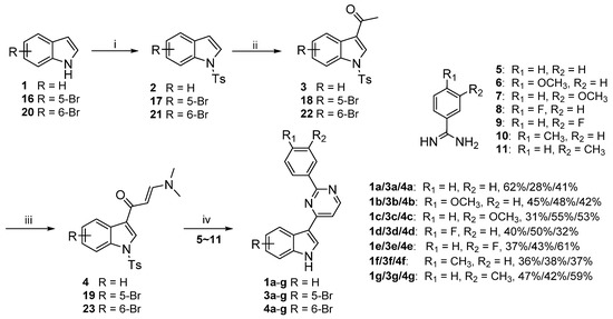
Scheme 1.
Synthesis of the final target compounds 1a–g, 3a–g and 4a–g. Reagents and conditions: (i) TsCl, NaOH, CH3CN, rt, 4 h; (ii) Ac2O, AlCl3, DCM, rt, 3 h; (iii) DMF-DMA, DMF, 110 °C, 3 h; (iv) K2CO3, 2-methoxyethanol, 120 °C, 12 h.
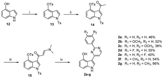
Scheme 2.
Synthesis of the final target compounds 2a–g. Reagents and conditions: (i) TsCl, NaH, DMF, 0 °C, 2 h; (ii) Ac2O, AlCl3, DCM, rt, 3 h; (iii) DMF-DMA, DMF, 110 °C, 3 h; (iv) 5–11, K2CO3, 2-methoxyethanol, 120 °C, 12 h.
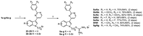
Scheme 3.
Synthesis of the final target compounds 5a–g and 6a–g. Reagents and conditions: (i) 1,6-dibromohexane, K2CO3, DMF, 50 °C 12 h; (ii) thiourea, isopropanol, 80 °C, 14 h.
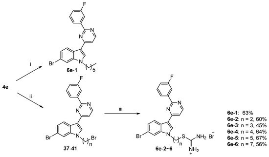
Scheme 4.
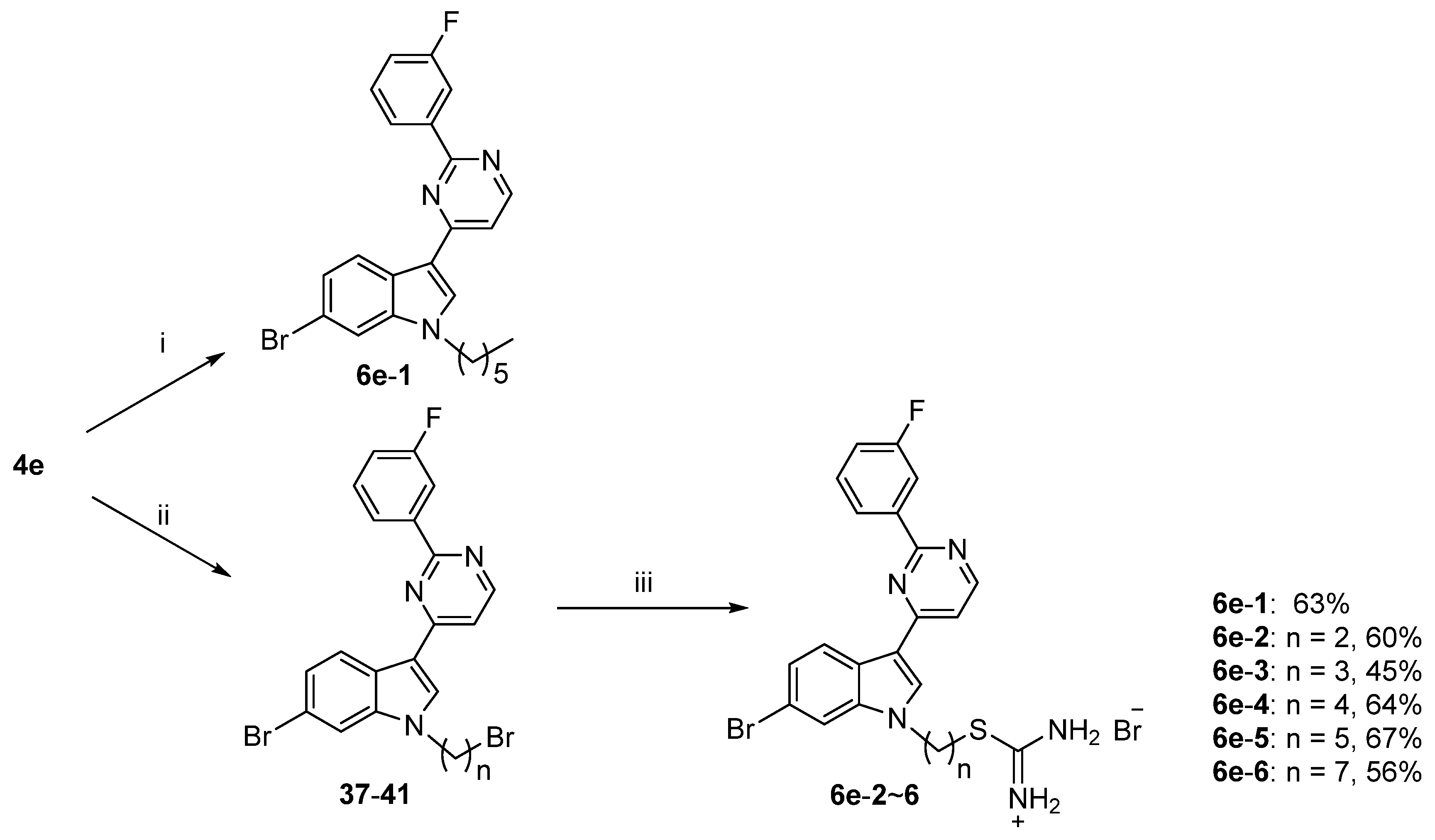
Synthesis of the final target compounds 6e-1–6. Reagents and conditions: (i) 1-bromohexane, K2CO3, DMF, 50 °C 12 h; (ii) dibromoalkyl chain, K2CO3, DMF, 50 °C 12 h; (iii) thiourea, isopropanol, 80 °C, 14 h.
The marine natural products indole alkaloids (meridianin A, C, D and G) were synthesized in four steps starting from commercially available indoles [27]. Firstly, the indolic nitrogen was protected by the reaction with tosyl chloride in the presence of NaOH and in acetonitrile, leading to the formation of compounds 2, 17 and 21 in 53–94% yields. Then, the C-3 position of indoles was acetylated using acetic anhydride and aluminum chloride inmethylene chloride to give derivatives 3, 18 and 22 in 73–79% yields. The enaminone intermediates proceeded with DMF/dimethylformamide-dimethylacetal (DMF-DMA) in 69–79% yield. Finally, compounds 1a–g, 3a–g and 4a–g were obtained from enaminone intermediates using 5–11 in 2-methoxyethanol in the presence of potassium carbonate in considerable yields (Scheme 1). As depicted in Scheme 2, the indolic nitrogen and 4-hydroxyl of compound 12 were tosyl chloride-protected to get compounds 13 in 85% yield. Then, the preparation of the corresponding 2a–g derivatives was undertaken using a similar synthetic pathway in 38–66% yields. As shown in Scheme 3 and Scheme 4, isothiouronium derivatives 5a–g, 6a–g and 6e-2–6 were synthesized in medium yield by introducing 1,6-dibromohexane or a dibromoalkyl chain with different carbon chains without any purification and then boiling isopropanol in the presence of thiourea. In addition, the compound 6e-1 was prepared by using introducing 1-bromohexane in 63% yield.
2.2. Biological Activity Assessments
2.2.1. Cell Viability Assay and SAR Analysis
To evaluate the antitumor activities of meridianin A, C, D, G and their derivatives, four JAK/STAT3 overactivated human cancer cell lines: HeLa, MDA-MB-231, A549 and DU145 were examined. Initially, the meridianins (A, C, D and G) and their derivatives 1a–g, 2a–g, 3a–g and 4a–g were prepared and evaluated for cell growth inhibitory activities against the cancer cell lines (Table 1). The results indicated that meridianins A, C, D and G displayed weak cancer cell growth inhibition in the four tested cell lines. By comparison, meridianin derivatives 1a–g, 2a–g, 3a–g and 4a–g showed significantly increased inhibitory activities. With the aim of improving the antitumor activity, compounds 5a–g and 6a–g were obtained by incorporating isothiourea groups at the C1 position of meridianins D and G with different lengths of carbon alkyl chains. Notably, the antitumor effects of the compounds were significantly enhanced, almost all compounds had a IC50 less than 10 μM (Table 2). Among them, the most potent compound 6e inhibited the growth of HeLa, MDA-MB-231, A549 and DU145 cells with IC50 values of 1.11, 1.22, 2.80 and 1.13 μM, which exhibited better activity than the positive control (Gefitinib), respectively. Based on the above results, the analogs of compound 6e with different carbon chain lengths or without isothiourea were obtained, and the antitumor activity results are shown in Table 3. The structure–activity relationship suggests that reducing or increasing the number of carbon atoms will lead to decreased antiproliferative activity. On the other hand, compound 6e-1 was found to be completely inactive with IC50 values greater than 100 μM, indicating that the substitution of the isothiourea group significantly contributed to the antitumor activity. The inhibitory effect of compound 6e on the proliferation of the four normal cell lines HUVEC, L02, L929 and MCF10A was determined by the MTT assay. The data showed that 6e had low toxicity compared to normal cells, predicting that it may be relatively safe in vivo (Table 4).

Table 1.
Antiproliferative activity of meridianins A, C, D, G and their derivatives against human cancer cell lines.
Table 2.
Antiproliferative activity of meridianin (D and G) derivatives against human cancer cell lines.
Table 3.
Antiproliferative activity of compound 6e analogs against human cancer cell lines.
Table 4.
Antiproliferative activity of compound 6e analogs against normal cell lines.
2.2.2. Compound 6e Inhibited Cancer Cell Proliferation and Induced Cell Apoptosis
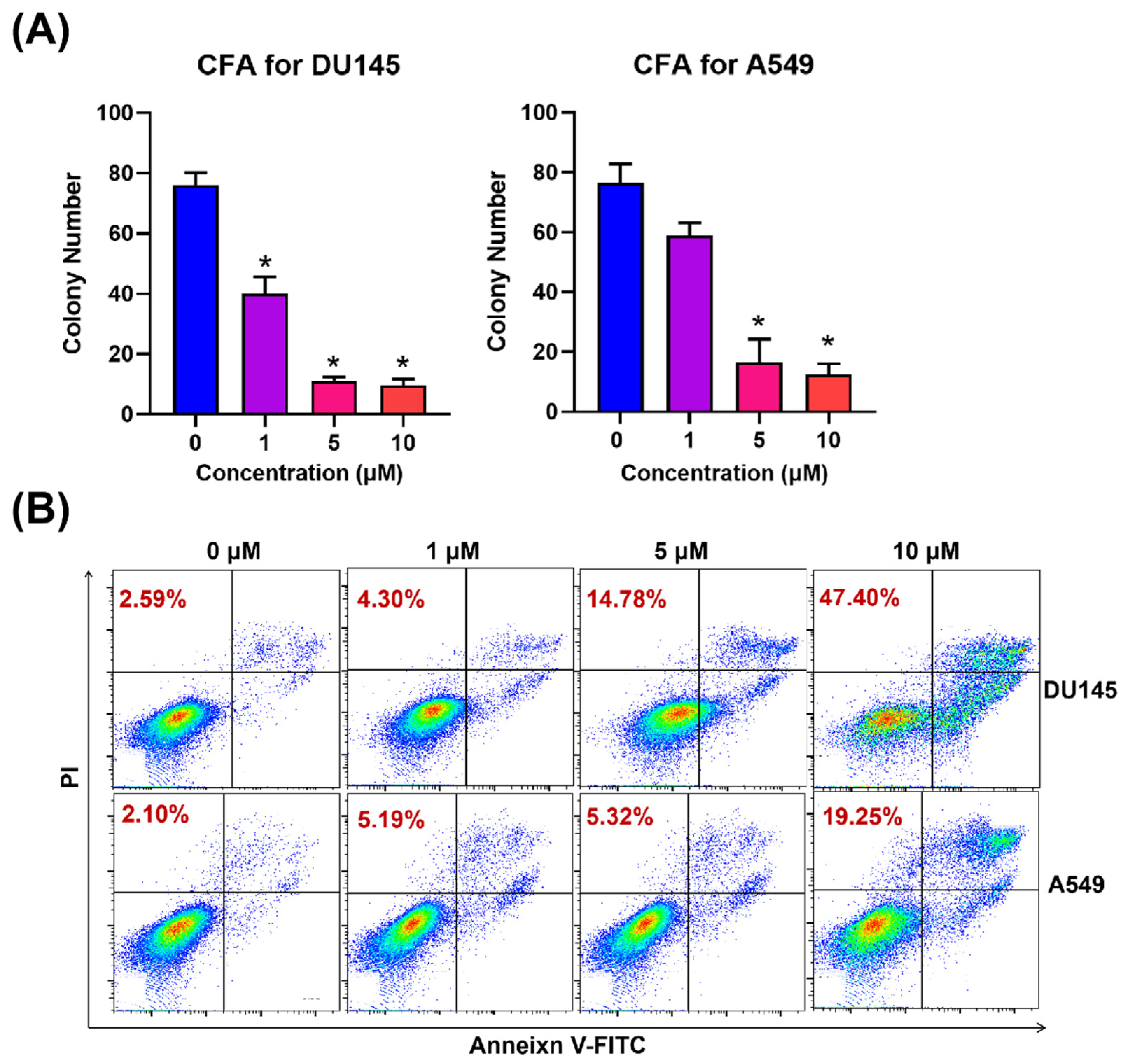
To evaluate the antiproliferative activities of compound 6e in the cell models, its effects on DU145 and A549 colony survival were evaluated. The results of the CFA analysis showed that 6e significantly inhibited the proliferation of both cells, and the effect was enhanced with the increasing 6e concentration. The effect of compound 6e on inducing tumor cell apoptosis was analyzed in Figure 1B. A549 and DU145 cells were incubated with 6e at different concentrations for 24 h. Annexin V-FITC/PI staining was carried out, and the percentage of apoptotic cells was further determined using flow cytometry. The results showed that the 6e dose dependently induced the apoptosis of the A549 and DU145 cells. As shown in Figure 1B, in A549, the induced apoptosis rates at 0, 1, 5 and 10 μM were 2.59%, 4.30%, 14.78% and 45.40%, respectively. In DU145, the induced apoptosis rates at 0, 1, 5 and 10 μM were 2.10%, 5.19%, 5.32% and 19.25%, respectively.
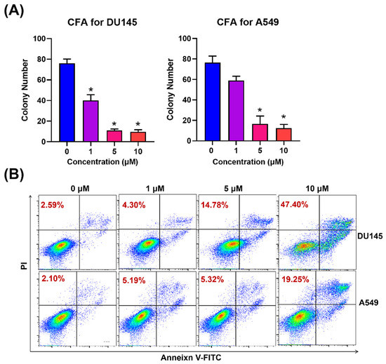
Figure 1.
(A) Effect of compound 6e on colony formation in DU145 and A549 cells. (B) Compound 6e induced A549 and DU145 cancer cells apoptosis in vitro. A549 and DU145 cells were incubated with 6e at different concentrations (0–10 μM) for 24 h. Annexin V/PI staining was carried out, and the percentage of apoptotic cells was further determined using flow cytometry. * Statistically significant (n = 2, p < 0.05).
2.2.3. Molecular Docking
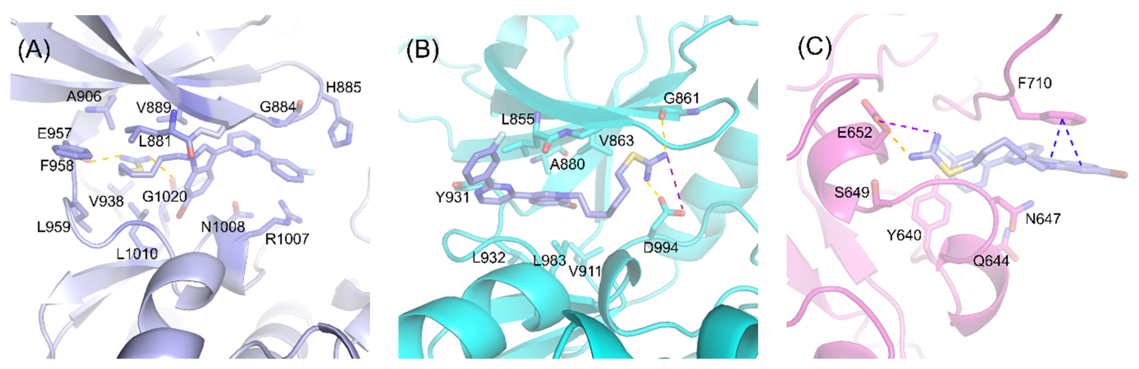
Molecular docking was performed for understanding the interaction mechanisms between compound 6e with JAK1 (PDB ID:4I5C), JAK2 (PDB ID:5CF5) and STAT3 (PDB ID:1BG1) [28], respectively. For JAK1, the results showed that the hydrophobic fatty chain was accommodated at a hydrophobic pocket mainly defined by residues Leu881, Val889, Ala906, Val938, Phe958, Leu959 and Leu1010 (Figure 2A). The hydrogen in the imine of 6e engages H-bond formation with Glu957 and Gly1020. For JAK2, the docking poses suggested that the imine group of 6e interacts with a carboxyl group of Asp994 and carbonyl group of Gly861 by forming two hydrogen bonds (Figure 2B). In addition, the nitrogen of imine on 6e also forms a salt bridge interaction with Asp994. While, for STAT3, Phe710 forms π–π interactions with the indole ring of 6e, Glu652 forms a hydrogen bond and a salt bridge with the H atom and the N atom on imine, respectively (Figure 2C) [29].

Figure 2.
Binding modes of the compound 6e with JAK1, JAK2 and STAT3. The compound 6e is shown in blue, while the JAK1 (A), JAK2 (B) and STAT3 (C) are shown in purple, blue and pink, respectively. The H-bonds, π–π and salt bridges were shown in dashed lines.
2.2.4. Compound 6e Inhibits the Expression of JAK/STAT3 Target Genes
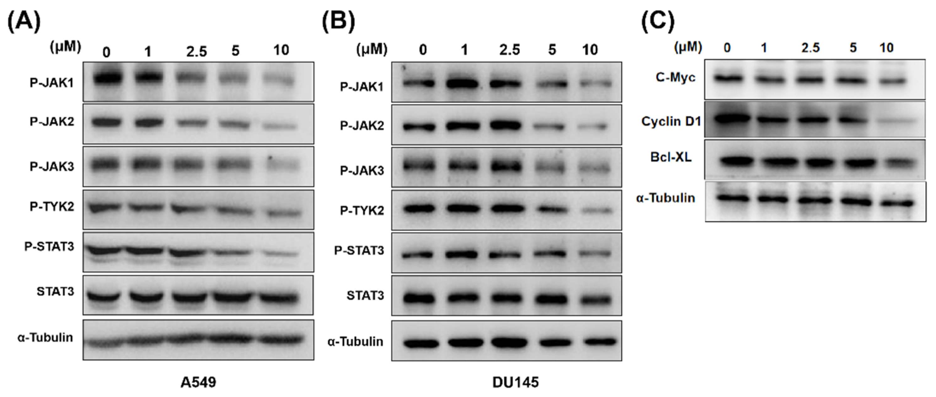
The ability of compound 6e to inhibit the phosphorylation of JAK/STAT3 was determined in A549 and DU145 cells. As shown in Figure 3A,B, after 24 h of treatment with 5-μM 6e, the decreased levels of JAK1, JAK2 and STAT3 were observed in both A549 and DU145 cells, but no significant change was seen with the total STAT3 protein expression. Moreover, compound 6e significantly inhibited the expression of JAK/STAT3 downstream genes c-Myc, Cyclin D1 and Bcl-XL at 10 μM after 24 h of treatment (Figure 3C). Therefore, pretreatment with compound 6e suppressed the JAK/STAT3 signaling pathway and its downstream gene expressions, which were consistent with the above results obtained in vitro.
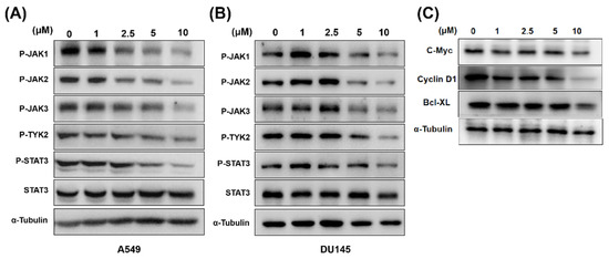
Figure 3.
The effect of 6e on overexpressed proteins in the JAK/STAT3 signaling pathway by Western blot analysis. (A) 6e decreased levels of JAK1, JAK2 and STAT3 in A549 cells. (B) 6e decreased levels of JAK1, JAK2 and STAT3 in DU145 cells. (C) 6e inhibited the expression of JAK/STAT3 downstream genes c-Myc, Cy-clin D1 and Bcl-XL in DU145 cells.
2.2.5. Compound 6e Inhibited Tumor Growth in a Mouse Breast Cancer Model
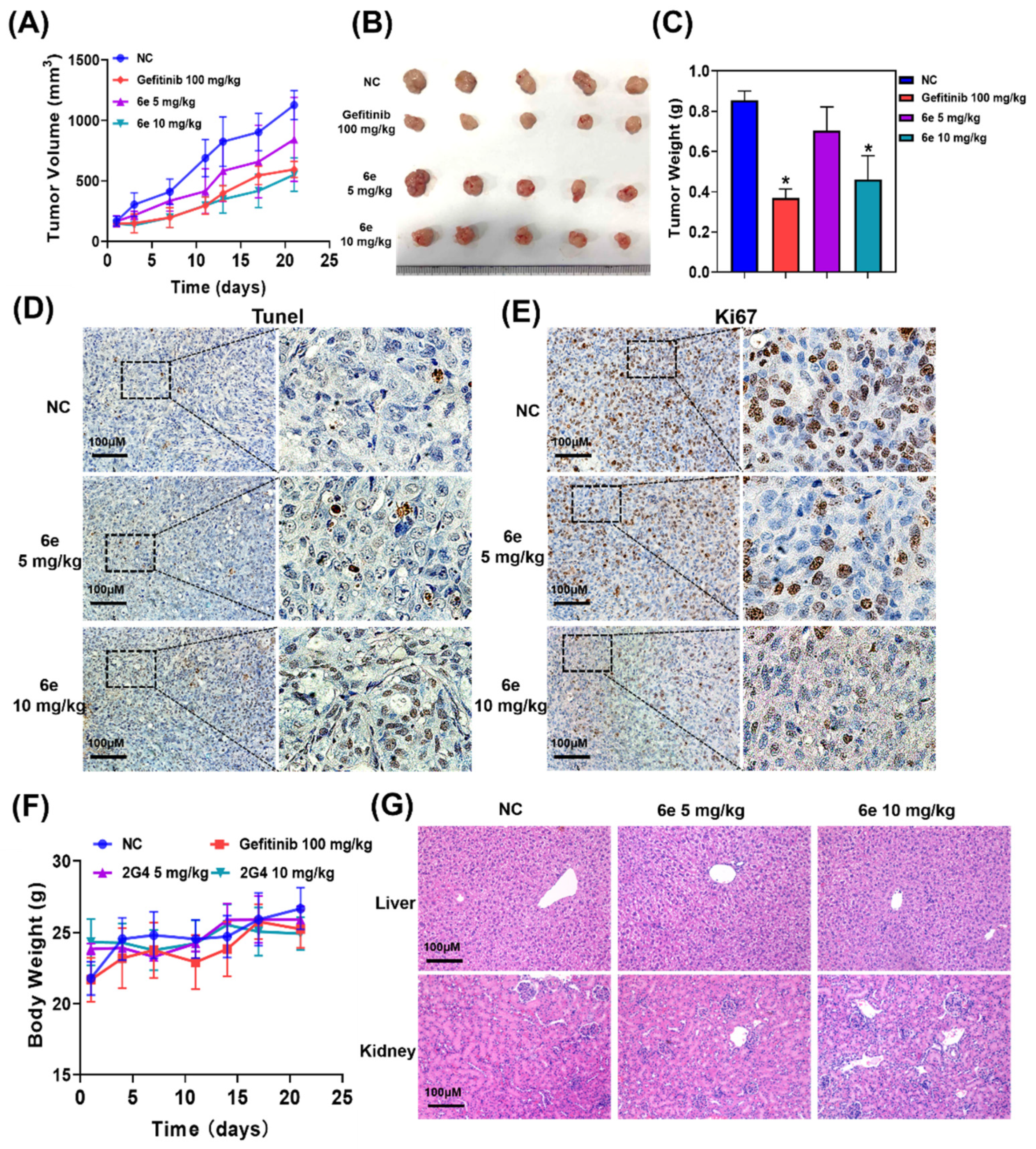
To further investigate the antitumor potential in vivo, we evaluated the effects of compound 6e in a nude mice tumor model (Figure 4). After the solid tumor was established, twenty-four nude mice were randomly divided into four groups, which were the vehicle control group, 6e groups (5 mg/kg and 10 mg/kg) and Gefitinib-positive control group. All the compounds were taken by intragastric gavage. Compound 6e at the dose of 10 mg/kg could significantly inhibit tumor growth, and the tumor inhibition rate of 6e was over 40%, which was comparable to that of the positive control (Figure 4A–C). Subsequently, Ki67 and Tunel staining were performed on tumor sections, which showed that tumor proliferation marker Ki67 was significantly inhibited, and the proportion of apoptotic cells that were marked by Tunel-positive staining also significantly increased with 6e treatment at a dose of 10 mg/kg (Figure 4D,E). Moreover, during the administration period, the weight of the nude mice did not increase or decrease significantly, indicating that there was no obvious biological toxicity of 6e (Figure 4F). H&E staining was performed on the liver and kidneys of each group of mice to observe the hepatic and renal toxicity of 6e. The results showed that no significant liver and kidney damage was observed in all the groups of mice, suggesting that 6e was less toxic at therapeutic doses (Figure 4G).
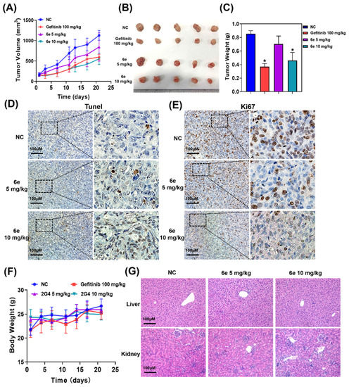
Figure 4.
The results of 6e on the DU145 tumor-bearing model in vivo therapeutic. (A) Growth curve of subcutaneous tumor-bearing DU145 treated with 6e. (B) Anatomical mice tumor tissues treated with 6e. (C) The weight of the tumors in each group treated with 6e. (D) Ki67 and (E) Tunel staining in DU145 tumors treated with different doses of 6e. (F) Changes in the body weights of mice in each group during administration. (G) H&E sections of the liver and kidneys of treated mice in each group. * Statistically significant (n = 5, p < 0.05).
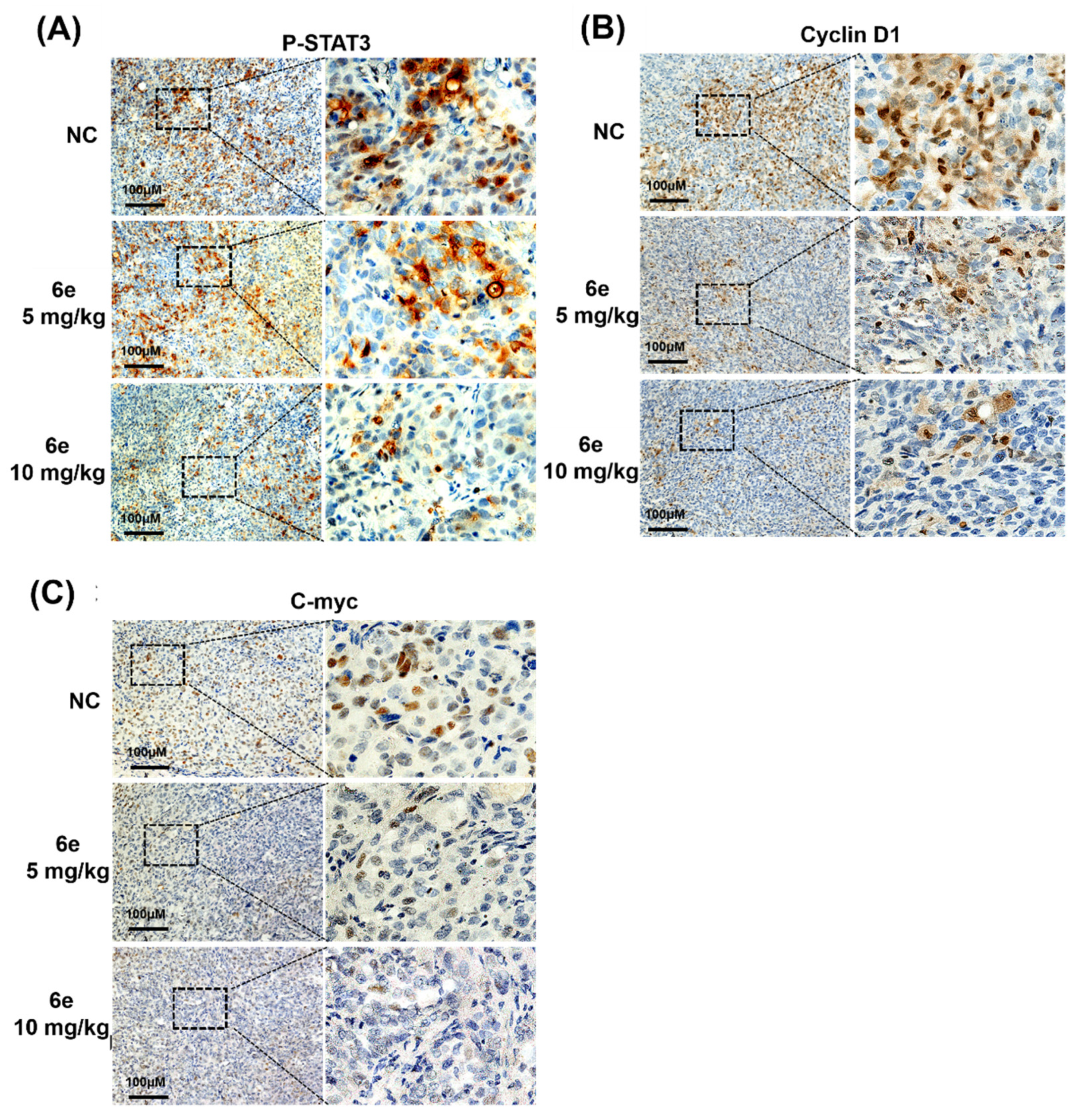
2.2.6. Immunohistochemical (IHC) Analysis
To further test the inhibitory effects of 6e on JAK/STAT3 signaling, the IHC analysis of nude mice inoculated with DU145 tumor cells was performed. As shown in Figure 5, after 6e treatment, the intratumoral staining of p-STAT3, cyclin D1 and c-Myc in DU145-inoculated mice was significantly lower than those in the NC group, and their staining levels decreased while the dose of 6e increased. Therefore, compound 6e may exert antitumor effects by inhibiting the JAK/STAT3 signaling pathway both in vitro and in vivo.

Figure 5.
Immunohistochemistry of the protein expressions in a DU145 xenograft model. Formalin-fixed paraffin-embedded sections from DU145 xenograft tumors were stained with H&E (A) p-STAT3, (B) cyclin D1 and (C) c-Myc antibodies.
3. Conclusions
In summary, a novel series of meridianin derivatives was obtained and biologically evaluated. Initially, the meridianins (A, C, D and G) and their four series derivatives of compounds 1a–g, 2a–g, 3a–g and 4a–g were prepared, and the results indicated that the meridianins (A, C, D and G) displayed weak inhibitory activity on four JAK/STAT3 overactivated human cancer cell lines: HeLa, MDA-MB-231, A549 and DU145, whereas most of the meridianin derivatives exerted promising inhibitory activity on the tested cell lines. To improve the antitumor activity, meridianin derivatives 5a–g and 6a–g were designed and synthesized by incorporating isothiourea groups at the N1 position with different lengths of carbon alkyl chains. Surprisingly, the antitumor effects of the isothiouronium-modified compounds were significantly enhanced, with IC50 less than 10 μM. Among them, the most potent compound, 6e with an alkyl chain of six carbon atoms, had an IC50 that ranged from 1.11 to 2.80 μM in various cancer cell lines, which was superior to the positive control, Gefitinib. The structure–activity relationship (SAR) study indicated that isothiouronium modified by N-alkylation with 6C alkyl chains may contribute the most to antitumor activity. It is worth noting that 6e had low toxicity to normal cells. The Western blotting assays suggested that treatment with compound 6e could decrease the phosphorylation level of JAK1, JAK2 and STAT3 at 5 μM but did not affect the total STAT3 level. Moreover, 6e also suppressed the expression of STAT3 downstream genes, including c-Myc, cyclin D1 and Bcl-XL. Consistently, 6e dose-dependently inhibited the proliferation and induced the apoptosis of A549 and DU145 cells. Molecular docking studies demonstrated that an H-bond is the main type of interaction between compound 6e and the JAK1/JAK2 kinases, and 6e could also tightly bind to the STAT3 SH2 domain. An additional in vivo study revealed that the application of 6e at a dose of 10 mg/kg could significantly inhibit the DU145 xenograft tumor growth without an obvious body weight loss, which was comparable to that of the positive control. Taken together, these results clearly indicated that 6e could be a highly potent antitumor agent by targeting the JAK/STAT3 signaling pathway. In addition, the pharmacokinetic properties of compound 6e will be further investigated in the future.
4. Materials and Methods
4.1. Chemistry
All commercially available starting materials and solvents were purchased from commercial vendors and used without further purification. Reactions were monitored using analytical thin-layer chromatography (TLC) on precoated silica gel GF254 plates (Qingdao Haiyang Chemical Plant, Qingdao, China) and visualized under ultraviolet light (254 nm and 365 nm). Column chromatography was performed on silica gel (200–300 mesh). Melting points were determined on a Mitamura-Riken micro-hot stage and uncorrected. 1H and 13C NMR spectra were recorded on the Broker AVANCE NEO and Agilent DD2 500 with 400 or 500 MHz for proton (1H NMR) and 100 or 125 MHz for carbon (13C NMR), respectively. The chemical shifts (δ) were expressed in parts per million (ppm) downfield, and the coupling constant (J) values were described as hertz. High-resolution (ESI) MS spectra were recorded using a QTOF-2 Micromass spectrometer. The purity of the final compounds for biological evaluation was higher than 95% by analytical HPLC analysis with the Primaide 1210 system.
Compounds 1–4, 16–19 and 20–23 were prepared according to the procedure published by Dong et al., and the spectroscopic data for the intermediates were identical to those described in the literature [30].
4.1.1. General Procedure for the Synthesis of 1a–g, 3a–g and 4a–g Meridianin Analogs
To the solution of intermediates 4, 19 and 23 (1.0 equiv.) in 2-methoxyethanol (5 mL) was added 5–11 (2.5 equiv.) and potassium carbonate (2.0 equiv.), respectively. The reaction mixture was stirred at 120 °C for 20 h under a nitrogen atmosphere. Then, the mixture was poured into ice water and extracted with ethyl acetate (3 × 50 mL). The combined organic layers were washed with brine and dried over magnesium sulfate anhydrous. After filtration, the solvent was removed under vacuum, and the residue was purified by silica gel column chromatography (petroleum ether/ethyl acetate 10:1) to give the final target compounds 1a–g, 3a–g and 4a–g.
3-(2-phenylpyrimidin-4-yl)-1H-indole (1a). Yield: 62%; pale yellow solid; m.p. 137–139 °C; 1H NMR (500 MHz, DMSO-d6): δ 11.94 (s, 1H), 8.75–8.71 (m, 1H), 8.68 (d, J = 7.6 Hz, 1H), 8.54 (d, J = 7.0 Hz, 2H), 8.50 (d, J = 2.5 Hz, 1H), 7.83 (d, J = 5.3 Hz, 1H), 7.60 (t, J = 7.2 Hz, 2H), 7.57–7.55 (m, 1H), 7.53 (d, J = 7.8 Hz, 1H), 7.31–7.22 (m, 2H); 13C NMR (125 MHz, DMSO-d6): δ 163.78, 162.85, 157.10, 138.72, 137.86, 131.09, 130.25, 129.25, 128.23, 125.80, 122.92, 122.25, 121.60, 114.68, 113.87, 112.81; HRMS: m/z [M + H]+ calcd for C18H13N3, 272.1182; found, 272.1189.
3-(2-(4-methoxyphenyl)pyrimidin-4-yl)-1H-indole (1b). Yield: 45%; white solid; m.p. 194–196 °C; 1H NMR (500 MHz, DMSO-d6): δ 11.90 (d, J = 8.4 Hz, 1H), 8.66 (s, 1H), 8.51–8.44 (m, 1H), 7.74 (dd, J = 5.5, 3.0 Hz, 1H), 7.54–7.49 (m, 1H), 7.27 (d, J = 7.0 Hz, 1H), 7.13 (dd, J = 8.4, 3.4 Hz, 1H), 3.85 (d, J = 3.2 Hz, 1H); 13C NMR (125 MHz, DMSO-d6): δ 163.49, 162.56, 161.76, 156.86, 137.71, 131.10, 129.95, 129.71, 125.69, 122.75, 122.14, 121.39, 114.47, 113.89, 113.81, 112.66, 55.75; HRMS: m/z [M + H]+ calcd for C19H15ON3, 302.1288; found, 302.1296.
3-(2-(3-methoxyphenyl)pyrimidin-4-yl)-1H-indole (1c). Yield: 31%; white solid; m.p. 188–190 °C; 1H NMR (500 MHz, DMSO-d6): δ 11.91 (s, 1H), 8.71 (d, J = 5.4 Hz, 1H), 8.68–8.63 (m, 1H), 8.49 (d, J = 2.9 Hz, 1H), 8.12 (d, J = 7.7 Hz, 1H), 8.08 (s, 1H), 7.82 (d, J = 5.4 Hz, 1H), 7.50 (dd, J = 9.3, 6.7 Hz, 2H), 7.26–7.20 (m, 2H), 7.12 (dd, J = 8.1, 2.2 Hz, 1H), 3.89 (s, 3H); 13C NMR (125 MHz, DMSO-d6): δ 163.38, 162.67, 160.00, 156.94, 140.06, 137.72, 130.23, 130.13, 125.68, 122.81, 122.08, 121.42, 120.46, 116.90, 114.64, 113.69, 112.98, 112.72, 55.56; HRMS: m/z [M + H]+ calcd for C19H15ON3, 302.1288; found, 302.1295.
3-(2-(4-fluorophenyl)pyrimidin-4-yl)-1H-indole (1d). Yield: 40%; white solid; m.p. 126–128 °C; 1H NMR (500 MHz, DMSO-d6): δ 11.94 (s, 1H), 8.70 (d, J = 5.4 Hz, 1H), 8.62 (dd, J = 6.4, 2.2 Hz, 1H), 8.56 (dd, J = 8.8, 5.8 Hz, 2H), 8.49 (d, J = 2.3 Hz, 1H), 7.81 (d, J = 5.4 Hz, 1H), 7.53–7.49 (m, 1H), 7.41 (t, J = 8.8 Hz, 2H), 7.24 (pd, J = 7.0, 3.5 Hz, 2H); 13C NMR (125 MHz, DMSO-d6): δ 165.24, 163.27, 162.75, 156.99, 137.73, 135.06, 130.44, 130.37, 130.23, 125.62, 122.81, 122.06, 121.48, 116.13, 115.96, 114.48, 113.65, 112.70; HRMS: m/z [M + H]+ calcd for C18H12N3F, 290.1088; found, 290.1096.
3-(2-(3-fluorophenyl)pyrimidin-4-yl)-1H-indole (1e). Yield: 37%; white solid; m.p. 128–130 °C; 1H NMR (500 MHz, DMSO-d6): δ 11.95 (s, 1H), 8.73 (d, J = 5.4 Hz, 1H), 8.61 (d, J = 7.5 Hz, 1H), 8.51 (d, J = 2.9 Hz, 1H), 8.38 (d, J = 7.8 Hz, 1H), 8.21 (d, J = 9.8 Hz, 1H), 7.85 (d, J = 5.5 Hz, 1H), 7.64 (dd, J = 14.1, 7.9 Hz, 1H), 7.52 (d, J = 7.4 Hz, 1H), 7.39 (td, J = 8.4, 2.4 Hz, 1H), 7.29–7.23 (m, 2H); 13C NMR (125 MHz, DMSO-d6): δ 163.97, 162.82, 162.43 (d, J = 3.2 Hz), 162.04, 157.05, 141.18 (d, J = 7.6 Hz), 137.75, 131.23 (d, J = 8.1 Hz), 130.39, 125.60, 124.12 (d, J = 2.5 Hz), 122.85, 121.94, 121.54, 117.87, 117.70, 115.04, 114.53, 114.34, 113.55, 112.76; HRMS: m/z [M + H]+ calcd for C18H12N3F, 290.1088; found, 290.1096.
3-(2-(p-tolyl)pyrimidin-4-yl)-1H-indole (1f). Yield: 36%; white solid; m.p. 193–195 °C; 1H NMR (500 MHz, DMSO-d6): δ 11.90 (s, 1H), 8.68 (d, J = 5.1 Hz, 1H), 8.64 (d, J = 7.2 Hz, 1H), 8.45 (s, 1H), 8.40 (d, J = 7.8 Hz, 2H), 7.77 (d, J = 5.0 Hz, 1H), 7.51 (d, J = 7.6 Hz, 1H), 7.38 (d, J = 7.5 Hz, 2H), 7.28–7.21 (m, 2H), 2.39 (s, 3H); 13C NMR (125 MHz, DMSO-d6): δ 163.71, 162.60, 156.93, 140.72, 137.70, 135.89, 129.96, 129.75, 128.08, 125.64, 122.80, 122.09, 121.46, 114.28, 113.78, 112.69, 21.48; HRMS: m/z [M + H]+ calcd for C19H15N3, 286.1339; found, 286.1346.
3-(2-(m-tolyl)pyrimidin-4-yl)-1H-indole (1g). Yield: 47%; white solid; m.p. 121–123 °C; 1H NMR (500 MHz, DMSO-d6): δ 11.92 (s, 1H), 8.71 (d, J = 5.4 Hz, 1H), 8.66 (d, J = 7.5 Hz, 1H), 8.48 (d, J = 2.5 Hz, 1H), 8.34 (d, J = 8.1 Hz, 2H), 7.81 (d, J = 5.4 Hz, 1H), 7.52 (d, J = 7.9 Hz, 1H), 7.47 (t, J = 7.5 Hz, 1H), 7.36 (d, J = 7.3 Hz, 1H), 7.25 (dd, J = 14.6, 7.3 Hz, 2H), 2.45 (s, 3H); 13C NMR (125 MHz, DMSO-d6): δ 163.73, 162.63, 156.94, 138.56, 138.14, 137.73, 131.58, 130.06, 129.02, 128.74, 125.67, 125.31, 122.77, 122.08, 121.44, 114.50, 113.79, 112.70, 21.71; HRMS: m/z [M + H]+ calcd for C19H15N3, 286.1339; found, 286.1347.
5-bromo-3-(2-phenylpyrimidin-4-yl)-1H-indole (3a). Yield: 28%; white solid; m.p. 201–203 °C; 1H NMR (500 MHz, DMSO-d6): δ 12.11 (s, 1H), 8.86 (d, J = 2.0 Hz, 1H), 8.75 (d, J = 5.4 Hz, 1H), 8.55 (d, J = 3.0 Hz, 1H), 8.50–8.46 (m, 2H), 7.83 (d, J = 5.4 Hz, 1H), 7.58 (tdd, J = 6.8, 3.8, 1.7 Hz, 3H), 7.49 (d, J = 8.6 Hz, 1H), 7.37 (dd, J = 8.6, 2.0 Hz, 1H); 13C NMR (125 MHz, DMSO-d6): δ 163.68, 162.17, 157.25, 138.52, 136.40, 131.39, 131.11, 129.14, 127.99, 127.43, 125.31, 124.51, 114.71, 114.61, 114.07, 113.30; HRMS: m/z [M + H]+ calcd for C18H12N3Br, 350.0287; found, 350.0287.
5-bromo-3-(2-(4-methoxyphenyl)pyrimidin-4-yl)-1H-indole (3b). Yield: 48%; pale yellow solid; m.p. 97–99 °C; 1H NMR (500 MHz, DMSO-d6): δ 12.08 (s, 1H), 8.84 (s, 1H), 8.68 (d, J = 5.4 Hz, 1H), 8.51 (s, 1H), 8.43 (d, J = 8.7 Hz, 2H), 7.75 (d, J = 5.4 Hz, 1H), 7.49 (d, J = 8.6 Hz, 1H), 7.36 (dd, J = 8.6, 1.6 Hz, 1H), 7.12 (d, J = 8.8 Hz, 2H), 3.86 (s, 3H); 13C NMR (125 MHz, DMSO-d6): δ 163.52, 162.01, 161.85, 157.13, 136.37, 131.23, 130.99, 129.59, 127.42, 125.26, 124.49, 114.68, 114.49, 114.00, 113.89, 113.41, 55.82; HRMS calcd for C19H14N381Br [M + H]+ 382.0373, found: 382.0382.
5-bromo-3-(2-(3-methoxyphenyl)pyrimidin-4-yl)-1H-indole (3c). Yield: 55%; white solid; m.p. 192–194 °C; 1H NMR (500 MHz, DMSO-d6): δ 12.11 (s, 1H), 8.91 (s, 1H), 8.73 (d, J = 5.4 Hz, 1H), 8.55 (d, J = 2.9 Hz, 1H), 8.08 (dd, J = 10.7, 4.5 Hz, 2H), 7.83 (d, J = 5.4 Hz, 1H), 7.52–7.45 (m, 2H), 7.37 (dd, J = 8.6, 1.7 Hz, 1H), 7.12 (dd, J = 8.1, 2.4 Hz, 1H), 3.95 (s, 3H); 13C NMR (125 MHz, DMSO-d6): δ 163.42, 162.18, 160.04, 157.14, 139.95, 136.40, 131.44, 130.20, 127.45, 125.33, 124.58, 120.45, 117.82, 114.72, 114.64, 114.07, 113.23, 112.01, 55.67; HRMS calcd for C19H14N381Br [M + H]+ 382.0373, found: 382.0382.
5-bromo-3-(2-(4-fluorophenyl)pyrimidin-4-yl)-1H-indole (3d). Yield: 50%; white solid; m.p. 88–90 °C; 1H NMR (500 MHz, DMSO-d6): δ 12.13 (s, 1H), 8.79 (d, J = 1.2 Hz, 1H), 8.73 (d, J = 5.4 Hz, 1H), 8.55 (d, J = 2.8 Hz, 1H), 8.51 (dd, J = 8.5, 5.9 Hz, 2H), 7.82 (d, J = 5.4 Hz, 1H), 7.49 (d, J = 8.6 Hz, 1H), 7.44–7.34 (m, 3H); 13C NMR (125 MHz, DMSO-d6): δ 165.29, 163.32, 162.80, 162.18, 157.28, 136.41, 135.03, 131.48, 130.27, 127.35, 125.33, 124.34, 116.17, 116.00, 114.74, 114.56, 114.09, 113.23; HRMS: m/z [M + H]+ calcd for C18H11N3BrF, 368.0193; found, 368.0189.
5-bromo-3-(2-(3-fluorophenyl)pyrimidin-4-yl)-1H-indole (3e). Yield: 43%; white solid; m.p. 205–206 °C; 1H NMR (500 MHz, DMSO-d6): δ 12.13 (s, 1H), 8.81 (s, 1H), 8.74 (d, J = 5.4 Hz, 1H), 8.55 (d, J = 2.8 Hz, 1H), 8.31 (d, J = 7.8 Hz, 1H), 8.17 (d, J = 10.3 Hz, 1H), 7.85 (d, J = 5.4 Hz, 1H), 7.62 (dd, J = 14.1, 7.8 Hz, 1H), 7.49 (d, J = 8.6 Hz, 1H), 7.43–7.33 (m, 2H); 13C NMR (125 MHz, DMSO-d6): δ 163.98, 162.45, 162.25, 162.05, 157.27, 141.09 (d, J = 7.6 Hz), 136.41, 131.59, 131.18 (d, J = 8.1 Hz), 127.36, 125.34, 124.42, 123.97 (d, J = 2.3 Hz), 117.95, 117.78, 115.06, 114.73, 114.51, 114.32, 114.14, 113.13; HRMS: m/z [M + H]+ calcd for C18H11N3BrF, 368.0193; found, 368.0198.
5-bromo-3-(2-(p-tolyl)pyrimidin-4-yl)-1H-indole (3f). Yield: 38%; pale yellow solid; m.p. 197–199 °C; 1H NMR (500 MHz, DMSO-d6): δ 12.09 (s, 1H), 8.86 (d, J = 1.7 Hz, 1H), 8.71 (d, J = 5.4 Hz, 1H), 8.53 (s, 1H), 8.37 (d, J = 8.1 Hz, 2H), 7.79 (d, J = 5.4 Hz, 1H), 7.49 (d, J = 8.6 Hz, 1H), 7.41–7.35 (m, 3H), 2.41 (s, 3H); 13C NMR (125 MHz, DMSO-d6): δ 163.75, 162.07, 157.19, 140.89, 136.38, 135.85, 131.27, 129.75, 127.97, 127.44, 125.27, 124.55, 114.69, 114.32, 114.03, 113.35, 21.44; HRMS: m/z [M + H]+ calcd for C19H14N3Br, 364.0444; found, 368.0444.
5-bromo-3-(2-(m-tolyl)pyrimidin-4-yl)-1H-indole (3g). Yield: 42%; pale yellow solid; m.p. 167–169 °C; 1H NMR (500 MHz, DMSO-d6): δ 12.09 (s, 1H), 8.94 (s, 1H), 8.73 (d, J = 5.4 Hz, 1H), 8.54 (d, J = 2.8 Hz, 1H), 8.35 (s, 1H), 8.27 (d, J = 7.7 Hz, 1H), 7.81 (d, J = 5.4 Hz, 1H), 7.51–7.44 (m, 2H), 7.40–7.35 (m, 2H), 2.47 (s, 3H); 13C NMR (125 MHz, DMSO-d6): δ 163.68, 162.08, 157.19, 138.44, 138.21, 136.37, 131.68, 131.32, 129.06, 128.73, 127.50, 125.25, 125.18, 124.75, 114.69, 114.45, 114.05, 113.30, 21.68; HRMS: m/z [M + H]+ calcd for C19H14N3Br, 364.0444; found, 368.0446.
6-bromo-3-(2-phenylpyrimidin-4-yl)-1H-indole (4a). Yield: 41%; pale yellow solid; m.p. 175–177 °C; 1H NMR (500 MHz, DMSO-d6): δ 12.03 (s, 1H), 8.74 (d, J = 5.4 Hz, 1H), 8.60 (d, J = 8.5 Hz, 1H), 8.51 (t, J = 4.4 Hz, 3H), 7.82 (d, J = 5.4 Hz, 1H), 7.70 (d, J = 1.5 Hz, 1H), 7.60–7.53 (m, 3H), 7.40 (dd, J = 8.5, 1.6 Hz, 1H); 13C NMR (125 MHz, DMSO-d6): δ 163.68, 162.26, 157.16, 138.58, 138.40, 131.05, 130.98, 129.15, 128.13, 124.70, 124.37, 123.85, 115.46, 115.27, 114.66, 113.90; HRMS: m/z [M + H]+ calcd for C18H13N3Br, 350.0287; found, 350.0288.
6-bromo-3-(2-(4-methoxyphenyl)pyrimidin-4-yl)-1H-indole (4b). Yield: 42%; white solid; m.p. >210 °C; 1H NMR (500 MHz, DMSO-d6): δ 12.00 (s, 1H), 8.68 (d, J = 5.4 Hz, 1H), 8.58 (d, J = 8.5 Hz, 1H), 8.47 (dd, J = 13.9, 5.7 Hz, 3H), 7.74 (d, J = 5.4 Hz, 1H), 7.69 (s, 1H), 7.38 (d, J = 8.5 Hz, 1H), 7.11 (d, J = 8.7 Hz, 2H), 3.85 (s, 3H); 13C NMR (125 MHz, DMSO-d6): δ 163.54, 162.08, 161.80, 157.07, 138.56, 130.93, 130.79, 129.75, 124.71, 124.27, 123.85, 115.41, 115.25, 114.49, 114.00, 113.92, 55.76; HRMS calcd for C19H14ON381Br [M + H]+ 382.0373, found: 382.0384.
6-bromo-3-(2-(3-methoxyphenyl)pyrimidin-4-yl)-1H-indole (4c). Yield: 53%; yellow solid; m.p. 180–181 °C; 1H NMR (500 MHz, DMSO-d6): δ 12.03 (s, 1H), 8.74 (d, J = 5.4 Hz, 1H), 8.60 (d, J = 8.5 Hz, 1H), 8.51 (d, J = 2.8 Hz, 1H), 8.09 (d, J = 7.7 Hz, 1H), 8.05 (s, 1H), 7.82 (d, J = 5.4 Hz, 1H), 7.70 (s, 1H), 7.49 (t, J = 7.9 Hz, 1H), 7.40 (d, J = 8.5 Hz, 1H), 7.12 (dd, J = 8.1, 1.6 Hz, 1H), 3.89 (s, 3H); 13C NMR (125 MHz, DMSO-d6): δ 163.45, 162.20, 160.00, 157.16, 139.90, 138.58, 130.96, 130.26, 124.71, 124.30, 123.76, 120.49, 117.11, 115.47, 115.31, 114.75, 113.85, 112.83, 55.58; HRMS calcd for C19H14ON381Br [M + H]+ 382.0373, found: 380.0377.
6-bromo-3-(2-(4-fluorophenyl)pyrimidin-4-yl)-1H-indole (4d). Yield: 32%; white solid; m.p. 187–188 °C; 1H NMR (500 MHz, DMSO-d6): δ 12.04 (s, 1H), 8.73 (d, J = 5.4 Hz, 1H), 8.59–8.52 (m, 3H), 8.52 (d, J = 2.9 Hz, 1H), 7.82 (d, J = 5.4 Hz, 1H), 7.70 (d, J = 1.7 Hz, 1H), 7.43–7.36 (m, 3H); 13C NMR (125 MHz, DMSO-d6): δ 165.27, 163.30, 162.81, 162.27, 157.22, 138.59, 130.46 (d, J = 8.7 Hz), 131.06, 130.50, 130.43, 124.65, 124.37, 123.79, 116.16, 115.99, 115.46, 115.29, 114.59, 113.83; HRMS: m/z [M + H]+ calcd for C18H13N3Br, 368.0193; found, 368.0193.
6-bromo-3-(2-(3-fluorophenyl)pyrimidin-4-yl)-1H-indole (4e). Yield: 61%; white solid; m.p. >210 °C; 1H NMR (500 MHz, DMSO-d6): δ 12.06 (s, 1H), 8.76 (d, J = 5.4 Hz, 1H), 8.57–8.52 (m, 2H), 8.35 (d, J = 7.8 Hz, 1H), 8.17 (d, J = 10.1 Hz, 1H), 7.86 (d, J = 5.4 Hz, 1H), 7.70 (d, J = 1.5 Hz, 1H), 7.63 (dd, J = 14.1, 7.9 Hz, 1H), 7.41 (dd, J = 8.5, 1.7 Hz, 2H); 13C NMR (125 MHz, DMSO-d6): δ 163.96, 162.50 (d, J = 3.2 Hz), 162.35, 162.03, 157.28, 138.60, 131.26, 130.77, 127.17, 124.62, 124.44, 124.18, 123.66, 115.50, 115.33, 115.16, 114.55, 114.37, 113.72; HRMS: m/z [M + H]+ calcd for C18H11N3BrF, 368.0193; found, 368.0192.
6-bromo-3-(2-(p-tolyl)pyrimidin-4-yl)-1H-indole (4f). Yield: 37%; yellow solid; m.p. 192–194 °C; 1H NMR (500 MHz, DMSO-d6): δ 12.02 (s, 1H), 8.71 (d, J = 5.4 Hz, 1H), 8.59 (d, J = 8.5 Hz, 1H), 8.49 (d, J = 2.8 Hz, 1H), 8.39 (d, J = 8.0 Hz, 2H), 7.78 (d, J = 5.4 Hz, 1H), 7.70 (d, J = 1.1 Hz, 1H), 7.39 (dd, J = 13.5, 4.7 Hz, 3H), 2.40 (s, 3H); 13C NMR (125 MHz, DMSO-d6): δ 163.75, 162.16, 157.09, 140.78, 138.57, 135.75, 130.87, 129.76, 128.11, 124.71, 124.31, 123.84, 115.43, 115.26, 114.38, 113.95, 21.49; HRMS: m/z [M + H]+ calcd for C19H14N3Br, 364.0444; found, 364.0443.
6-bromo-3-(2-(m-tolyl)pyrimidin-4-yl)-1H-indole (4g). Yield: 59%; pale yellow solid; m.p. 203–204 °C; 1H NMR (500 MHz, DMSO-d6): δ 12.02 (s, 1H), 8.73 (d, J = 5.4 Hz, 1H), 8.59 (d, J = 8.5 Hz, 1H), 8.50 (d, J = 2.7 Hz, 1H), 8.30 (d, J = 9.2 Hz, 2H), 7.81 (d, J = 5.4 Hz, 1H), 7.70 (d, J = 1.1 Hz, 1H), 7.46 (t, J = 7.5 Hz, 1H), 7.41 (dd, J = 8.5, 1.3 Hz, 1H), 7.36 (d, J = 7.5 Hz, 1H), 2.45 (s, 3H); 13C NMR (125 MHz, DMSO-d6): δ 163.78, 162.15, 157.17, 138.57, 138.40, 138.21, 131.67, 130.89, 129.07, 128.73, 125.32, 124.70, 124.34, 123.80, 115.44, 115.27, 114.60, 113.93, 21.69; HRMS: m/z [M + H]+ calcd for C19H14N3Br, 364.0444; found, 364.0443.
4.1.2. General Procedure for Synthesis of 2a–g Meridianin Analogs
To a stirring solution of 4-hydroxyindole (12) in dry DMF (10 mL) was added sodium hydride (5.0 equiv.) at 0 °C, and the mixture was stirred for 30 min. Then, p-toluenesulfonyl (3.0 equiv.) was added. After stirring for 4 h at room temperature. the reaction was quenched with saturated NaHCO3 solution and extracted with ethyl acetate (3 × 50 mL). The combined organic layers were washed with brine and dried over magnesium sulfate anhydrous. After filtration, the solvent was removed under vacuum, and the residue was purified by silica gel column chromatography (petroleum ether/ethyl acetate 10:1) to give compound 13.
1-tosyl-1H-indol-4-yl 4-methylbenzenesulfonate (13). Yield: 85%; pale yellow solid; m.p. 100–102 °C; 1H NMR (400 MHz, DMSO-d6): δ 7.89 (d, J = 8.5 Hz, 1H), 7.88 (d, J = 3.4 Hz, 1H), 7.85 (s, 1H), 7.78 (t, J = 4.1 Hz, 1H), 7.71 (s, 1H), 7.69 (s, 1H), 7.38 (dd, J = 15.4, 7.3 Hz, 4H), 7.32 (d, J = 8.2 Hz, 1H), 6.93 (d, J = 8.0 Hz, 1H), 6.51 (d, J = 3.7 Hz, 1H), 2.38 (s, 3H), 2.33 (s, 3H); 13C NMR (100 MHz, DMSO-d6): δ 146.42, 146.38, 141.98, 135.99, 134.31, 131.72, 130.84, 130.62, 128.71, 128.33, 127.28, 125.88, 124.78, 117.02, 112.81, 105.63, 21.62, 21.52; HRMS: m/z [M + H]+ calcd for C22H20O5NS2, 442.0777; found, 442.0789.
To a stirring solution of acetic anhydride (2.0 equiv.) in dry dichloromethane (8 mL) was added aluminum chloride (5.0 equiv.) at 0 °C. Then, compound 13 in dry dichloromethane (8 mL) was added dropwise, and the mixture was stirred for 2 h at room temperature. The reaction was quenched with saturated aqueous NH4Cl and extracted with ethyl acetate (3 × 50 mL). The combined organic layers were washed with brine and dried over magnesium sulfate anhydrous. After filtration, the solvent was removed under vacuum, and the residue was purified by silica gel column chromatography (petroleum ether/ethyl acetate 8:1) to give compound 14.
3-acetyl-1-tosyl-1H-indol-4-yl 4-methylbenzenesulfonate (14). Yield: 70%; pale yellow solid; m.p. 163–165 °C; 1H NMR (400 MHz, DMSO-d6): δ 8.66 (s, 1H), 8.04 (s, 1H), 8.02 (s, 1H), 7.95 (d, J = 7.9 Hz, 1H), 7.52 (s, 1H), 7.50 (s, 1H), 7.47 (s, 1H), 7.45 (s, 1H), 7.39 (t, J = 8.2 Hz, 1H), 7.34 (s, 1H), 7.32 (s, 1H), 6.90–6.85 (m, 1H), 2.48 (s, 3H), 2.38 (s, 3H), 2.36 (s, 3H); 13C NMR (100 MHz, DMSO-d6): δ 192.02, 147.08, 146.02, 142.26, 136.52, 134.25, 133.66, 131.87, 131.07, 130.27, 128.79, 128.67, 127.81, 126.84, 126.70, 122.26, 120.73, 119.30, 112.91, 105.45, 64.26, 29.61, 21.60; HRMS: m/z [M + H]+ calcd for C24H22O6NS2, 484.0883; found, 484.0891.
To a solution of compound 14 in DMF (5 mL) was added DMF-DMA (1.5 equiv.). The reaction mixture was stirred at 110 °C for 5 h under a nitrogen atmosphere. Then, the mixture was poured into ice water and extracted with ethyl acetate (3 × 50 mL). The combined organic layers were washed with brine and dried over magnesium sulfate anhydrous. After filtration, the solvent was removed under vacuum, and the residue was purified by silica gel column chromatography (petroleum ether/ethyl acetate 2:1) to give intermediate 15.
(E)-3-(3-(dimethylamino)acryloyl)-1-tosyl-1H-indol-4-yl 4-methylbenzenesulfonate (15). Yield: 64%; pale yellow solid; m.p. 129–131 °C; 1H NMR (400 MHz, DMSO-d6): δ 8.09 (s, 1H), 7.97 (s, 1H), 7.95 (d, J = 2.0 Hz, 2H), 7.93 (s, 1H), 7.60 (s, 1H), 7.58 (s, 1H), 7.43 (s, 2H), 7.41 (s, 1H), 7.36 (d, J = 8.2 Hz, 1H), 7.33 (s, 1H), 7.31 (s, 1H), 7.01 (d, J = 8.0 Hz, 1H), 5.40 (d, J = 12.5 Hz, 1H), 3.09 (d, J = 24.6 Hz, 3H), 2.81 (d, J = 16.5 Hz, 3H), 2.37 (s, 3H), 2.34 (d, J = 6.3 Hz, 3H); 13C NMR (100 MHz, DMSO-d6): δ 170.81, 162.77, 154.45, 146.69, 145.76, 142.23, 136.55, 134.01, 132.10, 130.94, 130.23, 129.12, 128.76, 127.58, 126.13, 121.74, 118.13, 112.70, 60.22, 45.73, 37.16, 36.24, 36.24, 31.23, 21.58, 21.22, 14.54; HRMS: m/z [M + H]+ calcd for C27H27O6N2S2, 539.1305; found, 539.1316.
To a solution of intermediate 15 in 2-methoxyethanol (5 mL) was added 5–11 (2.5 equiv.) and potassium carbonate (2.0 equiv.), respectively. The reaction mixture was stirred at 120 °C for 20 h under a nitrogen atmosphere. Then, the mixture was poured into ice water and extracted with ethyl acetate (3 × 50 mL). The combined organic layers were washed with brine and dried over magnesium sulfate anhydrous. After filtration, the solvent was removed under vacuum, and the residue was purified by silica gel column chromatography (petroleum ether/ethyl acetate 10:1) to give get the final target compounds 2a–g.
3-(2-phenylpyrimidin-4-yl)-1H-indol-4-ol (2a). Yield: 46%; pale yellow solid; m.p. 195–197 °C; 1H NMR (500 MHz, DMSO-d6): δ 13.44 (s, 1H), 12.07 (s, 1H), 8.74 (d, J = 5.6 Hz, 1H), 8.53 (s, 1H), 8.23 (dd, J = 6.7, 1.7 Hz, 2H), 8.00 (d, J = 5.6 Hz, 1H), 7.61 (dd, J = 9.7, 4.9 Hz, 3H), 7.06 (t, J = 7.9 Hz, 1H), 6.90 (d, J = 8.0 Hz, 1H), 6.50 (d, J = 7.7 Hz, 1H); 13C NMR (125 MHz, DMSO-d6): δ 163.56, 161.32, 157.80, 152.12, 139.93, 137.36, 131.38, 130.71, 129.39, 128.30, 125.23, 114.73, 114.59, 113.51, 106.42, 103.47; HRMS: m/z [M + H]+ calcd for C18H13ON3, 288.1131; found, 288.1129.
3-(2-(4-methoxyphenyl)pyrimidin-4-yl)-1H-indol-4-ol (2b). Yield: 52%; pale yellow solid; m.p. >210 °C; 1H NMR (500 MHz, DMSO-d6): δ 13.55 (s, 1H), 12.00 (s, 1H), 8.68 (d, J = 5.6 Hz, 1H), 8.50 (d, J = 2.8 Hz, 1H), 8.21 (d, J = 8.8 Hz, 2H), 7.91 (d, J = 5.6 Hz, 1H), 7.15 (d, J = 8.8 Hz, 2H), 7.05 (t, J = 7.9 Hz, 1H), 6.89 (d, J = 7.9 Hz, 1H), 6.49 (d, J = 7.6 Hz, 1H), 3.86 (s, 3H); 13C NMR (125 MHz, DMSO-d6): δ 163.24, 162.06, 161.15, 157.73, 152.16, 139.88, 130.49, 129.98, 129.64, 125.20, 114.79, 114.77, 113.83, 113.59, 106.32, 103.41, 55.86;HRMS: m/z [M + H]+ calcd for C19H15O2N3, 318.1238; found, 318.1237.
3-(2-(3-methoxyphenyl)pyrimidin-4-yl)-1H-indol-4-ol (2c). Yield: 38%; yellow solid; m.p. 207–209 °C; 1H NMR (500 MHz, DMSO-d6): δ 13.39 (s, 1H), 12.04 (s, 1H), 8.73 (d, J = 5.6 Hz, 1H), 8.53 (d, J = 2.7 Hz, 1H), 8.00 (d, J = 5.6 Hz, 1H), 7.81 (d, J = 7.8 Hz, 1H), 7.78 (s, 1H), 7.51 (t, J = 7.9 Hz, 1H), 7.16 (d, J = 8.1 Hz, 1H), 7.05 (t, J = 7.8 Hz, 1H), 6.89 (d, J = 8.0 Hz, 1H), 6.49 (d, J = 7.7 Hz, 1H), 3.87 (s, 3H); 13C NMR (125 MHz, DMSO-d6): δ 163.34, 161.31, 160.10, 157.75, 152.13, 139.92, 138.74, 130.73, 130.52, 125.24, 120.57, 117.21, 114.70, 113.50, 113.47, 106.46, 103.47, 55.68; HRMS: m/z [M + H]+ calcd for C19H15O2N3, 318.1237; found, 318.1231.
3-(2-(4-fluorophenyl)pyrimidin-4-yl)-1H-indol-4-ol (2d). Yield: 55%; yellow solid; m.p. 189–191 °C; 1H NMR (500 MHz, DMSO-d6): δ 13.39 (s, 1H), 12.04 (s, 1H), 8.72 (d, J = 5.6 Hz, 1H), 8.53 (d, J = 2.9 Hz, 1H), 8.27 (dd, J = 8.7, 5.6 Hz, 2H), 7.99 (d, J = 5.6 Hz, 1H), 7.44 (t, J = 8.8 Hz, 2H), 7.06 (t, J = 7.9 Hz, 1H), 6.90 (d, J = 8.0 Hz, 1H), 6.50 (d, J = 7.6 Hz, 1H); 13C NMR (125 MHz, DMSO-d6): δ 165.37, 163.39, 162.59, 161.34, 157.79, 152.06, 139.92, 133.85, 130.75, 125.26, 116.50, 116.33, 114.72, 114.53, 113.46, 106.42, 103.52; HRMS: m/z [M + H]+ calcd for C18H12ON3F, 306.1037; found, 306.1030.
3-(2-(3-fluorophenyl)pyrimidin-4-yl)-1H-indol-4-ol (2e). Yield: 40%; yellow solid; m.p. 159–161 °C; 1H NMR (500 MHz, DMSO-d6): δ 13.30 (s, 1H), 12.07 (s, 1H), 8.75 (d, J = 5.6 Hz, 1H), 8.55 (d, J = 3.1 Hz, 1H), 8.09–8.06 (m, 1H), 8.04 (d, J = 5.7 Hz, 1H), 7.67 (t, J = 8.0, 1H), 7.06 (t, J = 7.9, 2H), 6.89 (d, J = 8.1 Hz, 2H), 6.51 (d, J = 7.7 Hz, 1H); 13C NMR (125 MHz, DMSO-d6): δ 163.91, 162.28, 161.98, 161.45, 157.79, 152.02, 139.94, 136.91, 131.57, 130.97, 125.29, 118.37, 118.20, 115.10, 113.38, 106.83, 106.51, 103.57; HRMS: m/z [M + H]+ calcd for C18H12ON3F, 306.1037; found, 306.1033.
3-(2-(p-tolyl)pyrimidin-4-yl)-1H-indol-4-ol (2f). Yield: 54%; yellow solid; m.p. 175–177 °C; 1H NMR (500 MHz, DMSO-d6): δ 13.48 (s, 1H), 12.02 (s, 1H), 8.71 (d, J = 3.9 Hz, 1H), 8.51 (s, 1H), 8.12 (d, J = 6.9 Hz, 2H), 7.96 (d, J = 4.2 Hz, 1H), 7.40 (d, J = 7.3 Hz, 2H), 7.05 (t, J = 7.1 Hz, 1H), 6.89 (d, J = 7.2 Hz, 1H), 6.49 (d, J = 6.9 Hz, 1H), 2.40 (s, 3H); 13C NMR (125 MHz, DMSO-d6): δ 163.57, 161.23, 157.77, 152.15, 141.32, 139.90, 134.63, 130.57, 129.99, 128.27, 125.21, 114.74, 114.30, 113.56, 106.39, 103.42, 21.44; HRMS: m/z [M + H]+ calcd for C19H15ON3, 302.1288; found, 302.1281.
3-(2-(m-tolyl)pyrimidin-4-yl)-1H-indol-4-ol (2g). Yield: 66%; yellow solid; m.p. 144–146 °C; 1H NMR (500 MHz, DMSO-d6): δ 13.41 (s, 1H), 12.02 (s, 1H), 8.72 (d, J = 5.6 Hz, 1H), 8.52 (d, J = 3.0 Hz, 1H), 8.06 (s, 1H), 7.99 (t, J = 6.8 Hz, 2H), 7.48 (t, J = 7.6 Hz, 1H), 7.40 (d, J = 7.5 Hz, 1H), 7.05 (dd, J = 9.5, 6.2 Hz, 1H), 6.89 (d, J = 8.0 Hz, 1H), 6.49 (d, J = 7.7 Hz, 1H), 2.43 (s, 3H); 13C NMR (125 MHz, DMSO-d6): δ 163.67, 161.29, 157.75, 152.15, 139.92, 138.55, 137.34, 131.99, 130.64, 129.29, 129.03, 125.37, 125.22, 114.72, 114.52, 113.54, 106.44, 103.43, 21.61; HRMS: m/z [M + H]+ calcd for C19H15ON3, 302.1288; found, 302.1281.
4.1.3. General Procedure for Synthesis of 5a–g Meridianin Analogs
To the solution of 1a–g in DMF (5 mL) was added 1,6-dibromohexane (5.0 equiv.), and the mixture was stirred at 50 °C for 5 h. Then, the reaction mixture was removed under vacuum, and the residue was poured into ice water and extracted with ethyl acetate (3 × 50 mL). The combined organic layers were washed with brine, dried over magnesium sulfate anhydrous and concentrated to give intermediates 23–29 and used in the next step without further purification. To a stirring solution of compounds 23–29 in ethanol was added thiocarbamide (2.0 equiv.), and the mixture was stirred at 65 °C for 3 h. Then, the solvent was removed under vacuum, and the residue was purified by silica gel column chromatography (dichloromethane/methanol 10:1) to give the final target compounds 5a–g.
2-(6-(3-(2-phenylpyrimidin-4-yl)-1H-indol-1-yl)hexyl)isothiouronium (5a). Yield: 70%; yellow oily substance; 1H NMR (500 MHz, DMSO-d6): δ 8.97 (s, J = 28.0 Hz, 3H), 8.73 (d, J = 5.4 Hz, 1H), 8.68–8.64 (m, 1H), 8.56 (s, 1H), 8.53–8.49 (m, 2H), 7.77 (d, J = 5.4 Hz, 1H), 7.64–7.52 (m, 4H), 7.32–7.26 (m, 2H), 4.29 (t, J = 7.1 Hz, 2H), 3.11 (t, J = 7.3 Hz, 2H), 1.89–1.79 (m, 2H), 1.57 (dt, J = 14.9, 7.5 Hz, 2H), 1.46–1.36 (m, 2H), 1.31 (dt, J = 14.8, 7.3 Hz, 2H); 13C NMR (125 MHz, DMSO-d6): δ 170.30, 163.66, 162.26, 157.11, 138.51, 137.55, 133.02, 131.02, 129.13, 128.10, 126.13, 122.87, 122.35, 121.73, 114.51, 112.85, 111.20, 46.44, 30.49, 29.84, 28.71, 27.86, 26.06; HRMS calcd for C25H27N5S [M + H]+ 430.2060, found: 430.2065.
2-(6-(3-(2-(4-methoxyphenyl)pyrimidin-4-yl)-1H-indol-1-yl)hexyl)isothiouronium (5b). Yield: 78%; pale yellow solid; m.p. 191–193 °C; 1H NMR (500 MHz, DMSO-d6): δ 8.96 (s, 3H), 8.70 (d, J = 5.3 Hz, 1H), 8.64 (dd, J = 6.2, 2.7 Hz, 1H), 8.53 (s, 1H), 8.41 (d, J = 8.0 Hz, 2H), 7.73 (d, J = 5.4 Hz, 1H), 7.64–7.59 (m, 1H), 7.38 (d, J = 7.6 Hz, 2H), 7.33–7.25 (m, 2H), 4.29 (t, J = 6.6 Hz, 2H), 3.10 (t, J = 7.3 Hz, 2H), 2.40 (s, 3H), 1.88–1.80 (m, 2H), 1.56 (dd, J = 14.1, 6.9 Hz, 2H), 1.39 (dd, J = 14.0, 6.8 Hz, 2H), 1.35–1.26 (m, 2H); 13C NMR (125 MHz, DMSO-d6): δ 170.27, 163.72, 162.17, 157.06, 140.73, 137.54, 135.86, 132.91, 129.74, 128.08, 126.13, 122.83, 122.34, 121.69, 114.23, 112.90, 111.18, 46.43, 30.48, 29.85, 28.71, 27.86, 26.06, 21.51; HRMS calcd for C26H29ON5S [M + H]+ 460.2166, found: 460.2165.
2-(6-(3-(2-(3-methoxyphenyl)pyrimidin-4-yl)-1H-indol-1-yl)hexyl)isothiouronium (5c). Yield: 82%; pale yellow solid; m.p. 155–157 °C; 1H NMR (500 MHz, DMSO-d6): δ 8.99 (s, 3H), 8.73 (d, J = 5.4 Hz, 1H), 8.66 (dd, J = 5.7, 3.2 Hz, 1H), 8.55 (s, 1H), 8.11 (d, J = 7.7 Hz, 1H), 8.07 (s, 1H), 7.77 (d, J = 5.4 Hz, 1H), 7.64–7.60 (m, 1H), 7.50 (t, J = 7.9 Hz, 1H), 7.29 (dd, J = 6.1, 3.1 Hz, 2H), 7.13 (dd, J = 8.1, 1.9 Hz, 1H), 4.29 (t, J = 6.9 Hz, 2H), 3.89 (s, 3H), 3.11 (t, J = 7.3 Hz, 2H), 1.90-1.80 (m, 2H), 1.61–1.51 (m, 2H), 1.40 (dt, J = 14.5, 7.3 Hz, 2H), 1.31 (dt, J = 14.5, 7.5 Hz, 2H); 13C NMR (125 MHz, DMSO-d6): δ 170.31, 163.40, 162.23, 160.00, 157.06, 139.99, 137.55, 133.02, 130.25, 126.14, 122.88, 122.31, 121.68, 120.46, 116.88, 114.59, 113.05, 112.80, 111.23, 55.58, 46.44, 30.48, 29.84, 28.71, 27.86, 26.06; HRMS calcd for C26H29ON5S [M + H]+ 460.2166, found: 460.2166.
2-(6-(3-(2-(4-fluorophenyl)pyrimidin-4-yl)-1H-indol-1-yl)hexyl)isothiouronium (5d). Yield: 58%; yellow oily substance; 1H NMR (500 MHz, DMSO-d6): δ 8.99 (s, 3H), 8.71 (d, J = 5.4 Hz, 1H), 8.62 (dd, J = 6.0, 3.0 Hz, 1H), 8.58–8.53 (m, 3H), 7.77 (d, J = 5.5 Hz, 1H), 7.62 (dd, J = 6.0, 3.1 Hz, 1H), 7.40 (t, J = 8.8 Hz, 2H), 7.33 –7.26 (m, 2H), 4.28 (t, J = 7.0 Hz, 2H), 3.11 (t, J = 7.3 Hz, 2H), 1.84 (dt, J = 14.5, 7.2 Hz, 2H), 1.57 (dt, J = 14.7, 7.5 Hz, 2H), 1.40 (dt, J = 14.5, 7.3 Hz, 2H), 1.31 (dt, J = 15.1, 7.6 Hz, 2H); 13C NMR (125 MHz, DMSO-d6): δ 170.32, 165.26, 163.29, 162.76, 162.30, 157.12, 137.55, 134.99, 133.13, 130.42 (d, J = 8.7 Hz), 126.08, 122.88, 122.28, 121.75, 116.14, 115.97, 114.43, 112.77, 111.22, 46.45, 30.48, 29.83, 28.72, 27.85, 26.05; HRMS calcd for C25H26N5FS [M + H]; 448.1966, found: 448.1968.
2-(6-(3-(2-(3-fluorophenyl)pyrimidin-4-yl)-1H-indol-1-yl)hexyl)isothiouronium (5e). Yield: 77%; yellow oily substance; 1H NMR (500 MHz, DMSO-d6): δ 9.00 (s, 3H), 8.75 (d, J = 5.5 Hz, 1H), 8.62–8.57 (m, 2H), 8.36 (d, J = 7.8 Hz, 1H), 8.19 (dd, J = 6.4, 5.4 Hz, 1H), 7.81 (d, J = 5.5 Hz, 1H), 7.67–7.61 (m, 2H), 7.40 (td, J = 8.4, 2.0 Hz, 1H), 7.33–7.26 (m, 2H), 4.29 (t, J = 7.0 Hz, 2H), 3.11 (t, J = 7.3 Hz, 2H), 1.88–1.80 (m, 2H), 1.57 (dt, J = 14.8, 7.5 Hz, 2H), 1.40 (dt, J = 14.5, 7.3 Hz, 2H), 1.31 (dt, J = 14.9, 7.5 Hz, 2H); 13C NMR (125 MHz, DMSO-d6): δ 170.33, 163.97, 162.40 (d, J = 13.0 Hz), 162.04, 157.21, 141.11 (d, J = 7.6 Hz), 137.58, 133.28, 131.26 (d, J = 8.1 Hz), 126.05, 124.14, 122.91, 122.15, 121.80, 117.92, 117.76, 114.99, 114.53, 114.35, 112.66, 111.27, 46.47, 30.47, 29.84, 28.72, 27.85, 26.06; HRMS calcd for C25H26N5FS [M + H]+ 448.1966, found: 448.1974.
2-(6-(3-(2-(p-tolyl)pyrimidin-4-yl)-1H-indol-1-yl)hexyl)isothiouronium (5f). Yield: 60%; white solid; m.p. 205–207 °C; 1H NMR (500 MHz, DMSO-d6): δ 8.96 (s, 3H), 8.70 (d, J = 5.3 Hz, 1H), 8.64 (dd, J = 6.2, 2.7 Hz, 1H), 8.53 (s, 1H), 8.41 (d, J = 8.0 Hz, 2H), 7.73 (d, J = 5.4 Hz, 1H), 7.64–7.59 (m, 1H), 7.38 (d, J = 7.6 Hz, 2H), 7.33–7.25 (m, 2H), 4.29 (t, J = 6.6 Hz, 2H), 3.10 (t, J = 7.3 Hz, 2H), 2.40 (s, 3H), 1.88–1.80 (m, 2H), 1.56 (dd, J = 14.1, 6.9 Hz, 2H), 1.39 (dd, J = 14.0, 6.8 Hz, 2H), 1.35–1.26 (m, 2H); 13C NMR (125 MHz, DMSO-d6): δ 170.27, 163.72, 162.17, 157.06, 140.73, 137.54, 135.86, 132.91, 129.74, 128.08, 126.13, 122.83, 122.34, 121.69, 114.23, 112.90, 111.18, 46.43, 30.48, 29.85, 28.71, 27.86, 26.06, 21.51; HRMS calcd for C26H29N5S [M + H]+ 444.2216, found: 444.2228.
2-(6-(3-(2-(m-tolyl)pyrimidin-4-yl)-1H-indol-1-yl)hexyl)isothiouronium (5g). Yield: 74%; yellow solid; m.p. 125–127 °C; 1H NMR (500 MHz, DMSO-d6): δ 8.96 (s, 3H), 8.72 (d, J = 5.4 Hz, 1H), 8.68–8.63 (m, 1H), 8.54 (s, 1H), 8.32 (d, J = 7.2 Hz, 2H), 7.76 (d, J = 5.4 Hz, 1H), 7.65–7.59 (m, 1H), 7.47 (t, J = 7.7 Hz, 1H), 7.36 (d, J = 7.4 Hz, 1H), 7.32–7.26 (m, 2H), 4.29 (t, J = 6.9 Hz, 2H), 3.11 (t, J = 7.3 Hz, 2H), 2.45 (s, 3H), 1.89–1.80 (m, 2H), 1.59–1.53 (m, 2H), 1.40 (dt, J = 14.7, 7.3 Hz, 2H), 1.35–1.28 (m, 2H); 13C NMR (125 MHz, DMSO-d6): δ 170.30, 163.74, 162.19, 157.07, 138.48, 138.16, 137.54, 132.95, 131.64, 129.04, 128.72, 126.13, 125.31, 122.84, 122.31, 121.70, 114.45, 112.88, 111.20, 46.44, 30.49, 29.85, 28.71, 27.86, 26.06, 21.71; HRMS calcd for C26H29N5S [M + H]+ 444.2216, found: 444.2219.
4.1.4. General Procedure for Synthesis of 6a–g Meridianin Analogs
To the solution of 4a–g in DMF (5 mL) was added 1,6-dibromohexane (5.0 equiv.), and the mixture was stirred at 50 °C for 5 h. Then, the reaction mixture was removed under vacuum, and the residue was poured into ice water and extracted with ethyl acetate (3 × 50 mL). The combined organic layers were washed with brine, dried over magnesium sulfate anhydrous and concentrated to give intermediates 30–36 and used in the next step without further purification. To a stirring solution of compounds 30–36 in ethanol was added thiocarbamide (2.0 equiv.), and the mixture was stirred at 65 °C for 3 h. Then, the solvent was removed under vacuum, and the residue was purified by silica gel column chromatography (dichloromethane/methanol 10:1) to give the final target compounds 6a–g.
2-(6-(6-bromo-3-(2-phenylpyrimidin-4-yl)-1H-indol-1-yl)hexyl)isothiouronium (6a). Yield: 84%; white solid; m.p. 131–133 °C; 1H NMR (500 MHz, DMSO-d6): δ 9.20 (s, 3H), 8.75 (d, J = 5.4 Hz, 1H), 8.62–8.55 (m, 2H), 8.52–8.47 (m, 2H), 7.92 (d, J = 1.0 Hz, 1H), 7.78 (d, J = 5.4 Hz, 1H), 7.56 (p, J = 6.0 Hz, 3H), 7.43 (dd, J = 8.5, 1.4 Hz, 1H), 4.27 (t, J = 7.0 Hz, 2H), 3.06 (t, J = 7.2 Hz, 2H), 1.82 (dt, J = 14.5, 7.2 Hz, 2H), 1.57 (dt, J = 14.7, 7.3 Hz, 2H), 1.45–1.36 (m, 2H), 1.35–1.27 (m, 2H); 13C NMR (125 MHz, DMSO-d6): δ 169.33, 163.72, 161.83, 157.31, 138.45, 138.37, 133.72, 131.08, 129.16, 128.13, 125.07, 124.63, 124.02, 115.83, 114.62, 114.05, 113.10, 46.55, 30.35, 29.86, 28.85, 27.88, 26.02; HRMS: m/z [M + H]+ calcd for C25H26N5BrS, 510.1145; found, 510.1130.
2-(6-(6-bromo-3-(2-(4-methoxyphenyl)pyrimidin-4-yl)-1H-indol-1-yl)hexyl)isothiouronium (6b). Yield: 68%; pale yellow solid; m.p. 54–56 °C; 1H NMR (500 MHz, DMSO-d6): δ 9.32 (s, 3H), 8.74 (d, J = 4.6 Hz, 1H), 8.57 (dd, J = 26.4, 14.5 Hz, 2H), 8.13–7.99 (m, 2H), 7.92 (s, 1H), 7.78 (d, J = 4.3 Hz, 1H), 7.53–7.38 (m, 2H), 7.12 (d, J = 7.4 Hz, 1H), 4.27 (t, 2H), 3.88 (s, 3H), 3.03 (t, 2H), 1.81 (dt, 2H), 1.56 (dt, 2H), 1.40 (m, 2H), 1.30 (m, 2H); 13C NMR (125 MHz, DMSO-d6): δ 168.64, 163.46, 161.80, 160.00, 157.26, 139.86, 138.44, 133.73, 130.26, 125.09, 124.56, 123.94, 120.49, 117.08, 115.84, 114.73, 114.08, 113.05, 112.89, 55.58, 46.54, 30.25, 29.86, 28.94, 27.87, 26.02; HRMS: m/z [M + H]+ calcd for C26H28N581BrS, 540.1250; found, 540.1236.
2-(6-(6-bromo-3-(2-(3-methoxyphenyl)pyrimidin-4-yl)-1H-indol-1-yl)hexyl)isothiouronium (6c). Yield: 65%; yellow solid; m.p. 120–122 °C; 1H NMR (500 MHz, DMSO-d6): δ 9.32 (s, 3H), 8.68 (d, J = 5.3 Hz, 1H), 8.58 (d, J = 8.6 Hz, 1H), 8.54 (s, 1H), 8.44 (d, J = 8.8 Hz, 2H), 7.91 (s, 1H), 7.69 (d, J = 5.3 Hz, 1H), 7.41 (dd, J = 8.5, 1.4 Hz, 1H), 7.11 (d, J = 8.9 Hz, 2H), 4.26 (t, J = 6.9 Hz, 2H), 3.85 (s, 3H), 3.03 (t, J = 7.1 Hz, 2H), 1.80 (dd, J = 14.4, 7.2 Hz, 2H), 1.61–1.53 (m, 2H), 1.39 (dd, J = 14.3, 7.2 Hz, 2H), 1.31 (d, J = 6.6 Hz, 2H); 13C NMR (125 MHz, DMSO-d6): δ 168.51, 163.55, 161.81, 161.68, 157.18, 138.42, 133.58, 130.89, 129.76, 125.08, 124.52, 124.03, 115.78, 114.49, 114.01, 113.89, 113.20, 55.77, 46.52, 30.24, 29.87, 28.95, 27.88, 26.03; HRMS: m/z [M + H]+ calcd for C26H28N581BrS, 540.1250; found, 540.1234.
2-(6-(6-bromo-3-(2-(4-fluorophenyl)pyrimidin-4-yl)-1H-indol-1-yl)hexyl)isothiouronium (6d). Yield: 68%; yellow oily substance; 1H NMR (500 MHz, DMSO-d6): δ 9.33 (s, 3H), 8.73 (d, J = 5.4 Hz, 1H), 8.60–8.48 (m, 4H), 7.92 (d, J = 1.2 Hz, 1H), 7.77 (d, J = 5.4 Hz, 1H), 7.44–7.34 (m, 3H), 4.26 (t, J = 7.0 Hz, 2H), 3.03 (t, J = 7.1 Hz, 2H), 1.80 (dd, J = 14.3, 7.2 Hz, 2H), 1.56 (dd, J = 14.1, 7.2 Hz, 2H), 1.44–1.37 (m, 2H), 1.34–1.26 (m, 2H); 13C NMR (125 MHz, DMSO-d6): δ 168.61, 165.28, 163.30, 162.80, 161.86, 157.31, 138.44, 134.87, 133.83, 130.50, 130.43, 125.02, 124.62, 123.97, 116.15, 115.90 (d, J = 19.0 Hz), 114.56, 114.05, 113.02, 46.55, 30.24, 29.85, 28.95, 27.86, 26.02; HRMS: m/z [M + H]+ calcd for C25H25N581BrFS, 528.1050; found, 528.1035.
2-(6-(6-bromo-3-(2-(3-fluorophenyl)pyrimidin-4-yl)-1H-indol-1-yl)hexyl)isothiouronium (6e). Yield: 75%; brown solid; m.p. 124–126 °C; 1H NMR (500 MHz, DMSO-d6): δ 9.31 (s, 3H), 8.76 (d, J = 5.4 Hz, 1H), 8.59 (s, 1H), 8.53 (d, J = 8.5 Hz, 1H), 8.34 (d, J = 7.8 Hz, 1H), 8.17 (d, J = 10.4 Hz, 1H), 7.92 (d, J = 1.3 Hz, 1H), 7.81 (d, J = 5.4 Hz, 1H), 7.63 (dd, J = 14.0, 7.9 Hz, 1H), 7.45–7.35 (m, 2H), 4.27 (t, J = 7.1 Hz, 2H), 3.05 (t, J = 7.2 Hz, 2H), 1.81 (dd, J = 14.5, 7.3 Hz, 2H), 1.57 (dt, J = 14.5, 7.4 Hz, 2H), 1.40 (dt, J = 14.5, 7.3 Hz, 2H), 1.35–1.27 (m, 2H); 13C NMR (125 MHz, DMSO-d6): δ 168.99, 163.96, 162.50 (d, J = 3.0 Hz), 161.97 (d, J = 13.8 Hz), 157.38, 140.97 (d, J = 7.7 Hz), 138.46, 133.96, 131.26 (d, J = 8.1 Hz), 124.99, 124.69, 124.16, 123.83, 117.98, 117.81, 115.86, 115.12, 114.56, 114.38, 114.10, 112.92, 46.56, 30.29, 29.85, 28.90, 27.87, 26.02; HRMS calcd for C25H25N5BrFS [M + H]+ 528.1050, found: 528.1056.
2-(6-(6-bromo-3-(2-(p-tolyl)pyrimidin-4-yl)-1H-indol-1-yl)hexyl)isothiouronium (6f). Yield: 69%; yellow solid; m.p. 130–131 °C; 1H NMR (500 MHz, DMSO-d6): δ 9.33 (s, 3H), 8.71 (d, J = 5.3 Hz, 1H), 8.61–8.53 (m, 2H), 8.38 (d, J = 8.1 Hz, 2H), 7.91 (s, 1H), 7.74 (d, J = 5.3 Hz, 1H), 7.39 (dd, J = 25.4, 8.2 Hz, 3H), 4.26 (t, J = 6.8 Hz, 2H), 3.04 (t, J = 7.1 Hz, 2H), 2.40 (s, 3H), 1.86–1.75 (m, 2H), 1.55 (dd, J = 14.1, 7.1 Hz, 2H), 1.39 (dd, J = 14.2, 7.2 Hz, 2H), 1.31 (d, J = 6.7 Hz, 2H); 13C NMR (125 MHz, DMSO-d6): δ 168.93, 163.77, 161.74, 157.24, 140.80, 138.42, 135.73, 133.63, 129.76, 128.11, 125.08, 124.56, 124.02, 115.80, 114.36, 114.02, 113.15, 46.53, 30.26, 29.86, 28.91, 27.87, 26.02, 21.50; HRMS calcd for C26H29N581BrS [M + H]+ 524.1301, found: 524.1306.
2-(6-(6-bromo-3-(2-(m-tolyl)pyrimidin-4-yl)-1H-indol-1-yl)hexyl)isothiouronium (6g). Yield: 80%; brown solid; m.p. 125–127 °C; 1H NMR (500 MHz, DMSO-d6): δ 9.19 (s, 3H), 8.73 (d, J = 5.4 Hz, 1H), 8.61–8.53 (m, 2H), 8.29 (d, J = 7.6 Hz, 2H), 7.92 (d, J = 1.3 Hz, 1H), 7.76 (d, J = 5.4 Hz, 1H), 7.49–7.41 (m, 2H), 7.36 (d, J = 7.6 Hz, 1H), 4.27 (t, J = 7.1 Hz, 2H), 3.08 (t, J = 7.3 Hz, 2H), 2.44 (s, 3H), 1.82 (dt, J = 14.6, 7.3 Hz, 2H), 1.57 (dt, J = 14.7, 7.4 Hz, 2H), 1.40 (dt, J = 14.6, 7.3 Hz, 2H), 1.30 (dt, J = 14.8, 7.4 Hz, 2H); 13C NMR (125 MHz, DMSO-d6): δ 169.66, 163.80, 161.76, 157.26, 138.43, 138.35, 138.21, 133.65, 131.70, 129.07, 128.73, 125.34, 125.07, 124.60, 123.99, 115.82, 114.56, 114.04, 113.13, 46.54, 30.38, 29.86, 28.82, 27.87, 26.02, 21.69; HRMS calcd for C26H29N581BrS [M + H]+ 524.1301, found: 524.1310.
4.1.5. General Procedure for Synthesis of 6e-1
To a solution of 4e in DMF (5 mL) was added 1-bromohexane (5.0 equiv.), and the mixture was stirred at 50 °C for 5 h. Then, the reaction mixture was removed under vacuum, and the residue was poured into ice water and extracted with ethyl acetate (3 × 50 mL). The combined organic layers were washed with brine and dried over anhydrous magnesium sulfate. After filtration, the solvent was removed under vacuum, and the residue was purified by silica gel column chromatography (petroleum ether/ethyl acetate 5:1) to give get the final target compound 6e-1.
2-(6-(6-bromo-3-(2-(3-fluorophenyl)pyrimidin-4-yl)-1H-indol-1-yl)hexyl)isothiouronium bromide (6e-1). Yield: 63%; pale yellow solid; m.p. 85–87 °C; 1H NMR (400 MHz, DMSO-d6): δ 8.78 (d, J = 5.4 Hz, 1H), 8.60 (s, 1H), 8.54 (d, J = 8.6 Hz, 1H), 8.36 (d, J = 7.9 Hz, 1H), 8.19 (ddd, J = 10.6, 2.5, 1.5 Hz, 1H), 7.94 (d, J = 1.6 Hz, 1H), 7.82 (d, J = 5.5 Hz, 1H), 7.68–7.60 (m, 1H), 7.42 (ddd, J = 10.6, 8.3, 1.9 Hz, 2H), 4.29 (t, J = 7.1 Hz, 2H), 1.26 (dd, J = 16.6, 10.1 Hz, 8H), 0.84 (d, J = 7.0 Hz, 3H); 13C NMR (100 MHz, DMSO-d6): δ 161.96, 157.41, 141.01, 138.52, 133.99, 131.30, 124.99, 124.70, 124.19, 123.83, 118.04, 117.82, 115.87, 115.11, 114.50, 114.16, 112.91, 46.65, 31.23, 30.03, 26.24, 22.47, 14.33; HRMS: m/z [M + H]+ calcd for C24H24N3BrF, 452.1132; found, 452.1145.
4.1.6. General Procedure for Synthesis of 6e-2–6
To a solution of 4e in DMF (5 mL) was added 1,2-dibromoethane, 1,3-dibromopropane, 1,4-dibromobutane, 1,5-dibromopentane or 1,7-dibromoheptane (5.0 equiv.), respectively, and the mixture was stirred at 50 °C for 5 h. Then, the reaction mixture was removed under vacuum, and the residue was poured into ice water and extracted with ethyl acetate (3 × 50 mL). The combined organic layers were washed with brine, dried over anhydrous magnesium sulfate and concentrated to give intermediates 37–41 and used in the next step without further purification. To a stirring solution of compounds 37–41 in ethanol was added thiocarbamide (2.0 equiv.), and the mixture was stirred at 65 °C for 3 h. Then, the solvent was removed under vacuum, and the residue was purified by silica gel column chromatography (dichloromethane/methanol 10:1) to give the final target compounds 6e-2–6.
2-(2-(6-bromo-3-(2-(3-fluorophenyl)pyrimidin-4-yl)-1H-indol-1-yl)ethyl)isothiouronium bromide (6e-2). Yield: 60%; pale yellow solid; m.p. 194 °C; 1H NMR (400 MHz, DMSO-d6): δ 9.15 (s, 2H), 9.01 (d, J = 25.1 Hz, 2H), 8.83 (d, J = 5.4 Hz, 1H), 8.62 (s, 1H), 8.54 (d, J = 8.6 Hz, 1H), 8.36 (d, J = 7.9 Hz, 1H), 8.22–8.17 (m, 1H), 8.02 (d, J = 1.6 Hz, 1H), 7.81 (d, J = 5.4 Hz, 1H), 7.65 (td, J = 8.0, 6.1 Hz, 1H), 7.49 (dd, J = 8.6, 1.7 Hz, 1H), 7.43 (td, J = 8.3, 2.2 Hz, 1H), 4.64 (t, J = 6.4 Hz, 2H), 3.79–3.67 (m, 2H); 13C NMR (100 MHz, DMSO-d6): δ 169.30, 162.59, 161.71, 157.67, 140.89, 138.43, 134.21, 131.42, 131.33, 125.04, 124.22, 123.77, 118.15, 117.94, 116.20, 115.24, 114.62, 114.37, 113.46, 45.41, 30.84; HRMS: m/z [M + H]+ calcd for C21H18N581BrFS, 472.0424; found, 472.0427.
2-(3-(6-bromo-3-(2-(3-fluorophenyl)pyrimidin-4-yl)-1H-indol-1-yl)propyl)isothiouronium bromide (6e-3). Yield: 45%; pale yellow solid; m.p. 175–177 °C; 1H NMR (400 MHz, DMSO-d6): δ 9.12 (s, 2H), 8.98 (s, 2H), 8.80 (d, J = 5.4 Hz, 1H), 8.61 (s, 1H), 8.56 (d, J = 8.6 Hz, 1H), 8.36 (d, J = 7.9 Hz, 1H), 8.22–8.16 (m, 1H), 7.97 (d, J = 1.6 Hz, 1H), 7.83 (d, J = 5.5 Hz, 1H), 7.65 (td, J = 8.0, 6.1 Hz, 1H), 7.47 (dd, J = 8.6, 1.7 Hz, 1H), 7.42 (td, J = 8.3, 2.1 Hz, 1H), 4.41 (t, J = 6.9 Hz, 2H), 3.24–3.16 (m, 2H), 2.27–2.14 (m, 2H); 13C NMR (100 MHz, DMSO-d6): δ 170.02, 164,18, 162.52, 161.86, 157.51, 140.98, 140.83, 138.49, 133.91, 131.39, 124.99, 124.22, 123.91, 117.90, 116.06, 115.24, 114.06, 113.32, 45.32, 29.73, 27.85; HRMS: m/z [M + H]+ calcd for C22H20N581BrFS, 486.0581; found, 486.0583.
2-(4-(6-bromo-3-(2-(3-fluorophenyl)pyrimidin-4-yl)-1H-indol-1-yl)butyl)isothiouronium (6e-4). Yield: 64%; pale yellow solid; m.p. 171–173 °C; 1H NMR (400 MHz, DMSO-d6): δ 9.05 (s, 2H), 8.95 (s, 2H), 8.79 (d, J = 5.5 Hz, 1H), 8.64 (s, 1H), 8.55 (d, J = 8.6 Hz, 1H), 8.36 (d, J = 7.9 Hz, 1H), 8.19 (ddd, J = 10.5, 2.5, 1.4 Hz, 1H), 7.99 (d, J = 1.6 Hz, 1H), 7.83 (d, J = 5.5 Hz, 1H), 7.65 (td, J = 8.0, 6.1 Hz, 1H), 7.49–7.44 (m, 1H), 7.41 (dd, J = 8.5, 2.2 Hz, 1H), 4.40–4.30 (m, 2H), 3.23 (dd, J = 15.2, 8.0 Hz, 2H), 1.99–1.89 (m, 2H), 1.64 (dt, J = 14.9, 7.6 Hz, 2H); 13C NMR (100 MHz, DMSO): δ 170.17, 162.52, 161.92, 161.80, 157.42, 140.93, 138.48, 134.04, 131.38, 124.93, 124.23, 123.88, 118.06, 117.89, 116.00, 115.14, 114.59, 114.19, 113.05, 46.05, 30.06, 28.88, 26.37; HRMS: m/z [M + H]+ calcd for C23H22N581BrFS, 500.0737; found, 500.0740.
2-(5-(6-bromo-3-(2-(3-fluorophenyl)pyrimidin-4-yl)-1H-indol-1-yl)pentyl)isothiouronium (6e-5). Yield: 67%; pale yellow solid; m.p. 155–157 °C; 1H NMR (400 MHz, DMSO-d6): δ 9.02 (s, 2H), 8.92 (s, 2H), 8.78 (t, J = 7.4 Hz, 1H), 8.64 (s, 1H), 8.55 (d, J = 8.6 Hz, 1H), 8.36 (d, J = 7.9 Hz, 1H), 8.19 (ddd, J = 10.5, 2.5, 1.5 Hz, 1H), 7.96 (d, J = 1.6 Hz, 1H), 7.83 (d, J = 5.5 Hz, 1H), 7.65 (td, J = 8.0, 6.1 Hz, 1H), 7.45 (dd, J = 8.6, 1.7 Hz, 1H), 7.41 (dd, J = 8.3, 2.2 Hz, 1H), 4.37–4.28 (m, 2H), 3.15–3.11 (m, 3H), 1.87 (dd, J = 14.8, 7.3 Hz, 2H), 1.65 (dd, J = 14.5, 7.4 Hz, 3H), 1.43–1.37 (m, 2H); 13C NMR (101 MHz, DMSO-d6): δ 170.30, 162.53, 161.96, 157.41, 140.96, 138.52, 134.06, 131.37, 125.01, 124.77, 124.22, 123.85, 117.87, 115.93, 115.13, 114.41, 114.40, 114.13, 112.96, 46.48, 30.32, 29.47, 28.52, 25.56; HRMS: m/z [M + H]+ calcd for C24H24N581BrFS, 514.0894; found, 514.0898.
2-(7-(6-bromo-3-(2-(3-fluorophenyl)pyrimidin-4-yl)-1H-indol-1-yl)heptyl)isothiouronium (6e-6). Yield: 56%; pale yellow solid; m.p. 121–123 °C; 1H NMR (400 MHz, DMSO-d6): δ 9.00 (s, 2H), 8.90 (s, 2H), 8.79 (d, J = 5.4 Hz, 1H), 8.62 (s, 1H), 8.55 (d, J = 8.6 Hz, 1H), 8.36 (d, J = 7.9 Hz, 1H), 8.22–8.16 (m, 1H), 7.95 (d, J = 1.6 Hz, 1H), 7.83 (d, J = 5.5 Hz, 1H), 7.65 (td, J = 8.0, 6.1 Hz, 1H), 7.46 (dd, J = 8.6, 1.7 Hz, 1H), 7.41 (dd, J = 8.3, 2.2 Hz, 1H), 4.31 (t, J = 7.0 Hz, 2H), 3.11 (t, J = 7.3 Hz, 2H), 1.88–1.79 (m, 2H), 1.57 (d, J = 6.7 Hz, 2H), 1.32 (d, J = 11.1 Hz, 6H); 13C NMR (100 MHz, DMSO-d6): δ 170.27, 161.96, 157.45, 141.50, 140.90, 138.52, 134.05, 131.41, 125.24, 124.87, 124.22, 123.86, 118.15, 117.84, 115.90, 115.12, 114.63, 114.19, 112.91, 46.61, 30.47, 30.00, 28.70, 28.38, 28.16, 26.42; HRMS: m/z [M + H]+ calcd for C26H28N581BrFS, 542.1207; found, 542.1206.
4.2. Biological Evaluation
Antibodies against p-Tyr1022/1023-JAK1, p-Tyr1007/1008-JAK2, p-Tyr980/981-JAK3, p-Tyr1054/1055-TYK2, pTyr705-STAT3, STAT3, c-Myc, cyclin D1 and Bcl-XL were obtained from Cell Signaling Technology (Beverly, MA, USA), and antibodies against α-tubulin and GAPDH were purchased from Santa Cruz Biotechnology (Santa Cruz, CA, USA). Transfection reagent, protease inhibitor and phosphatase inhibitor were purchased from Millipore (Billerica, MA, USA). Polyvinylidene difluoride (PVDF) membranes and chemiluminescent horseradish peroxidase (HRP) substrate were purchased from Millipore (Billerica, MA, USA). Gefitinib was acquired from Selleckchem (Houston, TX, USA).
In vitro inhibitory activity. The resazurin indicator was used to evaluate the cell viability. HeLa, MDA-MB-231, A549, DU145, HUVEC, L02, L929 and MCF10A cells were seeded in 96-well plates in 50 µL at plating densities ranging from 4000 to 8000 cells/well, depending on the doubling time of individual cell lines. After incubation for 24 h, different concentrations of compounds were added, and then, the cells were further cultured for 72 h with 0.5% DMSO as the solvent control group, and 10 µL of resazurin solution (1 mg/mL) was directly added to each well as a redox indicator. Plates were incubated for 3 h to measure the absorbance of a SpectraMax@i3 (Molecular Devices, Madison, WI, USA) of each well at a 595-nm emission wavelength (549-nm excitation wavelength). Each treatment was performed in triplicate to reduce the experimental error. Results were analyzed with GraphPad Prism 6, and the data were shown as the mean ± SD.
Molecular Docking. All calculations were performed using the Molecular Docking program of MOE (version MOE 2020.09). The crystal structures of the proteins involved in this article were retrieved from the Protein Data Bank (PDB). Firstly, all compounds were treated through energy minimization. The parameters and charges were assigned with the MMFF94x force field. Secondly, after removing water molecules, each selected protein structure was treated by adding hydrogen atoms. Finally, the small molecules were docked into the pockets of the proteins defined by the originally bound ligands in the crystal structures, respectively. The poses were ranked by the scores from the GBVI/WSA-binding free energy calculations, and the results were analyzed using Pymol (1.8) (https://pymol.org/2/, accessed on 23 November 2021).
Colony formation assay. The colony formation assay was performed to examine the effect of compound 6e on cell colony survival. DU145 and A549 cells were seeded in 6-well plates with 500~1000 cells/well. The second day, various concentrations of 6e were added. After that, the cell culture medium was changed, and the corresponding concentration of 6e was added every 2 days until the colonies were visible. About 14 days later, the cells were fixed using 4% paraformaldehyde fix solution (Beyotime, Shanghai, China) and stained with crystal violet (Beyotime). Then, we observed and calculated the number of colonies.
Flow cytometry analysis of apoptotic cells. An Annexin V-FITC/PI apoptosis kit (Invitrogen) was used to detect cell apoptosis. A549 and DU145 cells were cells at a density of 5 × 10⁵ per well cultured in regular growth medium in 6-well plates for 24 h and disposed induplicate with various concentrations of compound 6e for 24 h. After 48 h later, A549 and DU145 were trypsinized, centrifuged and washed with precooled PBS twice with an Annexin V-FITC/PI apoptosis kit (Invitrogen) following the manufacturer’s instructions.
Western blot analysis. A549 and DU145 cells were plated in 6-well plates and cultured overnight, respectively, and different concentrations of compound 6e were added for 2 h. The corresponding cells were collected, washed with PBS and lysed with cell lysis buffer to extract the total proteins. The extracted protein was loaded and subjected to SDS-PAGE electrophoresis, and then, the protein was transferred to a PDVF membrane and incubated in the corresponding primary antibody overnight. The next day, the primary antibody was recovered and labeled, and the corresponding secondary antibody was incubated. The immune complexes were detected using chemiluminescence HRP substrate (Millipore) and visualized by the Tanon 5200 Chemiluminescence Imaging System (Bio-tanon, Shanghai, China).
In vivo studies. Six-week-old male nude mice (SPF degree, 17–20 g weight, nu/nu) were obtained from Beijing Vital River Laboratory Animal Technology Co. Ltd. (Beijing, China). Nude mice were injected into the back with DU145 tumor cells (about 15 × 106). After 2 weeks, the mice were randomly divided into four groups: blank control group (NC, DMSO), positive control Gefitinib group (PC, 100 mg/kg), compound 6e group (5 mg/kg) and compound 6e group (10 mg/kg), with 6 mice per group. The compound 6e groups and the PC group were intraperitoneally injected or intragastric-administered every two days until the mice were sacrificed. The body weights of the nude mice were recorded every three days, and the tumor weights were recorded on the day of death of the nude mice All of the procedures were approved by the Committee of Experimental Animals of the Ocean University of China and conformed to the Guide for the Care and Use of Laboratory Animals published by the United States National Institutes of Health (NIH Publication No. 85-23, revised 1996).
Immunohistochemistry (IHC) analyses. The mouse tumor tissues were collected, fixed in 4% PFA for 72 h at 4 °C, embedded in paraffin and cut into sections. The sections were deparaffinized in xylene, rehydrated in graded ethanol, boiled in antigen retrieval solution [31] and then incubated with fresh 3% H2O2 to inactivate endogenous peroxidase. After PBS washing, the slides were blocked with fatty free milk and incubated with the primary antibody at 4 °C overnight, followed by incubation with the HRP-conjugated secondary antibody at room temperature (Boster, Wuhan, China), according to the manufacturer’s instructions. Finally, DAB color developing solution was added dropwise. A brown color in the cell membrane indicated positive staining. Images were captured using an upright fluorescence microscope (Olympus BX53, Tokyo, Japan). For hematoxylin and eosin staining (H&E staining), the tumor sections were incubated in hematoxylin solution and then counterstained with eosin.
Statistical analysis. Data were reported as the mean ± SEM. Statistical analyses and significance as measured by repeated measures ANOVA (followed by Dunnett’s posttest or Friedman test) were obtained using GraphPad Prism version 7.0 (GraphPad Software Inc., San Diego, CA, USA). p < 0.05 was considered significant.
Supplementary Materials
The following are available online at https://www.mdpi.com/article/10.3390/ijms23042199/s1.
Author Contributions
Conceptualization, J.-Q.Z., R.L., X.-Y.D., C.-Y.Z. and T.J.; Methodology, J.-Q.Z., R.L., X.-Y.D., C.-Y.Z. and T.J.; Investigation, J.-Q.Z., R.L., N.H., R.-J.Y., M.-K.Y. and J.-Y.L.; Writing—original draft preparation, J.-Q.Z., R.L. and T.J.; Writing—review and editing, J.-Q.Z., R.L. and R.-L.Y.; Supervision, N.H., R.-J.Y. and M.-K.Y. and Funding acquisition, T.J. All authors have read and agreed to the published version of the manuscript.
Funding
This research was funded by the Natural Science Foundation of China (82073759), National Science and Technology Major Project for Significant New Drugs Development (2018ZX09735004) and Shandong Province Major Scientific and Technological Innovation Project (2020CXGC010503).
Institutional Review Board Statement
Not applicable.
Informed Consent Statement
Not applicable.
Data Availability Statement
The authors declare that (the/all other) data supporting the findings of this study are available within the article (and its Supplementary Materials).
Conflicts of Interest
The authors declare no other conflicts of interest.
References
- Aaronson, D.S.; Horvath, C.M. A road map for those who don’t know JAK-STAT. Science 2002, 296, 1653–1655. [Google Scholar] [CrossRef]
- Murray, P.J. The JAK-STAT signaling pathway: Input and output integration. J. Immunol. 2007, 178, 2623–2629. [Google Scholar] [CrossRef] [Green Version]
- Owen, K.L.; Brockwell, N.K.; Parker, B.S. JAK-STAT signaling: A double-edged sword of immune regulation and cancer progression. Cancers 2019, 11, 2002. [Google Scholar] [CrossRef] [Green Version]
- Staerk, J.; Constantinescu, S.N. The JAK-STAT pathway and hematopoietic stem cells from the JAK2 V617F perspective. JAK STAT 2012, 1, 184–190. [Google Scholar] [CrossRef] [Green Version]
- O’Shea, J.J.; Schwartz, D.M.; Villarino, A.V.; Gadina, M.; Mcinnes, I.B.; Laurence, A. The JAK-STAT pathway: Impact on human disease and therapeutic intervention. Annu. Rev. Med. 2015, 66, 311–328. [Google Scholar] [CrossRef] [Green Version]
- Li, H.X.; Zhao, W.; Shi, Y.; Li, Y.N.; Zhang, L.S.; Zhang, H.Q.; Wang, D. Retinoic acid amide inhibits JAK/STAT pathway in lung cancer which leads to apoptosis. Tumor Biol. 2015, 36, 8671–8678. [Google Scholar] [CrossRef]
- Yu, H.; Kortylewski, M.; Pardoll, D. Crosstalk between cancer and immune cells: Role of STAT3 in the tumour microenvironment. Nat. Rev. Immunol. 2007, 7, 41–51. [Google Scholar] [CrossRef]
- Yu, H.; Jove, R. The STATs of cancer-new molecular targets come of age. Nat. Rev. Cancer 2004, 4, 97–105. [Google Scholar] [CrossRef]
- Rawlings, J.S.; Rosler, K.M.; Harrison, D.A. The JAK/STAT signaling pathway. J. Cell. Sci. 2004, 117, 1281–1283. [Google Scholar] [CrossRef] [Green Version]
- Franco, L.H.; Joffé, E.B.K.; Puricelli, L.; Tatian, M.; Seldes, A.M.; Palermo, J.A.J. Indole alkaloids from the tunicate Aplidium meridianum. Nat. Prod. 1998, 61, 1130–1132. [Google Scholar] [CrossRef]
- Gompel, M.; Leost, M.; Joffe, E.; Puricelli, L.; Franco, L.H.; Palermo, J.; Meijer, L. Meridianins, a new family of protein kinase inhibitors isolated from the ascidian Aplidium meridianum. Bioorg. Med. Chem. Lett. 2004, 14, 1703–1707. [Google Scholar] [CrossRef]
- Radwan, M.A.; El-Sherbiny, M. Synthesis and antitumor activity of indolylpyrimidines: Marine natural product meridianin D analogues. Bioorg. Med. Chem. 2007, 15, 1206–1211. [Google Scholar] [CrossRef]
- Lebar, M.D.; Hahn, K.N.; Mutka, T.; Maiquan, P.; McClintock, J.B.; Amsler, C.D. CNS and antimalarial activity of synthetic meridianin and psammopemmin analogs. Bioorg. Med. Chem. 2011, 19, 5756–5762. [Google Scholar] [CrossRef]
- Yadav, R.R.; Khan, S.I.; Singh, S.; Khan, I.A.; Vishwakarma, R.A.; Bharate, S.B. Synthesis, antimalarial and antitubercular activities of meridianin derivatives. Eur. J. Med. Chem. 2015, 98, 160–169. [Google Scholar] [CrossRef]
- Llorach-Pares, L.; Nonell-Canals, A.; Sanchez-Martinez, M.; Avila, C. Computer-aided drug design applied to marine drug discovery: Meridianins as Alzheimer’s disease therapeutic agents. Mar. Drugs 2017, 15, 366. [Google Scholar] [CrossRef] [Green Version]
- Giraud, F.; Alves, G.; Debiton, E.; Nauton, L.; Théry, V.; Durieu, E.; Moreau, P. Synthesis, protein kinase inhibitory potencies and in vitro antiproliferative activities of meridianin derivatives. J. Med. Chem. 2011, 54, 4474–4489. [Google Scholar] [CrossRef]
- Bettayeb, K.; Tirado, O.M.; Marionneau-Lambot, S.; Ferandin, Y.; Lozach, O.; Morris, J.C.; Meijer, L. Meriolins, a new class of cell death inducing kinase inhibitors with enhanced selectivity for cyclin-dependent kinases. Cancer. Res. 2007, 67, 8325–8334. [Google Scholar] [CrossRef] [Green Version]
- Echalier, A.; Bettayeb, K.; Ferandin, Y.; Lozach, O.; Clement, M.; Valette, A.; Meijer, L. Meriolins (3-(pyrimidin-4-yl)-7-azaindoles): Synthesis, kinase inhibitory activity, cellular effects, and structure of a CDK2/cyclin A/meriolin complex. J. Med. Chem. 2008, 51, 737–751. [Google Scholar] [CrossRef]
- Bharate, S.B.; Yadav, R.R.; Battula, S.; Vishwakarma, R.A. Meridianins: Marine derived potent kinase inhibitors. Mini Rev. Med. Chem. 2012, 12, 618–631. [Google Scholar] [CrossRef]
- Regan, B.M.; Galysh, F.T.; Morris, R.N. The 2-thiopseudourea moiety, a newlocal anesthesiophore. J. Med. Chem. 1967, 10, 649–652. [Google Scholar] [CrossRef]
- Davis, P.D.; Elliott, L.H.; Harris, W.; Hill, C.H.; Hurst, S.A.; Keech, E.; Wilkinson, S.E. Inhibitors of protein kinase C. 2. Substituted bisindolylmaleimides with improved potency and selectivity. J. Med. Chem. 1992, 35, 994–1001. [Google Scholar] [CrossRef]
- Danilenko, V.N.; Simonov, A.Y.; Lakatosh, S.A.; Kubbutat, M.G.; Totzke, F.; Schächtele, C.; Preobrazhenskaya, M.N. Search for inhibitors of bacterial and human protein kinases among derivatives of diazepines[1,4] annelated with maleimide and indole cycles. J. Med. Chem. 2008, 51, 7731–7736. [Google Scholar] [CrossRef]
- Wang, H.; Yan, C. A small-molecule p53 activator induces apoptosis through inhibiting MDMX expression in breast cancer cells. Neoplasia 2011, 13, 611–619. [Google Scholar] [CrossRef] [Green Version]
- Heger, M.; van Golen, R.; Broekgaarden, M.; Michel, M.C. The molecular basis for the pharmacokinetics and pharmacodynamics of curcumin and its metabolites in relation to cancer. Pharmacol. Rev. 2014, 66, 222–307. [Google Scholar] [CrossRef]
- Alcolea, V.; Plano, D.; Karelia, D.N.; Palop, A.; Amin, S.; Sanmartín, C.; Sharma, A.K. Novel seleno- and thio-urea derivatives with potent in vitro activities against several cancer cell lines. Eur. J. Med. Chem. 2016, 2, 134–144. [Google Scholar] [CrossRef]
- Tong, S.; Zhang, M.; Wang, S.X.; Yin, R.J.; Yu, R.L.; Wan, S.B.; Zhang, L. Isothiouronium modification empowers pyrimidine-substituted curcumin analogs potent cytotoxicity and golgi localization. Eur. J. Med. Chem. 2016, 123, 849–857. [Google Scholar] [CrossRef]
- Dong, J.; Huang, S.S.; Hao, Y.N.; Wang, Z.W.; Liu, Y.X.; Li, Y.Q.; Wang, Q.M. Marine-natural-products for biocides development: First discovery of meridianin alkaloids as antiviral and ant-phytopathogenic-fungus agents. Pest. Manag. Sci. 2020, 76, 3369–3376. [Google Scholar] [CrossRef]
- Mehndiratta, S.; Wang, R.S.; Huang, H.L.; Su, C.J.; Hsu, C.M.; Wu, Y.W.; Liou, J.P. 4-Indolyl-N-hydroxyphenylacrylamides as potent HDAC class I and IIB inhibitors in vitro and in vivo. Eur. J. Med. Chem. 2017, 134, 13–23. [Google Scholar] [CrossRef]
- Corbel, B.; Michaud, F.; Meijer, L.; Simon, G.; Couthon-Gourves, H.; Haelters, J.P.; Kervarec, N. Towards the syntheses of N-H and N-alkylated derivatives of meridianins. J. Heterocycl. Chem. 2007, 44, 793–801. [Google Scholar] [CrossRef]
- Lee, S.Y.; Shin, W.R.; Sekhon, S.S.; Lee, J.P.; Kim, Y.C.; Ahn, J.Y.; Kim, Y.H. Molecular docking analysis and biochemical evaluation of levansucrase from sphingobium chungbukense DJ77. ACS Comb. Sci. 2018, 20, 414–422. [Google Scholar] [CrossRef]
- Shao, S.; Yu, R.; Yu, Y.; Li, Y. Dual-inhibitors of STAT5 and STAT3: Studies from molecular docking and molecular dynamics simulations. J. Mol. Model. 2014, 20, 2399. [Google Scholar] [CrossRef] [PubMed]


Publisher’s Note: MDPI stays neutral with regard to jurisdictional claims in published maps and institutional affiliations. |
© 2022 by the authors. Licensee MDPI, Basel, Switzerland. This article is an open access article distributed under the terms and conditions of the Creative Commons Attribution (CC BY) license (https://creativecommons.org/licenses/by/4.0/).