
The Emerging Role of Non-Coding RNAs in the Regulation of Virus Replication and Resultant Cellular Pathologies

 , ,
, ,
Abstract
:1. Introduction
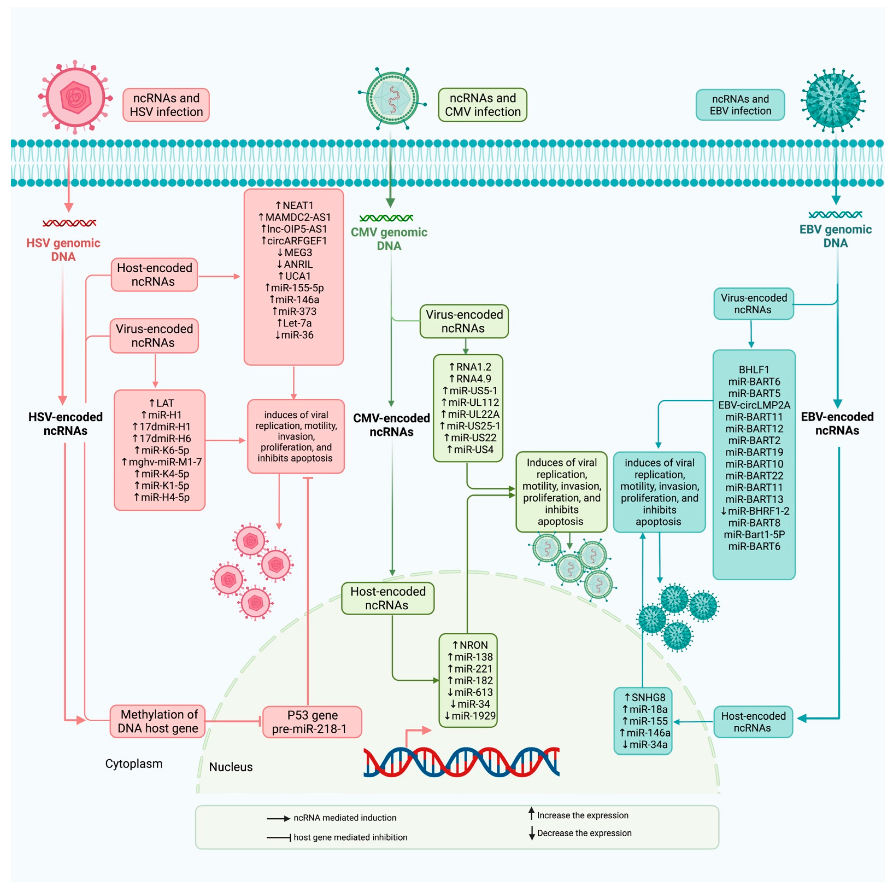
2. ncRNAs and HSV Infection
3. ncRNAs and CMV Infection
4. ncRNAs and EBV Infection
5. Impact of Drugs on the Expression of ncRNAs in Infected Patients
6. Diagnostic Value of Non-Coding RNAs in EBV-Infected Individuals
7. Discussion
Author Contributions
Funding
Institutional Review Board Statement
Informed Consent Statement
Data Availability Statement
Conflicts of Interest
References
- Palazzo, A.F.; Lee, E.S. Non-coding RNA: What is functional and what is junk? Front. Genetics 2015, 6, 2. [Google Scholar] [CrossRef] [PubMed] [Green Version]
- Wang, L.; Cho, K.B.; Li, Y.; Tao, G.; Xie, Z.; Guo, B. Long Noncoding RNA (lncRNA)-Mediated Competing Endogenous RNA Networks Provide Novel Potential Biomarkers and Therapeutic Targets for Colorectal Cancer. Int. J. Mol. Sci. 2019, 20, 5758. [Google Scholar] [CrossRef] [Green Version]
- Fang, Y.; Fullwood, M.J. Roles, functions, and mechanisms of long non-coding RNAs in cancer. Genom. Proteom. Bioinform. 2016, 14, 42–54. [Google Scholar] [CrossRef] [PubMed] [Green Version]
- Bartel, D.P. MicroRNAs: Target recognition and regulatory functions. Cell 2009, 136, 215–233. [Google Scholar] [CrossRef] [PubMed] [Green Version]
- Wang, P. The opening of pandora’s box: An emerging role of long noncoding RNA in viral infections. Front. Immunol. 2019, 9, 3138. [Google Scholar] [CrossRef] [PubMed] [Green Version]
- Josset, L.; Tchitchek, N.; Gralinski, L.E.; Ferris, M.T.; Eisfeld, A.J.; Green, R.R.; Thomas, M.J.; Tisoncik-Go, J.; Schroth, G.P.; Kawaoka, Y.; et al. Annotation of long non-coding RNAs expressed in collaborative cross founder mice in response to respiratory virus infection reveals a new class of interferon-stimulated transcripts. RNA Biol. 2014, 11, 875–890. [Google Scholar] [CrossRef] [Green Version]
- Sethuraman, S.; Gay, L.A.; Jain, V.; Haecker, I.; Renne, R. microRNA dependent and independent deregulation of long non-coding RNAs by an oncogenic herpesvirus. PLoS Pathog. 2017, 13, e1006508. [Google Scholar] [CrossRef] [Green Version]
- Wang, Z.; Fan, P.; Zhao, Y.; Zhang, S.; Lu, J.; Xie, W.; Jiang, Y.; Lei, F.; Xu, N.; Zhang, Y. NEAT1 modulates herpes simplex virus-1 replication by regulating viral gene transcription. Cell. Mol. Life Sci. 2017, 74, 1117–1131. [Google Scholar] [CrossRef] [PubMed] [Green Version]
- Imamura, K.; Imamachi, N.; Akizuki, G.; Kumakura, M.; Kawaguchi, A.; Nagata, K.; Kato, A.; Sato, H.; Yoneda, M.; Kai, C.; et al. Long noncoding RNA NEAT1-dependent SFPQ relocation from promoter region to paraspeckle mediates IL8 expression upon immune stimuli. Mol. Cell 2014, 53, 393–406. [Google Scholar] [CrossRef] [Green Version]
- Morchikh, M.; Cribier, A.; Raffel, R.; Amraoui, S.; Cau, J.; Severac, D.; Dubois, E.; Schwartz, O.; Bennasser, Y.; Benkirane, M. HEXIM1 and NEAT1 long non-coding RNA form a multi-subunit complex that regulates DNA-mediated innate immune response. Mol. Cell 2017, 67, 387–399.e5. [Google Scholar] [CrossRef]
- Shirahama, S.; Onoguchi-Mizutani, R.; Kawata, K.; Taniue, K.; Miki, A.; Kato, A.; Kawaguchi, Y.; Tanaka, R.; Kaburaki, T.; Kawashima, H.; et al. Long noncoding RNA U90926 is crucial for herpes simplex virus type 1 proliferation in murine retinal photoreceptor cells. Sci. Rep. 2020, 10, 19406. [Google Scholar] [CrossRef]
- Watson, Z.L.; Washington, S.D.; Phelan, D.M.; Lewin, A.S.; Tuli, S.S.; Schultz, G.S.; Neumann, D.M.; Bloom, D.C. In vivo knockdown of the herpes simplex virus 1 latency-associated transcript reduces reactivation from latency. J. Virol. 2018, 92, e00812–e00818. [Google Scholar] [CrossRef] [PubMed] [Green Version]
- Li, W.; Wang, Q.; Feng, Q.; Wang, F.; Yan, Q.; Gao, S.J.; Lu, C. Oncogenic KSHV-encoded interferon regulatory factor up-regulates HMGB2 and CMPK1 expression to promote cell invasion by disrupting a complex lncRNA-OIP5-AS1/miR-218-5p network. PLoS Pathog. 2019, 15, e1007578. [Google Scholar] [CrossRef]
- Zheng, K.; Liu, Q.; Wang, S.; Ren, Z.; Kitazato, K.; Yang, D.; Wang, Y. HSV-1-encoded microRNA miR-H1 targets Ubr1 to promote accumulation of neurodegeneration-associated protein. Virus Genes 2018, 54, 343–350. [Google Scholar] [CrossRef] [PubMed]
- Barrozo, E.R.; Nakayama, S.; Singh, P.; Vanni, E.A.; Arvin, A.M.; Neumann, D.M.; Bloom, D.C. Deletion of herpes simplex virus 1 microRNAs miR-H1 and miR-H6 impairs reactivation. J. Virol. 2020, 94, e00639-20. [Google Scholar] [CrossRef] [PubMed]
- Duan, Y.; Zeng, J.; Fan, S.; Liao, Y.; Feng, M.; Wang, L.; Zhang, Y.; Li, Q. Herpes Simplex Virus Type 1–Encoded miR-H2-3p Manipulates Cytosolic DNA–Stimulated Antiviral Innate Immune Response by Targeting DDX41. Viruses 2019, 11, 756. [Google Scholar] [CrossRef] [PubMed] [Green Version]
- Wang, Y.; Huang, L.; Wang, Y.; Luo, W.; Li, F.; Xiao, J.; Qin, S.; Wang, Z.; Song, X.; Jin, F.; et al. Single-cell RNA-sequencing analysis identifies host long noncoding RNA MAMDC2-AS1 as a co-factor for HSV-1 nuclear transport. Int. J. Biol. Sci. 2020, 16, 1586. [Google Scholar] [CrossRef] [Green Version]
- Yao, S.; Jia, X.; Wang, F.; Sheng, L.; Song, P.; Cao, Y.; Shi, H.; Fan, W.; Ding, X.; Gao, S.J.; et al. CircRNA ARFGEF1 functions as a ceRNA to promote oncogenic KSHV-encoded viral interferon regulatory factor induction of cell invasion and angiogenesis by up-regulating glutaredoxin 3. PLoS Pathog. 2021, 17, e1009294. [Google Scholar] [CrossRef] [PubMed]
- Wang, Z.; Li, K.; Wang, X.; Huang, W. MiR-155-5p modulates HSV-1 replication via the epigenetic regulation of SRSF2 gene expression. Epigenetics 2019, 14, 494–503. [Google Scholar] [CrossRef]
- Venuti, A.; Musarra-Pizzo, M.; Pennisi, R.; Tankov, S.; Medici, M.A.; Mastino, A.; Rebane, A.; Sciortino, M.T. HSV-1\EGFP stimulates miR-146a expression in a NF-κB-dependent manner in monocytic THP-1 cells. Sci. Rep. 2019, 9, 5157. [Google Scholar] [CrossRef] [Green Version]
- Qi, Y.; Zheng, G.; Di, C.; Zhang, J.; Wang, X.; Hong, Y.; Song, Y.; Chen, R.; Yang, Y.; Yan, Y.; et al. Latency-associated nuclear antigen inhibits lytic replication of Kaposi’s sarcoma-associated herpesvirus by regulating let-7a/RBPJ signaling. Virology 2019, 531, 69–78. [Google Scholar] [CrossRef] [PubMed]
- Hussein, H.A.; Akula, S.M. miRNA-36 inhibits KSHV, EBV, HSV-2 infection of cells via stifling expression of interferon induced transmembrane protein 1 (IFITM1). Sci. Rep. 2017, 7, 17972. [Google Scholar] [CrossRef] [PubMed]
- Xie, Y.; He, S.; Wang, J. MicroRNA-373 facilitates HSV-1 replication through suppression of type I IFN response by targeting IRF1. Biomed. Pharmacother. 2018, 97, 1409–1416. [Google Scholar] [CrossRef] [PubMed]
- Vanni, E.A.; Foley, J.W.; Davison, A.J.; Sommer, M.; Liu, D.; Sung, P.; Moffat, J.; Zerboni, L.; Arvin, A.M. The latency-associated transcript locus of herpes simplex virus 1 is a virulence determinant in human skin. PLoS Pathog. 2020, 16, e1009166. [Google Scholar] [CrossRef] [PubMed]
- Morrison, K.; Manzano, M.; Chung, K.; Schipma, M.J.; Bartom, E.T.; Gottwein, E. The Oncogenic Kaposi’s Sarcoma-Associated Herpesvirus Encodes a Mimic of the Tumor-Suppressive miR-15/16 miRNA Family. Cell Rep. 2019, 29, 2961–2969.e6. [Google Scholar] [CrossRef] [PubMed] [Green Version]
- Wang, Y.; Feldman, E.R.; Bullard, W.L.; Tibbetts, S.A. A gammaherpesvirus microRNA targets EWSR1 (Ewing sarcoma breakpoint region 1) in vivo to promote latent infection of germinal center B cells. Mbio 2019, 10, e00996-19. [Google Scholar] [CrossRef] [PubMed] [Green Version]
- Li, T.; Ju, E.; Gao, S.-J. Kaposi sarcoma–associated herpesvirus miRNAs suppress CASTOR1-mediated mTORC1 inhibition to promote tumorigenesis. J. Clin. Investig. 2019, 129, 3310–3323. [Google Scholar] [CrossRef] [PubMed] [Green Version]
- Zhao, Y.; Yang, J.; Liu, Y.; Fan, J.; Yang, H. HSV-2-encoded miRNA-H4 regulates cell cycle progression and Act-D-induced apoptosis in HeLa Cells by targeting CDKL2 and CDKN2A. Virol. Sin. 2019, 34, 278–286. [Google Scholar] [CrossRef] [PubMed]
- Wang, X.; Liu, S.; Zhou, Z.; Yan, H.; Xiao, J. A herpes simplex virus type 2–encoded microRNA promotes tumor cell metastasis by targeting suppressor of cytokine signaling 2 in lung cancer. Tumor Biol. 2017, 39. [Google Scholar] [CrossRef] [PubMed] [Green Version]
- Lau, B.; Kerr, K.; Gu, Q.; Nightingale, K.; Antrobus, R.; Suárez, N.M.; Stanton, R.J.; Wang, E.C.; Weekes, M.P.; Davison, A.J. Human cytomegalovirus long non-coding RNA1. 2 suppresses extracellular release of the pro-inflammatory cytokine IL-6 by blocking NF-κB activation. Front. Cell. Infect. Microbiol. 2020, 10, 361. [Google Scholar] [CrossRef] [PubMed]
- Tai-Schmiedel, J.; Karniely, S.; Lau, B.; Ezra, A.; Eliyahu, E.; Nachshon, A.; Kerr, K.; Suárez, N.; Schwartz, M.; Davison, A.J.; et al. Human cytomegalovirus long noncoding RNA4. 9 regulates viral DNA replication. PLoS Pathog. 2020, 16, e1008390. [Google Scholar] [CrossRef] [PubMed] [Green Version]
- Kumar, A.; Tripathy, M.K.; Pasquereau, S.; Al Moussawi, F.; Abbas, W.; Coquard, L.; Khan, K.A.; Russo, L.; Algros, M.-P.; Valmary-Degano, S.; et al. The human cytomegalovirus strain DB activates oncogenic pathways in mammary epithelial cells. EBioMedicine 2018, 30, 167–183. [Google Scholar] [CrossRef] [Green Version]
- Wang, Y.-H.; Yu, X.-H.; Qu, G.-J.; Qiao, F.-F.; Han, H. Reduced expression of the lncRNA NRON is a potential hallmark of the CMV-amplified CD8+ T cell accumulations commonly seen in older humans. Exp. Gerontol. 2019, 115, 46–54. [Google Scholar] [CrossRef] [PubMed]
- Zhou, W.; Xi, D.; Shi, Y.; Wang, L.; Zhong, H.; Huang, Z.; Liu, Y.; Tang, Y.; Lu, N.; Wang, Y.; et al. MicroRNA-1929-3p participates in murine cytomegalovirus-induced hypertensive vascular remodeling through Ednra/NLRP3 inflammasome activation. Int. J. Mol. Med. 2021, 47, 719–731. [Google Scholar] [CrossRef]
- Hancock, M.H.; Crawford, L.B.; Perez, W.; Struthers, H.M.; Mitchell, J.; Caposio, P. Human cytomegalovirus UL7, miR-US5-1, and miR-UL112-3p inactivation of FOXO3a protects CD34+ hematopoietic progenitor cells from apoptosis. Msphere 2021, 6, e00986-20. [Google Scholar] [CrossRef] [PubMed]
- Liang, Q.; Wang, K.; Wang, B.; Cai, Q. HCMV-encoded miR-UL112-3p promotes glioblastoma progression via tumour suppressor candidate 3. Sci. Rep. 2017, 7, 44705. [Google Scholar] [CrossRef] [Green Version]
- Zhang, S.; Liu, L.; Wang, R.; Tuo, H.; Guo, Y.; Yi, L.; Wang, D.; Wang, J. miR-138 promotes migration and tube formation of human cytomegalovirus-infected endothelial cells through the SIRT1/p-STAT3 pathway. Arch. Virol. 2017, 162, 2695–2704. [Google Scholar] [CrossRef]
- Shi, L.; Fan, B.; Chen, D.; Guo, C.; Xiang, H.; Nie, Y.; Zhong, D.; Shi, X. Human cytomegalovirus protein UL136 activates the IL-6/STAT3 signal through MiR-138 and MiR-34c in gastric cancer cells. Int. J. Clin. Oncol. 2020, 25, 1936–1944. [Google Scholar] [CrossRef]
- Yan, B.; Ma, H.; Jiang, S.; Shi, J.; Yang, Z.; Zhu, W.; Kong, C.; Chen, L.; Yan, H.; Ma, C. microRNA-221 restricts human cytomegalovirus replication via promoting type I IFN production by targeting SOCS1/NF-κB pathway. Cell Cycle 2019, 18, 3072–3084. [Google Scholar] [CrossRef]
- He, X.; Teng, J.; Cui, C.; Li, D.; Wen, L. MicroRNA-182 inhibits HCMV replication through activation of type I IFN response by targeting FOXO3 in neural cells. Exp. Cell Res. 2018, 369, 197–207. [Google Scholar] [CrossRef]
- Wang, Y.; Zhao, P.; Qian, D.; Hu, M.; Zhang, L.; Shi, H.; Wang, B. MicroRNA-613 is downregulated in HCMV-positive glioblastoma and inhibits tumour progression by targeting arginase-2. Tumor Biol. 2017, 39. [Google Scholar] [CrossRef] [Green Version]
- Hancock, M.H.; Mitchell, J.; Goodrum, F.D.; Nelson, J.A. Human cytomegalovirus miR-US5-2 downregulation of GAB1 regulates cellular proliferation and UL138 expression through modulation of epidermal growth factor receptor signaling pathways. Msphere 2020, 5, e00582-20. [Google Scholar] [CrossRef] [PubMed]
- Hancock, M.H.; Crawford, L.B.; Pham, A.H.; Mitchell, J.; Struthers, H.M.; Yurochko, A.D.; Caposio, P.; Nelson, J.A. Human cytomegalovirus miRNAs regulate TGF-β to mediate myelosuppression while maintaining viral latency in CD34+ hematopoietic progenitor cells. Cell Host Microbe 2020, 27, 104–114.e4. [Google Scholar] [CrossRef] [PubMed]
- Mikell, I.; Crawford, L.B.; Hancock, M.H.; Mitchell, J.; Buehler, J.; Goodrum, F.; Nelson, J.A. HCMV miR-US22 down-regulation of EGR-1 regulates CD34+ hematopoietic progenitor cell proliferation and viral reactivation. PLoS Pathog. 2019, 15, e1007854. [Google Scholar] [CrossRef] [Green Version]
- Skinner, C.M.; Ivanov, N.S.; Barr, S.A.; Chen, Y.; Skalsky, R.L. An Epstein-Barr virus microRNA blocks interleukin-1 (IL-1) signaling by targeting IL-1 receptor 1. J. Virol. 2017, 91, e00530-17. [Google Scholar] [CrossRef] [Green Version]
- Shao, Y.; Qi, Y.; Huang, Y.; Liu, Z.; Ma, Y.; Guo, X.; Jiang, S.; Sun, Z.; Ruan, Q. Human cytomegalovirus miR-US4-5p promotes apoptosis via downregulation of p21-activated kinase 2 in cultured cells. Mol. Med. Rep. 2017, 16, 4171–4178. [Google Scholar] [CrossRef] [PubMed]
- Verhoeven, R.J.; Tong, S.; Zhang, G.; Zong, J.; Chen, Y.; Jin, D.Y.; Chen, M.R.; Pan, J.; Chen, H. NF-κB signaling regulates expression of Epstein-Barr virus BART microRNAs and long noncoding RNAs in nasopharyngeal carcinoma. J. Virol. 2016, 90, 6475–6488. [Google Scholar] [CrossRef] [Green Version]
- Yetming, K.D.; Lupey-Green, L.N.; Biryukov, S.; Hughes, D.J.; Marendy, E.M.; Miranda, J.L.; Sample, J.T. The BHLF1 locus of Epstein-Barr virus contributes to viral latency and B-cell immortalization. J. Virol. 2020, 94, e01215–e01220. [Google Scholar] [CrossRef] [PubMed]
- He, B.; Li, W.; Wu, Y.; Wei, F.; Gong, Z.; Bo, H.; Wang, Y.; Li, X.; Xiang, B.; Guo, C.; et al. Epstein-Barr virus-encoded miR-BART6-3p inhibits cancer cell proliferation through the LOC553103-STMN1 axis. FASEB J. 2020, 34, 8012–8027. [Google Scholar]
- Zheng, X.; Wang, J.; Wei, L.; Peng, Q.; Gao, Y.; Fu, Y.; Lu, Y.; Qin, Z.; Zhang, X.; Lu, J.; et al. Epstein-Barr virus microRNA miR-BART5-3p inhibits p53 expression. J. Virol. 2018, 92, e01022-18. [Google Scholar] [CrossRef] [Green Version]
- Huang, T.; Ji, Y.; Hu, D.; Chen, B.; Zhang, H.; Li, C.; Chen, G.; Luo, X.; Zheng, X.W.; Lin, X. SNHG8 is identified as a key regulator of epstein-barr virus (EBV)-associated gastric cancer by an integrative analysis of lncRNA and mRNA expression. Oncotarget 2016, 7, 80990. [Google Scholar] [CrossRef]
- Mai, S.; Xiao, R.; Shi, L.; Zhou, X.; Yang, T.; Zhang, M.; Weng, N.; Zhao, X.; Wang, R.; Liu, J.; et al. MicroRNA-18a promotes cancer progression through SMG1 suppression and mTOR pathway activation in nasopharyngeal carcinoma. Cell Death Dis. 2019, 10, 819. [Google Scholar] [CrossRef] [Green Version]
- Shi, Q.; Zhang, Y.; Liu, W.; Xiao, H.; Qi, Y.; Li, J.; Luo, B. Latent membrane protein 2A inhibits expression level of Smad2 through regulating miR-155-5p in EBV-associated gastric cancer cell lines. J. Med. Virol. 2020, 92, 96–106. [Google Scholar] [CrossRef] [PubMed]
- Wang, W.; Zhang, Y.; Liu, W.; Xiao, H.; Zhang, Q.; Wang, J.; Luo, B. LMP1–miR-146a–CXCR4 axis regulates cell proliferation, apoptosis and metastasis. Virus Res. 2019, 270, 197654. [Google Scholar] [CrossRef]
- Anastasiadou, E.; Stroopinsky, D.; Alimperti, S.; Jiao, A.L.; Pyzer, A.R.; Cippitelli, C.; Pepe, G.; Severa, M.; Rosenblatt, J.; Etna, M.P.; et al. Epstein− Barr virus-encoded EBNA2 alters immune checkpoint PD-L1 expression by downregulating miR-34a in B-cell lymphomas. Leukemia 2019, 33, 132–147. [Google Scholar] [CrossRef] [PubMed] [Green Version]
- Kim, S.-M.; Hur, D.Y.; Hong, S.-W.; Kim, J.H. EBV-encoded EBNA1 regulates cell viability by modulating miR34a-NOX2-ROS signaling in gastric cancer cells. Biochem. Biophys. Res. Commun. 2017, 494, 550–555. [Google Scholar] [CrossRef] [PubMed]
- Gong, L.P.; Chen, J.N.; Dong, M.; Xiao, Z.D.; Feng, Z.Y.; Pan, Y.H.; Zhang, Y.; Du, Y.; Zhang, J.Y.; Bi, Y.H.; et al. Epstein–Barr virus-derived circular RNA LMP 2A induces stemness in EBV-associated gastric cancer. EMBO Rep. 2020, 21, e49689. [Google Scholar] [CrossRef] [PubMed]
- Kase, K.; Saito, M.; Nakajima, S.; Takayanagi, D.; Saito, K.; Yamada, L.; Ashizawa, M.; Nakano, H.; Hanayama, H.; Onozawa, H.; et al. ARID1A deficiency in EBV-positive gastric cancer is partially regulated by EBV-encoded miRNAs, but not by DNA promotor hypermethylation. Carcinogenesis 2021, 42, 21–30. [Google Scholar] [CrossRef] [PubMed]
- Wu, Y.; Wang, D.; Wei, F.; Xiong, F.; Zhang, S.; Gong, Z.; Shi, L.; Li, X.; Xiang, B.; Ma, J.; et al. EBV-miR-BART12 accelerates migration and invasion in EBV-associated cancer cells by targeting tubulin polymerization-promoting protein 1. FASEB J. 2020, 34, 16205–16223. [Google Scholar] [CrossRef] [PubMed]
- Lung, R.W.M.; Tong, J.H.M.; Ip, L.M.; Lam, K.H.; Chan, A.W.H.; Chak, W.P.; Chung, L.; Yeung, W.W.; Hau, P.; Chau, S.; et al. EBV–encoded miRNAs can sensitize nasopharyngeal carcinoma to chemotherapeutic drugs by targeting BRCA1. J. Cell. Mol. Med. 2020, 24, 13523–13535. [Google Scholar] [CrossRef]
- Zhang, Q.; Luo, D.; Xie, Z.; He, H.; Duan, Z. The Oncogenic Role of miR-BART19-3p in Epstein-Barr Virus-Associated Diseases. BioMed Res. Int. 2020, 2020, 5217039. [Google Scholar] [CrossRef] [PubMed]
- Min, K.; Lee, S.K. EBV miR-BART10-3p promotes cell proliferation and migration by targeting DKK1. Int. J. Biol. Sci. 2019, 15, 657. [Google Scholar] [CrossRef] [PubMed]
- Dong, M.; Gong, L.P.; Chen, J.N.; Zhang, X.F.; Zhang, Y.W.; Hui, D.Y.; Zhao, X.X.; Wu, X.Y.; Shao, C.K. EBV-miR-BART10-3p and EBV-miR-BART22 promote metastasis of EBV-associated gastric carcinoma by activating the canonical Wnt signaling pathway. Cell. Oncol. 2020, 43, 901–913. [Google Scholar] [CrossRef] [PubMed]
- Song, Y.; Li, Q.; Liao, S.; Zhong, K.; Jin, Y.; Zeng, T. Epstein-Barr virus-encoded miR-BART11 promotes tumor-associated macrophage-induced epithelial-mesenchymal transition via targeting FOXP1 in gastric cancer. Virology 2020, 548, 6–16. [Google Scholar] [CrossRef]
- Liu, J.; Zhang, Y.; Liu, W.; Zhang, Q.; Xiao, H.; Song, H.; Luo, B. MiR-BART1-5p targets core 2β-1, 6-acetylglucosaminyltransferase GCNT3 to inhibit cell proliferation and migration in EBV-associated gastric cancer. Virology 2020, 541, 63–74. [Google Scholar] [CrossRef]
- Huang, J.; Qin, Y.; Yang, C.; Wan, C.; Dai, X.; Sun, Y.; Meng, J.; Lu, Y.; Li, Y.; Zhang, Z.; et al. Downregulation of ABI2 expression by EBV-miR-BART13-3p induces epithelial-mesenchymal transition of nasopharyngeal carcinoma cells through upregulation of c-JUN/SLUG signaling. Aging 2020, 12, 340. [Google Scholar] [CrossRef]
- Cristino, A.S.; Nourse, J.; West, R.A.; Sabdia, M.B.; Law, S.C.; Gunawardana, J.; Vari, F.; Mujaj, S.; Thillaiyampalam, G.; Snell, C.; et al. EBV microRNA-BHRF1-2-5p targets the 3′ UTR of immune checkpoint ligands PD-L1 and PD-L2. Blood 2019, 134, 2261–2270. [Google Scholar] [CrossRef]
- Zhou, X.; Zheng, J.; Tang, Y.; Lin, Y.; Wang, L.; Li, Y.; Liu, C.; Wu, D.; Cai, L. EBV encoded miRNA BART8-3p promotes radioresistance in nasopharyngeal carcinoma by regulating ATM/ATR signaling pathway. Biosci. Rep. 2019, 39, BSR20190415. [Google Scholar] [CrossRef] [Green Version]
- Lin, C.; Zong, J.; Lin, W.; Wang, M.; Xu, Y.; Zhou, R.; Lin, S.; Guo, Q.; Chen, H.; Ye, Y.; et al. EBV-miR-BART8-3p induces epithelial-mesenchymal transition and promotes metastasis of nasopharyngeal carcinoma cells through activating NF-κB and Erk1/2 pathways. J. Exp. Clin. Cancer Res. 2018, 37, 283. [Google Scholar] [CrossRef]
- Xu, Y.J.; Zhou, R.; Zong, J.F.; Lin, W.S.; Tong, S.; Guo, Q.J.; Lin, C.; Lin, S.J.; Chen, Y.X.; Chen, M.R.; et al. Epstein-Barr virus-coded miR-BART13 promotes nasopharyngeal carcinoma cell growth and metastasis via targeting of the NKIRAS2/NF-κB pathway. Cancer Lett. 2019, 447, 33–40. [Google Scholar] [CrossRef]
- Lyu, X.; Wang, J.; Guo, X.; Wu, G.; Jiao, Y.; Faleti, O.D.; Liu, P.; Liu, T.; Long, Y.; Chong, T.; et al. EBV-miR-BART1-5P activates AMPK/mTOR/HIF1 pathway via a PTEN independent manner to promote glycolysis and angiogenesis in nasopharyngeal carcinoma. PLoS Pathog. 2018, 14, e1007484. [Google Scholar] [CrossRef] [PubMed] [Green Version]
- Lu, Y.; Qin, Z.; Wang, J.; Zheng, X.; Lu, J.; Zhang, X.; Wei, L.; Peng, Q.; Zheng, Y.; Ou, C.; et al. Epstein-Barr virus miR-BART6-3p inhibits the RIG-I pathway. J. Innate Immun. 2017, 9, 574–586. [Google Scholar] [CrossRef]
- Liu, Y.; Jiang, Q.; Liu, X.; Lin, X.; Tang, Z.; Liu, C.; Zhou, J.; Zhao, M.; Li, X.; Cheng, Z.; et al. Cinobufotalin powerfully reversed EBV-miR-BART22-induced cisplatin resistance via stimulating MAP2K4 to antagonize non-muscle myosin heavy chain IIA/glycogen synthase 3β/β-catenin signaling pathway. EBioMedicine 2019, 48, 386–404. [Google Scholar] [CrossRef] [PubMed] [Green Version]
- Wang, J.; Zheng, X.; Qin, Z.; Wei, L.; Lu, Y.; Peng, Q.; Gao, Y.; Zhang, X.; Zhang, X.; Li, Z.; et al. Epstein–Barr virus miR-BART3-3p promotes tumorigenesis by regulating the senescence pathway in gastric cancer. J. Biol. Chem. 2019, 294, 4854–4866. [Google Scholar] [CrossRef] [PubMed] [Green Version]
- Lu, T.; Guo, Q.; Lin, K.; Chen, H.; Chen, Y.; Xu, Y.; Lin, C.; Su, Y.; Chen, Y.; Chen, M.; et al. Circulating Epstein-Barr virus microRNAs BART7-3p and BART13-3p as novel biomarkers in nasopharyngeal carcinoma. Cancer Sci. 2020, 111, 1711. [Google Scholar] [CrossRef]

| ncRNA | Expression Pattern | Clinical Samples/Animal Model | Cell Culture | Targets | Regulators | Signaling Pathways | Description | Reference |
|---|---|---|---|---|---|---|---|---|
| NEAT1 | ↑ | - | HeLa, MEF | TK, ICP0 | - | - | PSPC1 binds to STAT3 and increases STAT3 binding to the viral promoter, leading to increased viral expression. Knocking down STAT3 or NEAT1 improves skin healing. | [8] |
| ↑ | - | HeLa, HeLa S3, HEK293T, HUVEC, | HDP-RNP, HEXIM1 | - | - | NEAT1 assembles HDP-RNP, which regulates innate immune response against foreign DNA through stabilizing the host genome. | [10] | |
| U90926 | ↑ | - | 661 W, Vero | ICP0, ICP4 | - | - | After HSV-1 infection, upregulation of U90926 results in increased viral proliferation. | [11] |
| MAMDC2-AS1 | ↑ | - | 293T, A549, HaCaT, HepG2, HFF, HeLa | Hsp90α, VP16 | YY1 | - | This lncRNA increases HSV-1 infection in human cells through interacting with Hsp90α and importing HSV-1 into the nucleus. | [17] |
| MEG3 | ↓ | - | TIVE, HUVEC, HeLa, iSLK, BCBL-1 | - | miR-K12-3, miR-K12-5, miR-K12-6-5p, miR-K12-8, miR-K12-9 | - | During the active infection and latency phase, HSV dysregulates lncRNAs expression using its miRNAs and proteins. Moreover, UCA1 induction promotes cell migration and proliferation. | [7] |
| ANRIL | ↓ | - | miR-K12-1, miR-K12-6-5p, miR-K12-2, miR-K12-11 | |||||
| UCA1 | ↑ | - | Kaposin and vCyclin | |||||
| lnc-OIP5-AS1 | ↑ | - | iSLK, HEK293T, HUVECs | miR-218-5p, HMGB2, CMPK1 | vIRF1 | - | cIRF1 induces lnc-OIP5-AS1 upregulation, which diminishes miR-218-5p. As a result, in KSHV cells, migration and proliferation would be increased. | [13] |
| circARFGEF1 | ↑ | - | iSLK-RGB-BAC16, iSLK-RGB-K9, HUVECs, HEK293T | miR-125a-3p, GLRX3 | vIRF1 | - | As a circRNA, ARFGEF1is induced by vIRF1. Moreover, ARFGEF1 increases cell motility, invasion, proliferation, and angiogenesis in Kaposi’s sarcoma-associated herpesvirus infected cells. | [18] |
| miR-155-5p | ↑ | - | HeLa | SRSF2 | - | - | This miRNA induces improved levels of viral replication and gene expression by regulating SRSF2 promoter histones. | [19] |
| miR-146a | ↑ | - | THP-1, HEp-2 | IRAK1 | - | NF-κB | miR-146a activates NF-κB signaling pathway in HSV-1 infected cells. | [20] |
| let-7a | ↑ | 293T, iSLK.219 | RBPJ | LANA, LIN28B | NF-κB | In Kaposi’s sarcoma-associated herpesvirus infected cells, let-7a inhibits the lytic reactivated phase. | [21] | |
| miR-36 | ↓ | - | BJAB, HMVEC-d, HFFs, HEK293 | IFITM1 | - | - | miR-36 overexpression lowers IFITM1 levels, a protein induced by Kaposi’s sarcoma herpes virus, leading to decreased viral infection. | [22] |
| miR-373 | ↑ | Serum samples from 10 herpetic gingivostomatitis and 10 normal cases | HeLa | IRF1 | - | - | miR-373 is overexpressed upon HSV-1 infection, leading to increased viral replication and infection. | [23] |
| ncRNA | Expression Pattern | Clinical Samples/Animal Model | Cell Culture | Targets | Signaling Pathways | Description | Reference |
|---|---|---|---|---|---|---|---|
| LAT | ↑ | Rabbit skin cells | primary rabbit kidney cells | - | - | LAT knockdown diminishes HSV-1 reactivation in the latency phase. Therefore, this lncRNA could be used as a therapeutic target for herpes stromal keratitis. | [12] |
| ↑ | Human fetal skin tissue, SCID C.B-17 male mice | - | - | - | LAT is a remarkable factor in increasing HSV-1 replication and lesion formation. | [24] | |
| miR-H1 | ↑ | - | SH-SY5Y, HEK293T | - | miR-H1, encoded by HSV-1, lowers Ubr1, a ubiquitin-protein ligase, which leads to β-amyloid accumulation. | [14] | |
| 17dmiR-H1 | ↑ | female ND-40 Swiss-Webster mice, Male and female New Zealand White rabbits | CCL-13, HEK293T | LAT | - | Upon deleting these miRNAs, HSV-1 loses the ability to reactivate. | [15] |
| 17dmiR-H6 | ↑ | ||||||
| miR-K6-5p | ↑ | - | HEK293T, BC-3, BC-1, JSC-1, BCBL-1 | CCND3, CDC25A | - | In Kaposi’s sarcoma-associated herpesvirus, this miRNA is expressed and as a tumor suppressor lowers cell cycle progression. | [25] |
| miR-H2-3p | ↑ | - | HEK293T, THP-1, HFF | DDX41, IFN-β, MxI | - | miR-H2-3p lowers innate immune response in infected cells and intensifies viral DNA replication and proliferation. | [16] |
| mghv-miR-M1-7-5p | ↑ | C57BL/6J mice | NIH3T12 | EWSR1 | - | This miRNA is encoded by gamma-herpesviruses and is a necessary factor for its infection and splenic latency. | [21] |
| miR-K4-5p | ↑ | - | TIVE, KTIVE, MM, KMM | CASTOR1 | mTORC1 | These two miRNAs are encoded by Kaposi’s sarcoma herpes virus and could activate the mTORC1 signaling pathway and improve cell proliferation and colony formation. | [26] |
| miR-K1-5p | ↑ | ||||||
| miR-H4-5p | ↑ | - | HeLa | CDKN2A, CDKL2 | - | miR-H4-5p is transcribed by HSV-2 and induces cell cycle progression, proliferation, and inhibits apoptosis. | [27] |
| miR-H9-5p | ↑ | 10 cancerous lung tissues and 10 control tissues | LTEP-α-2, SPC-α-1 | SOCS2 | Transcribed by HSV-2 infection, miR-H9-5p increases migration, proliferation, and invasion of lung cancer cells. | [28] |
| ncRNA | Expression Pattern | Clinical Samples/Animal Model | Cell Culture | Targets | Signaling Pathways | Description | Reference |
|---|---|---|---|---|---|---|---|
| NRON | ↑ | 40 elderly CMV positive cases | - | NFAT | - | Early CMV infection results in increased NRON expression in all B cell types. | [32] |
| miR-1929-3p | ↓ | C57BL/6 J mice | - | ET-1, Ednra, NLRP3 | - | CMV downregulates miR-1929-3p to enhance blood pressure, endothelial cell injury, and vascular remodeling in mice. | [33] |
| miR-138 | ↑ | - | HUVECs, MRC-5 | SIRT1, p-STAT3 | - | Endothelial cells infected by CMV indicate a higher miR-138 expression, which leads to tube formation and migration. | [36] |
| ↑ | Male BALB/c nude mice | MNK-45, SGC-7901 | IL6R | - | UL136 induces cell invasion and proliferation by activating IL6/STAT3 signaling, causing fluctuation in these miRNAs’ expression. | [37] | |
| miR-34 | ↓ | ||||||
| miR-221 | ↑ | C57BL/6 mice | Neural Precursor Cells | SOCS1, IFN | NF-κB | miR-221 hampers CMV replication in cells through modulating the NF-κB signaling pathway. | [38] |
| miR-182 | ↑ | Female Balb/c mice | U-251MG, HFFs, NPCs | IRF7, FOXO3, IFN-I | - | CMV infection increases miR-182 expression, which itself produces IFN-I to restrict CMV replication. | [39] |
| miR-613 | ↓ | 10 CMV-positive and 10 CMV-negative glioblastoma tissues and their adjacent normal tissues | U87, U251 | Arginase-2 | - | In glioblastoma infected cells and tissues, miR-613 is negatively correlated with tumor size, stage, and patients’ survival rates. Moreover, it could diminish colony formation, migration, and invasion. | [40] |
| ncRNA | Expression Pattern | Clinical Samples/Animal Model | Cell Culture | Targets | Signaling Pathways | Description | Reference |
|---|---|---|---|---|---|---|---|
| RNA1.2 | ↑ | - | HFFF2, HEK293T, HFT | TPRG1L, IL-6, MCP-1, CXCL1 | NF-κB | RNA1.2, encoded by CMV, modifies the expression of many genes, which is a conspicuous indication of modulating the NF-κB signaling pathway. | [29] |
| RNA4.9 | ↑ | - | MEF | ssDBP | - | Located in the nucleus, RNA4.9 improves viral growth and DNA replication. | [30] |
| ↑ | female NOD/SCID Gamma mice | MDA-MB-231, MCF-7, HMECs | - | - | CMV infection induces RNA4.9 expression, which leads to tumor formation. | [31] | |
| miR-US5-1 | ↑ | - | CD34+ HPCs of fetal liver tissues, THF, NHDF, HEK293 | FOXO3a, BCL2L11 | MAPK | CMV downregulates pro-apoptotic proteins and increases CD34+ progenitor cells. | [34] |
| miR-UL112-3p | ↑ | ||||||
| ↑ | 40 CMV-positive glioblastoma and adjacent normal tissues | Glioblastoma primary culture, HEK293 | TUSC3 | Akt | miR-UL112-3p improves glioblastoma cells proliferation, migration, invasion, and colony formation. | [35] | |
| miR-US5-2 | ↓ | - | NHDF, hAECs, 293T | GAB1, UL138, EGR1 | PI3k/ERK | This miRNA can thwart the normal EGF signaling pathway that boosts cell proliferation. | [41] |
| ↑ | - | NHDF, HEK293T, CC-2535 | TGF-β, NAB1 | - | HCMV miR-US5-2 is a latent miRNA that suppresses uninfected CD34+ hematopoietic progenitor cells. | [42] | |
| miR-UL22A | ↑ | SMAD3 | By diminishing SMAD3, this miRNA reactivates CMV in CD34+ cells. | ||||
| miR-US22 | ↑ | - | HEK293T, NHDF, AEC | EGR1 | - | miR-US22 lowers hematopoietic stem cells proliferation, self-renewal, and colony formation. | [43] |
| miR-US25-1-5p | ↑ | - | HFF, U251, HEK293 | CD147, IFN-β | NF-κB | miR-US25-1-5p diminishes antiviral immune response and CD147 but induces the lytic phase of CMV virus. | [44] |
| miR-US4-5p | ↑ | - | HELF, HEK293, THP-1 | PAK2 | - | miR-US4-5p, encoded by CMV, inhibits apoptosis by downregulating PAK2. | [45] |
| ncRNA | Expression Pattern | Clinical Samples/Animal Model | Cell Culture | Targets | Regulators | Signaling Pathways | Description | Reference |
|---|---|---|---|---|---|---|---|---|
| SNHG8 | ↑ | 88 gastric cancer and adjacent normal tissues | - | TRIM28, EIF4A2, NAP1L1, PLD3, RPL18A, TRPM7 | - | SNHG8 is up-regulated in gastric cancer tissues afflicted with the EBV virus and may trigger cancer initiation by altering various proteins. | [50] | |
| miR-18a | ↑ | 21 cancerous and 14 non-cancerous tissues, male BALB/c nude mice | CNE1, CNE2, S-18, S-26, 5-8F, 6-10B, SUNE2, C666-1 | SMG1 | LAT | NF-κB | In EBV-positive cases, miR-182 positively correlates with nasopharyngeal tumor size and tumor stage. Moreover, it enhances cell migration, invasion, and proliferation. | [51] |
| miR-155-5p | ↑ | - | GT38, GT39, SNU719, HGC27, SGC7901, BGC823 | Smad2 | LMP2A | NF-κB, TGF-β | In EBV-infected gastric cancer cells, miR-155-5p reduces cell proliferation and boosts cell cycle arrest and apoptosis. | [52] |
| miR-146a | ↑ | - | GT38, GT39, AGS, BGC823, SGC7901 | CXCR4 | LMP1 | - | EBV induces miR-146a expression, which leads to lower cell proliferation, migration, and cell cycle progression. | [53] |
| miR-34a | ↓ | 27 diffuse large B-cell lymphoma cases | LCL, OMA4, DG75, BL41, U2932, SUDHL5, ER/EB 2.5, Mutu I, Mutu III, Daudi, Jijoye | PD-L1 | EBF1 | - | In Burkitt lymphoma and diffuse large B-cell lymphomas, miR-34a acts as a tumor suppressor due to downregulating PD-L1 and activating T cells. | [54] |
| ↓ | - | SNU719, SNU638 | NOX2 | EBNA1 | - | In gastric cancer cell lines, miR-34a downregulation leads to increased cell viability and reduced apoptosis rate. | [55] |
| ncRNA | Expression Pattern | Clinical Samples/Animal Model | Cell Culture | Targets | Signaling Pathways | Description | Reference |
|---|---|---|---|---|---|---|---|
| BHLF1 | ↑ | - | A.21, Kem I, Mutu I, BX1, HEK293 | - | - | After being up-regulated by SM protein, BHLF1 encodes ncRNAs that induce a latency phase, immortalize EBV, and generate B cells growth. | [47] |
| miR-BART6-3p | ↑ | female BALB/c nude mice | 5-8 F, HNE2, C666-1, HEK293T | LOC553103, STMN1 | - | Invasion, cell cycle progression, and metastasis of tumors are all diminished by this miRNA. | [48] |
| miR-BART5-5p | ↑ | - | CNE2, HeLa-Bx1, HEK293T, C666-1 | LMP1 | NF-κB | Through the NF-κB pathway, LMP1 activates the BART promoters in nasopharyngeal carcinoma. BART itself could downregulate LMP1 expression as well. | [46] |
| ↑ | 24 gastric cancer patients, nude mice | SGC7901, GES1, HNE1, 6-10B, AGS | TP53, CDKN1A, BAX, FAS | - | Aside from increasing the resistance to chemotherapeutic agents, miR-BART5-5p improves cell cycle progression and growth in cancerous tissues. | [49] | |
| EBV-circLMP2A | ↑ | 78 gastric cancer patients, female NOD/SCID mice | SNU719, YCCEL1, HEK293T | miR-3908, TRIM59, p53 | - | This circRNA is encoded by EBV and has a remarkable association with poor prognosis and metastasis in EBV-associated gastric cancer cases. Furthermore, it induces stemness in cancerous cells. | [56] |
| miR-BART11-3p | ↑ | 20 EBV positive and 2 EBV negative cases | MKN7, NCI-N87 | ARID1A | - | These two miRNAs bind ARID1A, a tumor suppressor, and decrease its level in gastric cancer tumors. | [57] |
| miR-BART12 | ↑ | ||||||
| ↑ | 27 cancerous and 13 nasopharyngeal epithelial tissues, male BALB/c nude mice | C666-1, 5-8F, AGS | TPPP1 | - | This miRNA increases the invasion, EMT, and migration rate in EBV-related tumors by hampering α-tubulin acetylation and remodeling the cytoskeleton. It is also correlated with a poor prognosis rate. | [58] | |
| ↑ | 55 nasopharyngeal tumors and 21 normal tissues | C666-1, NPC43, C17, HK1, NP69, C15, HeLa, 293FT | BRCA1 | - | These miRNAs downregulate BRCA1 and potentiate cancerous cells to chemotherapy. | [59] | |
| miR-BART2-3p | ↑ | ||||||
| miR-BART17-5p | ↑ | ||||||
| miR-BART19-3p | ↑ | ||||||
| ↑ | 20 chronic active EBV infection, 20 EBV-associated hemophagocytic lymphohistiocytosis, 10 healthy cases | AGS, C666-1, B95-8, Akata-Bx1 | APC | - | miR-BART19-3p is increased in these diseases and induces cell proliferation, and inhibits cell apoptosis. | [60] | |
| miR-BART10-3p | ↑ | - | AGS, SNU-719, HEK293T | DKK1 | - | In gastric cancer cells infected by EBV, this miRNA increases cell proliferation and migration. | [61] |
| ↑ | 874 gastric cancerous tissues | SNU719, YCCEL1, BGC823, AGS | APC, DKK1, Twist | Wnt | These miRNAs induce invasion and migration and are indicators of a lower survival rate. | [62] | |
| miR-BART22 | ↑ | ||||||
| miR-BART11 | ↑ | 36 gastric cancer tissues, blood samples from 102 gastric patients, and 112 healthy controls | AGS, THP-1, MKN-45, SGC-7901, 293T | FOXP1 | - | EMT is significantly increased by this miRNA. Moreover, it negatively correlates with a lower survival rate. | [63] |
| miR-BART1-5p | ↑ | 28 EBV-positive and 31 EBV-negative gastric cancer patients. | GT38, GT39, SNU719, AGS, HGC27, BGC823, SGC7901, HEK293T | GCNT3 | NF-kB | miR-BART1-5p inhibits cell migration and invasion in EBV-infected gastric cancer cells. | [64] |
| miR-BART13-3p | ↑ | 24 cancerous tissues, BALB/c nude mice | CNE1, S26, CNE2, 5-8F, HEK293T | ABI2 | c-JUN/SLUG | This miRNA could induce EMT, migration, and invasion in nasopharyngeal carcinoma through the c-JUN/SLUG pathway. | [65] |
| miR-BHRF1-2-5p | ↓ | - | PBMCs from healthy cases | PD-L1, PD-L2, LMP1 | - | In progenitor B cells, miR-BHRF1-2-5p manipulates apoptosis-related molecules, insinuating its significant role in B cell lymphomas. | [66] |
| ↓ | - | BJAB, HEK293T, isolated primary B lymphocytes | IL1R1, TYW3 | NF-κB | miR-BHRF1-2-5p blocks NF-κB activation and IL-1 signaling. | [44] | |
| miR-BART8-3p | ↑ | male nude mice | HONE1, 5-8F | γ-H2AX, CCNB1, CDK1, CHK1, CHK2 | ATM/ATR | miR-BART8-3p, encoded by EBV, enhances tumor size, cell proliferation, and resistance to radiation therapy by activating the ATM/ATR pathway. | [53] |
| ↑ | 19 cancerous and 10 normal nasopharyngeal tissues, female BALB/c nude mice | CNE-1, SUNE-1, HEK293T, C666–1 | RNF38 | NF-κB, Erk1/2 | miR-BART8-3p escalates migration, EMT, invasion, and metastasis in nasopharyngeal cancerous cells. | [67] | |
| miR-BART13 | ↑ | 36 nasopharyngeal cancerous and 25 normal tissues, female BALB/c nude mice | CNE-1, 293T, C666-1, SUNE-1 | NKIRAS2 | NF-κB | miR-BART13 improves EMT, metastasis, tumor growth, and cell proliferation in cancerous cells by activating the NF-κB signaling pathway. | [68] |
| miR-Bart1-5P | ↑ | 55 cancerous and 15 normal nasopharyngeal tissues, nude mice | HONE1, HK1, CNE1, 5-8F, 6-10B, SUNE1, HNE1 and CNE2, HEK293T | AMPKα1 | AMPK/mTOR | miR-Bart1-5P augments glycosis, proliferation, and angiogenesis levels in nasopharyngeal carcinoma cells. | [69] |
| miR-BART6-3p | ↑ | - | HK-1, C666-1, BJAB, B95.8 | IFN-β, RIG-I | In EBV infected cells, miR-BART6-3p impedes immune response and by downregulating a pattern recognition receptor. | [70] |
| ncRNA | Expression Pattern | Drug | Assessed Sample | Description | Reference |
|---|---|---|---|---|---|
| miR-BART22 | ↓ | Cinobufotalin | HONE1 and 5-8F cells, 61 cancerous and 36 non-cancerous nasopharyngeal tissues, female BALB/c nude mice | Cinobufotalin downregulates miR-BART22, an ncRNA that enhances metastasis and tumor stemness in EBV-infected cases suffering from nasopharyngeal cancer. This miRNA increases cisplatin resistance by increasing MYH9 levels and activating PI3K/AKT/c-Jun pathway. | [71] |
| miR-BART3-3p | ↑ | RASG12V/irinotecan | SGC7901, KATOIII, AGS, and HEK293 cells lines. 20 positive and 20 negative gastric cancer tissues + nude mice | These two drugs up-regulate BART3-3p expression to hamper gastric cancer cells’ senescence and lower NK cells and macrophages infiltration. That is to say, BART3-3p reduces TP53, TP21, and inflammatory cytokines such as IL-1A, IL-1B, IL-6, and IL-8. | [72] |
| ncRNA | Clinical Cases | AUC | Sensitivity | Specificity | Description | Reference |
|---|---|---|---|---|---|---|
| miR-BART7-3p | 483 cases with nasopharyngeal carcinoma and 243 healthy cases | 0.964 | 96.1 | 96.7 | Advanced stage nasopharyngeal carcinoma is markedly correlated with these miRNAs overexpression. Therefore, they have the potential to be used as biomarkers for predicting patients’ outcomes. | [73] |
| miR-BART13-3p | 0.973 | 97.3 | 99.6 | |||
| miR-BART13-3p | Serum samples from 39 nasopharyngeal carcinoma cases, 33 healthy controls, and 29 non-nasopharyngeal cases | 0.91 | 74 | 97 | Combining BART13-3p with BART7-3p results in an AUC equal to 0.93, indicating the significance of these miRNAs in diagnosing nasopharyngeal carcinoma. | [74] |
| miR- BART7-3p | 0.90 | 85 | 90 | |||
| miR- BART9-3p | 0.87 | 97 | 82 | |||
| miR-BART2-5p | 148 nasopharyngeal cases and 118 healthy controls | 0.972 | 93.9 | 89.8 | - | [75] |
Publisher’s Note: MDPI stays neutral with regard to jurisdictional claims in published maps and institutional affiliations. |
© 2022 by the authors. Licensee MDPI, Basel, Switzerland. This article is an open access article distributed under the terms and conditions of the Creative Commons Attribution (CC BY) license (https://creativecommons.org/licenses/by/4.0/).
Share and Cite
Ghafouri-Fard, S.; Hussen, B.M.; Jamal, H.H.; Taheri, M.; Sharifi, G. The Emerging Role of Non-Coding RNAs in the Regulation of Virus Replication and Resultant Cellular Pathologies. Int. J. Mol. Sci. 2022, 23, 815. https://doi.org/10.3390/ijms23020815
Ghafouri-Fard S, Hussen BM, Jamal HH, Taheri M, Sharifi G. The Emerging Role of Non-Coding RNAs in the Regulation of Virus Replication and Resultant Cellular Pathologies. International Journal of Molecular Sciences. 2022; 23(2):815. https://doi.org/10.3390/ijms23020815
Chicago/Turabian StyleGhafouri-Fard, Soudeh, Bashdar Mahmud Hussen, Hazha Hadayat Jamal, Mohammad Taheri, and Guive Sharifi. 2022. "The Emerging Role of Non-Coding RNAs in the Regulation of Virus Replication and Resultant Cellular Pathologies" International Journal of Molecular Sciences 23, no. 2: 815. https://doi.org/10.3390/ijms23020815
APA StyleGhafouri-Fard, S., Hussen, B. M., Jamal, H. H., Taheri, M., & Sharifi, G. (2022). The Emerging Role of Non-Coding RNAs in the Regulation of Virus Replication and Resultant Cellular Pathologies. International Journal of Molecular Sciences, 23(2), 815. https://doi.org/10.3390/ijms23020815