2.1. Fabrication and Characterization of AuDNP-LY@Gel with Increasing Gelatin Shells
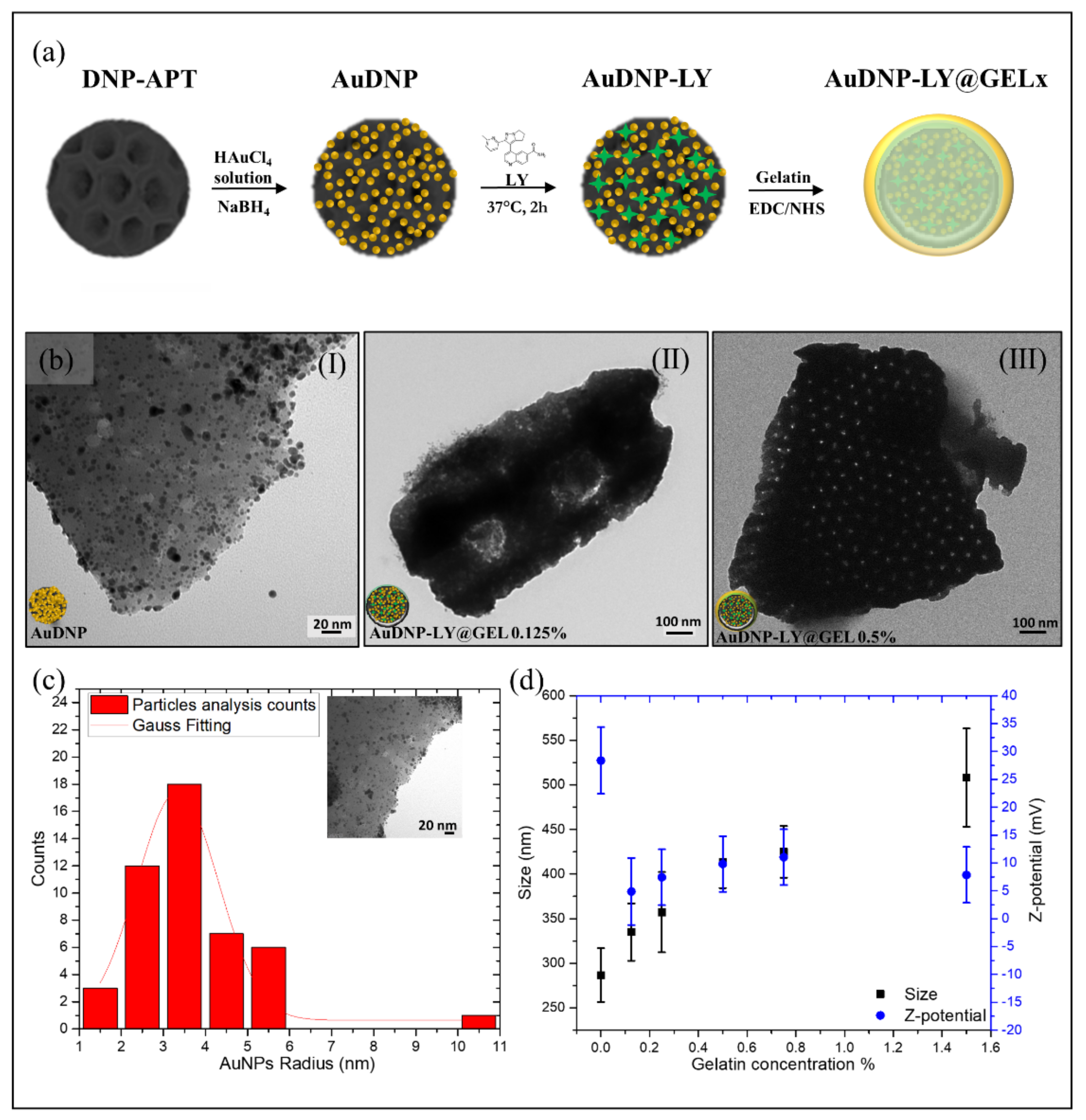
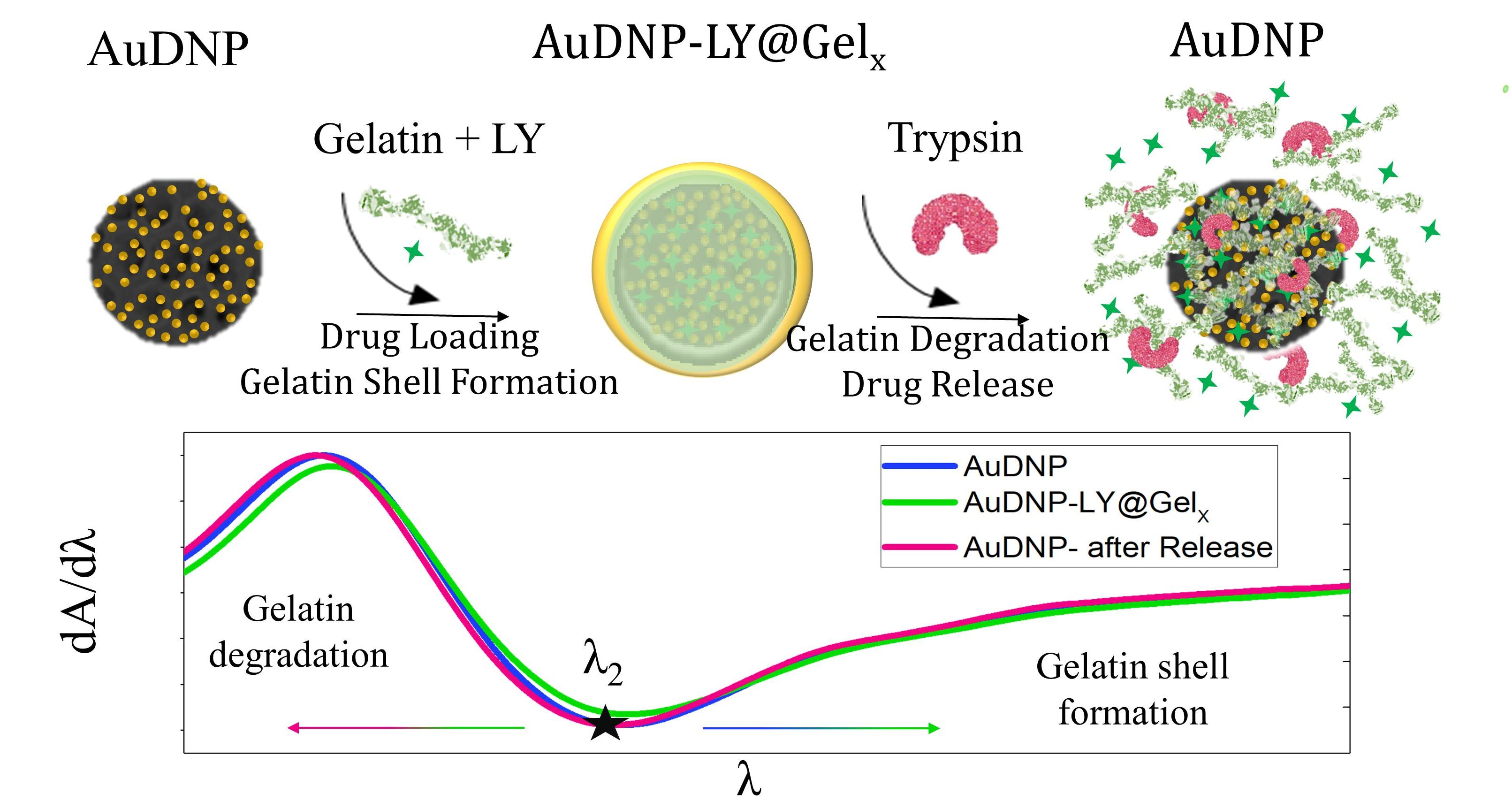
The development of a hybrid nanosystem with increasing shells of cross-linked gelatin was performed according to the functionalization procedure reported in
Figure 1a.
The growth of AuNPs on the surface of DNP (AuDNP) was performed through a liquid phase approach in which DNPs were suspended in a tetrachloroauric acid solution (HAuCl
4) and the reduction of AuNPs occurred in-situ on the surface of DNPs. Before the growth of AuNPs, the surface of DNPs was modified with a 10%
v/v APTES solution to anchor positive groups NH
3+ on the biosilica surface (DNP-APT) and promote the electrostatic interactions between gold precursor ions and DNPs. The electrostatic attraction between negative gold precursors and the positive charged DNP (DNP-APT) granted an in-situ accumulation of AuNPs on the DNP-APT substrate. Briefly, DNP-APT were dispersed in 0.1 M HAuCl
4 solution and stirred for ten minutes in presence of 0.025% gelatin as a stabilizing agent. The major advantage of gelatin as a stabilizer is that it can both tailor the nanocomposite properties and provide AuNPs with long-term stability preventing particle aggregation [
26]. Furthermore, since gelatin is a natural biocompatible material its use as a stabilizing agent did not introduce any environmental toxicity or biological hazards in the final nanocarrier. Generally, gelatin-stabilized AuNPs are obtained by the reduction of gold salts in AuNPs under thermal heating until the dispersion color turns from yellow to red [
27]. Here, for the first time, the reduction of gelatin-stabilized gold ions was performed by adding to the above dispersion a solution (0.1 M) of sodium borohydride (NaBH
4). The size of the obtained AuNPs can be controlled by NaBH
4 easier and faster than heating since the reducing agent is added dropwise. The formation of the DNP-AuNPs complex was immediately appreciated by a colorimetric change of the dispersion, which turned from light yellow (before reduction with NaBH
4) to deep red (after reduction). The dispersion containing the hybrid system (AuDNPs) was washed intensively to remove excess reagents and resuspended in PBS solution. Since, in the complex AuDNP, the electrostatic attraction between gelatin-stabilized AuNPs and DNP-APT is not stable under harsh stirring and pH conditions, we operated a cross-linking of the gelatin stabilizing AuNPs to ensure their stability on the surface of DNP-APT. To this aim, the system AuDNP was dispersed in 1-ethyl-3-(3-dimethyl aminopropyl) carbodiimide (EDC)/N-hydroxysulfosuccinimide (NHS) solution to promote the peptide bond between amino (NH
2) and carboxyl groups (COOH) on the gelatin backbone. The crosslinking of the gelatin stabilizing AuDNP dispersion strengthened the presence of AuNPs on the biosilica surface, allowing to further functionalize the complex and prevent loss of AuNPs on the substrate. At this stage, gelatin must be considered only as a surfactant that inhibits particle aggregation and improves the AuNP stability on biosilica. Then, Galunisertib (LY) was loaded in the hybrid complex through both entrapping and physisorption methods. To prevent the burst release effect attributed to porous drug carriers [
28], the complex AuDNP-LY was mixed with high-concentrated solutions of gelatin to grow a shell around the whole system. To this purpose, the AuDNP-LY complex was dispersed in gelatin solutions (0.125%, 0.25%, 0.50%, 0.75%, 1.5%
p/v) and crosslinked via EDC/NHS chemistry that favored the development of shells with different thicknesses. At the end of the functionalization procedure, the gelatin shells covered the entire hybrid system composed of DNP decorated by gelatin stabilized AuNPs.
To assess the presence of the outer gelatin shell in the AuDNP-LY@Gel complex, transmission electron microscopy (TEM), Dynamic Light Scattering (DLS), and
-Potential analysis were performed. According to
Figure 1b(I), the irregular surface of DNPs was decorated by a carpet of AuNPs with a mean radius
r of ~ 3 nm (
Figure 1c). The presence of the gelatin shell covering the final AuDNP-LY@Gel
x complex was confirmed by the surface of the complex becoming darker and smoother upon an increment of the gelatin concentration [
29,
30]. As evident from
Figure 1b(II,III), the gelatin shell on the biosilica surface covered the carpet of AuNPs, which were no longer visible by TEM analysis. Additionally, the porous structure of DNPs in
Figure 1b(III) is more evident than in
Figure 1b(II), due to the heterogeneity of the diatomite powder that is constituted by various species of diatoms having diverse pore-pattern (see also
Figure S1). According to DLS and
-potential investigations (
Figure 1d), DNPs-APT were characterized by a mean diameter of 290 (30) nm and a charge of 28 (6) mV due to the presence of protonated NH
3+ groups on their surface. Increasing concentrations in the gelatin shell caused an increment in the particle size mainly ascribed to the steaky nature of gelatin and the formation of agglomerates between the DNPs. In the sample AuDNP-LY@Gel
0.125% and AuDNP-LY@Gel
0.25%, the size increment observed was almost similar to the control without the gelatin shell with a variation of about 30 nm. For the AuDNP-LY@Gel
0.5% sample, a significant variation of size from 290 (30) to 420 (60) nm was appreciated and even a greater variation was detected in the AuDNP-LY@Gel
1.5% sample. In fact, by increasing the amount of gelatin in the external shell, the internal cross-linking reactions occurring between the DNPs arise and cause particle agglomeration in the samples [
31]. Therefore, in the sample with the highest amount of gelatin in the shell a size increment of about 250 nm was observed compared to the control sample. The size evolution observed after the formation of the gelatin shell was also justified by the surface charge of the samples, which was close to zero [
31]. The type B gelatin used in this study has an Isoelectric Point (IEP) between 4.8 and 5.4, therefore it was expected for AuDNPs-LY@Gel samples to have a negative surface charge in an aqueous solution pH 6.5–7.0 (pH
). However, a shift of the IEP of the gelatin toward 6–7 can be observed as a consequence of the crosslinking mechanism between COOH and NH
2 groups in the polymer backbone [
32]. Due to this slight shift, the surface charge of AuDNP-LY@Gel samples was expected to be neutral at the IEP, or slightly positive. The charge of AuDNP-LY@Gel complex (0.125%, 0.25%, 0.5%, 1%, and 1,5%) was comparable to each other’s and decreased from 28 (6) (DNPs-APT) mV to a mean value of 7 (5) mV in an aqueous solution, confirming the successful cross-linking of the gelatin shell. The small
-potential value caused in the samples particle aggregation and flocculation that lowered the repulsion forces between AuDNPs-LY@Gel
x complex and the physical colloidal stability as well. Since the Gel-AuNPs composing the system were firmly stabilized on the DNP surface, these aggregation phenomena did not affect the stability and plasmonic response of AuNPs.
2.2. Reverse Engineering-Based Modeling of the LSPR of AuDNPs-LY@Gel Drug Delivery System
The morphological characterization reported in the previous section provided the first evidence of the increase of cross-linked gelatin shells on AuDNPs-LY@Gel systems. Unfortunately, an accurate estimation of this parameter, which could be crucial in the design of DNPs-based drug delivery systems with enhanced drug loading capacity, could not be provided by TEM micrographs or DLS measurements since the dimensions of DNPs, on the one hand, gelatin-stabilized AuNPs (Gel-AuNPs) and shell thicknesses, on the other hand, were significantly different. For this reason, to extrapolate our missing information (i.e., gelatin shell thickness
t), we exploited the LSPR optical response of AuNPs providing full modeling to our systems, starting from a reverse engineering approach. The small AuNPs, grown in-situ on the DNP surface and stabilized in presence of gelatin, exhibited a mean radius
r of ~3 nm, as highlighted in
Figure 1c. Moreover, TEM micrographs clearly show that, on average, their spacing was sufficiently larger than their size. For this reason, they can be considered as non-interacting NPs in good approximation (
Figure 2b). However, the application of the simple Mie theory model to very small AuNPs dispersed in water would result in an LSPR peak (
) located at a wavelength of
nm in the visible spectrum. Instead, from the absorbance spectroscopy performed on the AuDNPs-LY systems with no further gelatin coatings, the typical plasmonic response of AuNPs was observed, but with a huge redshift (~25 nm) of the resonance peak (
~540 nm) (
Figure S2). We attributed this shift to the combination of two parameters. First, the reduction of AuCl
4− ions in presence of the gelatin, which led to the formation of inhomogeneous AuNPs with a significant percentage of gelatin remaining entrapped in the volume during nucleation and growth processes (
Figure S1b). This resulted in a first resonance shift to 525 nm (
Figure S2). Secondly, the presence of DNPs as substrates in the surroundings of the plasmonic NPs, which possess an effective refractive index higher than water, caused a further resonance shift to 540 nm. Although Gustav Mie found the analytical solution of Maxwell’s equations for the scattering and absorption cross-sections of a pure metallic nanosphere [
33,
34], his description did not consider the use of stabilizing agents and inhomogeneities occurring during the bottom-up synthesis of NPs. Stabilizers, as gelatin, having their refractive index, are generally involved in the chemical synthesis of colloidal NPs. Their use affects the effective dielectric constant and optical properties of the hybrid NPs, making an accurate description of their absorption spectra difficult. The effect of inhomogeneities, denoted here as inclusions, on the optical constants of the hybrid material can be predicted by Maxwell-Garnett’s homogenization theory [
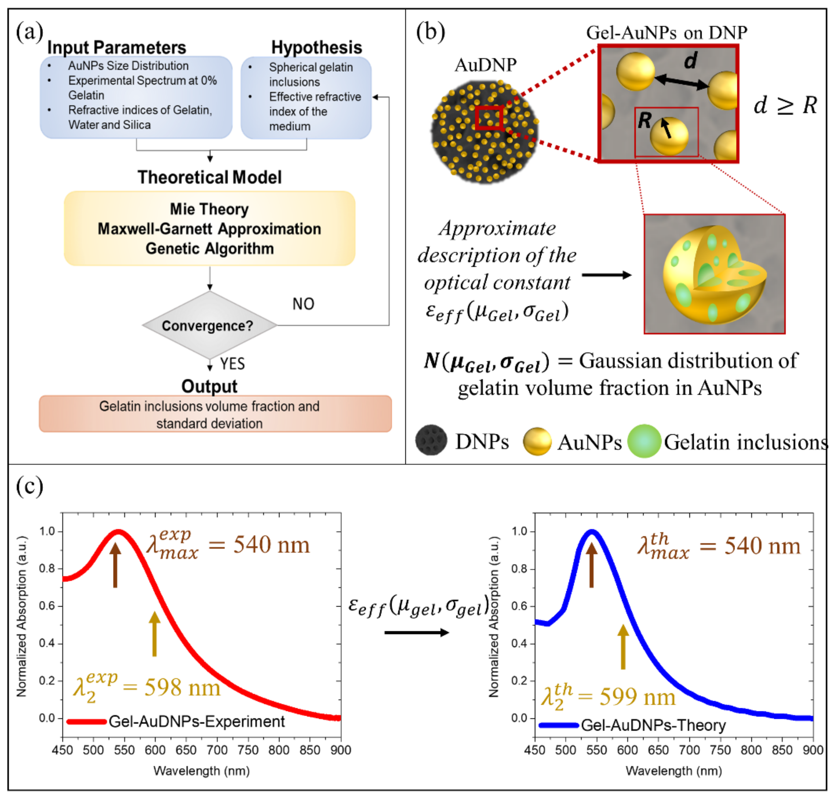
35]. Therefore, we combined Mie Theory and Maxwell-Garnett approximation to predict the absorption and scattering cross-sections of our Gel-AuNPs [
36]. We introduced an effective dielectric constant
describing the hybrid nature of Gel-AuNPs, in which the gelatin inclusions are assumed as spherical for sake of simplicity, and an effective refractive index of the medium, which considered the anchoring of the Gel-AuNPs on DNPs.
More precisely, we assumed the gelatin inclusions as a gaussian distribution of spherical particles within the AuNPs, as schematized in
Figure 2b, and we optimized the mean
and standard deviation
of the volume fraction occupied by the inclusions with a simple optimization strategy. We used the Genetic Algorithm (GA) to minimize the objective function reported in Equation (4), imposing a stopping condition below 0.1 and extracting the two fitting parameters, namely the mean volume fraction percentage occupied by gelatin inclusions (modeled as spheres) within Gel-AuNPs (
) and the standard deviation of these spheres (
). The root mean square value between theoretical and experimental absorbance spectra of AuDNPs-LY returned by GA was 0.09 (<0.1) and the fitting values were 0.11 and 0.03 for
and
, respectively. This optimization gave us three important results: first, Gel-AuNPs contained a gelatin volume fraction of
, which was in good agreement with the initial weight
ratio (
w/w) between gelatin and HAuCl
4 of the reaction volume; secondly, we obtained an estimation of the medium refractive index from which Gel-AuNPs optical response was affected, corresponding to a 10% silica (DNPs) and 90% water; finally, accurate optical modeling of the hybrid AuDNPs-LY system immersed in water was obtained, confirming the consistency of our initial hypotheses. The model is strongly based on the homogenization approach, which was possible due to the very small size of the AuNPs compared to the operating wavelength. The description of Gel-AuNPs would safely hold also in case of the presence of gelatin only on the surface of the AuNPs and not also within, thus offering an accurate description of the hybrid system (see also
Figure S2).
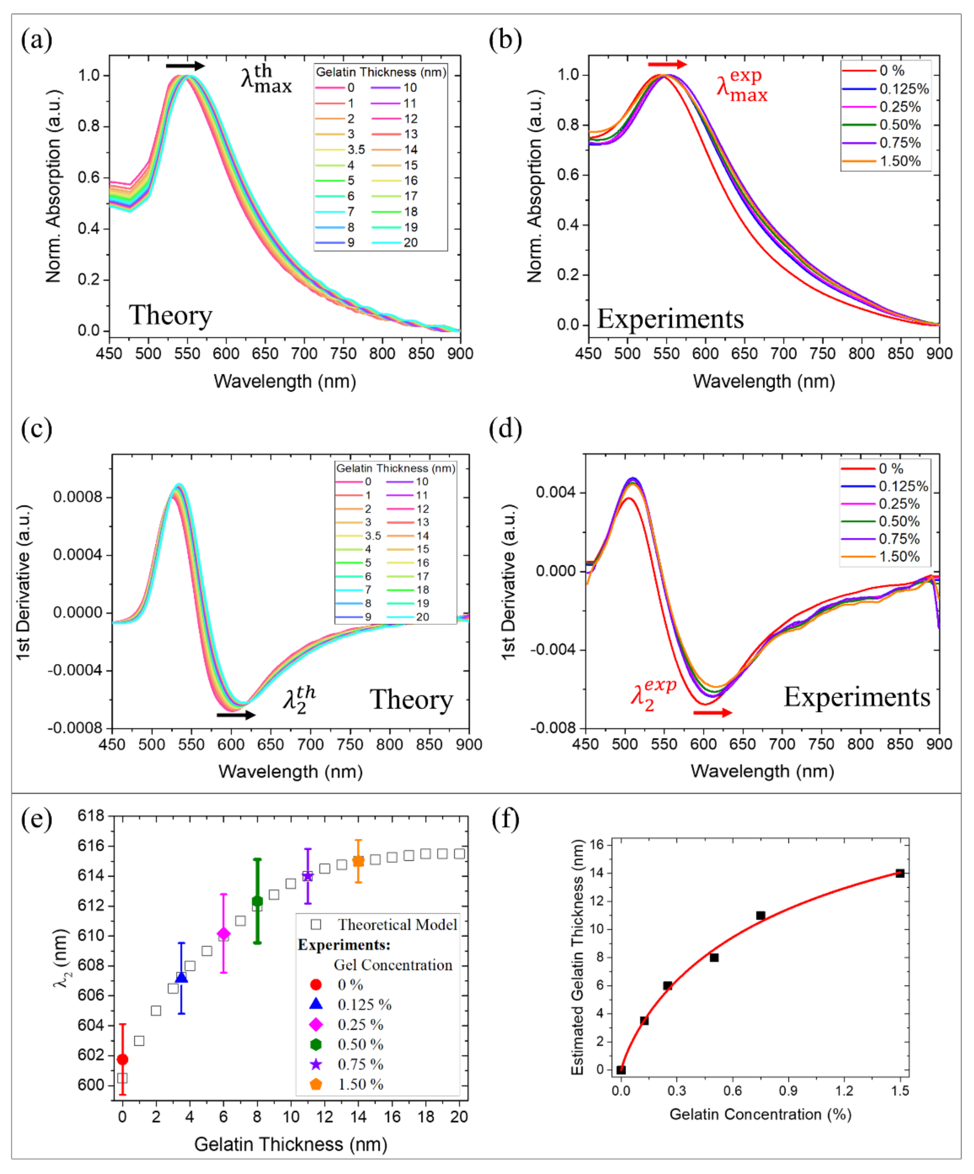
As shown in
Figure 2c, the theoretical modeling of the hybrid AuDNPs system is in very good agreement with both mean LSP resonance
and the inflection point
of the experimental absorption spectra, corresponding to 540 nm and 598 nm, respectively. The inflection point
is simply the minimum of the first derivative of plasmonic absorbance spectra and will be crucial in the next section to increase the sensitivity of AuDNPs-LY@Gel systems. The variation of 9% between theory and experiments resulting from these simulations was attributed to the different absorption intensities between 450 and 500 nm, which arose experimentally from both LY and gelatin showing non-negligible absorptions in the UV and visible region (450–500 nm) of the spectrum. However, both
and
were not affected by absorptions in that region and, consequently, they could be considered negligible. The small molecule LY, having a refractive index of 1.75, was not considered in the model optimization since the low molecular weight and tiny concentration could not result in LSPR shifts. This hypothesis was confirmed by experimental absorption measurements, which are reported in
Figure S3 of the
Supplementary Materials.
2.3. Correlation of Gelatin Concentration with Mean Shell Thickness on AuDNPs-LY@Gel Drug Delivery System Based on a Validated Optical Model
Once the goodness of our model was assessed in terms of accurate prediction of the optical response (
and
) of Gel-AuNPs on DNPs in presence of LY, we applied it to the AuDNPs-LY@Gel
x systems to give an estimation of the gelatin shell thicknesses as a function of the increasing gelatin concentrations. The correlation between gel concentrations and effective gelatin shell’s average thicknesses (
) can be a crucial parameter for the design of a nanocarrier and, employing our optical model, we provided our system with an efficient strategy to monitor the shell formation. First, we simulated the LSPR response of the AuDNPs-LY systems with shells of increasing thicknesses (
t) in the NPs surroundings. By doing this, we modified the effective refractive index of Gel-AuNPs on the DNPs surface observing, as expected, a plasmon redshift with the increasing gelatin shell thickness. Indeed, crosslinked gelatin possesses a refractive index higher than water and silica, thus causing a redshift of the plasmon resonance
. We performed the simulation for average thicknesses of gelatin shells in the Gel-AuNPs surroundings in the interval (0–20) nm and the results are reported in
Figure 3a. Differently, we experimentally monitored the plasmon resonance shift corresponding to the chemical crosslinking of different gelatin concentrations on the AuDNPs-LY@Gel
x systems (
Figure 3b). Since we were dealing with very tiny AuNPs, we also considered the inflection point
of both theoretical and experimental absorption spectra, which enabled the enhancement of the plasmonic response sensitivity [
37,
38]. To do this, we calculated the first derivatives of the spectra and took, as
, the minima of these curves (
Figure 3c,d). Although there was another inflection point (
) corresponding to the maxima of the first derivatives, it has been already demonstrated that the highest sensitivity for refractive index sensing can be obtained by considering the
values [
37,
38]. Moreover, as explained earlier, the first inflection points lie near the regions in which the theoretical prediction was not as accurate as in the other regions due to the neglected absorptions of LY and gelatin in those regions (
Figure S4). For these reasons, from this point on, we only consider the inflection points for our discussion. From the theoretical point of view,
underwent a redshift as a function of the gelatin thickness. The
exhibited a red shift from 600 to 616 nm following a saturation curve with a linear range from 0 to ~11 nm of gelatin thickness and achieving saturation at ~18 nm (white squares in
Figure 3e). This saturation behavior can be easily explained by the rapidly decaying field enhancement of Gel-AuNPs, whose plasmonic effect is localized to the NPs surroundings.
This means that, even if thicker gelatin shells could be easily simulated, the inflection points of the absorption spectra would have been not affected anymore by the increase of the gelatin shell. Therefore, we stopped our simulations at 20 nm gelatin thickness. Meanwhile, the experimental evaluation of
was performed by measuring the absorption spectra of the AuDNP-LY@Gel systems obtained by crosslinking different gelatin concentrations: 0%, 0.125%, 0.25%, 0.5%, 0.75%, and 1.50% on the AuDNP-LY systems. Accordingly, a redshift of the inflection points was observed for the different gelatin concentrations. The theoretical and experimental results reported in
Figure 3c–e allowed the introduction of a correlation between gelatin concentration and gelatin thickness on the hybrid AuDNPs-LY delivery system. This relationship between gelatin concentration
and gelatin thickness (t) exhibited a saturation behavior due to the electromagnetic field decay in the surroundings of AuNPs, which could be described by a Michaelis-Menten type equation in the investigated thickness range (
Figure 3f):
where
~ 18 nm is the maximum gelatin thickness at which a redshift of the inflection point
of the absorption spectra of Gel-AuNPs was still detectable and
is the gelatin concentration at which the shell thickness on the AuDNP-LY@Gel system corresponded to half of the
(~ 9 nm). Therefore, we found a strong analogy between the formation of gelatin shells on the Gel-AuDNPs and the typical kinetics of substrate-enzyme reactions. In this analogy, the gelatin concentration acts as the substrate, while the electromagnetic field decay in the surroundings of AuNPs acts as the limiting factor of the process (enzyme). While saturation in a typical enzyme-substrate reaction is achieved when the enzyme is exhausted, in our case saturation is achieved when the electromagnetic field enhancement in the Gel-AuNPs surroundings is decayed. At fixed times, we correlated the shell thickness directly with the gelatin concentration according to the same equation. After a linear increase of the estimated thickness as a function of the gelatin concentration, we observed a saturation, which is not due to the gelatin formation itself but to the plasmonic response of Gel-AuNPs on DNP, which is not sensitive any more to the gelatin formation. The modeling and estimation of the average gelatin thickness of our drug delivery system was revealed as fundamental in the nanocarrier design. Indeed, the model provided the reverse design of a system in which the gelatin concentration could be tuned to achieve the desired average coating thickness.
2.4. Evaluation of the Loading Capacity of the Nanosystem AuDNP-LY@Gelx
The burst release of a significant fraction of payload in the medium within a few minutes represents the most frequent drawback of porous drug carriers. Such uncontrolled release reduces the effective nanocarrier drug loading capacity and, in turn, the amount of drug delivered to the desired site of action [
39]. To avoid this limitation, porous drug carriers can be encapsulated in polymeric matrices able to retain the therapeutic compound and control the drug release in response to external
stimuli. We already assessed the ability of a gelatin shell embedding drug-loaded DNPs to allow a sustained release of the drug in response to the acidic microenvironment [
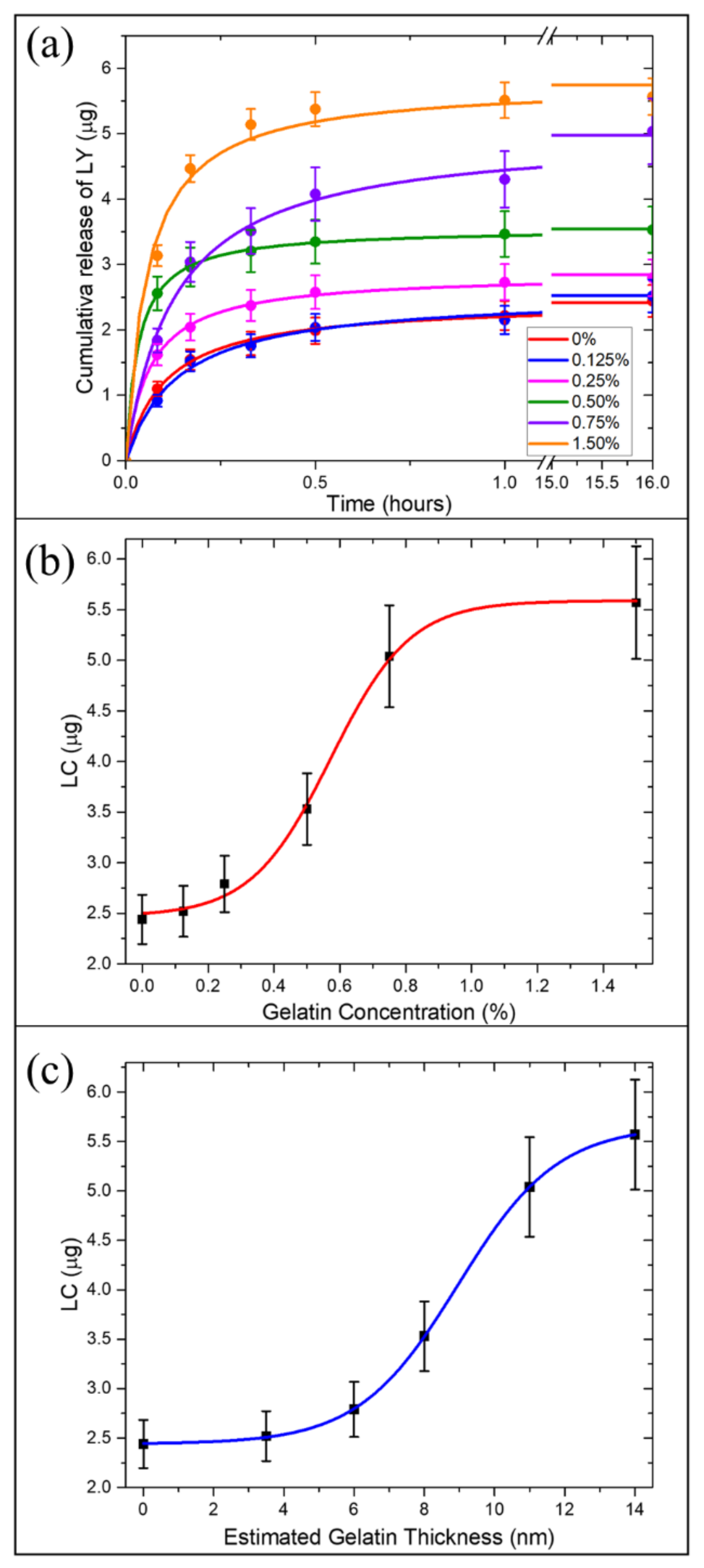
21]. However, we did not investigate the loading capacity (LC) tunability of porous DNPs as a function of the amount of gelatin in the external shell. To this aim, in this study, in vitro-drug release analysis was performed in triplicate and the amount of drug released in the solution was quantified through Reversed-Phase High-Performance Liquid Chromatography (RP-HPLC) (
Figure 4a). Drug release investigations were carried out in Phosphate Buffered Saline (PBS) solutions with a large excess of trypsin enzyme for 16 h. Recent findings suggested that gelatin is preferentially degraded by trypsin compared to enzymes such as lipase and amylase [
40]. Moreover, since trypsin overexpression is involved in tumor progression and metastasis, in vitro release tests were carried out in presence of a large amount of trypsin to simulate the cancer microenvironment [
41]. The LC and encapsulation efficiency (EE) of AuDNP-LY@Gel
x with increasing gelatin amount in the outer shells was compared to the AuDNP control sample (in which gelatin was used only as a stabilizer agent in the AuNP synthesis, but not as the external shell). We evaluated the influence of the shell thickness (estimated in previous sections) on the system loading capacity. First, we measured the LC and EE of the control AuDNP and found that, even without the external gelatin shell, both LC and EE (
and 0.48% respectively) were higher than the previous LY delivery system [
21]. This improvement can be ascribed to the presence of gelatin as AuNP stabilizer and its ability to establish strong interactions with drug molecules [
42]. Therefore, it was already possible to appreciate an improvement of the DNP drug LC and EE by modifying the AuNP synthesis approach. Later, we studied the variation of the LC and EE as the concentration of gelatin increased, figuring out a way to modulate the desired LC to the shell grown on the DNP surface. According to
Figure 4a, the increase of gelatin concentration/thickness in the shell solution resulted in a higher amount of LY entrapped in the system and available for delivery to the desired site of action. The AuDNP-LY@Gel
0.125% exhibited an EE of 0.50% (LC was
similar to the control, probably because the used gelatin concentration was too low to improve the LC of the nanosystem. Therefore, no differences in the amount of loaded and released drug were detected compared to the control sample. Conversely, when the gelatin concentration increased from 0.125% to 0.25%, the ability of the gelatin shell to retain LY increased, and the LC of the sample was improved up to
of the drug, with an EE of 0.56%. An even higher LC and EE of LY were observed in the samples AuDNP-LY@Gel
0.5% and AuDNP-LY@Gel
0.75%, respectively
and
(
), confirming that the higher was the concentration in the gelatin shell, the higher the amount of drug entrapped in the delivery system.
The LC swiftly approached the saturation point in the sample AuDNP-LY@Gel
1.5%, which released about
of LY (
). All the samples displayed a similar release profile with 50% of the drug released within 20 min, due to the rapid degradation of the gelatin shell by the serine-protease enzyme trypsin. The excess of trypsin in the medium digested the gelatin shell rapidly and did not allow us to appreciate the release kinetics of LY from the gelatin-capped samples. However, our group already investigated the drug release profile of gelatin-capped drug carriers in previous investigations [
21]. However, we demonstrated that the presence of gelatin capping the biosilica surface provided the system with pH-responsive properties (
Figure S5), due to the enhanced extension of the gelatin chains in acidic microenvironments [
43]. The therapeutic dose of a drug to encapsulate in carriers is generally a non-modifiable parameter, and it is not obvious that NPs have the adequate LC for encapsulating the therapeutic dose. In this scenario, to reach the wanted amount of drug released, the quantity of administered NPs must be raised. The opportunity to select the desired amount of drug by varying the gelatin shell is a great opportunity when DNPs are employed as carriers of chemotherapeutic molecules. Therefore, the tunability of LC as a function of the gelatin shell represents the means to improve the LC of DNPs without increasing the mass of administered biosilica.
The variation of the LC in response to the increasing concentration of gelatin in the outer shell followed a typical sigmoidal behavior (
Figure 4b). The amount of LY retained in the system did not show significant variation for gelatin concentrations below 0.125% (toe region), since the gelatin shell was not thick enough to appreciate an improvement of the loading capacity compared to the control. As is obvious from
Figure 4c, the drug loading capacity of AuDNP-LY@Gel
x systems followed the same sigmoidal curve as a function of the gelatin estimated average thickness. No significant enhanced retaining efficiency was observed for gelatin average thickness below ~6nm. At higher gelatin concentrations, instead, a direct proportionality with the nanosystem drug loading capacity was observed, reaching a plateau region in the sample with a gelatin concentration of 1.5%.
The increment of the LY retention in the system can be explained by considering the non-covalent interactions between the gelatin molecules and LY. The type-B gelatin shows an IEP value between 4.7–5.2 and displays an almost neutral charge at a pH value of 4.5. When the system was dispersed in the gelatin solution pH 4.0, the LY molecules physisorbed on the surface only exhibited a negligible positive surface charge on the quinoline-carboxamide group. Therefore, by increasing the gelatin concentration, the strength of Van der Walls interactions occurring between the drug molecules and polymer chains increased on the surface of the system, allowing better retention of the LY. Moreover, by raising the gelatin concentration, a larger polymer matrix was crosslinked as well, thus inhibiting the diffusion of the drug molecules out of the system. The sigmoidal relationship between the drug LC (μg) of AuDNPs-LY@Gel
x systems and the estimated gelatin shell thickness (t, nm) can be expressed as a Boltzmann equation:
where
and
are the maximum and minimum amounts of LY loaded on the AuDNP-LY@Gel
x systems reported previously,
t is the estimated gelatin thickness,
, and
is:
The
trend of the AuDNP-LY@Gel
x systems as a function of the estimated gelatin thickness reported in
Figure 4c exhibited three different regimes, which can be described with the Boltzmann type equation (Equation (2)): a toe region in which the gelatin thickness did not affect the
capacity of the system (0–5 nm); a linear region in which the
was directly proportional to the estimated gelatin thickness (5–11 nm) due to the increase of the interactions between the drug and gelatin molecules; and a saturation region (11–14 nm) in which the drug, whose experimental concentration was kept constant (
), represents the limiting factor of the entrapment efficiency in the gelatin matrix. It is worth mentioning that the drug loading capacity enhancement due to the increased shell thickness could be fully considered in the estimation model we described earlier.
Surprisingly, the saturation achieved by the drug loading enhancement fell in the range of the estimated gelatin thickness (0–20 nm). As stated in the previous section, this was not obvious since our system could have reached saturation in drug loading capacity for gelatin thicknesses higher than 18 nm, in which no plasmon resonance shifts would have been observed. In that case, our optical description of the system would have been valid only for a limited part of the work. Instead, we provided our system with complete optical modeling, in which the estimated gelatin thickness could be experimentally verified and tuned in time to monitor two parameters: the gelatin shell formation (with an estimation of its thickness) via simple spectroscopic measurements, and the simultaneous gelatin degradation and drug release. This second part will be discussed in-depth in the next section.
2.5. Application of the Model to the Monitoring of the In-Situ LY Drug Release
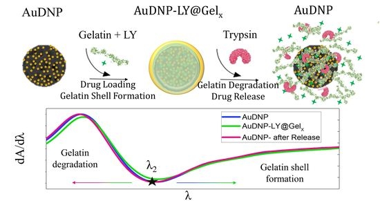
As discussed in the previous sections, the complete optical modeling of the AuDNP-LY@Gel
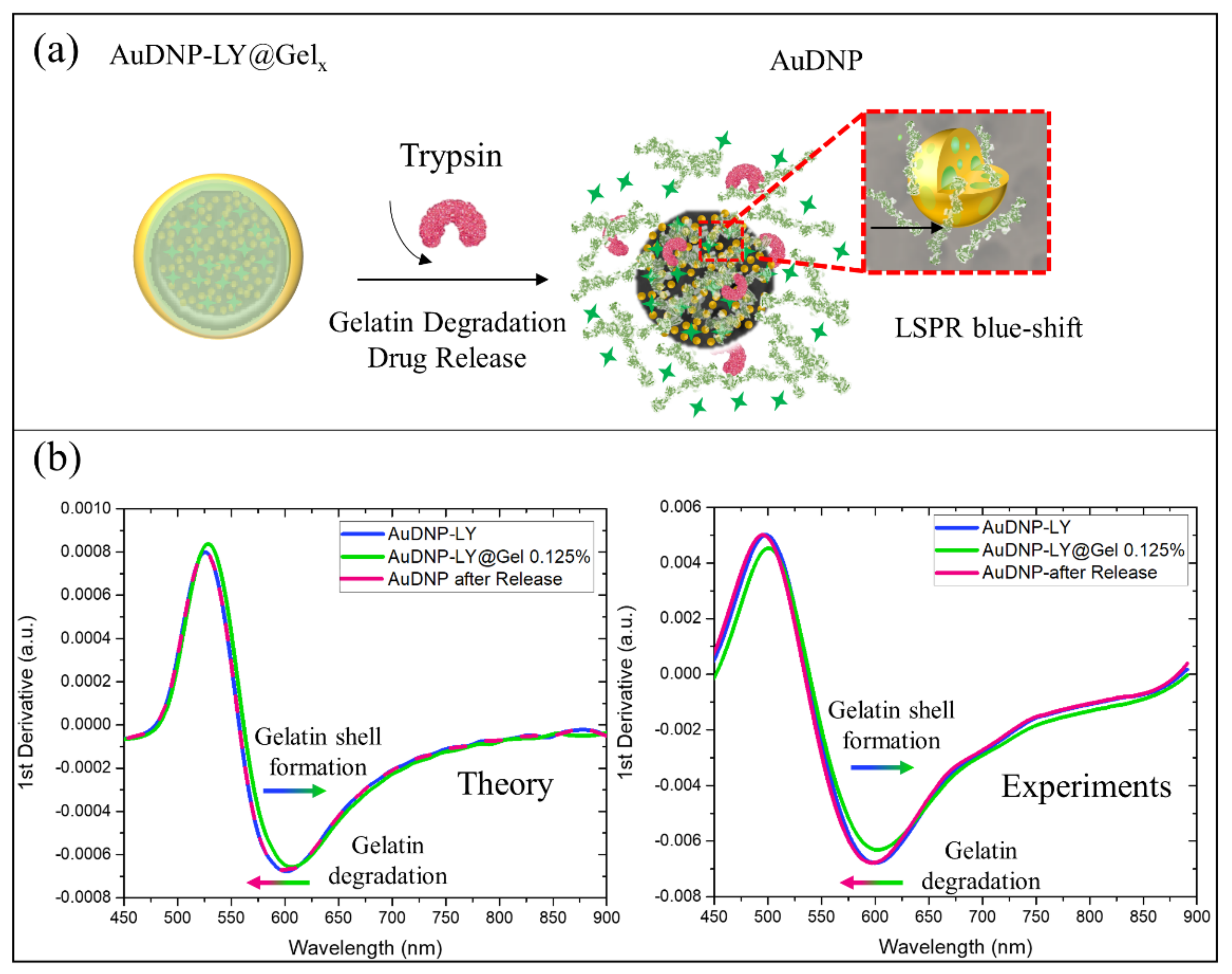
x system could be used to monitor the gelatin shell formation on the surface of the nanocarrier in time. Furthermore, the presence of AuNPs on DNP provided our system with another important functionality: to monitor the degradation of the outer gelatin shell in presence of trypsin enzyme (
Figure 5a) directly and to trace the drug release of LY indirectly. It was expected that once half of the gelatin thickness was degraded by trypsin, about half of the loaded drug in the nanocarrier was released; this could be evaluated by a spectroscopic measurement without the need for expensive equipment. As a proof of concept, the blue shift associated with the gelatin degradation of the AuDNP-LY@Gel
0.125% system was reported in
Figure 5b.
From the theoretical point of view, trypsin enzymes used during the release studies reduced the medium effective refractive index from 1.54 (in presence of gelatin) to 1.33 (water refractive index) in the surroundings of AuNPs on the DNPs. For this reason, 100% of gelatin shell degradation would result in a blue shift in both
and
of the Gel-AuNPs absorption spectra. Ideally, the blue shift due to the gelatin degradation should be equal and opposite to the redshift caused by the gelatin shell formation on the AuDNP system. To show this, we reported in
Figure 5b the theoretical first derivatives of the absorption spectra highlighting the perfect overlapping between blue and purple lines corresponding to the AuDNP system before the gelatin shell formation and after gelatin degradation, respectively.
To further validate our modeling for the gelatin degradation, we optically monitored the AuDNP-LY@Gel
0.125% system after the in-vitro release in presence of the trypsin enzyme. The experimental first derivatives of the absorption spectra were accurately predicted by our model (
Figure 5b) since a blue shift of the inflection point in the first derivative occurred. However, from the experimental point of view, the overlap between the AuDNP-LY system (blue line) and AuDNP system after the drug release study (purple line), was not observed. The inflection wavelength of the AuDNP system underwent a slight blue shift compared to its initial position, after gelatin degradation. We attributed this phenomenon to the intrinsic hybrid nature of the Gel-AuNPs, which has been theoretically hypothesized and experimentally validated in this study. Indeed, as highlighted in the inset of
Figure 5a, the AuNPs of this work contained gelatin inclusions, which could be partially available on their surface for trypsin recognition and degradation. Therefore, since the effective relative dielectric constant of Gel-AuNPs was strictly dependent on the gelatin volume fraction, also their optical properties could be affected by gelatin degradation. However, due to the very slight variation root mean square between theoretical and experimental results, this unwanted effect could be safely neglected.


 ,
, 

