
Dual-Specificity, Tyrosine Phosphorylation-Regulated Kinases (DYRKs) and cdc2-Like Kinases (CLKs) in Human Disease, an Overview


Abstract
1. Introduction
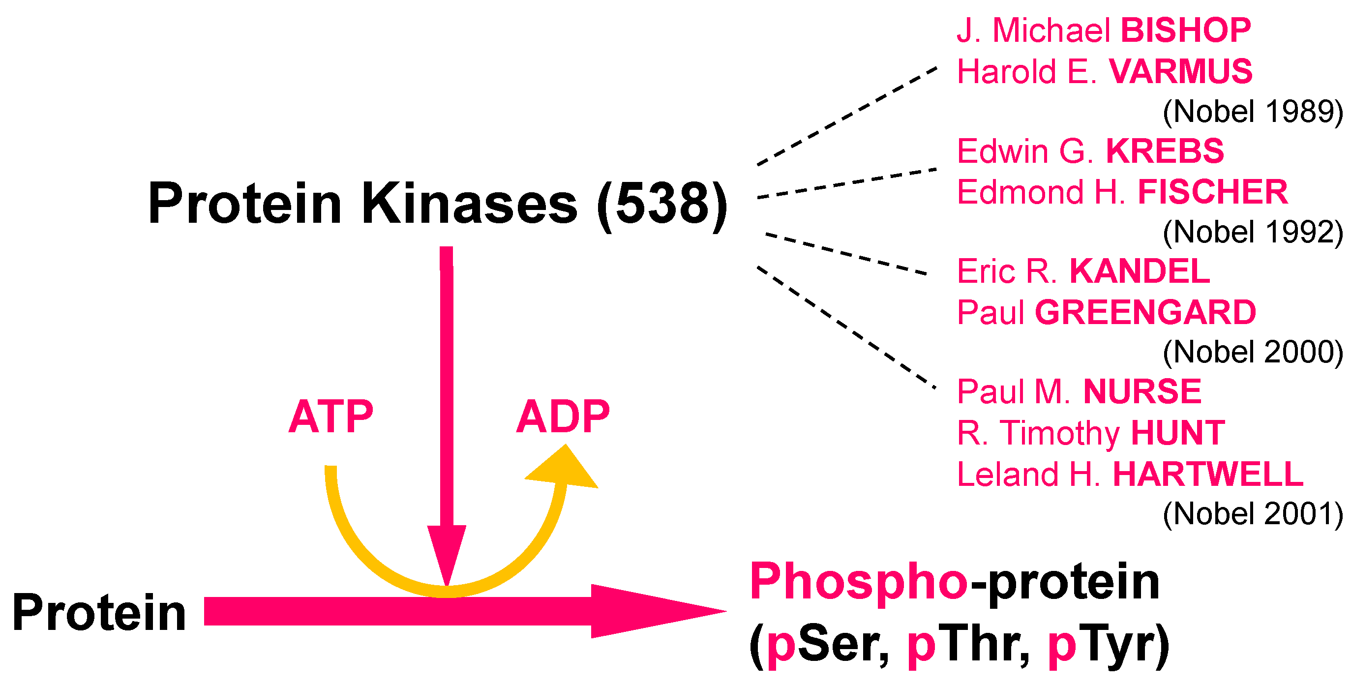
1.1. Protein Phosphorylation, Protein Kinases, Kinase Inhibitors, and Human Disease
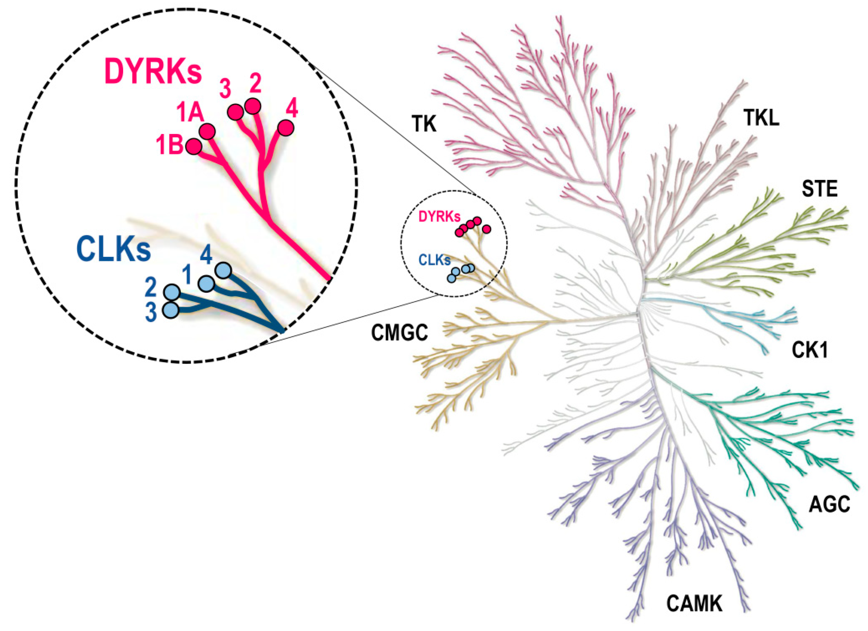
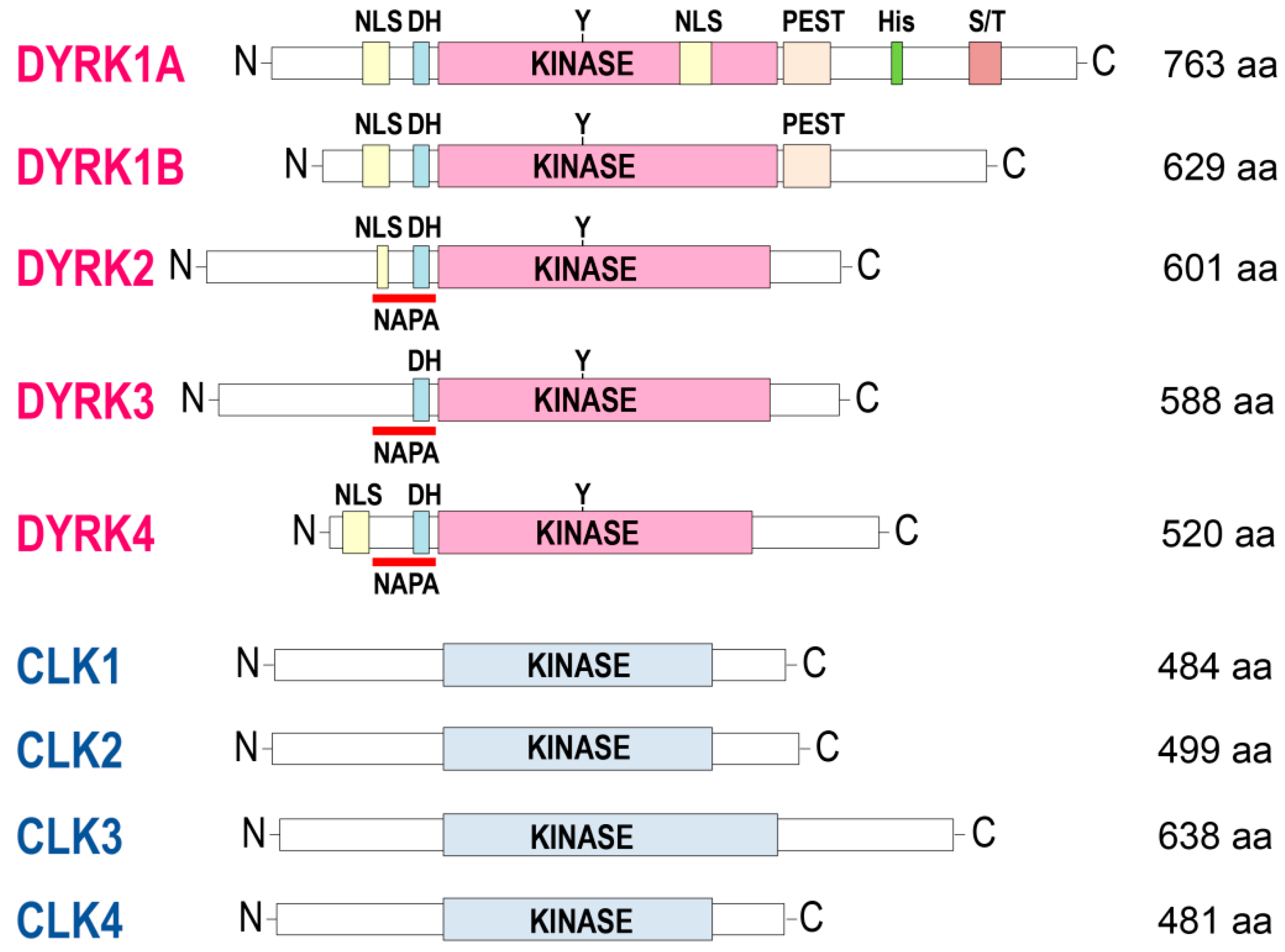
1.2. DYRKs and CLKs: Structure, Activation, Interactors, and Substrates


| Kinase | Ligand | PDB | Reference |
|---|---|---|---|
| DYRK1A | DJM2005 | 2VX3, 2WO6 | [18] |
| Leucettine L41 | 4AZE | [43] | |
| Harmine | 3ANR | [44] | |
| INDY | 3ANQ | [44] | |
| Compounds 3 and 23 | 4MQ1, 4MQ2 | [45] | |
| LDN-211898 | 5AIK | Elkins, unpublished | |
| PKC412 | 4NCT | [46] | |
| Inhibitor 5t, 5s | 4YLL, 4YLK | [47] | |
| Compound 32, 14 | 6A1G, 6A1F | [48] | |
| XMD7-112, JWD-065 | 6EJ4, 6EIV | [49] | |
| [b]-annulated chloro-substituted indole 13 | 4YLJ | [50] | |
| KuFal319 | 6T6A | [50] | |
| AnnH75 | 4YU2 | [51] | |
| compound 2-2 (harmine derivative) | 6UWY | [52] | |
| GNF2133 | 6UIP | [53] | |
| DJM2005 (DB07608) | 2WO6 | [18] | |
| DYRK2 | - | 3K2L | [18] |
| Leucettine L41 | 4AZF | [43] | |
| Indirubin 6i | 3KVW | [54] | |
| EHT 5372, EHT 1610 | 5LXC, 5LXD | [55] | |
| DYRK3 | Harmine | 5Y86 | [56] |
| CLK1 | - | 6TW2 | [57] |
| compounds 8g, 16 | 6FT8, 6FT9 | [58] | |
| debromohymenialdisine | 1Z57 | [59] | |
| KH-CB19 | 2VAG | [60,61] | |
| Pyrido [3, 4-G] quinazolines 13, 14 | 5J1V, 5J1W | [62] | |
| Compound 25 | 5X8I | [63] | |
| CX-4945 | 6KHD | [64] | |
| CX-4945 | 6FYO | [65] | |
| Compounds 9m, 10i | 6Q8P, 6Q8K | [66] | |
| 5-iodotubercidin | 6G33 | [67] | |
| furanopyrimidines VN412, VN316, VN345 | 6I5H, 6I5L, 6I5K | [68] | |
| ETH1610 (Cpd 17) | 6YTI | [69] | |
| KH-CARB13 (Cpd 3) | 6YTG | [69] | |
| Tg003 (Cpd 2) | 6YTE | [69] | |
| GW807982X (Cpd 8) | 6ZLN | [69] | |
| imidazopyridazine (Cpd 1) | 6YTA | [69] | |
| CAF052 | 7AK3 | [70] | |
| TbCLK1 | AB1 | 6Q2A | [40] |
| CLK2 | 1RO, NR9 | 3NR9 | Knapp, unpublished |
| CX-4945 | 6KHE | [64] | |
| CX-4945 | 6FYL | [65] | |
| CLK3 | - | 2EU9, 2EXE | [59] |
| KH-CB19 | 2WU7 | [60] | |
| K00546 | 2WU6 | [60] | |
| Leucettine L41 | 3RAW | [71] | |
| CX-4945 | 6KHF | [64] | |
| CX-4945 | 6FYP | [65] | |
| KH-CARB13 (Cpd 3) | 6YU1 | [69] | |
| Tg003 | 6YTW | [70] | |
| compound 8a | 6FT7 | [58] | |
| CLK4 | CX-4945 | 6FYV | [65] |
2. DYRKs and Human Disease
2.1. DYRK1A and Down Syndrome (DS)
2.2. DYRK1A and Alzheimer’s Disease (AD)
2.3. DYRK1A and Parkinson’s Disease (PD)
2.4. DYRK1A and Mental Retardation Disease 7 (MRD7)
2.5. DYRK1A and Viral Infections
2.6. DYRK1A and Diabetes
2.7. DYRK1A and Cancers and Leukemias
2.8. Other DYRKs and Human Disease
| Kinase Target | Disease | References |
|---|---|---|
| DYRK1A | Down syndrome (DS) | [127,128,129,130,131,132,133,134,135,136,137,138,139,140,141,142,143,144,145,146,147] |
| DYRK1A | Alzheimer’s disease (AD) and other Taupathies | [96,98,128,129,131,148,149,150,151,152,153,154,155,156,157,158,159,160,161,162,163] |
| DYRK1A | Parkinson’s disease | [99,100,101,131,164,165,166,167,168] |
| DYRK1A | Pick’s disease | [101] |
| DYRK1A | CDKL5 Deficiency Disorder | [169] |
| DYRK1A | Diabetes | [52,53,105,106,170,171,172,173,174,175,176,177,178,179] |
| DYRK1A | Regulation of folate and methionine metabolism | [180] |
| DYRK1A | Cancers (review) | [109] |
| DYRK1A | Glioblastoma | [181] |
| DYRK1A | Head and neck squamous cell carcinoma | [182] |
| DYRK1A | Pancreatic ductal adenocarcinoma | [183,184,185] |
| DYRK1A | Hepatocellular carcinoma | [186] |
| DYRK1A | Ovarian cancer | [187,188] |
| DYRK1A | Acute megakaryoblastic leukemia (AMKL) | [110,189] |
| DYRK1A | Acute lymphoblastic leukemia (ALL) | [111,190,191] |
| DYRK1A | Psoriasis | [192] |
| DYRK1A | Knee osteoarthritis | [193,194] |
| DYRK1A | Tendinopathy | [195] |
| DYRK1A | Human immunodeficiency virus type 1 (HIV-1) | [196,197,198] |
| DYRK1A DYRK1B | Human cytomegalovirus (HCMV) | [199] |
| DYRK1B | Hepatitis C virus (HCV), Chikungunya virus, Dengue virus, and severe acute respiratory syndrome (SARS) coronavirus Cytomegalovirus (CMV) Human papillomavirus (HPV) | [199,200,201] |
| DYRK1B | Diabetes | [105] |
| DYRK1B | Neuroinflammation | [115] |
| DYRK1B | Oral squamous cell carcinoma Liposarcoma Breast cancer Hedgehog/GLI-dependent cancer | [117,202,203,204,205] |
| DYRK2 | Cancers (reviews) | [119,120,206,207] |
| DYRK2 | Triple-negative breast cancer (TNBC) and multiple myeloma (MM) | [208,209] |
| DYRK2 | Lung adenocarcinoma | [210] |
| DYRK2 | Chronic myeloid leukemia (CML) | [211,212] |
| DYRK2 | Gliblastoma | [213] |
| DYRK2 | Colorectal cancer (tumor suppressor) | [214] |
| DYRK2 | Liver cancer (predictive marker) | [215] |
| DYRK2 | Trypanosoma cruzi | [216] |
| DYRK3 | Hepatocellular carcinoma | [121] |
| DYRK3 | Glioblastoma | [122] |
| DYRK3 | Influenza virus replication | [123] |
| DYRK3 | Anemia | [217] |
| DYRK3 | Osteoarthritis | [218] |
| DYRK4 | Breast cancer | [219] |
| DYRKs | Glioblastoma | [220] |
| DYRKs | Herpes virus, rhesus macaque cytomegalovirus, varicella-zoster virus, and herpes simplex virus (HSV-1) | [221] |
| LmDYRK1 | Leishmaniasis | [39] |
| TbDYRK | Trypanosoma brucei (sleeping sickness) | [35,36,37] |
| DYRKs/CLKs | Glioblastoma | [220] |
3. CLKs and Human Disease
| Kinase Target | Disease | References |
|---|---|---|
| CLK1 | Glioblastoma Small-cell lung cancer | [229] [230] |
| CLK1 | Duchenne muscular dystrophy | [231] |
| CLK1 | Influenza A West Nile and Chikungunya viruses | [232,233,234,235,236] [61] |
| CLK1/CLK2 | Triple-negative breast cancer | [237] |
| CLK2 | HIV-1 | [238] |
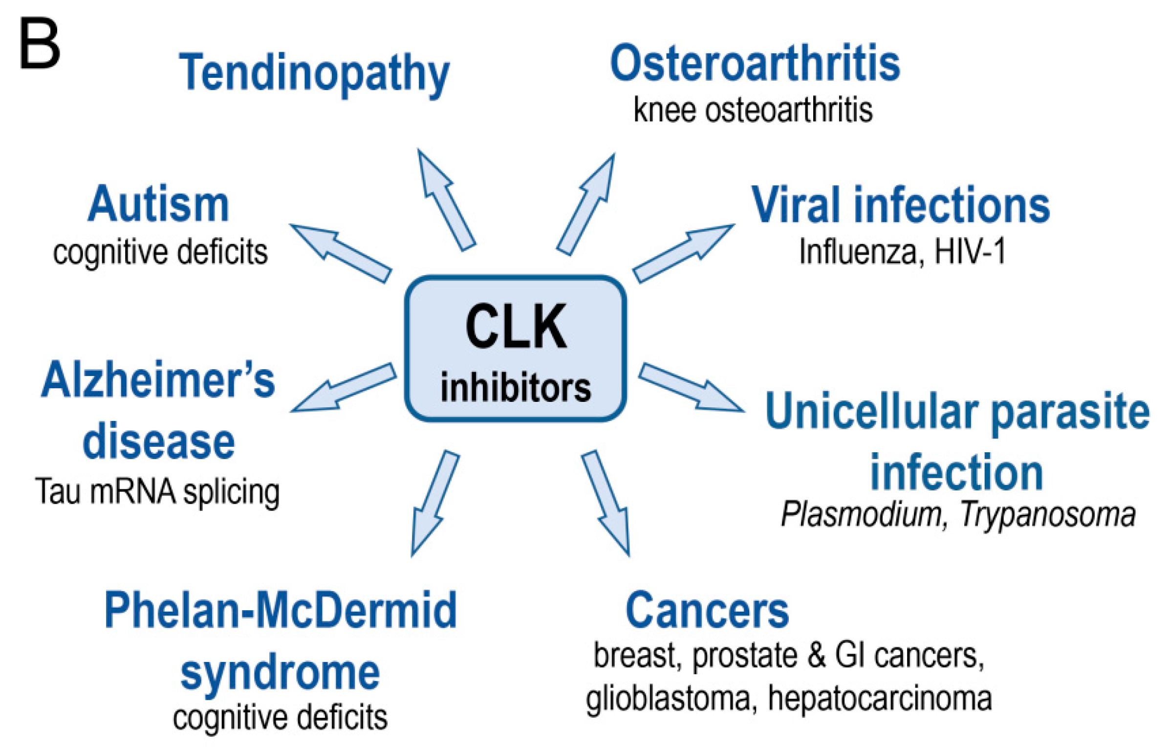
| CLK2 | Autism Phelan-McDermid syndrome (PMDS) | [239] [65] |
| CLK2 | Knee osteoarthritis Tendinopathy | [193,194] [195] |
| CLK2 | Breast cancer Triple-negative breast cancer Glioblastoma | [240,241] [242,243] [244,245] |
| CLK2 | Alzheimer’s disease (alternative splicing of Tau exon 10) | [224,225] |
| CLK3 | Hepatocellular carcinoma Prostate cancer Cholangiocarcinoma | [226] [227] [228] |
| CLKs | Body temperature | [57] |
| CLKs | Prostate cancer Gastrointestinal cancer Colorectal, ovarian cancers | [227] [246] [247] |
| PfCLKs | Plasmodium falciparum (malaria) | [248,249,250,251,252,253] |
| Tb CLK1/2 | Trypanosoma brucei (sleeping sickness) | [38,40] |
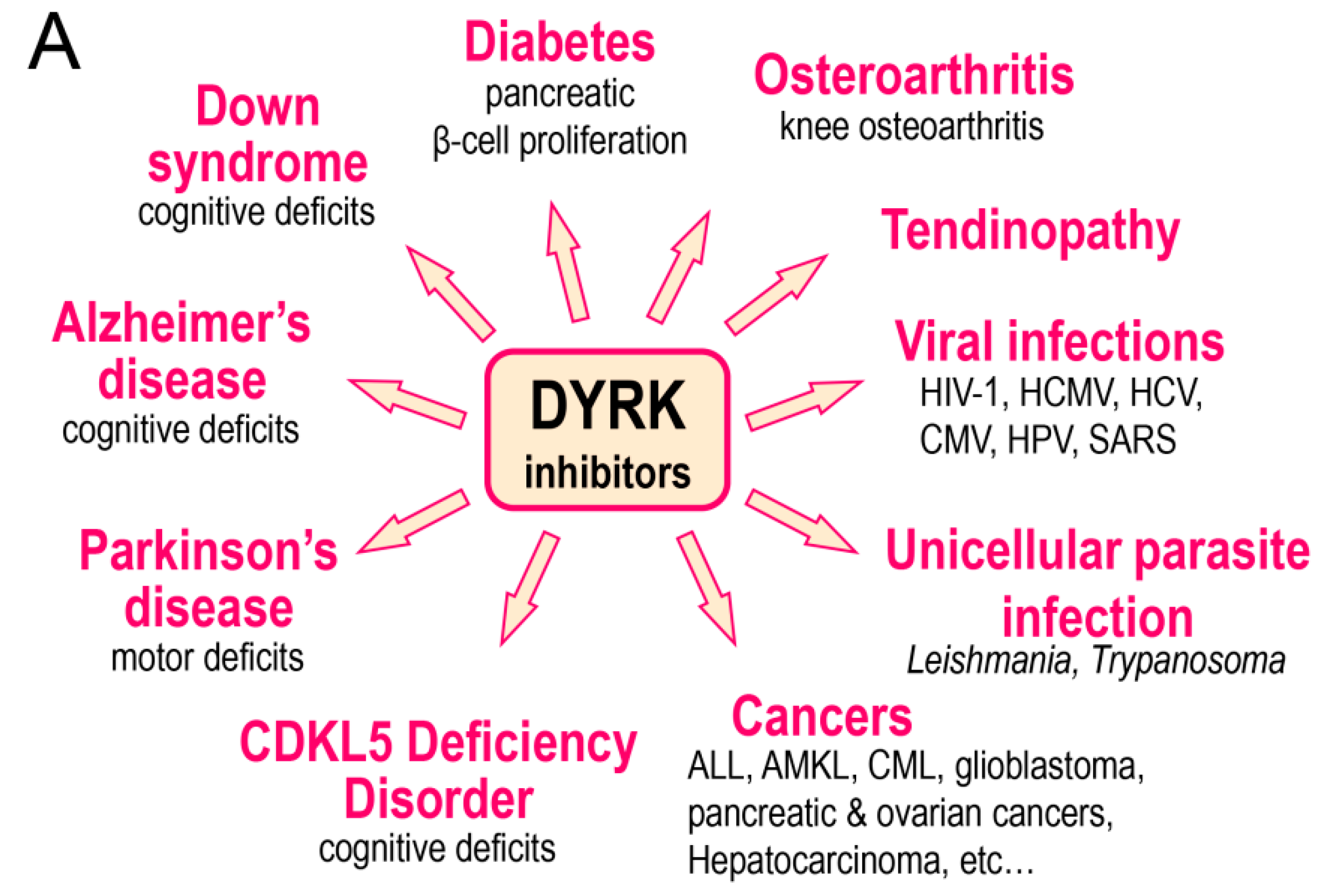
4. Therapeutic Potential of DYRK and CLK Inhibitors
5. Conclusions
Author Contributions
Funding
Conflicts of Interest
Abbreviations
| AD | Alzheimer’s disease |
| AGC | PKA, PKG, and PKC family |
| ALL | acute lymphoblastic leukemia |
| AMKL | acute megakaryoblastic leukemia |
| APP | amyloid precursor protein |
| CAMK | Ca2+/calmodulin-dependent kinases |
| CDKs | cyclin-dependent kinases |
| CSNK1/CK1 | casein kinases 1 |
| CK2 | casein kinase 2 |
| CLKs | cdc2-like kinases |
| CML | chronic myeloid leukemia |
| DCAF7 | DDB1-associated and CUL4-associated factor 7 |
| DH | DYRK homology box |
| DS | Down syndrome |
| DYRKs | dual specificity, tyrosine phosphorylation regulated kinases |
| GSK3 | glycogen synthase kinase 3 |
| GWAS | genome-wide association studies |
| HAN11 | human homolog of the Petunia hybrida an11 gene |
| Hap1 | Huntington-associated protein 1 |
| HCMV | human cytomegalovirus |
| HCV | hepatitis C virus |
| HIV-1 | human immunodeficiency virus type 1 |
| HIPK2 | Homeodomain-interacting protein kinase 2 |
| HPV | human papillomavirus |
| HSV-1 | herpes simplex virus 1 |
| IMAC | immobilized metal-affinity chromatography |
| MAPKs | mitogen-activated protein kinases |
| MRD7 | mental retardation disease 7 |
| NAPA | N-terminal autophosphorylation accessory domain |
| NLS | nuclear localization signals domain |
| PD | Parkinson’s disease |
| PEST | region enriched in proline (P), glutamic acid (E), serine (S), and threonine (T) residues |
| PKA | cAMP-dependent protein kinase |
| PKC | protein kinase C |
| PKG | cGMP-dependent protein kinase |
| PMDS | Phelan-McDermid syndrome |
| STE | homologs of yeast STE7, STE11, and STE20 kinases |
| TK | tyrosine kinases |
| TKL | tyrosine kinase-like kinases |
| WDR68 | WD40-repeat protein 68 |
References
- Zhang, H.; Cao, X.; Tang, M.; Zhong, G.; Si, Y.; Li, H.; Zhu, F.; Liao, Q.; Li, L.; Zhao, J.; et al. A Subcellular Map of the Human Kinome. eLife 2021, 10. [Google Scholar] [CrossRef]
- Wilson, L.J.; Linley, A.; Hammond, D.E.; Hood, F.E.; Coulson, J.M.; MacEwan, D.J.; Ross, S.J.; Slupsky, J.R.; Smith, P.D.; Eyers, P.A.; et al. New Perspectives, Opportunities, and Challenges in Exploring the Human Protein Kinome. Cancer Res. 2018, 78, 15–29. [Google Scholar] [CrossRef]
- Fischer, E.H.; Krebs, E.G. Conversion of Phosphorylase b to Phosphorylase a in Muscle Extracts. J. Biol. Chem. 1955, 216, 121–132. [Google Scholar] [CrossRef]
- Roskoski, R. A Historical Overview of Protein Kinases and Their Targeted Small Molecule Inhibitors. Pharmacol. Res. 2015, 100, 1–23. [Google Scholar] [CrossRef]
- Ferguson, F.M.; Gray, N.S. Kinase Inhibitors: The Road Ahead. Nat. Rev. Drug Discov. 2018, 17, 353–377. [Google Scholar] [CrossRef]
- Wu, P.; Nielsen, T.E.; Clausen, M.H. Small-Molecule Kinase Inhibitors: An Analysis of FDA-Approved Drugs. Drug Discov. Today 2016, 21, 5–10. [Google Scholar] [CrossRef]
- Klaeger, S.; Heinzlmeir, S.; Wilhelm, M.; Polzer, H.; Vick, B.; Koenig, P.-A.; Reinecke, M.; Ruprecht, B.; Petzoldt, S.; Meng, C.; et al. The Target Landscape of Clinical Kinase Drugs. Science 2017, 358. [Google Scholar] [CrossRef] [PubMed]
- Wu, P.; Nielsen, T.E.; Clausen, M.H. FDA-Approved Small-Molecule Kinase Inhibitors. Trends Pharmacol. Sci. 2015, 36, 422–439. [Google Scholar] [CrossRef]
- Roskoski, R. Properties of FDA-Approved Small Molecule Protein Kinase Inhibitors. Pharmacol. Res. 2019, 144, 19–50. [Google Scholar] [CrossRef]
- Roskoski, R. Properties of FDA-Approved Small Molecule Protein Kinase Inhibitors: A 2020 Update. Pharmacol. Res. 2020, 152, 104609. [Google Scholar] [CrossRef]
- Roskoski, R. Properties of FDA-Approved Small Molecule Protein Kinase Inhibitors: A 2021 Update. Pharmacol. Res. 2021, 165, 105463. [Google Scholar] [CrossRef]
- Aranda, S.; Laguna, A.; de la Luna, S. DYRK Family of Protein Kinases: Evolutionary Relationships, Biochemical Properties, and Functional Roles. FASEB J. 2011, 25, 449–462. [Google Scholar] [CrossRef]
- Becker, W.; Sippl, W. Activation, Regulation, and Inhibition of DYRK1A. FEBS J. 2011, 278, 246–256. [Google Scholar] [CrossRef]
- Arbones, M.L.; Thomazeau, A.; Nakano-Kobayashi, A.; Hagiwara, M.; Delabar, J.M. DYRK1A and Cognition: A Lifelong Relationship. Pharmacol. Ther. 2019, 194, 199–221. [Google Scholar] [CrossRef]
- Martín Moyano, P.; Němec, V.; Paruch, K. Cdc-Like Kinases (CLKs): Biology, Chemical Probes, and Therapeutic Potential. Int. J. Mol. Sci. 2020, 21, 7549. [Google Scholar] [CrossRef]
- Widowati, E.W.; Bamberg-Lemper, S.; Becker, W. Mutational Analysis of Two Residues in the DYRK Homology Box of the Protein Kinase DYRK1A. BMC Res. Notes 2018, 11, 297. [Google Scholar] [CrossRef]
- Himpel, S.; Panzer, P.; Eirmbter, K.; Czajkowska, H.; Sayed, M.; Packman, L.C.; Blundell, T.; Kentrup, H.; Grötzinger, J.; Joost, H.G.; et al. Identification of the Autophosphorylation Sites and Characterization of Their Effects in the Protein Kinase DYRK1A. Biochem. J. 2001, 359, 497–505. [Google Scholar] [CrossRef] [PubMed]
- Soundararajan, M.; Roos, A.K.; Savitsky, P.; Filippakopoulos, P.; Kettenbach, A.N.; Olsen, J.V.; Gerber, S.A.; Eswaran, J.; Knapp, S.; Elkins, J.M. Structures of Down Syndrome Kinases, DYRKs, Reveal Mechanisms of Kinase Activation and Substrate Recognition. Structure 2013, 21, 986–996. [Google Scholar] [CrossRef]
- Lee, S.B.; Ko, A.; Oh, Y.T.; Shi, P.; D’Angelo, F.; Frangaj, B.; Koller, A.; Chen, E.I.; Cardozo, T.; Iavarone, A.; et al. Proline Hydroxylation Primes Protein Kinases for Autophosphorylation and Activation. Mol. Cell 2020, 79, 376–389.e8. [Google Scholar] [CrossRef] [PubMed]
- Chang, C.-C.; Hsia, K.-C. More than a Zip Code: Global Modulation of Cellular Function by Nuclear Localization Signals. FEBS J. 2020. [Google Scholar] [CrossRef] [PubMed]
- Kinstrie, R.; Luebbering, N.; Miranda-Saavedra, D.; Sibbet, G.; Han, J.; Lochhead, P.A.; Cleghon, V. Characterization of a Domain That Transiently Converts Class 2 DYRKs into Intramolecular Tyrosine Kinases. Sci. Signal 2010, 3, ra16. [Google Scholar] [CrossRef] [PubMed]
- Rechsteiner, M.; Rogers, S.W. PEST Sequences and Regulation by Proteolysis. Trends Biochem. Sci. 1996, 21, 267–271. [Google Scholar] [CrossRef]
- Salichs, E.; Ledda, A.; Mularoni, L.; Albà, M.M.; de la Luna, S. Genome-Wide Analysis of Histidine Repeats Reveals Their Role in the Localization of Human Proteins to the Nuclear Speckles Compartment. PLoS Genet. 2009, 5, e1000397. [Google Scholar] [CrossRef]
- Bornhorst, J.A.; Falke, J.J. Purification of Proteins Using Polyhistidine Affinity Tags. Methods Enzymol. 2000, 326, 245–254. [Google Scholar] [CrossRef]
- Raducanu, V.-S.; Isaioglou, I.; Raducanu, D.-V.; Merzaban, J.S.; Hamdan, S.M. Simplified Detection of Polyhistidine-Tagged Proteins in Gels and Membranes Using a UV-Excitable Dye and a Multiple Chelator Head Pair. J. Biol. Chem. 2020, 295, 12214–12223. [Google Scholar] [CrossRef]
- Kettenbach, A.N.; Deng, L.; Wu, Y.; Baldissard, S.; Adamo, M.E.; Gerber, S.A.; Moseley, J.B. Quantitative Phosphoproteomics Reveals Pathways for Coordination of Cell Growth and Division by the Conserved Fission Yeast Kinase Pom1. Mol. Cell Proteom. 2015, 14, 1275–1287. [Google Scholar] [CrossRef]
- Bhattacharjee, R.; Mangione, M.C.; Wos, M.; Chen, J.-S.; Snider, C.E.; Roberts-Galbraith, R.H.; McDonald, N.A.; Presti, L.L.; Martin, S.G.; Gould, K.L. DYRK Kinase Pom1 Drives F-BAR Protein Cdc15 from the Membrane to Promote Medial Division. Mol. Biol. Cell 2020, 31, 917–929. [Google Scholar] [CrossRef] [PubMed]
- Kim, D.; Ntui, V.O.; Zhang, N.; Xiong, L. Arabidopsis Yak1 Protein (AtYak1) Is a Dual Specificity Protein Kinase. FEBS Lett. 2015, 589, 3321–3327. [Google Scholar] [CrossRef] [PubMed]
- Huang, W.-Y.; Wu, Y.-C.; Pu, H.-Y.; Wang, Y.; Jang, G.-J.; Wu, S.-H. Plant Dual-Specificity Tyrosine Phosphorylation-Regulated Kinase Optimizes Light-Regulated Growth and Development in Arabidopsis. Plant Cell Environ. 2017, 40, 1735–1747. [Google Scholar] [CrossRef]
- Iwabuchi, K.; Ohnishi, H.; Tamura, K.; Fukao, Y.; Furuya, T.; Hattori, K.; Tsukaya, H.; Hara-Nishimura, I. ANGUSTIFOLIA Regulates Actin Filament Alignment for Nuclear Positioning in Leaves. Plant Physiol. 2019, 179, 233–247. [Google Scholar] [CrossRef]
- Barrada, A.; Djendli, M.; Desnos, T.; Mercier, R.; Robaglia, C.; Montané, M.-H.; Menand, B. A TOR-YAK1 Signaling Axis Controls Cell Cycle, Meristem Activity and Plant Growth in Arabidopsis. Development 2019, 146. [Google Scholar] [CrossRef]
- Forzani, C.; Duarte, G.T.; Van Leene, J.; Clément, G.; Huguet, S.; Paysant-Le-Roux, C.; Mercier, R.; De Jaeger, G.; Leprince, A.-S.; Meyer, C. Mutations of the AtYAK1 Kinase Suppress TOR Deficiency in Arabidopsis. Cell Rep. 2019, 27, 3696–3708.e5. [Google Scholar] [CrossRef]
- Colina, F.; Carbó, M.; Meijón, M.; Cañal, M.J.; Valledor, L. Low UV-C Stress Modulates Chlamydomonas Reinhardtii Biomass Composition and Oxidative Stress Response through Proteomic and Metabolomic Changes Involving Novel Signalers and Effectors. Biotechnol. Biofuels 2020, 13, 110. [Google Scholar] [CrossRef] [PubMed]
- Schulz-Raffelt, M.; Chochois, V.; Auroy, P.; Cuiné, S.; Billon, E.; Dauvillée, D.; Li-Beisson, Y.; Peltier, G. Hyper-Accumulation of Starch and Oil in a Chlamydomonas Mutant Affected in a Plant-Specific DYRK Kinase. Biotechnol. Biofuels 2016, 9, 55. [Google Scholar] [CrossRef] [PubMed]
- Han, J.; Miranda-Saavedra, D.; Luebbering, N.; Singh, A.; Sibbet, G.; Ferguson, M.A.J.; Cleghon, V. Deep Evolutionary Conservation of an Intramolecular Protein Kinase Activation Mechanism. PLoS ONE 2012, 7, e29702. [Google Scholar] [CrossRef]
- De Hiller, N.J.; Silva, N.A.A.E.; Faria, R.X.; Souza, A.L.A.; Resende, J.A.L.C.; Borges Farias, A.; Correia Romeiro, N.; de Luna Martins, D. Synthesis and Evaluation of the Anticancer and Trypanocidal Activities of Boronic Tyrphostins. ChemMedChem 2018, 13, 1395–1404. [Google Scholar] [CrossRef]
- Cayla, M.; McDonald, L.; MacGregor, P.; Matthews, K. An Atypical DYRK Kinase Connects Quorum-Sensing with Posttranscriptional Gene Regulation in Trypanosoma Brucei. eLife 2020, 9. [Google Scholar] [CrossRef] [PubMed]
- Ishii, M.; Akiyoshi, B. Characterization of Unconventional Kinetochore Kinases KKT10 and KKT19 in Trypanosoma Brucei. J. Cell Sci. 2020, 133. [Google Scholar] [CrossRef] [PubMed]
- Rocha, V.P.C.; Dacher, M.; Young, S.A.; Kolokousi, F.; Efstathiou, A.; Späth, G.F.; Soares, M.B.P.; Smirlis, D. Leishmania Dual-Specificity Tyrosine-Regulated Kinase 1 (DYRK1) Is Required for Sustaining Leishmania Stationary Phase Phenotype. Mol. Microbiol. 2020, 113, 983–1002. [Google Scholar] [CrossRef]
- Saldivia, M.; Fang, E.; Ma, X.; Myburgh, E.; Carnielli, J.B.T.; Bower-Lepts, C.; Brown, E.; Ritchie, R.; Lakshminarayana, S.B.; Chen, Y.-L.; et al. Targeting the Trypanosome Kinetochore with CLK1 Protein Kinase Inhibitors. Nat. Microbiol. 2020, 5, 1207–1216. [Google Scholar] [CrossRef] [PubMed]
- Sievers, F.; Wilm, A.; Dineen, D.; Gibson, T.J.; Karplus, K.; Li, W.; Lopez, R.; McWilliam, H.; Remmert, M.; Söding, J.; et al. Fast, Scalable Generation of High-Quality Protein Multiple Sequence Alignments Using Clustal Omega. Mol. Syst. Biol. 2011, 7, 539. [Google Scholar] [CrossRef]
- Waterhouse, A.M.; Procter, J.B.; Martin, D.M.A.; Clamp, M.; Barton, G.J. Jalview Version 2—A Multiple Sequence Alignment Editor and Analysis Workbench. Bioinformatics 2009, 25, 1189–1191. [Google Scholar] [CrossRef]
- Tahtouh, T.; Elkins, J.M.; Filippakopoulos, P.; Soundararajan, M.; Burgy, G.; Durieu, E.; Cochet, C.; Schmid, R.S.; Lo, D.C.; Delhommel, F.; et al. Selectivity, Cocrystal Structures, and Neuroprotective Properties of Leucettines, a Family of Protein Kinase Inhibitors Derived from the Marine Sponge Alkaloid Leucettamine B. J. Med. Chem. 2012, 55, 9312–9330. [Google Scholar] [CrossRef]
- Ogawa, Y.; Nonaka, Y.; Goto, T.; Ohnishi, E.; Hiramatsu, T.; Kii, I.; Yoshida, M.; Ikura, T.; Onogi, H.; Shibuya, H.; et al. Development of a Novel Selective Inhibitor of the Down Syndrome-Related Kinase Dyrk1A. Nat. Commun. 2010, 1, 1–9. [Google Scholar] [CrossRef]
- Anderson, K.; Chen, Y.; Chen, Z.; Dominique, R.; Glenn, K.; He, Y.; Janson, C.; Luk, K.-C.; Lukacs, C.; Polonskaia, A.; et al. Pyrido[2,3-d]Pyrimidines: Discovery and Preliminary SAR of a Novel Series of DYRK1B and DYRK1A Inhibitors. Bioorg. Med. Chem. Lett. 2013, 23, 6610–6615. [Google Scholar] [CrossRef] [PubMed]
- Alexeeva, M.; Åberg, E.; Engh, R.A.; Rothweiler, U. The Structure of a Dual-Specificity Tyrosine Phosphorylation-Regulated Kinase 1A–PKC412 Complex Reveals Disulfide-Bridge Formation with the Anomalous Catalytic Loop HRD(HCD) Cysteine. Acta Crystallogr. D Biol. Crystallogr. 2015, 71, 1207–1215. [Google Scholar] [CrossRef] [PubMed]
- Falke, H.; Chaikuad, A.; Becker, A.; Loaëc, N.; Lozach, O.; Abu Jhaisha, S.; Becker, W.; Jones, P.G.; Preu, L.; Baumann, K.; et al. 10-Iodo-11H-Indolo[3,2-c]Quinoline-6-Carboxylic Acids Are Selective Inhibitors of DYRK1A. J. Med. Chem. 2015, 58, 3131–3143. [Google Scholar] [CrossRef]
- Fukuda, T.; Uchida, K.; Nakayama, H.; Ano, Y. Short-Term Administration of Iso-α-Acids Increases Transthyretin Transcription in the Hippocampus. Biochem. Biophys. Res. Commun. 2018, 507, 471–475. [Google Scholar] [CrossRef]
- Czarna, A.; Wang, J.; Zelencova, D.; Liu, Y.; Deng, X.; Choi, H.G.; Zhang, T.; Zhou, W.; Chang, J.W.; Kildalsen, H.; et al. Novel Scaffolds for Dual Specificity Tyrosine-Phosphorylation-Regulated Kinase (DYRK1A) Inhibitors. J. Med. Chem. 2018, 61, 7560–7572. [Google Scholar] [CrossRef] [PubMed]
- Lechner, C.; Flaßhoff, M.; Falke, H.; Preu, L.; Loaëc, N.; Meijer, L.; Knapp, S.; Chaikuad, A.; Kunick, C. [B]-Annulated Halogen-Substituted Indoles as Potential DYRK1A Inhibitors. Molecules 2019, 24, 4090. [Google Scholar] [CrossRef]
- Wurzlbauer, A.; Rüben, K.; Gürdal, E.; Chaikuad, A.; Knapp, S.; Sippl, W.; Becker, W.; Bracher, F. How to Separate Kinase Inhibition from Undesired Monoamine Oxidase a Inhibition-The Development of the DYRK1A Inhibitor AnnH75 from the Alkaloid Harmine. Molecules 2020, 25, 5962. [Google Scholar] [CrossRef] [PubMed]
- Kumar, K.; Wang, P.; Wilson, J.; Zlatanic, V.; Berrouet, C.; Khamrui, S.; Secor, C.; Swartz, E.A.; Lazarus, M.; Sanchez, R.; et al. Synthesis and Biological Validation of a Harmine-Based, Central Nervous System (CNS)-Avoidant, Selective, Human β-Cell Regenerative Dual-Specificity Tyrosine Phosphorylation-Regulated Kinase A (DYRK1A) Inhibitor. J. Med. Chem. 2020, 63, 2986–3003. [Google Scholar] [CrossRef] [PubMed]
- Liu, Y.A.; Jin, Q.; Zou, Y.; Ding, Q.; Yan, S.; Wang, Z.; Hao, X.; Nguyen, B.; Zhang, X.; Pan, J.; et al. Selective DYRK1A Inhibitor for the Treatment of Type 1 Diabetes: Discovery of 6-Azaindole Derivative GNF2133. J. Med. Chem. 2020, 63, 2958–2973. [Google Scholar] [CrossRef]
- Myrianthopoulos, V.; Kritsanida, M.; Gaboriaud-Kolar, N.; Magiatis, P.; Ferandin, Y.; Durieu, E.; Lozach, O.; Cappel, D.; Soundararajan, M.; Filippakopoulos, P.; et al. Novel Inverse Binding Mode of Indirubin Derivatives Yields Improved Selectivity for DYRK Kinases. ACS Med. Chem. Lett. 2012, 4, 22–26. [Google Scholar] [CrossRef]
- Chaikuad, A.; Diharce, J.; Schröder, M.; Foucourt, A.; Leblond, B.; Casagrande, A.-S.; Désiré, L.; Bonnet, P.; Knapp, S.; Besson, T. An Unusual Binding Model of the Methyl 9-Anilinothiazolo[5,4-f] Quinazoline-2-Carbimidates (EHT 1610 and EHT 5372) Confers High Selectivity for Dual-Specificity Tyrosine Phosphorylation-Regulated Kinases. J. Med. Chem. 2016, 59, 10315–10321. [Google Scholar] [CrossRef]
- Kim, K.; Cha, J.S.; Cho, Y.-S.; Kim, H.; Chang, N.; Kim, H.-J.; Cho, H.-S. Crystal Structure of Human Dual-Specificity Tyrosine-Regulated Kinase 3 Reveals New Structural Features and Insights into Its Auto-Phosphorylation. J. Mol. Biol. 2018, 430, 1521–1530. [Google Scholar] [CrossRef]
- Haltenhof, T.; Kotte, A.; De Bortoli, F.; Schiefer, S.; Meinke, S.; Emmerichs, A.-K.; Petermann, K.K.; Timmermann, B.; Imhof, P.; Franz, A.; et al. A Conserved Kinase-Based Body-Temperature Sensor Globally Controls Alternative Splicing and Gene Expression. Mol. Cell 2020, 78, 57–69.e4. [Google Scholar] [CrossRef] [PubMed]
- Walter, A.; Chaikuad, A.; Helmer, R.; Loaëc, N.; Preu, L.; Ott, I.; Knapp, S.; Meijer, L.; Kunick, C. Molecular Structures of Cdc2-like Kinases in Complex with a New Inhibitor Chemotype. PLoS ONE 2018, 13, e0196761. [Google Scholar] [CrossRef]
- Bullock, A.N.; Das, S.; Debreczeni, J.É.; Rellos, P.; Fedorov, O.; Niesen, F.H.; Guo, K.; Papagrigoriou, E.; Amos, A.L.; Cho, S.; et al. Kinase Domain Insertions Define Distinct Roles of CLK Kinases in SR Protein Phosphorylation. Structure 2009, 17, 352–362. [Google Scholar] [CrossRef] [PubMed]
- Fedorov, O.; Huber, K.; Eisenreich, A.; Filippakopoulos, P.; King, O.; Bullock, A.N.; Szklarczyk, D.; Jensen, L.J.; Fabbro, D.; Trappe, J.; et al. Specific CLK Inhibitors from a Novel Chemotype for Regulation of Alternative Splicing. Chem. Biol. 2011, 18, 67–76. [Google Scholar] [CrossRef]
- Dekel, N.; Eisenberg-Domovich, Y.; Karlas, A.; Meyer, T.F.; Bracher, F.; Lebendiker, M.; Danieli, T.; Livnah, O. Expression, Purification and Crystallization of CLK1 Kinase—A Potential Target for Antiviral Therapy. Protein Expr. Purif. 2020, 176, 105742. [Google Scholar] [CrossRef]
- Esvan, Y.J.; Zeinyeh, W.; Boibessot, T.; Nauton, L.; Théry, V.; Knapp, S.; Chaikuad, A.; Loaëc, N.; Meijer, L.; Anizon, F.; et al. Discovery of Pyrido[3,4-g]Quinazoline Derivatives as CMGC Family Protein Kinase Inhibitors: Design, Synthesis, Inhibitory Potency and X-Ray Co-Crystal Structure. Eur. J. Med. Chem. 2016, 118, 170–177. [Google Scholar] [CrossRef]
- Sun, Q.-Z.; Lin, G.-F.; Li, L.-L.; Jin, X.-T.; Huang, L.-Y.; Zhang, G.; Yang, W.; Chen, K.; Xiang, R.; Chen, C.; et al. Discovery of Potent and Selective Inhibitors of Cdc2-Like Kinase 1 (CLK1) as a New Class of Autophagy Inducers. J. Med. Chem. 2017, 60, 6337–6352. [Google Scholar] [CrossRef]
- Lee, J.Y.; Yun, J.-S.; Kim, W.-K.; Chun, H.-S.; Jin, H.; Cho, S.; Chang, J.H. Structural Basis for the Selective Inhibition of Cdc2-Like Kinases by CX-4945. Biomed. Res. Int. 2019. [Google Scholar] [CrossRef]
- Kallen, J.; Bergsdorf, C.; Arnaud, B.; Bernhard, M.; Brichet, M.; Cobos-Correa, A.; Elhajouji, A.; Freuler, F.; Galimberti, I.; Guibourdenche, C.; et al. X-Ray Structures and Feasibility Assessment of CLK2 Inhibitors for Phelan-McDermid Syndrome. ChemMedChem 2018, 13, 1997–2007. [Google Scholar] [CrossRef] [PubMed]
- Tazarki, H.; Zeinyeh, W.; Esvan, Y.J.; Knapp, S.; Chatterjee, D.; Schröder, M.; Joerger, A.C.; Khiari, J.; Josselin, B.; Baratte, B.; et al. New Pyrido[3,4-g]Quinazoline Derivatives as CLK1 and DYRK1A Inhibitors: Synthesis, Biological Evaluation and Binding Mode Analysis. Eur. J. Med. Chem. 2019, 166, 304–317. [Google Scholar] [CrossRef]
- Heroven, C.; Georgi, V.; Ganotra, G.K.; Brennan, P.; Wolfreys, F.; Wade, R.C.; Fernández-Montalván, A.E.; Chaikuad, A.; Knapp, S. Halogen-Aromatic π Interactions Modulate Inhibitor Residence Times. Angew. Chem. Int. Ed. Engl. 2018, 57, 7220–7224. [Google Scholar] [CrossRef] [PubMed]
- Němec, V.; Hylsová, M.; Maier, L.; Flegel, J.; Sievers, S.; Ziegler, S.; Schröder, M.; Berger, B.-T.; Chaikuad, A.; Valčíková, B.; et al. Furo[3,2-b]Pyridine: A Privileged Scaffold for Highly Selective Kinase Inhibitors and Effective Modulators of the Hedgehog Pathway. Angew. Chem. Int. Ed. Engl. 2019, 58, 1062–1066. [Google Scholar] [CrossRef]
- Schröder, M.; Bullock, A.N.; Fedorov, O.; Bracher, F.; Chaikuad, A.; Knapp, S. DFG-1 Residue Controls Inhibitor Binding Mode and Affinity, Providing a Basis for Rational Design of Kinase Inhibitor Selectivity. J. Med. Chem. 2020, 63, 10224–10234. [Google Scholar] [CrossRef] [PubMed]
- Schröder, M.; Filippakopoulos, P.; Schwalm, M.P.; Ferrer, C.A.; Drewry, D.H.; Knapp, S.; Chaikuad, A. Crystal Structure and Inhibitor Identifications Reveal Targeting Opportunity for the Atypical MAPK Kinase ERK3. Int. J. Mol. Sci. 2020, 21, 7953. [Google Scholar] [CrossRef]
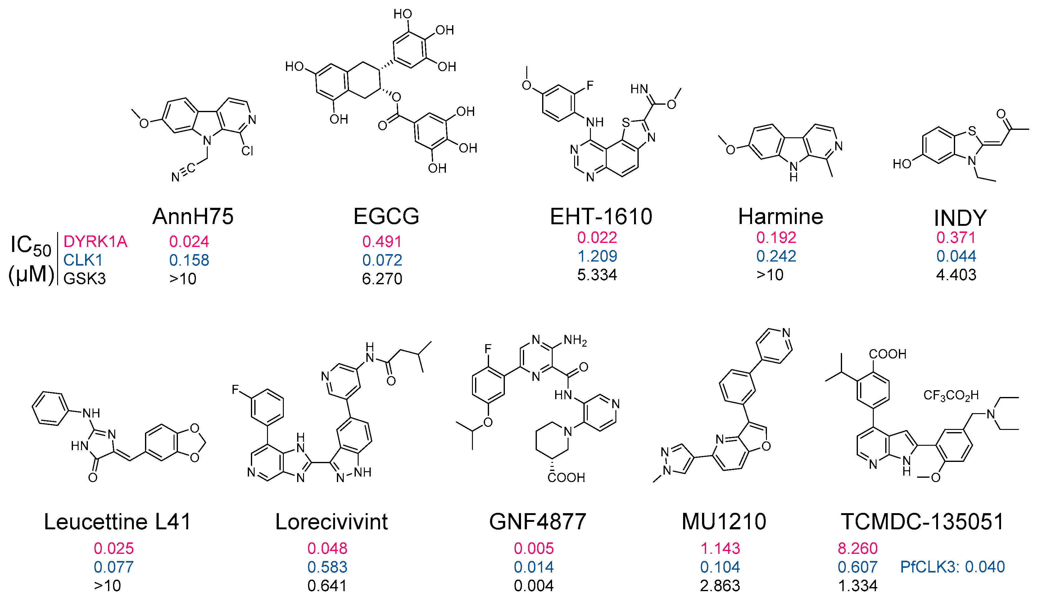
- Debdab, M.; Carreaux, F.; Renault, S.; Soundararajan, M.; Fedorov, O.; Filippakopoulos, P.; Lozach, O.; Babault, L.; Tahtouh, T.; Baratte, B.; et al. Leucettines, a Class of Potent Inhibitors of Cdc2-Like Kinases and Dual Specificity, Tyrosine Phosphorylation Regulated Kinases Derived from the Marine Sponge Leucettamine B: Modulation of Alternative Pre-RNA Splicing. J. Med. Chem. 2011, 54, 4172–4186. [Google Scholar] [CrossRef]
- Guard, S.E.; Poss, Z.C.; Ebmeier, C.C.; Pagratis, M.; Simpson, H.; Taatjes, D.J.; Old, W.M. The Nuclear Interactome of DYRK1A Reveals a Functional Role in DNA Damage Repair. Sci. Rep. 2019, 9, 6539. [Google Scholar] [CrossRef]
- Guard, S.E.; Ebmeier, C.C.; Old, W.M. Label-Free Immunoprecipitation Mass Spectrometry Workflow for Large-Scale Nuclear Interactome Profiling. J. Vis. Exp. 2019. [Google Scholar] [CrossRef]
- Roewenstrunk, J.; Di Vona, C.; Chen, J.; Borras, E.; Dong, C.; Arató, K.; Sabidó, E.; Huen, M.S.Y.; de la Luna, S. A Comprehensive Proteomics-Based Interaction Screen That Links DYRK1A to RNF169 and to the DNA Damage Response. Sci. Rep. 2019, 9, 6014. [Google Scholar] [CrossRef] [PubMed]
- Varjosalo, M.; Keskitalo, S.; Van Drogen, A.; Nurkkala, H.; Vichalkovski, A.; Aebersold, R.; Gstaiger, M. The Protein Interaction Landscape of the Human CMGC Kinase Group. Cell Rep. 2013, 3, 1306–1320. [Google Scholar] [CrossRef]
- Huttlin, E.L.; Ting, L.; Bruckner, R.J.; Gebreab, F.; Gygi, M.P.; Szpyt, J.; Tam, S.; Zarraga, G.; Colby, G.; Baltier, K.; et al. The BioPlex Network: A Systematic Exploration of the Human Interactome. Cell 2015, 162, 425–440. [Google Scholar] [CrossRef] [PubMed]
- Hein, M.Y.; Hubner, N.C.; Poser, I.; Cox, J.; Nagaraj, N.; Toyoda, Y.; Gak, I.A.; Weisswange, I.; Mansfeld, J.; Buchholz, F.; et al. A Human Interactome in Three Quantitative Dimensions Organized by Stoichiometries and Abundances. Cell 2015, 163, 712–723. [Google Scholar] [CrossRef]
- Song, R.; Wang, Z.-D.; Schapira, M. Disease Association and Druggability of WD40 Repeat Proteins. J. Proteome Res. 2017, 16, 3766–3773. [Google Scholar] [CrossRef]
- Miyata, Y.; Nishida, E. DYRK1A Binds to an Evolutionarily Conserved WD40-Repeat Protein WDR68 and Induces Its Nuclear Translocation. Biochim. Biophys. Acta BBA Mol. Cell Res. 2011, 1813, 1728–1739. [Google Scholar] [CrossRef] [PubMed]
- Miyata, Y.; Shibata, T.; Aoshima, M.; Tsubata, T.; Nishida, E. The Molecular Chaperone TRiC/CCT Binds to the Trp-Asp 40 (WD40) Repeat Protein WDR68 and Promotes Its Folding, Protein Kinase DYRK1A Binding, and Nuclear Accumulation. J. Biol. Chem. 2014, 289, 33320–33332. [Google Scholar] [CrossRef] [PubMed]
- Glenewinkel, F.; Cohen, M.J.; King, C.R.; Kaspar, S.; Bamberg-Lemper, S.; Mymryk, J.S.; Becker, W. The Adaptor Protein DCAF7 Mediates the Interaction of the Adenovirus E1A Oncoprotein with the Protein Kinases DYRK1A and HIPK2. Sci. Rep. 2016, 6, 28241. [Google Scholar] [CrossRef] [PubMed]
- Yu, D.; Cattoglio, C.; Xue, Y.; Zhou, Q. A Complex between DYRK1A and DCAF7 Phosphorylates the C-Terminal Domain of RNA Polymerase II to Promote Myogenesis. Nucleic Acids Res. 2019, 47, 4462–4475. [Google Scholar] [CrossRef] [PubMed]
- Wang, B.; Doan, D.; Roman Petersen, Y.; Alvarado, E.; Alvarado, G.; Bhandari, A.; Mohanty, A.; Mohanty, S.; Nissen, R.M. Wdr68 Requires Nuclear Access for Craniofacial Development. PLoS ONE 2013, 8, e54363. [Google Scholar] [CrossRef]
- Alvarado, E.; Yousefelahiyeh, M.; Alvarado, G.; Shang, R.; Whitman, T.; Martinez, A.; Yu, Y.; Pham, A.; Bhandari, A.; Wang, B.; et al. Wdr68 Mediates Dorsal and Ventral Patterning Events for Craniofacial Development. PLoS ONE 2016, 11, e0166984. [Google Scholar] [CrossRef] [PubMed]
- Xiang, J.; Yang, S.; Xin, N.; Gaertig, M.A.; Reeves, R.H.; Li, S.; Li, X.-J. DYRK1A Regulates Hap1–Dcaf7/WDR68 Binding with Implication for Delayed Growth in Down Syndrome. Proc. Natl. Acad. Sci. USA 2017, 114, E1224–E1233. [Google Scholar] [CrossRef]
- Yousefelahiyeh, M.; Xu, J.; Alvarado, E.; Yu, Y.; Salven, D.; Nissen, R.M. DCAF7/WDR68 Is Required for Normal Levels of DYRK1A and DYRK1B. PLoS ONE 2018, 13, e0207779. [Google Scholar] [CrossRef]
- Rueda, N.; Flórez, J.; Dierssen, M.; Martínez-Cué, C. Translational Validity and Implications of Pharmacotherapies in Preclinical Models of Down Syndrome. Prog. Brain Res. 2020, 251, 245–268. [Google Scholar] [CrossRef]
- Jarhad, D.B.; Mashelkar, K.K.; Kim, H.-R.; Noh, M.; Jeong, L.S. Dual-Specificity Tyrosine Phosphorylation-Regulated Kinase 1A (DYRK1A) Inhibitors as Potential Therapeutics. J. Med. Chem. 2018, 61, 9791–9810. [Google Scholar] [CrossRef]
- Feki, A.; Hibaoui, Y. DYRK1A Protein, A Promising Therapeutic Target to Improve Cognitive Deficits in Down Syndrome. Brain Sci. 2018, 8, 187. [Google Scholar] [CrossRef] [PubMed]
- Kay, L.J.; Smulders-Srinivasan, T.K.; Soundararajan, M. Understanding the Multifaceted Role of Human Down Syndrome Kinase DYRK1A. Adv. Protein Chem. Struct. Biol. 2016, 105, 127–171. [Google Scholar] [CrossRef] [PubMed]
- Duchon, A.; Herault, Y. DYRK1A, a Dosage-Sensitive Gene Involved in Neurodevelopmental Disorders, Is a Target for Drug Development in Down Syndrome. Front. Behav. Neurosci. 2016, 10. [Google Scholar] [CrossRef]
- Becker, W.; Soppa, U.; Tejedor, F.J. DYRK1A: A Potential Drug Target for Multiple Down Syndrome Neuropathologies. CNS Neurol. Disord. Drug Targets 2014, 13, 26–33. [Google Scholar] [CrossRef] [PubMed]
- Nguyen, T.L.; Fruit, C.; Hérault, Y.; Meijer, L.; Besson, T. Dual-Specificity Tyrosine Phosphorylation-Regulated Kinase 1A (DYRK1A) Inhibitors: A Survey of Recent Patent Literature. Expert Opin. Ther. Patents 2017, 27, 1183–1199. [Google Scholar] [CrossRef] [PubMed]
- Stotani, S.; Giordanetto, F.; Medda, F. DYRK1A Inhibition as Potential Treatment for Alzheimer’s Disease. Future Med. Chem. 2016, 8, 681–696. [Google Scholar] [CrossRef]
- Pathak, A.; Rohilla, A.; Gupta, T.; Akhtar, M.J.; Haider, M.R.; Sharma, K.; Haider, K.; Yar, M.S. DYRK1A Kinase Inhibition with Emphasis on Neurodegeneration: A Comprehensive Evolution Story-Cum-Perspective. Eur. J. Med. Chem. 2018, 158, 559–592. [Google Scholar] [CrossRef] [PubMed]
- Jin, N.; Yin, X.; Gu, J.; Zhang, X.; Shi, J.; Qian, W.; Ji, Y.; Cao, M.; Gu, X.; Ding, F.; et al. Truncation and Activation of Dual Specificity Tyrosine Phosphorylation-Regulated Kinase 1A by Calpain I: A molecular mechanism linked to tau pathology in alzheimer disease. J. Biol. Chem. 2015, 290, 15219–15237. [Google Scholar] [CrossRef]
- Yin, X.; Jin, N.; Gu, J.; Shi, J.; Zhou, J.; Gong, C.-X.; Iqbal, K.; Grundke-Iqbal, I.; Liu, F. Dual-Specificity Tyrosine Phosphorylation-Regulated Kinase 1A (Dyrk1A) Modulates Serine/Arginine-Rich Protein 55 (SRp55)-Promoted Tau Exon 10 Inclusion. J. Biol. Chem. 2012, 287, 30497–30506. [Google Scholar] [CrossRef]
- Yin, X.; Jin, N.; Shi, J.; Zhang, Y.; Wu, Y.; Gong, C.-X.; Iqbal, K.; Liu, F. Dyrk1A Overexpression Leads to Increase of 3R-Tau Expression and Cognitive Deficits in Ts65Dn Down Syndrome Mice. Sci. Rep. 2017, 7, 619. [Google Scholar] [CrossRef]
- Nalls, M.A.; Blauwendraat, C.; Vallerga, C.L.; Heilbron, K.; Bandres-Ciga, S.; Chang, D.; Tan, M.; Kia, D.A.; Noyce, A.J.; Xue, A.; et al. Identification of Novel Risk Loci, Causal Insights, and Heritable Risk for Parkinson’s Disease: A Meta-Analysis of Genome-Wide Association Studies. Lancet Neurol. 2019, 18, 1091–1102. [Google Scholar] [CrossRef]
- Chiu, C.-C.; Yeh, T.-H.; Chen, R.-S.; Chen, H.-C.; Huang, Y.-Z.; Weng, Y.-H.; Cheng, Y.-C.; Liu, Y.-C.; Cheng, A.-J.; Lu, Y.-C.; et al. Upregulated Expression of MicroRNA-204-5p Leads to the Death of Dopaminergic Cells by Targeting DYRK1A-Mediated Apoptotic Signaling Cascade. Front. Cell. Neurosci. 2019, 13. [Google Scholar] [CrossRef]
- Ferrer, I.; Barrachina, M.; Puig, B.; Martínez de Lagrán, M.; Martí, E.; Avila, J.; Dierssen, M. Constitutive Dyrk1A Is Abnormally Expressed in Alzheimer Disease, Down Syndrome, Pick Disease, and Related Transgenic Models. Neurobiol. Dis. 2005, 20, 392–400. [Google Scholar] [CrossRef] [PubMed]
- Ji, J.; Lee, H.; Argiropoulos, B.; Dorrani, N.; Mann, J.; Martinez-Agosto, J.A.; Gomez-Ospina, N.; Gallant, N.; Bernstein, J.A.; Hudgins, L.; et al. DYRK1A Haploinsufficiency Causes a New Recognizable Syndrome with Microcephaly, Intellectual Disability, Speech Impairment, and Distinct Facies. Eur. J. Hum. Genet. 2015, 23, 1473–1481. [Google Scholar] [CrossRef] [PubMed]
- Evers, J.M.G.; Laskowski, R.A.; Bertolli, M.; Clayton-Smith, J.; Deshpande, C.; Eason, J.; Elmslie, F.; Flinter, F.; Gardiner, C.; Hurst, J.A.; et al. Structural Analysis of Pathogenic Mutations in the DYRK1A Gene in Patients with Developmental Disorders. Hum. Mol. Genet. 2017, 26, 519–526. [Google Scholar] [CrossRef]
- Widowati, E.W.; Ernst, S.; Hausmann, R.; Müller-Newen, G.; Becker, W. Functional Characterization of DYRK1A Missense Variants Associated with a Syndromic Form of Intellectual Deficiency and Autism. Biol. Open 2018, 7. [Google Scholar] [CrossRef]
- Ackeifi, C.; Swartz, E.; Kumar, K.; Liu, H.; Chalada, S.; Karakose, E.; Scott, D.K.; Garcia-Ocaña, A.; Sanchez, R.; DeVita, R.J.; et al. Pharmacologic and Genetic Approaches Define Human Pancreatic β Cell Mitogenic Targets of DYRK1A Inhibitors. JCI Insight 2020, 5. [Google Scholar] [CrossRef] [PubMed]
- Kumar, K.; Suebsuwong, C.; Wang, P.; Garcia-Ocana, A.; Stewart, A.F.; DeVita, R.J. DYRK1A Inhibitors as Potential Therapeutics for β-Cell Regeneration for Diabetes. J. Med. Chem. 2021. [Google Scholar] [CrossRef]
- Abbassi, R.; Johns, T.G.; Kassiou, M.; Munoz, L. DYRK1A in Neurodegeneration and Cancer: Molecular Basis and Clinical Implications. Pharmacol. Ther. 2015, 151, 87–98. [Google Scholar] [CrossRef]
- Boni, J.; Rubio-Perez, C.; López-Bigas, N.; Fillat, C.; de la Luna, S. The DYRK Family of Kinases in Cancer: Molecular Functions and Therapeutic Opportunities. Cancers 2020, 12, 2106. [Google Scholar] [CrossRef]
- Laham, A.J.; Saber-Ayad, M.; El-Awady, R. DYRK1A: A down Syndrome-Related Dual Protein Kinase with a Versatile Role in Tumorigenesis. Cell Mol. Life Sci. 2021, 78, 603–619. [Google Scholar] [CrossRef]
- Malinge, S.; Bliss-Moreau, M.; Kirsammer, G.; Diebold, L.; Chlon, T.; Gurbuxani, S.; Crispino, J.D. Increased Dosage of the Chromosome 21 Ortholog Dyrk1a Promotes Megakaryoblastic Leukemia in a Murine Model of Down Syndrome. J. Clin. Investig. 2012, 122, 948–962. [Google Scholar] [CrossRef]
- Bhansali, R.S.; Rammohan, M.; Lee, P.; Laurent, A.P.; Wen, Q.; Suraneni, P.; Yip, B.H.; Tsai, Y.-C.; Jenni, S.; Bornhauser, B.; et al. DYRK1A Regulates B Cell Acute Lymphoblastic Leukemia through Phosphorylation of FOXO1 and STAT3. J. Clin. Investig. 2021, 131. [Google Scholar] [CrossRef]
- Lee, S.B.; Frattini, V.; Bansal, M.; Castano, A.M.; Sherman, D.; Hutchinson, K.; Bruce, J.N.; Califano, A.; Liu, G.; Cardozo, T.; et al. An ID2-Dependent Mechanism for VHL Inactivation in Cancer. Nature 2016, 529, 172–177. [Google Scholar] [CrossRef] [PubMed]
- Fernández-Martínez, P.; Zahonero, C.; Sánchez-Gómez, P. DYRK1A: The Double-Edged Kinase as a Protagonist in Cell Growth and Tumorigenesis. Mol. Cell Oncol. 2015, 2. [Google Scholar] [CrossRef]
- Birger, Y.; Izraeli, S. DYRK1A in Down Syndrome: An Oncogene or Tumor Suppressor? J. Clin. Investig. 2012, 122, 807–810. [Google Scholar] [CrossRef] [PubMed][Green Version]
- He, M.; Gu, J.; Zhu, J.; Wang, X.; Wang, C.; Duan, C.; Ni, Y.; Lu, X.; Li, J. Up-Regulation of Dyrk1b Promote Astrocyte Activation Following Lipopolysaccharide-Induced Neuroinflammation. Neuropeptides 2018, 69, 76–83. [Google Scholar] [CrossRef] [PubMed]
- Becker, W. A Wake-up Call to Quiescent Cancer Cells—Potential Use of DYRK1B Inhibitors in Cancer Therapy. FEBS J. 2018, 285, 1203–1211. [Google Scholar] [CrossRef] [PubMed]
- Kokkorakis, N.; Gaitanou, M. Minibrain-Related Kinase/Dual-Specificity Tyrosine-Regulated Kinase 1B Implication in Stem/Cancer Stem Cells Biology. World J. Stem Cells 2020, 12, 1553–1575. [Google Scholar] [CrossRef]
- Woo, Y.; Kim, S.J.; Suh, B.K.; Kwak, Y.; Jung, H.-J.; Nhung, T.T.M.; Mun, D.J.; Hong, J.-H.; Noh, S.-J.; Kim, S.; et al. Sequential Phosphorylation of NDEL1 by the DYRK2-GSK3β Complex Is Critical for Neuronal Morphogenesis. eLife 2019, 8. [Google Scholar] [CrossRef]
- Yoshida, S.; Yoshida, K. Multiple Functions of DYRK2 in Cancer and Tissue Development. FEBS Lett. 2019, 593, 2953–2965. [Google Scholar] [CrossRef]
- Correa-Sáez, A.; Jiménez-Izquierdo, R.; Garrido-Rodríguez, M.; Morrugares, R.; Muñoz, E.; Calzado, M.A. Updating Dual-Specificity Tyrosine-Phosphorylation-Regulated Kinase 2 (DYRK2): Molecular Basis, Functions and Role in Diseases. Cell. Mol. Life Sci. 2020, 77, 4747–4763. [Google Scholar] [CrossRef]
- Ma, F.; Zhu, Y.; Liu, X.; Zhou, Q.; Hong, X.; Qu, C.; Feng, X.; Zhang, Y.; Ding, Q.; Zhao, J.; et al. Dual-Specificity Tyrosine Phosphorylation-Regulated Kinase 3 Loss Activates Purine Metabolism and Promotes Hepatocellular Carcinoma Progression. Hepatology 2019, 70, 1785–1803. [Google Scholar] [CrossRef] [PubMed]
- Kim, K.; Lee, S.; Kang, H.; Shin, E.; Kim, H.Y.; Youn, H.; Youn, B. Dual Specificity Kinase DYRK3 Promotes Aggressiveness of Glioblastoma by Altering Mitochondrial Morphology and Function. Int. J. Mol. Sci. 2021, 22, 2982. [Google Scholar] [CrossRef] [PubMed]
- Bakre, A.; Andersen, L.E.; Meliopoulos, V.; Coleman, K.; Yan, X.; Brooks, P.; Crabtree, J.; Tompkins, S.M.; Tripp, R.A. Identification of Host Kinase Genes Required for Influenza Virus Replication and the Regulatory Role of MicroRNAs. PLoS ONE 2013, 8, e66796. [Google Scholar] [CrossRef]
- Wippich, F.; Bodenmiller, B.; Trajkovska, M.G.; Wanka, S.; Aebersold, R.; Pelkmans, L. Dual Specificity Kinase DYRK3 Couples Stress Granule Condensation/Dissolution to MTORC1 Signaling. Cell 2013, 152, 791–805. [Google Scholar] [CrossRef]
- Rai, A.K.; Chen, J.-X.; Selbach, M.; Pelkmans, L. Kinase-Controlled Phase Transition of Membraneless Organelles in Mitosis. Nature 2018, 559, 211–216. [Google Scholar] [CrossRef]
- Slepak, T.I.; Salay, L.D.; Lemmon, V.P.; Bixby, J.L. Dyrk Kinases Regulate Phosphorylation of Doublecortin, Cytoskeletal Organization, and Neuronal Morphology. Cytoskeleton 2012, 69, 514–527. [Google Scholar] [CrossRef]
- Nguyen, T.L.; Duchon, A.; Manousopoulou, A.; Loaëc, N.; Villiers, B.; Pani, G.; Karatas, M.; Mechling, A.E.; Harsan, L.-A.; Limanton, E.; et al. Correction of Cognitive Deficits in Mouse Models of Down Syndrome by a Pharmacological Inhibitor of DYRK1A. Dis. Models Mech. 2018, 11. [Google Scholar] [CrossRef] [PubMed]
- Souchet, B.; Audrain, M.; Billard, J.M.; Dairou, J.; Fol, R.; Orefice, N.S.; Tada, S.; Gu, Y.; Dufayet-Chaffaud, G.; Limanton, E.; et al. Inhibition of DYRK1A Proteolysis Modifies Its Kinase Specificity and Rescues Alzheimer Phenotype in APP/PS1 Mice. Acta Neuropathol. Commun. 2019, 7. [Google Scholar] [CrossRef]
- Sharma, A.; Chunduri, A.; Gopu, A.; Shatrowsky, C.; Crusio, W.E.; Delprato, A. Common Genetic Signatures of Alzheimer’s Disease in Down Syndrome. F1000Research 2020, 9, 1299. [Google Scholar] [CrossRef]
- Duchon, A.; Del Mar Muñiz Moreno, M.; Lorenzo, S.M.; de Souza, M.P.S.; Chevalier, C.; Nalesso, V.; Meziane, H.; de Sousa, P.L.; Noblet, V.; Armspach, J.-P.; et al. Multi-Influential Genetic Interactions Alter Behaviour and Cognition through Six Main Biological Cascades in Down Syndrome Mouse Models. Hum. Mol. Genet. 2021. [Google Scholar] [CrossRef]
- Kargbo, R.B. Selective DYRK1A Inhibitor for the Treatment of Neurodegenerative Diseases: Alzheimer, Parkinson, Huntington, and Down Syndrome. ACS Med. Chem. Lett. 2020, 11, 1795–1796. [Google Scholar] [CrossRef]
- Goodlett, C.R.; Stringer, M.; LaCombe, J.; Patel, R.; Wallace, J.M.; Roper, R.J. Evaluation of the Therapeutic Potential of Epigallocatechin-3-Gallate (EGCG) via Oral Gavage in Young Adult Down Syndrome Mice. Sci. Rep. 2020, 10, 1–17. [Google Scholar] [CrossRef] [PubMed]
- Gu, Y.; Moroy, G.; Paul, J.-L.; Rebillat, A.-S.; Dierssen, M.; de la Torre, R.; Cieuta-Walti, C.; Dairou, J.; Janel, N. Molecular Rescue of Dyrk1A Overexpression Alterations in Mice with Fontup® Dietary Supplement: Role of Green Tea Catechins. Int. J. Mol. Sci. 2020, 21, 1404. [Google Scholar] [CrossRef] [PubMed]
- Chang, P.; Bush, D.; Schorge, S.; Good, M.; Canonica, T.; Shing, N.; Noy, S.; Wiseman, F.K.; Burgess, N.; Tybulewicz, V.L.J.; et al. Altered Hippocampal-Prefrontal Neural Dynamics in Mouse Models of Down Syndrome. Cell Rep. 2020, 30, 1152–1163.e4. [Google Scholar] [CrossRef] [PubMed]
- Sachse, S.M.; Lievens, S.; Ribeiro, L.F.; Dascenco, D.; Masschaele, D.; Horré, K.; Misbaer, A.; Vanderroost, N.; De Smet, A.S.; Salta, E.; et al. Nuclear Import of the DSCAM-Cytoplasmic Domain Drives Signaling Capable of Inhibiting Synapse Formation. EMBO J. 2019, 38. [Google Scholar] [CrossRef]
- Neumann, F.; Gourdain, S.; Albac, C.; Dekker, A.D.; Bui, L.C.; Dairou, J.; Schmitz-Afonso, I.; Hue, N.; Rodrigues-Lima, F.; Delabar, J.M.; et al. DYRK1A Inhibition and Cognitive Rescue in a Down Syndrome Mouse Model Are Induced by New Fluoro-DANDY Derivatives. Sci. Rep. 2018, 8, 2859. [Google Scholar] [CrossRef] [PubMed]
- García-Cerro, S.; Vidal, V.; Lantigua, S.; Berciano, M.T.; Lafarga, M.; Ramos-Cabrer, P.; Padro, D.; Rueda, N.; Martínez-Cué, C. Cerebellar Alterations in a Model of Down Syndrome: The Role of the Dyrk1A Gene. Neurobiol. Dis. 2018, 110, 206–217. [Google Scholar] [CrossRef] [PubMed]
- Stringer, M.; Goodlett, C.R.; Roper, R.J. Targeting Trisomic Treatments: Optimizing Dyrk1a Inhibition to Improve Down Syndrome Deficits. Mol. Genet. Genom. Med. 2017, 5, 451–465. [Google Scholar] [CrossRef]
- McElyea, S.D.; Starbuck, J.M.; Tumbleson-Brink, D.M.; Harrington, E.; Blazek, J.D.; Ghoneima, A.; Kula, K.; Roper, R.J. Influence of Prenatal EGCG Treatment and Dyrk1a Dosage Reduction on Craniofacial Features Associated with Down Syndrome. Hum. Mol. Genet. 2016, 25, 4856–4869. [Google Scholar] [CrossRef]
- Kim, H.; Lee, K.-S.; Kim, A.-K.; Choi, M.; Choi, K.; Kang, M.; Chi, S.-W.; Lee, M.-S.; Lee, J.-S.; Lee, S.-Y.; et al. A Chemical with Proven Clinical Safety Rescues Down-Syndrome-Related Phenotypes in through DYRK1A Inhibition. Dis. Models Mech. 2016, 9, 839–848. [Google Scholar] [CrossRef] [PubMed]
- Blazek, J.D.; Abeysekera, I.; Li, J.; Roper, R.J. Rescue of the Abnormal Skeletal Phenotype in Ts65Dn Down Syndrome Mice Using Genetic and Therapeutic Modulation of Trisomic Dyrk1a. Hum. Mol. Genet. 2015, 24, 5687–5696. [Google Scholar] [CrossRef]
- García-Cerro, S.; Martínez, P.; Vidal, V.; Corrales, A.; Flórez, J.; Vidal, R.; Rueda, N.; Arbonés, M.L.; Martínez-Cué, C. Overexpression of Dyrk1A Is Implicated in Several Cognitive, Electrophysiological and Neuromorphological Alterations Found in a Mouse Model of Down Syndrome. PLoS ONE 2014, 9, e106572. [Google Scholar] [CrossRef] [PubMed]
- De la Torre, R.; De Sola, S.; Pons, M.; Duchon, A.; de Lagran, M.M.; Farré, M.; Fitó, M.; Benejam, B.; Langohr, K.; Rodriguez, J.; et al. Epigallocatechin-3-Gallate, a DYRK1A Inhibitor, Rescues Cognitive Deficits in Down Syndrome Mouse Models and in Humans. Mol. Nutr. Food Res. 2014, 58, 278–288. [Google Scholar] [CrossRef] [PubMed]
- Altafaj, X.; Martín, E.D.; Ortiz-Abalia, J.; Valderrama, A.; Lao-Peregrín, C.; Dierssen, M.; Fillat, C. Normalization of Dyrk1A Expression by AAV2/1-ShDyrk1A Attenuates Hippocampal-Dependent Defects in the Ts65Dn Mouse Model of Down Syndrome. Neurobiol. Dis. 2013, 52, 117–127. [Google Scholar] [CrossRef]
- Park, J.; Oh, Y.; Chung, K.C. Two Key Genes Closely Implicated with the Neuropathological Characteristics in Down Syndrome: DYRK1A and RCAN1. BMB Rep. 2009, 42, 6–15. [Google Scholar] [CrossRef] [PubMed]
- Ortiz-Abalia, J.; Sahún, I.; Altafaj, X.; Andreu, N.; Estivill, X.; Dierssen, M.; Fillat, C. Targeting Dyrk1A with AAVshRNA Attenuates Motor Alterations in TgDyrk1A, a Mouse Model of Down Syndrome. Am. J. Hum. Genet. 2008, 83, 479–488. [Google Scholar] [CrossRef]
- Shi, J.; Zhang, T.; Zhou, C.; Chohan, M.O.; Gu, X.; Wegiel, J.; Zhou, J.; Hwang, Y.-W.; Iqbal, K.; Grundke-Iqbal, I.; et al. Increased Dosage of Dyrk1A Alters Alternative Splicing Factor (ASF)-Regulated Alternative Splicing of Tau in Down Syndrome. J. Biol. Chem. 2008, 283, 28660–28669. [Google Scholar] [CrossRef] [PubMed]
- Naert, G.; Ferré, V.; Meunier, J.; Keller, E.; Malmström, S.; Givalois, L.; Carreaux, F.; Bazureau, J.-P.; Maurice, T. Leucettine L41, a DYRK1A-Preferential DYRKs/CLKs Inhibitor, Prevents Memory Impairments and Neurotoxicity Induced by Oligomeric Aβ25-35 Peptide Administration in Mice. Eur. Neuropsychopharmacol. 2015, 25, 2170–2182. [Google Scholar] [CrossRef]
- Lee, Y.H.; Im, E.; Hyun, M.; Park, J.; Chung, K.C. Protein Phosphatase PPM1B Inhibits DYRK1A Kinase through Dephosphorylation of PS258 and Reduces Toxic Tau Aggregation. J. Biol. Chem. 2020. [Google Scholar] [CrossRef]
- Lee, H.; Woo, H.; Lee, H.-E.; Jeon, H.; Ryu, K.-Y.; Nam, J.H.; Jeon, S.G.; Park, H.; Lee, J.-S.; Han, K.-M.; et al. The Novel DYRK1A Inhibitor KVN93 Regulates Cognitive Function, Amyloid-Beta Pathology, and Neuroinflammation. Free Radic. Biol. Med. 2020, 160, 575–595. [Google Scholar] [CrossRef]
- Delabar, J.M.; Ortner, M.; Simon, S.; Wijkhuisen, A.; Feraudet-Tarisse, C.; Pegon, J.; Vidal, E.; Hirschberg, Y.; Dubois, B.; Potier, M.-C. Altered Age-Linked Regulation of Plasma DYRK1A in Elderly Cognitive Complainers (INSIGHT-PreAD Study) with High Brain Amyloid Load. Alzheimers Dement. 2020, 6, e12046. [Google Scholar] [CrossRef] [PubMed]
- Liu, Y.; Wang, L.; Xie, F.; Wang, X.; Hou, Y.; Wang, X.; Liu, J. Overexpression of MiR-26a-5p Suppresses Tau Phosphorylation and Aβ Accumulation in Alzheimer’s Disease Mice by Targeting DYRK1A. Curr. Neurovasc. Res. 2020. [Google Scholar] [CrossRef]
- Velazquez, R.; Meechoovet, B.; Ow, A.; Foley, C.; Shaw, A.; Smith, B.; Oddo, S.; Hulme, C.; Dunckley, T. Chronic Dyrk1 Inhibition Delays the Onset of AD-Like Pathology in 3xTg-AD Mice. Mol. Neurobiol. 2019, 56, 8364–8375. [Google Scholar] [CrossRef] [PubMed]
- Branca, C.; Shaw, D.M.; Belfiore, R.; Gokhale, V.; Shaw, A.Y.; Foley, C.; Smith, B.; Hulme, C.; Dunckley, T.; Meechoovet, B.; et al. Dyrk1 Inhibition Improves Alzheimer’s Disease-like Pathology. Aging Cell 2017, 16, 1146–1154. [Google Scholar] [CrossRef]
- García-Cerro, S.; Rueda, N.; Vidal, V.; Lantigua, S.; Martínez-Cué, C. Normalizing the Gene Dosage of Dyrk1A in a Mouse Model of Down Syndrome Rescues Several Alzheimer’s Disease Phenotypes. Neurobiol. Dis. 2017, 106, 76–88. [Google Scholar] [CrossRef] [PubMed]
- Kawakubo, T.; Mori, R.; Shirotani, K.; Iwata, N.; Asai, M. Neprilysin Is Suppressed by Dual-Specificity Tyrosine-Phosphorylation Regulated Kinase 1A (DYRK1A) in Down-Syndrome-Derived Fibroblasts. Biol. Pharm. Bull. 2017, 40, 327–333. [Google Scholar] [CrossRef]
- Janel, N.; Alexopoulos, P.; Badel, A.; Lamari, F.; Camproux, A.C.; Lagarde, J.; Simon, S.; Feraudet-Tarisse, C.; Lamourette, P.; Arbones, M.; et al. Combined Assessment of DYRK1A, BDNF and Homocysteine Levels as Diagnostic Marker for Alzheimer’s Disease. Transl. Psychiatry 2017, 7, e1154. [Google Scholar] [CrossRef]
- Coutadeur, S.; Benyamine, H.; Delalonde, L.; de Oliveira, C.; Leblond, B.; Foucourt, A.; Besson, T.; Casagrande, A.-S.; Taverne, T.; Girard, A.; et al. A Novel DYRK1A (Dual Specificity Tyrosine Phosphorylation-Regulated Kinase 1A) Inhibitor for the Treatment of Alzheimer’s Disease: Effect on Tau and Amyloid Pathologies in Vitro. J. Neurochem. 2015, 133, 440–451. [Google Scholar] [CrossRef] [PubMed]
- Fant, X.; Durieu, E.; Chicanne, G.; Payrastre, B.; Sbrissa, D.; Shisheva, A.; Limanton, E.; Carreaux, F.; Bazureau, J.-P.; Meijer, L. Cdc-Like/Dual-Specificity Tyrosine Phosphorylation–Regulated Kinases Inhibitor Leucettine L41 Induces MTOR-Dependent Autophagy: Implication for Alzheimer’s Disease. Mol. Pharmacol. 2014, 85, 441–450. [Google Scholar] [CrossRef]
- Ryu, Y.S.; Park, S.Y.; Jung, M.-S.; Yoon, S.-H.; Kwen, M.-Y.; Lee, S.-Y.; Choi, S.-H.; Radnaabazar, C.; Kim, M.-K.; Kim, H.; et al. Dyrk1A-Mediated Phosphorylation of Presenilin 1: A Functional Link between Down Syndrome and Alzheimer’s Disease. J. Neurochem. 2010, 115, 574–584. [Google Scholar] [CrossRef]
- Ryoo, S.-R.; Cho, H.-J.; Lee, H.-W.; Jeong, H.K.; Radnaabazar, C.; Kim, Y.-S.; Kim, M.-J.; Son, M.-Y.; Seo, H.; Chung, S.-H.; et al. Dual-Specificity Tyrosine(Y)-Phosphorylation Regulated Kinase 1A-Mediated Phosphorylation of Amyloid Precursor Protein: Evidence for a Functional Link between Down Syndrome and Alzheimer’s Disease. J. Neurochem. 2008, 104, 1333–1344. [Google Scholar] [CrossRef] [PubMed]
- Ryoo, S.-R.; Jeong, H.K.; Radnaabazar, C.; Yoo, J.-J.; Cho, H.-J.; Lee, H.-W.; Kim, I.-S.; Cheon, Y.-H.; Ahn, Y.S.; Chung, S.-H.; et al. DYRK1A-Mediated Hyperphosphorylation of Tau. A Functional Link between Down Syndrome and Alzheimer Disease. J. Biol. Chem. 2007, 282, 34850–34857. [Google Scholar] [CrossRef] [PubMed]
- Kimura, R.; Kamino, K.; Yamamoto, M.; Nuripa, A.; Kida, T.; Kazui, H.; Hashimoto, R.; Tanaka, T.; Kudo, T.; Yamagata, H.; et al. The DYRK1A Gene, Encoded in Chromosome 21 Down Syndrome Critical Region, Bridges between β-Amyloid Production and Tau Phosphorylation in Alzheimer Disease. Hum. Mol. Genet. 2007, 16, 15–23. [Google Scholar] [CrossRef] [PubMed]
- Fang, L.; Tang, B.-S.; Fan, K.; Wan, C.-M.; Yan, X.-X.; Guo, J.-F. Alzheimer’s Disease Susceptibility Genes Modify the Risk of Parkinson Disease and Parkinson’s Disease-Associated Cognitive Impairment. Neurosci. Lett. 2018, 677, 55–59. [Google Scholar] [CrossRef] [PubMed]
- Cen, L.; Xiao, Y.; Wei, L.; Mo, M.; Chen, X.; Li, S.; Yang, X.; Huang, Q.; Qu, S.; Pei, Z.; et al. Association of DYRK1A Polymorphisms with Sporadic Parkinson’s Disease in Chinese Han Population. Neurosci. Lett. 2016, 632, 39–43. [Google Scholar] [CrossRef]
- Im, E.; Chung, K.C. Dyrk1A Phosphorylates Parkin at Ser-131 and Negatively Regulates Its Ubiquitin E3 Ligase Activity. J. Neurochem. 2015, 134, 756–768. [Google Scholar] [CrossRef]
- Jones, E.L.; Aarsland, D.; Londos, E.; Ballard, C. A Pilot Study Examining Associations between DYRK1A and α-Synuclein Dementias. Neurodegener. Dis. 2012, 10, 229–231. [Google Scholar] [CrossRef]
- Sitz, J.H.; Baumgärtel, K.; Hämmerle, B.; Papadopoulos, C.; Hekerman, P.; Tejedor, F.J.; Becker, W.; Lutz, B. The Down Syndrome Candidate Dual-Specificity Tyrosine Phosphorylation-Regulated Kinase 1A Phosphorylates the Neurodegeneration-Related Septin 4. Neuroscience 2008, 157, 596–605. [Google Scholar] [CrossRef]
- Trovò, L.; Fuchs, C.; De Rosa, R.; Barbiero, I.; Tramarin, M.; Ciani, E.; Rusconi, L.; Kilstrup-Nielsen, C. The Green Tea Polyphenol Epigallocatechin-3-Gallate (EGCG) Restores CDKL5-Dependent Synaptic Defects in Vitro and in Vivo. Neurobiol. Dis. 2020, 104791. [Google Scholar] [CrossRef]
- Liu, Y.A.; Jin, Q.; Ding, Q.; Hao, X.; Mo, T.; Yan, S.; Zou, Y.; Huang, Z.; Zhang, X.; Gao, W.; et al. A Dual Inhibitor of DYRK1A and GSK3β for Β-Cell Proliferation: Aminopyrazine Derivative GNF4877. ChemMedChem 2020, 15, 1562–1570. [Google Scholar] [CrossRef]
- Kumar, K.; Wang, P.; Swartz, E.A.; Khamrui, S.; Secor, C.; Lazarus, M.B.; Sanchez, R.; Stewart, A.F.; DeVita, R.J. Structure-Activity Relationships and Biological Evaluation of 7-Substituted Harmine Analogs for Human β-Cell Proliferation. Molecules 2020, 25, 1983. [Google Scholar] [CrossRef]
- Hohmeier, H.E.; Zhang, L.; Taylor, B.; Stephens, S.; Lu, D.; McNamara, P.; Laffitte, B.; Newgard, C.B. Identification of a Small Molecule That Stimulates Human β-Cell Proliferation and Insulin Secretion, and Protects against Cytotoxic Stress in Rat Insulinoma Cells. PLoS ONE 2020, 15, e0224344. [Google Scholar] [CrossRef] [PubMed]
- Brial, F.; Alzaid, F.; Sonomura, K.; Kamatani, Y.; Meneyrol, K.; Le Lay, A.; Péan, N.; Hedjazi, L.; Sato, T.-A.; Venteclef, N.; et al. The Natural Metabolite 4-Cresol Improves Glucose Homeostasis and Enhances β-Cell Function. Cell Rep. 2020, 30, 2306–2320.e5. [Google Scholar] [CrossRef]
- Scavuzzo, M.A.; Borowiak, M. Two Drugs Converged in a Pancreatic β Cell. Sci. Transl. Med. 2020, 12. [Google Scholar] [CrossRef] [PubMed]
- Ackeifi, C.; Wang, P.; Karakose, E.; Manning Fox, J.E.; González, B.J.; Liu, H.; Wilson, J.; Swartz, E.; Berrouet, C.; Li, Y.; et al. GLP-1 Receptor Agonists Synergize with DYRK1A Inhibitors to Potentiate Functional Human β Cell Regeneration. Sci. Transl. Med. 2020, 12. [Google Scholar] [CrossRef]
- Lu, M.; Ma, L.; Shan, P.; Liu, A.; Yu, X.; Jiang, W.; Wang, X.; Zhao, X.; Ye, X.; Wang, T. DYRK1A Aggravates β Cell Dysfunction and Apoptosis by Promoting the Phosphorylation and Degradation of IRS2. Exp. Gerontol. 2019, 125, 110659. [Google Scholar] [CrossRef] [PubMed]
- Wang, P.; Karakose, E.; Liu, H.; Swartz, E.; Ackeifi, C.; Zlatanic, V.; Wilson, J.; González, B.J.; Bender, A.; Takane, K.K.; et al. Combined Inhibition of DYRK1A, SMAD, and Trithorax Pathways Synergizes to Induce Robust Replication in Adult Human Beta Cells. Cell Metab. 2019, 29, 638–652.e5. [Google Scholar] [CrossRef]
- Wang, P.; Alvarez-Perez, J.-C.; Felsenfeld, D.P.; Liu, H.; Sivendran, S.; Bender, A.; Kumar, A.; Sanchez, R.; Scott, D.K.; Garcia-Ocaña, A.; et al. A High-Throughput Chemical Screen Reveals That Harmine-Mediated Inhibition of DYRK1A Increases Human Pancreatic Beta Cell Replication. Nat. Med. 2015, 21, 383–388. [Google Scholar] [CrossRef] [PubMed]
- Shen, W.; Taylor, B.; Jin, Q.; Nguyen-Tran, V.; Meeusen, S.; Zhang, Y.-Q.; Kamireddy, A.; Swafford, A.; Powers, A.F.; Walker, J.; et al. Inhibition of DYRK1A and GSK3B Induces Human β-Cell Proliferation. Nat. Commun. 2015, 6, 8372. [Google Scholar] [CrossRef]
- Zheng, Y.; Ramsamooj, S.; Li, Q.; Johnson, J.L.; Yaron, T.M.; Sharra, K.; Cantley, L.C. Regulation of Folate and Methionine Metabolism by Multisite Phosphorylation of Human Methylenetetrahydrofolate Reductase. Sci. Rep. 2019, 9, 4190. [Google Scholar] [CrossRef] [PubMed]
- Pozo, N.; Zahonero, C.; Fernández, P.; Liñares, J.M.; Ayuso, A.; Hagiwara, M.; Pérez, A.; Ricoy, J.R.; Hernández-Laín, A.; Sepúlveda, J.M.; et al. Inhibition of DYRK1A Destabilizes EGFR and Reduces EGFR-Dependent Glioblastoma Growth. J. Clin. Investig. 2013, 123, 2475–2487. [Google Scholar] [CrossRef] [PubMed]
- Radhakrishnan, A.; Nanjappa, V.; Raja, R.; Sathe, G.; Puttamallesh, V.N.; Jain, A.P.; Pinto, S.M.; Balaji, S.A.; Chavan, S.; Sahasrabuddhe, N.A.; et al. A Dual Specificity Kinase, DYRK1A, as a Potential Therapeutic Target for Head and Neck Squamous Cell Carcinoma. Sci. Rep. 2016, 6, 36132. [Google Scholar] [CrossRef]
- Bai, Z.; Du, Y.; Cong, L.; Cheng, Y. The USP22 Promotes the Growth of Cancer Cells through the DYRK1A in Pancreatic Ductal Adenocarcinoma. Gene 2020, 758, 144960. [Google Scholar] [CrossRef] [PubMed]
- Zhao, C.; Wang, D.; Gao, Z.; Kan, H.; Qiu, F.; Chen, L.; Li, H. Licocoumarone Induces BxPC-3 Pancreatic Adenocarcinoma Cell Death by Inhibiting DYRK1A. Chem. Biol. Interact. 2020, 316, 108913. [Google Scholar] [CrossRef]
- Luna, J.; Boni, J.; Cuatrecasas, M.; Bofill-De Ros, X.; Núñez-Manchón, E.; Gironella, M.; Vaquero, E.C.; Arbones, M.L.; de la Luna, S.; Fillat, C. DYRK1A Modulates C-MET in Pancreatic Ductal Adenocarcinoma to Drive Tumour Growth. Gut 2019, 68, 1465–1476. [Google Scholar] [CrossRef]
- Li, L.; Wei, J.-R.; Song, Y.; Fang, S.; Du, Y.; Li, Z.; Zeng, T.-T.; Zhu, Y.-H.; Li, Y.; Guan, X.-Y. TROAP Switches DYRK1 Activity to Drive Hepatocellular Carcinoma Progression. Cell Death Dis. 2021, 12, 125. [Google Scholar] [CrossRef]
- Mauro, L.J.; Seibel, M.I.; Diep, C.H.; Spartz, A.; Perez Kerkvliet, C.; Singhal, H.; Swisher, E.M.; Schwartz, L.E.; Drapkin, R.; Saini, S.; et al. Progesterone Receptors Promote Quiescence & Ovarian Cancer Cell Phenotypes via DREAM in P53-Mutant Fallopian Tube Models. J. Clin. Endocrinol. Metab. 2021. [Google Scholar] [CrossRef]
- Iness, A.N.; Rubinsak, L.; Meas, S.J.; Chaoul, J.; Sayeed, S.; Pillappa, R.; Temkin, S.M.; Dozmorov, M.G.; Litovchick, L. Oncogenic B-Myb Is Associated with Deregulation of the DREAM-Mediated Cell Cycle Gene Expression Program in High Grade Serous Ovarian Carcinoma Clinical Tumor Samples. Front. Oncol. 2021, 11, 637193. [Google Scholar] [CrossRef]
- Jang, S.M.; Azebi, S.; Soubigou, G.; Muchardt, C. DYRK1A Phoshorylates Histone H3 to Differentially Regulate the Binding of HP1 Isoforms and Antagonize HP1-Mediated Transcriptional Repression. EMBO Rep. 2014, 15, 686–694. [Google Scholar] [CrossRef]
- Kim, J.-H.; Li, L.; Resar, L.M. Doubling up on Function: Dual-Specificity Tyrosine-Regulated Kinase 1A (DYRK1A) in B Cell Acute Lymphoblastic Leukemia. J. Clin. Investig. 2021, 131. [Google Scholar] [CrossRef]
- Lee, P.; Bhansali, R.; Izraeli, S.; Hijiya, N.; Crispino, J.D. The Biology, Pathogenesis and Clinical Aspects of Acute Lymphoblastic Leukemia in Children with Down Syndrome. Leukemia 2016, 30, 1816–1823. [Google Scholar] [CrossRef]
- Liu, A.; Zhang, B.; Zhao, W.; Tu, Y.; Wang, Q.; Li, J. MicroRNA-215-5p Inhibits the Proliferation of Keratinocytes and Alleviates Psoriasis-like Inflammation by Negatively Regulating DYRK1A and Its Downstream Signaling Pathways. Exp. Dermatol. 2020. [Google Scholar] [CrossRef]
- Deshmukh, V.; O’Green, A.L.; Bossard, C.; Seo, T.; Lamangan, L.; Ibanez, M.; Ghias, A.; Lai, C.; Do, L.; Cho, S.; et al. Modulation of the Wnt Pathway through Inhibition of CLK2 and DYRK1A by Lorecivivint as a Novel, Potentially Disease-Modifying Approach for Knee Osteoarthritis Treatment. Osteoarthr. Cartil. 2019, 27, 1347–1360. [Google Scholar] [CrossRef]
- Yazici, Y.; McAlindon, T.E.; Gibofsky, A.; Lane, N.E.; Lattermann, C.; Skrepnik, N.; Swearingen, C.J.; Simsek, I.; Ghandehari, H.; DiFrancesco, A.; et al. A Phase 2b Randomized Trial of Lorecivivint, a Novel Intra-Articular CLK2/DYRK1A Inhibitor and Wnt Pathway Modulator for Knee Osteoarthritis. Osteoarthr. Cartil. 2021. [Google Scholar] [CrossRef]
- Deshmukh, V.; Seo, T.; Lauren O’Green, A.; Ibanez, M.; Hofilena, B.; Sunil, K.; Stewart, J.; Dellamary, L.; Chiu, K.; Ghias, A.; et al. SM04755, a Small-Molecule Inhibitor of the Wnt Pathway, as a Potential Topical Treatment for Tendinopathy. J. Orthop. Res. 2020. [Google Scholar] [CrossRef]
- Kisaka, J.K.; Ratner, L.; Kyei, G.B. The Dual-Specificity Kinase DYRK1A Modulates the Levels of Cyclin L2 To Control HIV Replication in Macrophages. J. Virol. 2020, 94. [Google Scholar] [CrossRef] [PubMed]
- Booiman, T.; Loukachov, V.V.; van Dort, K.A.; van’t Wout, A.B.; Kootstra, N.A. DYRK1A Controls HIV-1 Replication at a Transcriptional Level in an NFAT Dependent Manner. PLoS ONE 2015, 10, e0144229. [Google Scholar] [CrossRef] [PubMed]
- Bol, S.M.; Moerland, P.D.; Limou, S.; van Remmerden, Y.; Coulonges, C.; van Manen, D.; Herbeck, J.T.; Fellay, J.; Sieberer, M.; Sietzema, J.G.; et al. Genome-Wide Association Study Identifies Single Nucleotide Polymorphism in DYRK1A Associated with Replication of HIV-1 in Monocyte-Derived Macrophages. PLoS ONE 2011, 6, e17190. [Google Scholar] [CrossRef] [PubMed]
- Hamilton, S.T.; Hutterer, C.; Egilmezer, E.; Steingruber, M.; Milbradt, J.; Marschall, M.; Rawlinson, W.D. Human Cytomegalovirus Utilises Cellular Dual-Specificity Tyrosine Phosphorylation-Regulated Kinases during Placental Replication. Placenta 2018, 72, 10–19. [Google Scholar] [CrossRef]
- Dirmeier, S.; Dächert, C.; van Hemert, M.; Tas, A.; Ogando, N.S.; van Kuppeveld, F.; Bartenschlager, R.; Kaderali, L.; Binder, M.; Beerenwinkel, N. Host Factor Prioritization for Pan-Viral Genetic Perturbation Screens Using Random Intercept Models and Network Propagation. PLoS Comput. Biol. 2020, 16, e1007587. [Google Scholar] [CrossRef]
- Zhou, N.; Yuan, S.; Wang, R.; Zhang, W.; Chen, J.J. Role of Dual Specificity Tyrosine-Phosphorylation-Regulated Kinase 1B (Dyrk1B) in S-Phase Entry of HPV E7 Expressing Cells from Quiescence. Oncotarget 2015, 6, 30745–30761. [Google Scholar] [CrossRef] [PubMed][Green Version]
- Saluja, T.S.; Kumar, V.; Agrawal, M.; Tripathi, A.; Meher, R.K.; Srivastava, K.; Gupta, A.; Singh, A.; Chaturvedi, A.; Singh, S.K. Mitochondrial Stress-Mediated Targeting of Quiescent Cancer Stem Cells in Oral Squamous Cell Carcinoma. Cancer Manag. Res. 2020, 12, 4519–4530. [Google Scholar] [CrossRef]
- Chen, H.; Shen, J.; Choy, E.; Hornicek, F.J.; Shan, A.; Duan, Z. Targeting DYRK1B Suppresses the Proliferation and Migration of Liposarcoma Cells. Oncotarget 2018, 9, 13154–13166. [Google Scholar] [CrossRef]
- Chen, Y.; Wang, S.; He, Z.; Sun, F.; Huang, Y.; Ni, Q.; Wang, H.; Wang, Y.; Cheng, C. Dyrk1B Overexpression Is Associated with Breast Cancer Growth and a Poor Prognosis. Hum. Pathol. 2017, 66, 48–58. [Google Scholar] [CrossRef] [PubMed]
- Gruber, W.; Hutzinger, M.; Elmer, D.P.; Parigger, T.; Sternberg, C.; Cegielkowski, L.; Zaja, M.; Leban, J.; Michel, S.; Hamm, S.; et al. DYRK1B as Therapeutic Target in Hedgehog/GLI-Dependent Cancer Cells with Smoothened Inhibitor Resistance. Oncotarget 2016, 7, 7134–7148. [Google Scholar] [CrossRef]
- Tandon, V.; de la Vega, L.; Banerjee, S. Emerging Roles of DYRK2 in Cancer. J. Biol. Chem. 2020. [Google Scholar] [CrossRef]
- Mehnert, M.; Ciuffa, R.; Frommelt, F.; Uliana, F.; van Drogen, A.; Ruminski, K.; Gstaiger, M.; Aebersold, R. Multi-Layered Proteomic Analyses Decode Compositional and Functional Effects of Cancer Mutations on Kinase Complexes. Nat. Commun. 2020, 11, 3563. [Google Scholar] [CrossRef]
- Moreno, R.; Banerjee, S.; Jackson, A.W.; Quinn, J.; Baillie, G.; Dixon, J.E.; Dinkova-Kostova, A.T.; Edwards, J.; de la Vega, L. The Stress-Responsive Kinase DYRK2 Activates Heat Shock Factor 1 Promoting Resistance to Proteotoxic Stress. Cell Death Differ. 2020. [Google Scholar] [CrossRef]
- Banerjee, S.; Wei, T.; Wang, J.; Lee, J.J.; Gutierrez, H.L.; Chapman, O.; Wiley, S.E.; Mayfield, J.E.; Tandon, V.; Juarez, E.F.; et al. Inhibition of Dual-Specificity Tyrosine Phosphorylation-Regulated Kinase 2 Perturbs 26S Proteasome-Addicted Neoplastic Progression. Proc. Natl. Acad. Sci. USA 2019, 116, 24881–24891. [Google Scholar] [CrossRef] [PubMed]
- Koike, C.; Okudela, K.; Matsumura, M.; Mitsui, H.; Suzuki, T.; Arai, H.; Kataoka, T.; Ishikawa, Y.; Umeda, S.; Tateishi, Y.; et al. Frequent DYRK2 Gene Amplification in Micropapillary Element of Lung Adenocarcinoma—An Implication in Progression in EGFR-Mutated Lung Adenocarcinoma. Histol. Histopathol. 2020, 18294. [Google Scholar] [CrossRef]
- Park, C.S.; Lacorazza, H.D. DYRK2 Controls a Key Regulatory Network in Chronic Myeloid Leukemia Stem Cells. Exp. Mol. Med. 2020, 52, 1663–1672. [Google Scholar] [CrossRef]
- Park, C.S.; Lewis, A.H.; Chen, T.J.; Bridges, C.S.; Shen, Y.; Suppipat, K.; Puppi, M.; Tomolonis, J.A.; Pang, P.D.; Mistretta, T.-A.; et al. A KLF4-DYRK2-Mediated Pathway Regulating Self-Renewal in CML Stem Cells. Blood 2019, 134, 1960–1972. [Google Scholar] [CrossRef] [PubMed]
- Shen, Y.; Zhang, L.; Wang, D.; Bao, Y.; Liu, C.; Xu, Z.; Huang, W.; Cheng, C. Regulation of Glioma Cells Migration by DYRK2. Neurochem. Res. 2017, 42, 3093–3102. [Google Scholar] [CrossRef] [PubMed]
- Kumamoto, T.; Yamada, K.; Yoshida, S.; Aoki, K.; Hirooka, S.; Eto, K.; Yanaga, K.; Yoshida, K. Impairment of DYRK2 by DNMT1-mediated Transcription Augments Carcinogenesis in Human Colorectal Cancer. Int. J. Oncol. 2020, 56, 1529–1539. [Google Scholar] [CrossRef] [PubMed]
- Yokoyama-Mashima, S.; Yogosawa, S.; Kanegae, Y.; Hirooka, S.; Yoshida, S.; Horiuchi, T.; Ohashi, T.; Yanaga, K.; Saruta, M.; Oikawa, T.; et al. Forced Expression of DYRK2 Exerts Anti-Tumor Effects via Apoptotic Induction in Liver Cancer. Cancer Lett. 2019, 451, 100–109. [Google Scholar] [CrossRef]
- Wozniak, J.M.; Silva, T.A.; Thomas, D.; Siqueira-Neto, J.L.; McKerrow, J.H.; Gonzalez, D.J.; Calvet, C.M. Molecular Dissection of Chagas Induced Cardiomyopathy Reveals Central Disease Associated and Druggable Signaling Pathways. PLoS Negl. Trop. Dis. 2020, 14, e0007980. [Google Scholar] [CrossRef]
- Bogacheva, O.; Bogachev, O.; Menon, M.; Dev, A.; Houde, E.; Valoret, E.I.; Prosser, H.M.; Creasy, C.L.; Pickering, S.J.; Grau, E.; et al. DYRK3 Dual-Specificity Kinase Attenuates Erythropoiesis during Anemia. J. Biol. Chem. 2008, 283, 36665–36675. [Google Scholar] [CrossRef]
- Reed, K.S.M.; Ulici, V.; Kim, C.; Chubinskaya, S.; Loeser, R.F.; Phanstiel, D.H. Transcriptional Response of Human Articular Chondrocytes Treated with Fibronectin Fragments: An in Vitro Model of the Osteoarthritis Phenotype. Osteoarth. Cartil. 2021, 29, 235–247. [Google Scholar] [CrossRef] [PubMed]
- Liu, C.-C.; Veeraraghavan, J.; Tan, Y.; Kim, J.-A.; Wang, X.; Loo, S.K.; Lee, S.; Hu, Y.; Wang, X.-S. A Novel Neoplastic Fusion Transcript, RAD51AP1-DYRK4, Confers Sensitivity to the MEK Inhibitor Trametinib in Aggressive Breast Cancers. Clin. Cancer Res. 2021, 27, 785–798. [Google Scholar] [CrossRef] [PubMed]
- Zhou, Q.; Phoa, A.F.; Abbassi, R.H.; Hoque, M.; Reekie, T.A.; Font, J.S.; Ryan, R.M.; Stringer, B.W.; Day, B.W.; Johns, T.G.; et al. Structural Optimization and Pharmacological Evaluation of Inhibitors Targeting Dual-Specificity Tyrosine Phosphorylation-Regulated Kinases (DYRK) and CDC-like Kinases (CLK) in Glioblastoma. J. Med. Chem. 2017, 60, 2052–2070. [Google Scholar] [CrossRef]
- Hutterer, C.; Milbradt, J.; Hamilton, S.; Zaja, M.; Leban, J.; Henry, C.; Vitt, D.; Steingruber, M.; Sonntag, E.; Zeitträger, I.; et al. Inhibitors of Dual-Specificity Tyrosine Phosphorylation-Regulated Kinases (DYRK) Exert a Strong Anti-Herpesviral Activity. Antiviral Res. 2017, 143, 113–121. [Google Scholar] [CrossRef] [PubMed]
- Dominguez, D.; Tsai, Y.-H.; Weatheritt, R.; Wang, Y.; Blencowe, B.J.; Wang, Z. An Extensive Program of Periodic Alternative Splicing Linked to Cell Cycle Progression. eLife 2016, 5. [Google Scholar] [CrossRef]
- Uzor, S.; Zorzou, P.; Bowler, E.; Porazinski, S.; Wilson, I.; Ladomery, M. Autoregulation of the Human Splice Factor Kinase CLK1 through Exon Skipping and Intron Retention. Gene 2018, 670, 46–54. [Google Scholar] [CrossRef]
- Hartmann, A.M.; Rujescu, D.; Giannakouros, T.; Nikolakaki, E.; Goedert, M.; Mandelkow, E.-M.; Gao, Q.S.; Andreadis, A.; Stamm, S. Regulation of Alternative Splicing of Human Tau Exon 10 by Phosphorylation of Splicing Factors. Mol. Cell. Neurosci. 2001, 18, 80–90. [Google Scholar] [CrossRef]
- Glatz, D.C.; Rujescu, D.; Tang, Y.; Berendt, F.J.; Hartmann, A.M.; Faltraco, F.; Rosenberg, C.; Hulette, C.; Jellinger, K.; Hampel, H.; et al. The Alternative Splicing of Tau Exon 10 and Its Regulatory Proteins CLK2 and TRA2-BETA1 Changes in Sporadic Alzheimer’s Disease. J. Neurochem. 2006, 96, 635–644. [Google Scholar] [CrossRef]
- Li, H.; Cui, X.; Hu, Q.; Chen, X.; Zhou, P. CLK3 Is A Direct Target of MiR-144 And Contributes To Aggressive Progression In Hepatocellular Carcinoma. Onco Targets Ther. 2019, 12, 9201–9213. [Google Scholar] [CrossRef]
- Bowler, E.; Porazinski, S.; Uzor, S.; Thibault, P.; Durand, M.; Lapointe, E.; Rouschop, K.M.A.; Hancock, J.; Wilson, I.; Ladomery, M. Hypoxia Leads to Significant Changes in Alternative Splicing and Elevated Expression of CLK Splice Factor Kinases in PC3 Prostate Cancer Cells. BMC Cancer 2018, 18, 355. [Google Scholar] [CrossRef] [PubMed]
- Zhou, Q.; Lin, M.; Feng, X.; Ma, F.; Zhu, Y.; Liu, X.; Qu, C.; Sui, H.; Sun, B.; Zhu, A.; et al. Targeting CLK3 Inhibits the Progression of Cholangiocarcinoma by Reprogramming Nucleotide Metabolism. J. Exp. Med. 2020, 217. [Google Scholar] [CrossRef] [PubMed]
- Zhang, L.; Yang, H.; Zhang, W.; Liang, Z.; Huang, Q.; Xu, G.; Zhen, X.; Zheng, L.T. Clk1-Regulated Aerobic Glycolysis Is Involved in Glioma Chemoresistance. J. Neurochem. 2017, 142, 574–588. [Google Scholar] [CrossRef]
- Liu, B.; Kong, X.; Wang, R.; Xin, C. CLK2 Promotes Occurrence and Development of Non-Small Cell Lung Cancer. J. BUON 2021, 26, 58–64. [Google Scholar]
- Sako, Y.; Ninomiya, K.; Okuno, Y.; Toyomoto, M.; Nishida, A.; Koike, Y.; Ohe, K.; Kii, I.; Yoshida, S.; Hashimoto, N.; et al. Development of an Orally Available Inhibitor of CLK1 for Skipping a Mutated Dystrophin Exon in Duchenne Muscular Dystrophy. Sci. Rep. 2017, 7, 46126. [Google Scholar] [CrossRef]
- Artarini, A.; Meyer, M.; Shin, Y.J.; Huber, K.; Hilz, N.; Bracher, F.; Eros, D.; Orfi, L.; Keri, G.; Goedert, S.; et al. Regulation of Influenza A Virus MRNA Splicing by CLK1. Antiviral Res. 2019, 168, 187–196. [Google Scholar] [CrossRef] [PubMed]
- Li, C.; Xu, L.-J.; Lian, W.-W.; Pang, X.-C.; Jia, H.; Liu, A.-L.; Du, G.-H. Anti-Influenza Effect and Action Mechanisms of the Chemical Constituent Gallocatechin-7-Gallate from Pithecellobium Clypearia Benth. Acta Pharmacol. Sin. 2018, 39, 1913–1922. [Google Scholar] [CrossRef]
- Zu, M.; Li, C.; Fang, J.-S.; Lian, W.-W.; Liu, A.-L.; Zheng, L.-S.; Du, G.-H. Drug Discovery of Host CLK1 Inhibitors for Influenza Treatment. Molecules 2015, 20, 19735–19747. [Google Scholar] [CrossRef] [PubMed]
- An, J.; Nakajima, T.; Shibata, H.; Arimura, T.; Yasunami, M.; Kimura, A. A Novel Link of HLA Locus to the Regulation of Immunity and Infection: NFKBIL1 Regulates Alternative Splicing of Human Immune-Related Genes and Influenza Virus M Gene. J. Autoimmun. 2013, 47, 25–33. [Google Scholar] [CrossRef] [PubMed]
- Karlas, A.; Machuy, N.; Shin, Y.; Pleissner, K.-P.; Artarini, A.; Heuer, D.; Becker, D.; Khalil, H.; Ogilvie, L.A.; Hess, S.; et al. Genome-Wide RNAi Screen Identifies Human Host Factors Crucial for Influenza Virus Replication. Nature 2010, 463, 818–822. [Google Scholar] [CrossRef] [PubMed]
- Zhu, D.; Xu, S.; Deyanat-Yazdi, G.; Peng, S.X.; Barnes, L.A.; Narla, R.K.; Tran, T.; Mikolon, D.; Ning, Y.; Shi, T.; et al. Synthetic Lethal Strategy Identifies a Potent and Selective TTK and CLK1/2 Inhibitor for Treatment of Triple-Negative Breast Cancer with a Compromised G1-S Checkpoint. Mol. Cancer Ther. 2018, 17, 1727–1738. [Google Scholar] [CrossRef] [PubMed]
- Wong, R.; Balachandran, A.; Mao, A.Y.; Dobson, W.; Gray-Owen, S.; Cochrane, A. Differential Effect of CLK SR Kinases on HIV-1 Gene Expression: Potential Novel Targets for Therapy. Retrovirology 2011, 8, 47. [Google Scholar] [CrossRef] [PubMed]
- Bidinosti, M.; Botta, P.; Krüttner, S.; Proenca, C.C.; Stoehr, N.; Bernhard, M.; Fruh, I.; Mueller, M.; Bonenfant, D.; Voshol, H.; et al. CLK2 Inhibition Ameliorates Autistic Features Associated with SHANK3 Deficiency. Science 2016, 351, 1199–1203. [Google Scholar] [CrossRef]
- Iwai, K.; Yaguchi, M.; Nishimura, K.; Yamamoto, Y.; Tamura, T.; Nakata, D.; Dairiki, R.; Kawakita, Y.; Mizojiri, R.; Ito, Y.; et al. Anti-Tumor Efficacy of a Novel CLK Inhibitor via Targeting RNA Splicing and MYC-Dependent Vulnerability. EMBO Mol. Med. 2018, 10. [Google Scholar] [CrossRef]
- Salvador, F.; Gomis, R.R. CLK2 Blockade Modulates Alternative Splicing Compromising MYC-Driven Breast Tumors. EMBO Mol. Med. 2018, 10. [Google Scholar] [CrossRef]
- Riggs, J.R.; Nagy, M.; Elsner, J.; Erdman, P.; Cashion, D.; Robinson, D.; Harris, R.; Huang, D.; Tehrani, L.; Deyanat-Yazdi, G.; et al. The Discovery of a Dual TTK Protein Kinase/CDC2-Like Kinase (CLK2) Inhibitor for the Treatment of Triple Negative Breast Cancer Initiated from a Phenotypic Screen. J. Med. Chem. 2017, 60, 8989–9002. [Google Scholar] [CrossRef] [PubMed]
- Yoshida, T.; Kim, J.H.; Carver, K.; Su, Y.; Weremowicz, S.; Mulvey, L.; Yamamoto, S.; Brennan, C.; Mei, S.; Long, H.; et al. CLK2 Is an Oncogenic Kinase and Splicing Regulator in Breast Cancer. Cancer Res. 2015, 75, 1516–1526. [Google Scholar] [CrossRef] [PubMed]
- Park, S.Y.; Piao, Y.; Thomas, C.; Fuller, G.N.; de Groot, J.F. Cdc2-like Kinase 2 Is a Key Regulator of the Cell Cycle via FOXO3a/P27 in Glioblastoma. Oncotarget 2016, 7, 26793–26805. [Google Scholar] [CrossRef]
- Park, S.Y.; Mittal, S.; Dong, J.; Jeong, K.; Martinez-Ledesma, E.; Piao, Y.; Khan, S.; Henry, V.; Verhaak, R.G.; Majd, N.; et al. Depletion of CLK2 Sensitizes Glioma Stem-like Cells to PI3K/MTOR and FGFR Inhibitors. Am. J. Cancer Res. 2020, 10, 3765–3783. [Google Scholar]
- Tam, B.Y.; Chiu, K.; Chung, H.; Bossard, C.; Nguyen, J.D.; Creger, E.; Eastman, B.W.; Mak, C.C.; Ibanez, M.; Ghias, A.; et al. The CLK Inhibitor SM08502 Induces Anti-Tumor Activity and Reduces Wnt Pathway Gene Expression in Gastrointestinal Cancer Models. Cancer Lett. 2020, 473, 186–197. [Google Scholar] [CrossRef] [PubMed]
- Murai, A.; Ebara, S.; Sasaki, S.; Ohashi, T.; Miyazaki, T.; Nomura, T.; Araki, S. Synergistic Apoptotic Effects in Cancer Cells by the Combination of CLK and Bcl-2 Family Inhibitors. PLoS ONE 2020, 15, e0240718. [Google Scholar] [CrossRef]
- Solyakov, L.; Halbert, J.; Alam, M.M.; Semblat, J.-P.; Dorin-Semblat, D.; Reininger, L.; Bottrill, A.R.; Mistry, S.; Abdi, A.; Fennell, C.; et al. Global Kinomic and Phospho-Proteomic Analyses of the Human Malaria Parasite Plasmodium Falciparum. Nat. Commun. 2011, 2, 565. [Google Scholar] [CrossRef]
- Agarwal, S.; Kern, S.; Halbert, J.; Przyborski, J.M.; Baumeister, S.; Dandekar, T.; Doerig, C.; Pradel, G. Two Nucleus-Localized CDK-like Kinases with Crucial Roles for Malaria Parasite Erythrocytic Replication Are Involved in Phosphorylation of Splicing Factor. J. Cell Biochem. 2011, 112, 1295–1310. [Google Scholar] [CrossRef]
- Kern, S.; Agarwal, S.; Huber, K.; Gehring, A.P.; Strödke, B.; Wirth, C.C.; Brügl, T.; Abodo, L.O.; Dandekar, T.; Doerig, C.; et al. Inhibition of the SR Protein-Phosphorylating CLK Kinases of Plasmodium Falciparum Impairs Blood Stage Replication and Malaria Transmission. PLoS ONE 2014, 9, e105732. [Google Scholar] [CrossRef]
- Alam, M.M.; Sanchez-Azqueta, A.; Janha, O.; Flannery, E.L.; Mahindra, A.; Mapesa, K.; Char, A.B.; Sriranganadane, D.; Brancucci, N.M.B.; Antonova-Koch, Y.; et al. Validation of the Protein Kinase PfCLK3 as a Multistage Cross-Species Malarial Drug Target. Science 2019, 365. [Google Scholar] [CrossRef] [PubMed]
- Mahindra, A.; Janha, O.; Mapesa, K.; Sanchez-Azqueta, A.; Alam, M.M.; Amambua-Ngwa, A.; Nwakanma, D.C.; Tobin, A.B.; Jamieson, A.G. Development of Potent PfCLK3 Inhibitors Based on TCMDC-135051 as a New Class of Antimalarials. J. Med. Chem. 2020, 63, 9300–9315. [Google Scholar] [CrossRef] [PubMed]
- Mahmud, F.; Lee, P.C.; Abdul Wahab, H.; Mustaffa, K.M.F.; Leow, C.H.; Azhar, R.; Lai, N.S. Plasmodium Falciparum Protein Kinase as a Potential Therapeutic Target for Antimalarial Drugs Development. Trop. Biomed. 2020, 37, 822–841. [Google Scholar] [CrossRef] [PubMed]
- Kii, I.; Sumida, Y.; Goto, T.; Sonamoto, R.; Okuno, Y.; Yoshida, S.; Kato-Sumida, T.; Koike, Y.; Abe, M.; Nonaka, Y.; et al. Selective Inhibition of the Kinase DYRK1A by Targeting Its Folding Process. Nat. Commun. 2016, 7. [Google Scholar] [CrossRef] [PubMed]
- Bain, J.; McLauchlan, H.; Elliott, M.; Cohen, P. The Specificities of Protein Kinase Inhibitors: An Update. Biochem. J. 2003, 371, 199–204. [Google Scholar] [CrossRef]
- Grabher, P.; Durieu, E.; Kouloura, E.; Halabalaki, M.; Skaltsounis, L.A.; Meijer, L.; Hamburger, M.; Potterat, O. Library-Based Discovery of DYRK1A/CLK1 Inhibitors from Natural Product Extracts. Planta Med. 2012, 78, 951–956. [Google Scholar] [CrossRef]
- Debdab, M.; Renault, S.; Lozach, O.; Meijer, L.; Paquin, L.; Carreaux, F.; Bazureau, J.-P. Synthesis and Preliminary Biological Evaluation of New Derivatives of the Marine Alkaloid Leucettamine B as Kinase Inhibitors. Eur. J. Med. Chem. 2010, 45, 805–810. [Google Scholar] [CrossRef]
- Deshmukh, V.; Hu, H.; Barroga, C.; Bossard, C.; Kc, S.; Dellamary, L.; Stewart, J.; Chiu, K.; Ibanez, M.; Pedraza, M.; et al. A Small-Molecule Inhibitor of the Wnt Pathway (SM04690) as a Potential Disease Modifying Agent for the Treatment of Osteoarthritis of the Knee. Osteoarth. Cartil. 2018, 26, 18–27. [Google Scholar] [CrossRef]
- Yazici, Y.; McAlindon, T.E.; Gibofsky, A.; Lane, N.E.; Clauw, D.; Jones, M.; Bergfeld, J.; Swearingen, C.J.; DiFrancesco, A.; Simsek, I.; et al. Lorecivivint, a Novel Intraarticular CDC-like Kinase 2 and Dual-Specificity Tyrosine Phosphorylation-Regulated Kinase 1A Inhibitor and Wnt Pathway Modulator for the Treatment of Knee Osteoarthritis: A Phase II Randomized Trial. Arthritis Rheumatol. 2020, 72, 1694–1706. [Google Scholar] [CrossRef]


| %Identify | CLK | DYRK | ||||||||
|---|---|---|---|---|---|---|---|---|---|---|
| %Similarity | 1 | 2 | 3 | 4 | 1A | 1B | 2 | 3 | 4 | |
| CLK | 1 | 100 | 67 | 62 | 87 | 30 | 33 | 36 | 36 | 33 |
| 2 | 84 | 100 | 73 | 68 | 32 | 31 | 32 | 30 | 32 | |
| 3 | 77 | 87 | 100 | 64 | 31 | 31 | 33 | 33 | 35 | |
| 4 | 93 | 84 | 79 | 100 | 30 | 31 | 34 | 35 | 32 | |
| DYRK | 1A | 48 | 51 | 50 | 48 | 100 | 85 | 45 | 43 | 45 |
| 1B | 49 | 49 | 48 | 48 | 93 | 100 | 45 | 44 | 45 | |
| 2 | 55 | 52 | 53 | 53 | 60 | 61 | 100 | 79 | 59 | |
| 3 | 55 | 51 | 52 | 54 | 63 | 64 | 89 | 100 | 57 | |
| 4 | 54 | 56 | 55 | 55 | 62 | 63 | 74 | 72 | 100 | |
Publisher’s Note: MDPI stays neutral with regard to jurisdictional claims in published maps and institutional affiliations. |
© 2021 by the authors. Licensee MDPI, Basel, Switzerland. This article is an open access article distributed under the terms and conditions of the Creative Commons Attribution (CC BY) license (https://creativecommons.org/licenses/by/4.0/).
Share and Cite
Lindberg, M.F.; Meijer, L. Dual-Specificity, Tyrosine Phosphorylation-Regulated Kinases (DYRKs) and cdc2-Like Kinases (CLKs) in Human Disease, an Overview. Int. J. Mol. Sci. 2021, 22, 6047. https://doi.org/10.3390/ijms22116047
Lindberg MF, Meijer L. Dual-Specificity, Tyrosine Phosphorylation-Regulated Kinases (DYRKs) and cdc2-Like Kinases (CLKs) in Human Disease, an Overview. International Journal of Molecular Sciences. 2021; 22(11):6047. https://doi.org/10.3390/ijms22116047
Chicago/Turabian StyleLindberg, Mattias F., and Laurent Meijer. 2021. "Dual-Specificity, Tyrosine Phosphorylation-Regulated Kinases (DYRKs) and cdc2-Like Kinases (CLKs) in Human Disease, an Overview" International Journal of Molecular Sciences 22, no. 11: 6047. https://doi.org/10.3390/ijms22116047
APA StyleLindberg, M. F., & Meijer, L. (2021). Dual-Specificity, Tyrosine Phosphorylation-Regulated Kinases (DYRKs) and cdc2-Like Kinases (CLKs) in Human Disease, an Overview. International Journal of Molecular Sciences, 22(11), 6047. https://doi.org/10.3390/ijms22116047