Abstract
The crystal and molecular structures of [Ni(Im)6](dtp)2 (Im = imidazole, dtp = O,O′-diphenyldithiophosphate) have been determined by X-ray crystallography. It crystallizes in the triclinic system, space group Pī, with cell parameters a = 9.375 (2), b = 12.324(3), c = 13.285(3) Å, α = 107.86(3), β = 102.28(3), γ = 109.24(3), and Z = 1. The crystal structure of the title compound is built up of discrete monomeric molecules of [Ni(Im)6](dtp)2. The nickel (II) ion is hexacoordinated by six imidazole molecules and the coordination environment of Ni (II) is of octahedral geometry. In the solid state, a network of N-H∙∙∙S intermolecular hydrogen bonds connect the Ni(Im)6 moieties and O,O′-diphenyl- dithiophosphate molecules, forming a three-dimensional structure.
Introduction
Imidazole is of considerable interest as a ligand in that its presence in many biological systems provides a potential binding site for metal ions [1,2]. Imidazole itself is usually a unidentate ligand and forms complexes with metal ions through its tertiary nitrogen atom. Some complexes of imidazole and its derivatives with transition-metal ions have been reported [3,4,5].
Complexes of nickel (II) and imidazole ligand have been studied as models for copper proteins that contain both functionalities in the side chain [6]. Some of these nickel (II) complexes were found to exhibit a variety of pharmacological activity and superoxide dismutase activities [7]. The adducts and their formation reactions have also been found useful in a variety of ways, such as in biological applications and as both ultraviolet absorbers and antioxidants. Recently, the structure of hexakis(imidazole) nickel (II) disalicylate, [Ni(Im)6](sal)2, has been reported [8]. In this paper, we describe the synthesis, and crystal structure of the title compound, hexakis(imidazole) nickel (II) O,O′-diphenyldithiophosphate, [Ni(Im)6](Ph2O2PS2)2.
Results and Discussion
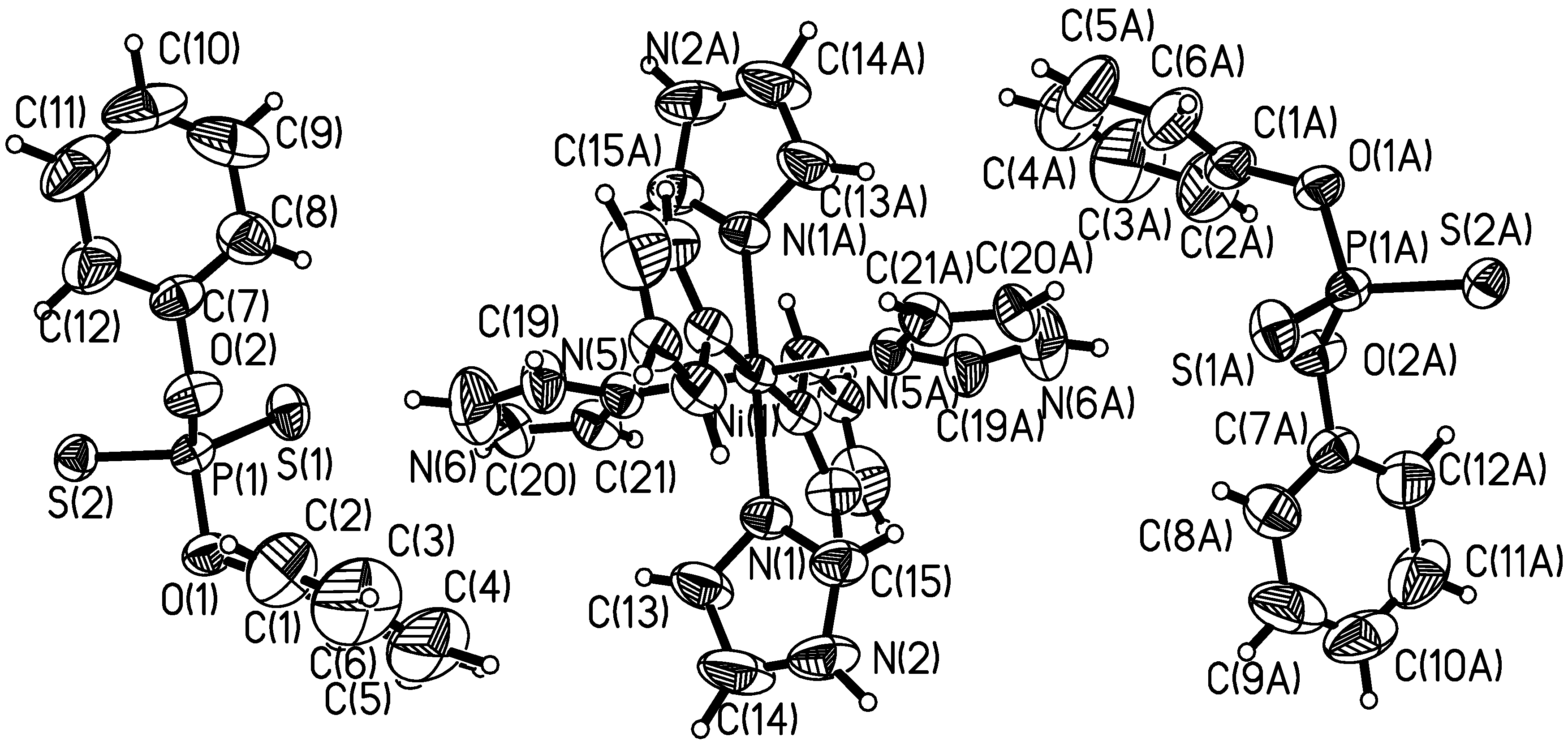
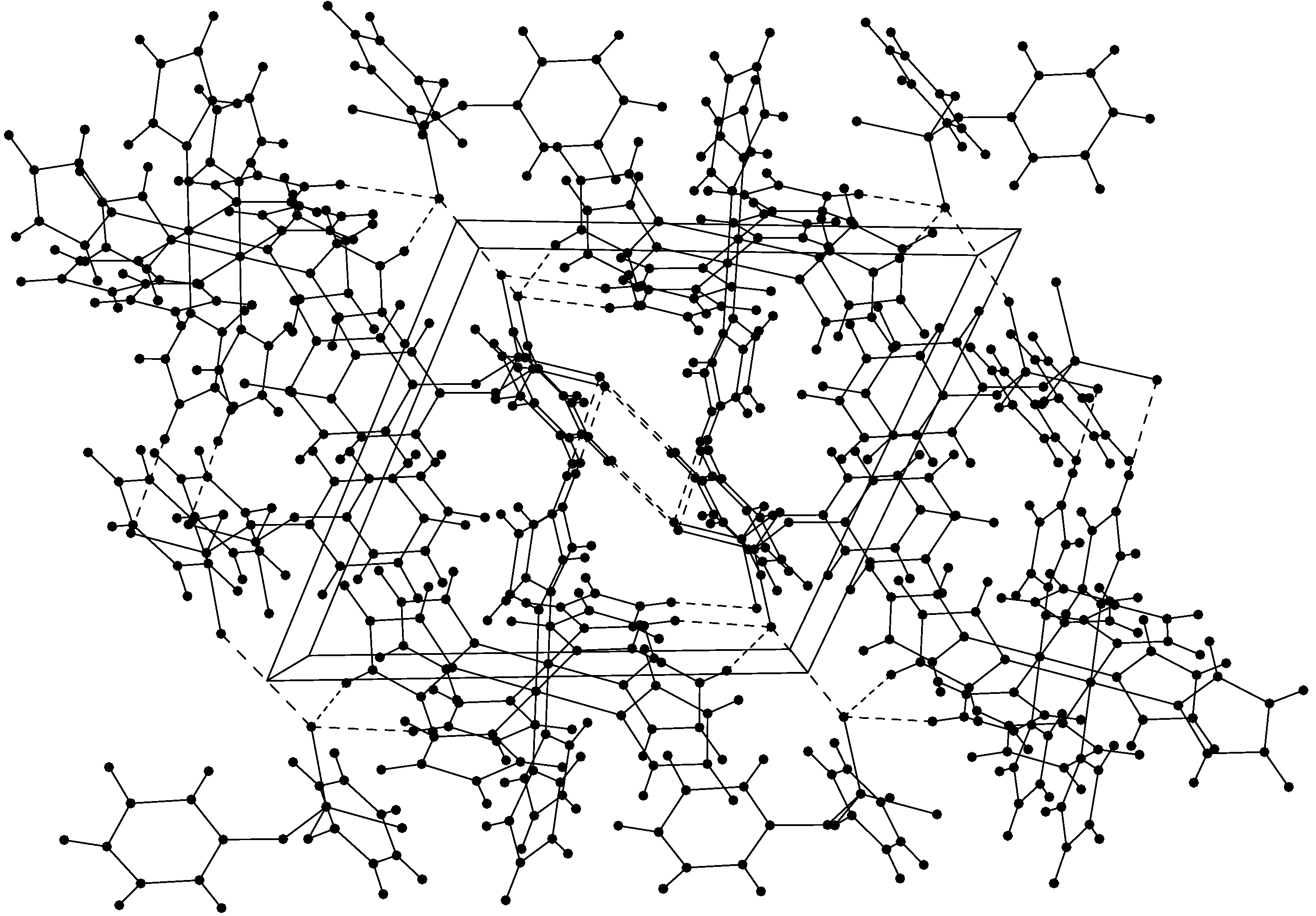
X-ray Crystal Structure of the Title Compound
The X-ray structure of the complex [Ni(Im)6](dtp)2 is built up of discrete monomeric molecules. Figure 1 shows a perspective view of the title compound with atomic numbering scheme and Figure 2 a perspective view of the crystal packing in the unit cell. Table 1 contains atomic positions and equivalent temperature factors for non-hydrogen atoms. Selected bond lengths and angles are presented in Table 2.
Figure 1.
Molecular structure for [Ni(Im)6](dtp)2 with the atomic numbering scheme
The crystal structure of the complex consists of [Ni(Im)6]2+ cation and two dtp anions, linked by electrostatic forces and hydrogen bonds (shown as dashed lines in Figure 2). The coordination model of the nickel (II) can be described as being of octahedral geometry. Six imidazole molecules are coordinated through their tertiary nitrogen atoms to each nickel (II) ion, and two dtp anions are out of the coordination sphere and balance the charge. The bond distances of Ni(1)-N(1), Ni(1)-N(3) and Ni(1)-N(5) of [Ni(Im)6](dtp)2 are 2.133(3), 2.132(3) and 2.125(3) Å, respectively. These distances are in agreement with those reported for octahedral [Ni(Im)6]2+ complex [2.120(1), 2.124(1), 2.141(1) Å], [Ni(Im)6](sal)2 (sal = salicylate) [8]. In the solid state, one dtp sulfur atom forms a N-H∙∙∙S intermolecular hydrogen bond with the nitrogen atom of Ni(Im)62+ (Figure 2), and the donor and acceptor distances are N(2)∙∙∙S(1) 3.6547, C(17)∙∙∙S(2) ( 2-x, 1-y, 1-z) 3.3052, C(11)∙∙∙S(2) ( 2-x, 1-y, 1-z) 3.7945 and C(20)∙∙∙S(1) (2-x, -y, 1-z) 3.3807 Å (Table 3), the whole complexes forming a 3-dimensional network structure.
Figure 2.
A view of the crystal packing down the z axis for [Ni(Im)6](dtp)2. The dashed lines represent hydrogen bonds between dtp anions and imidazoles.
IR Spectroscopy
The IR spectrum of the complex shows characteristic absorption bands at 3401 and 552 cm-1, assigned to ν(N-H) and ν(Ni-N) vibrations, respectively. This indicates that the atom coordinated with Ni(II) is the double bond nitrogen (=N) in the imidazole molecule. This result is in agreement with that of X-ray crystal structure determination.
Table 1.
Atomic coordinates and equivalent isotropic displacement parameters
| Atom | x | y | z | Ueq |
| Ni1 | 0.50000 | 0.00000 | 0.50000 | 0.03408 |
| S1 | -0.02504 | 0.11075 | 0.11362 | 0.05583 |
| S2 | 0.23540 | 0.33035 | 0.36706 | 0.04651 |
| P1 | 0.11983 | 0.28764 | 0.20898 | 0.04016 |
| O1 | 0.25663 | 0.34717 | 0.16152 | 0.05387 |
| O2 | 0.02372 | 0.37457 | 0.19719 | 0.05210 |
| N1 | 0.70819 | 0.07677 | 0.45909 | 0.04516 |
| N2 | 0.86587 | 0.12438 | 0.36401 | 0.07807 |
| N3 | 0.49052 | 0.17820 | 0.55780 | 0.04184 |
| N4 | 0.44719 | 0.34158 | 0.64827 | 0.09284 |
| C1 | 0.22540 | 0.34804 | 0.05361 | 0.06202 |
| C2 | 0.20081 | 0.44556 | 0.03942 | 0.09705 |
| C3 | 0.17904 | 0.45204 | -0.06446 | 0.14530 |
| C4 | 0.18613 | 0.36170 | -0.15079 | 0.15496 |
| C5 | 0.20809 | 0.25992 | -0.13735 | 0.13347 |
| C6 | 0.22857 | 0.25275 | -0.03206 | 0.09396 |
| C7 | -0.10835 | 0.36540 | 0.23377 | 0.05025 |
| C8 | -0.25918 | 0.27493 | 0.16484 | 0.07322 |
Ueq is defined as one third of the trace of the orthogonalized Ueq tensor.
Table 2.
Selected bond distances (Å) and bond angles (°) of title compound
| Ni1-N1 | 2.133(3) | S1-P1 | 1.955 (2) |
| Ni1- N3 | 2.132(3) | S2-P1 | 1.959 (2) |
| Ni1-N5 | 2.125(3) | P1-O1 | 1.609(2) |
| C1-C2 | 1.355(7) | P1-O2 | 1.632(2) |
| C1-C6 | 1.376(7) | O1-C1 | 1.405(4) |
| O1-P1-O2 | 97.47(13) | C1-C2-C3 | 119.7(6) |
| O1-P1-S2 | 105.29(11) | C4-C3-C2 | 119.6(7) |
| S1-P1-S2 | 117.31(7) | N5-Ni1-N3 | 90.40(11) |
| N5-Ni1-N1 | 90.81(11) | N3-Ni1-N1 | 91.05(11) |
| C14-C13-N1 | 109.2(4) | N1-C15-N2 | 111.6(4) |
Symmetry transformations used to generate equivalent atoms: $1 -x+1, -y, -z+1
Table 3.
Hydrogen bond distances (Å) and angles of title compound
| D | H | A | symm | D-H | H…A | D…A | D-H…A Bond angle |
| N(2) | H(8A) (2a) | S(1) | 0.9287 | 2.7403 | 3.6547 | 168.35 | |
| C(17) | H(4A) | S(2) | 2766 | 0.9224 | 2.3836 | 3.3052 | 177.03 |
| C(11) | H(11A) | S(2) | 2766 | 0.9467 | 2.8717 | 3.7945 | 165.15 |
| C(20) | H(20A) | S(1) | 2756 | 0.9170 | 2.5156 | 3.3807 | 157.42 |
[2766]=2-x,1-y,1-z; [2756]=2-x,-y,1-z
Experimental
General
All commercially available chemicals were of analytical reagent grade and used directly without further purification. The C, N and H contents were determined by using an Elementar Vario EL analyzer. Infrared spectra were recorded from KBr pellets on a Midac Prospet IR spectrometer. All operations were carried out under ambient conditions.
Synthesis of the complex [Ni(Im)6](dtp)2
To a warm solution of excess imidazole (1.0 g, 15 mmol) in EtOH (50 mL) was added with stirring nickel (II) O, O′-diphenyl dithiophosphate, and the mixture was refluxed for 40 min. The blue solution was filtered and the filtrate was left to stand undisturbed. Upon slow evaporation at room temperature, a blue crystalline solid suitable for X-ray structure determination appeared several weeks later and was separated by filtration. Main IR absorption bands (KBr, cm-1): 3401 (N-H), 1491 (C=N), 1591, 1530 (phenyl ring stretch), 552 (Ni-N). Anal. Data: Calcd for C42H44N12O4P2S4Ni1 C, 48.94; H, 4.27; N, 16.31 %. Found: C, 48.93; H, 4.30; N, 16.21 %.
Crystallographic Data and Structure Determination [9]
The selected crystal of [Ni(Im)6](dtp)2 was mounted on a Nonius CAD4 diffractometer, reflection data for the unit cell determination were measured at 293 K using MoKα radiation (λ= 0.71073 Å) with a graphite monochromator. The technique used was ω-scan with 0.995<θ<25.04° for the complex. Absorption correction was made with π-scans. The structure was solved by direct method and refined by full-matrix least-squares method on F2obs by using the SHELXTL software package [10]. All non-H atoms were anisotropically refined. The hydrogen atoms were geometrically fixed and allowed to ride on the parent atoms to which they are attached. The final conventional R = 0.0604, wR = 0.1388 for 3843 reflections with I > 2σ(I); w = 1/[σ2(F2o)+(0.0600P)2+1.4652P] where P = (F2o+2(F2c))/3. The molecular graphics were created by SHELXTL. Atomic scattering factors and anomalous dispersion correction were taken from International Table for X-Ray Crystallography [11]. A summary of the key crystallographic information is given in Table 4.
Table 4.
Summary of crystallographic results for [Ni(Im)6](dtp)2
| Formula | C42H44N12O4P2S4Ni1 |
| Formula weight | 1029.78 |
| Color/shape | Blue/prism |
| Crystal system | Triclinic |
| Space group | Pī |
| a / Å | 9.375(2) |
| b/ Å | 12.324(3) |
| c/ Å | 13.285(3) |
| α/° | 107.86(3) |
| β/° | 102.28(3) |
| γ/° | 109.24(3) |
| V/ Å3 | 1292(5) |
| Z value | 1 |
| D(calcd.)/g.cm-3 | 1.307 |
| μ/ mm-1 | 0.65 |
| F (000) | 534 |
| Crystal size/mm | 0.20×0.24×0.20 |
| Temp. /K | 293(2) |
| θ ranges/° | 0.995-25.04 |
| h/k/l | -10,11/-14,0/-15,15 |
| Reflections collected | 4551 |
| Independent reflections | 3843 |
| Absorption correction | Empirical |
| No. restrains/ No. parameters | 0/295 |
| GOF | 1.085 |
| Final R indices[I>2σ(I)] | R1=0.0473 wR2=0.1246 |
| R indices(all data) | R1=0.0604 wR2=0.1388 |
| Largest peak and hole/e·nm-3 | 660 and -490 |
Acknowledgements
We thank the National Natural Science Foundation of China (Project 20275020) and the Natural Science Foundation of Shandong Province (Project Z2002B02) for financial support.
References and Notes
- Brooks, P.; Davidson, N. J. Amer. Chem. Soc. 1960, 82, 2118.
- Davis, W. J.; Smith, J. J. Chem. Soc. (A) 1971, 317.
- Inova, M.; Kishita, M.; Kabo, M. Bull. Chem. Soc. Jpn. 1969, 39, 1352.
- Eillbeck, W.J.; Holmes, F.; Underhill, A.E. J. Chem. Soc. (A) 1967, 757.
- Xu, L. Z.; Zhao, P. S.; Zhang, S. S. Chin. J. Chem. 2001, 19, 436.
- Bernarducci, E. E.; Bharadwij, P. K. Inorg. Chem. 1983, 22, 3911.
- Tamura, H.; Imai, H.; Kuwahra, J.; Sugiura, Y. J. Amer. Chem. Soc. 1987, 109, 6870.
- Jian, F. F.; Wang, Z.X.; Bai, Z. P.; You, X. Z. J. Chem. Cryst. 1999, 29, 359.
- CCDC 212159 contains the supplementary crystallographic data for this paper. These data can be obtained free of charge via www.ccdc.cam.ac.uk/conts/retrieving.html (or from the CCDC, 12 Union Road, Cambridge CB2 1EZ, UK; fax: +44 1223 336033; e-mail: deposit@ccdc.cam.ac.uk).
- Sheldrick, G. M. SHELXTL V5 Reference Manual, Siemens Analytical X-ray Systems; Inc., Madison: Wisconsin (USA), 1996. [Google Scholar]
- Wilson, A.J. International Table for X-Ray Crystallography; Vol C, Kluwer Academic Publishers: Dordrecht, 1992. [Google Scholar]
- Samples Availability: Available from the corresponding author.
© 2003 by MDPI ( http://www.mdpi.org). Reproduction is permitted for noncommercial purposes.