
Isolation of the Main Biologically Active Substances and Phytochemical Analysis of Ginkgo biloba Callus Culture Extracts

 ,
,  ,
,  and
and
Abstract
1. Introduction
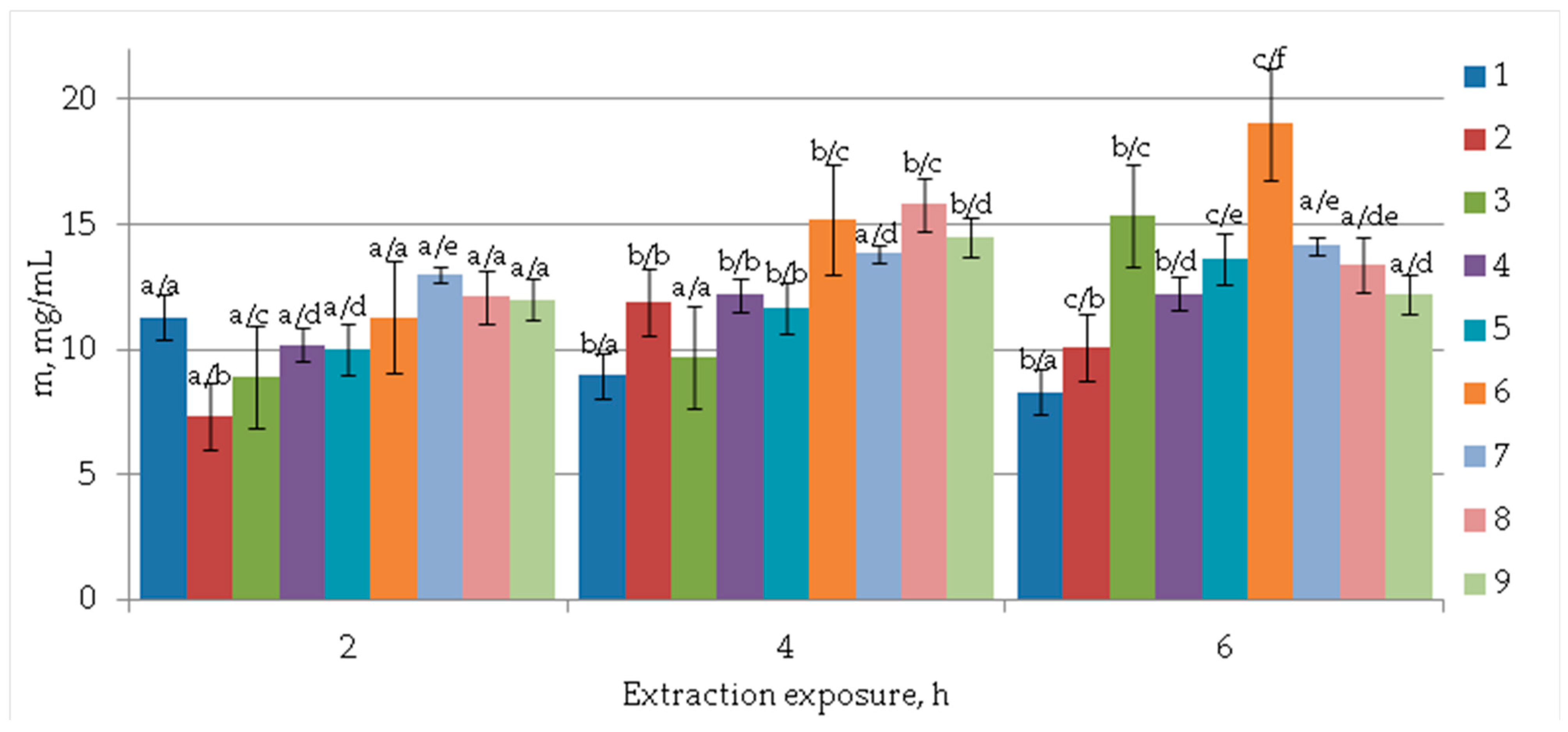
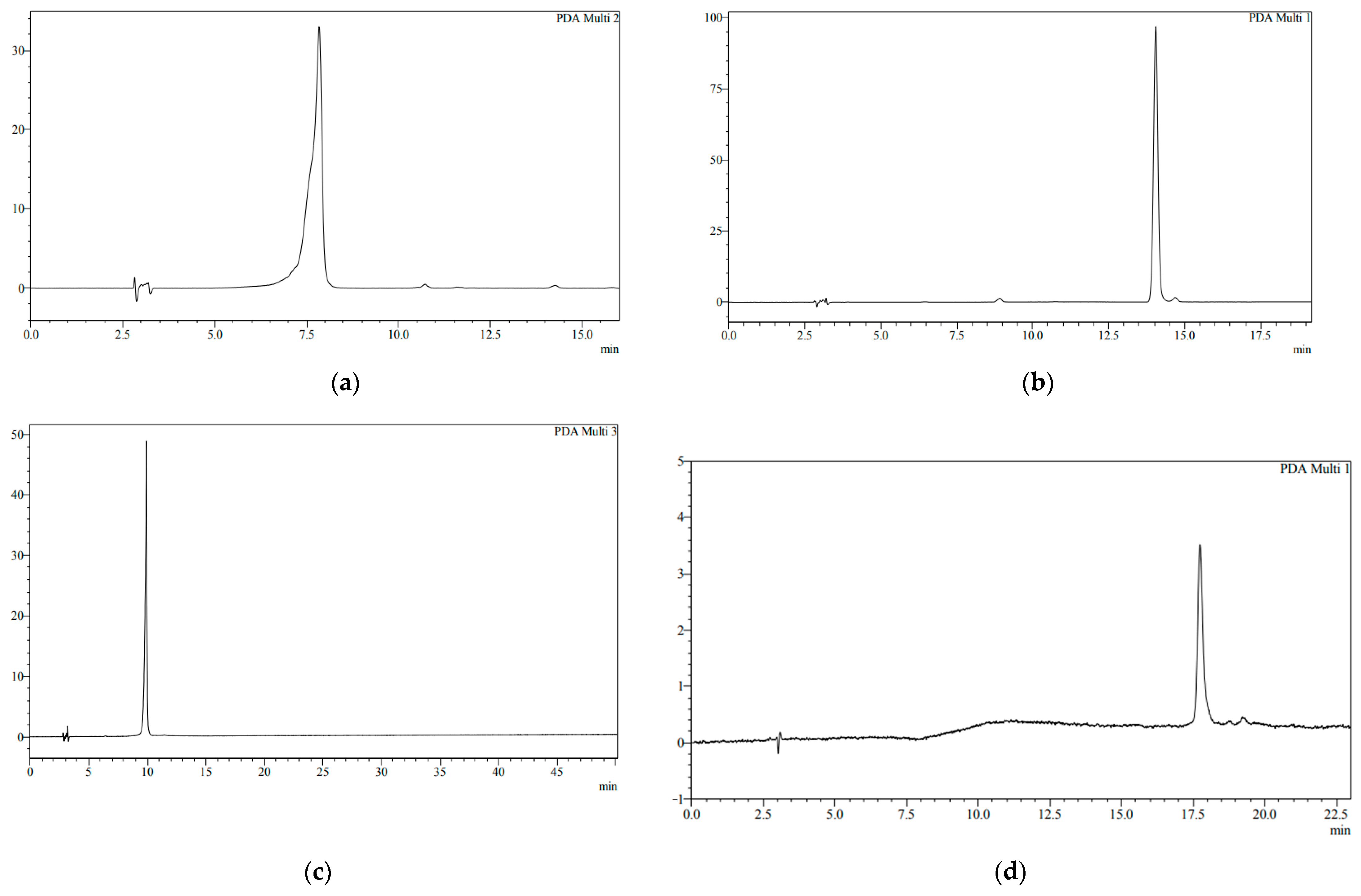
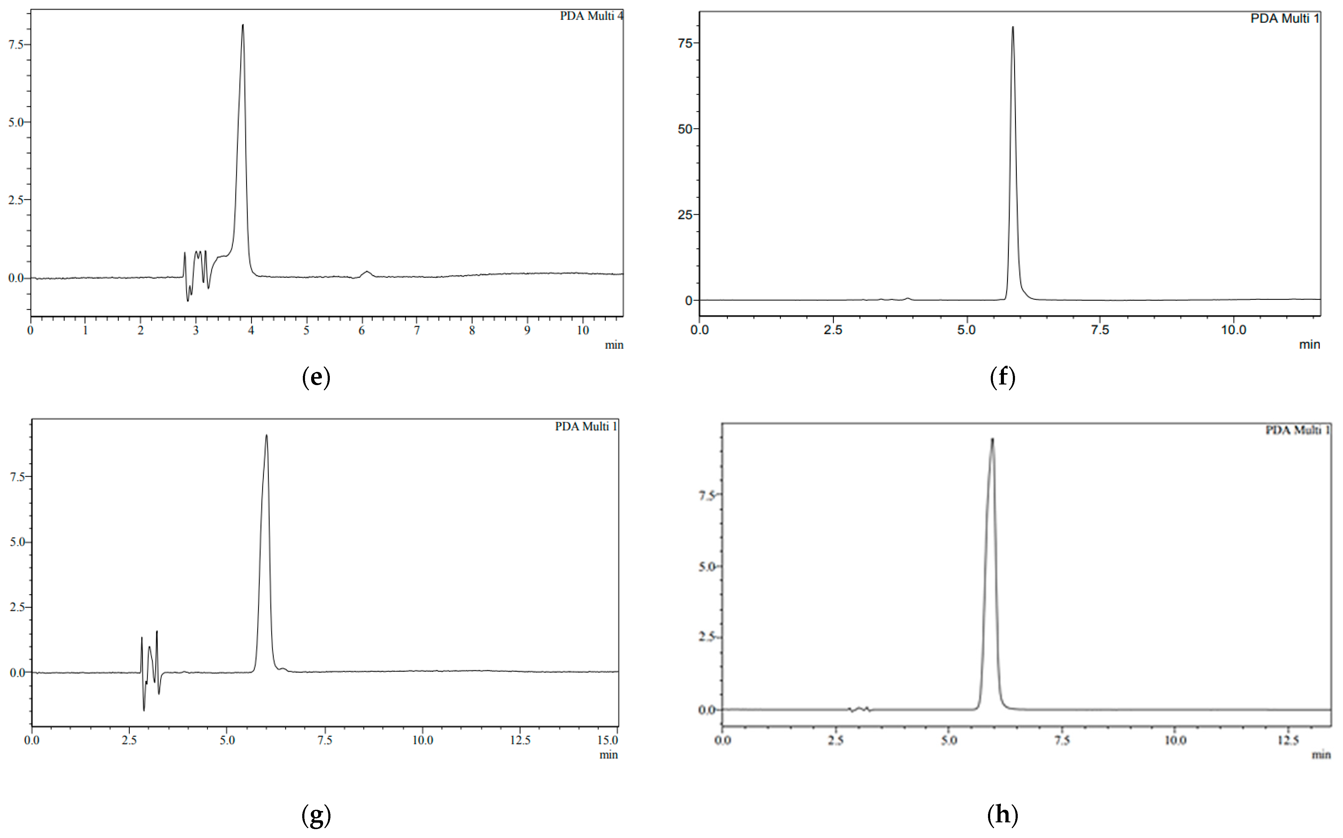
2. Results
3. Discussion
4. Materials and Methods
4.1. Seed Culture for Callus Induction
4.2. Extraction of Biologically Active Components
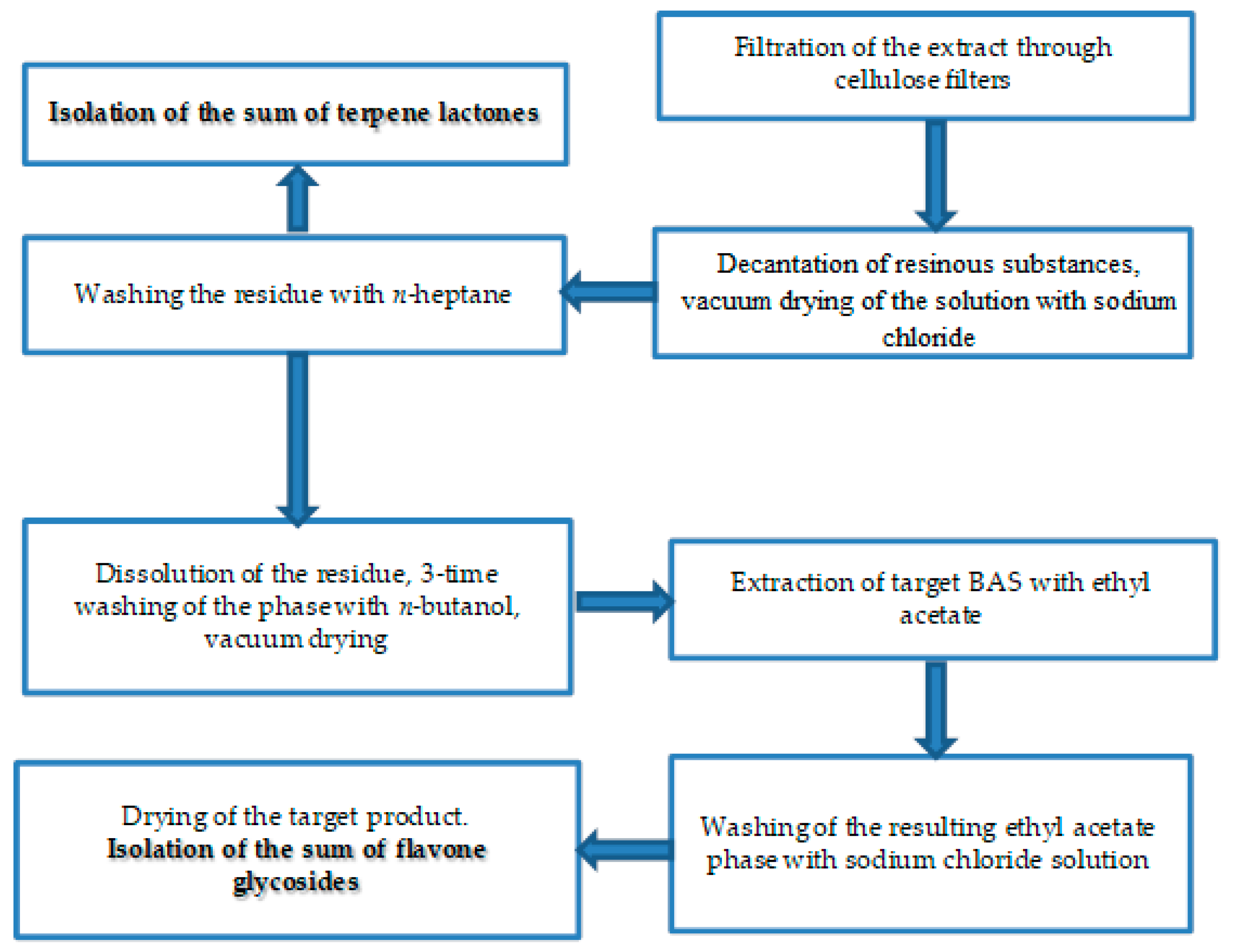
4.3. Method of Isolation of Biologically Active Substances
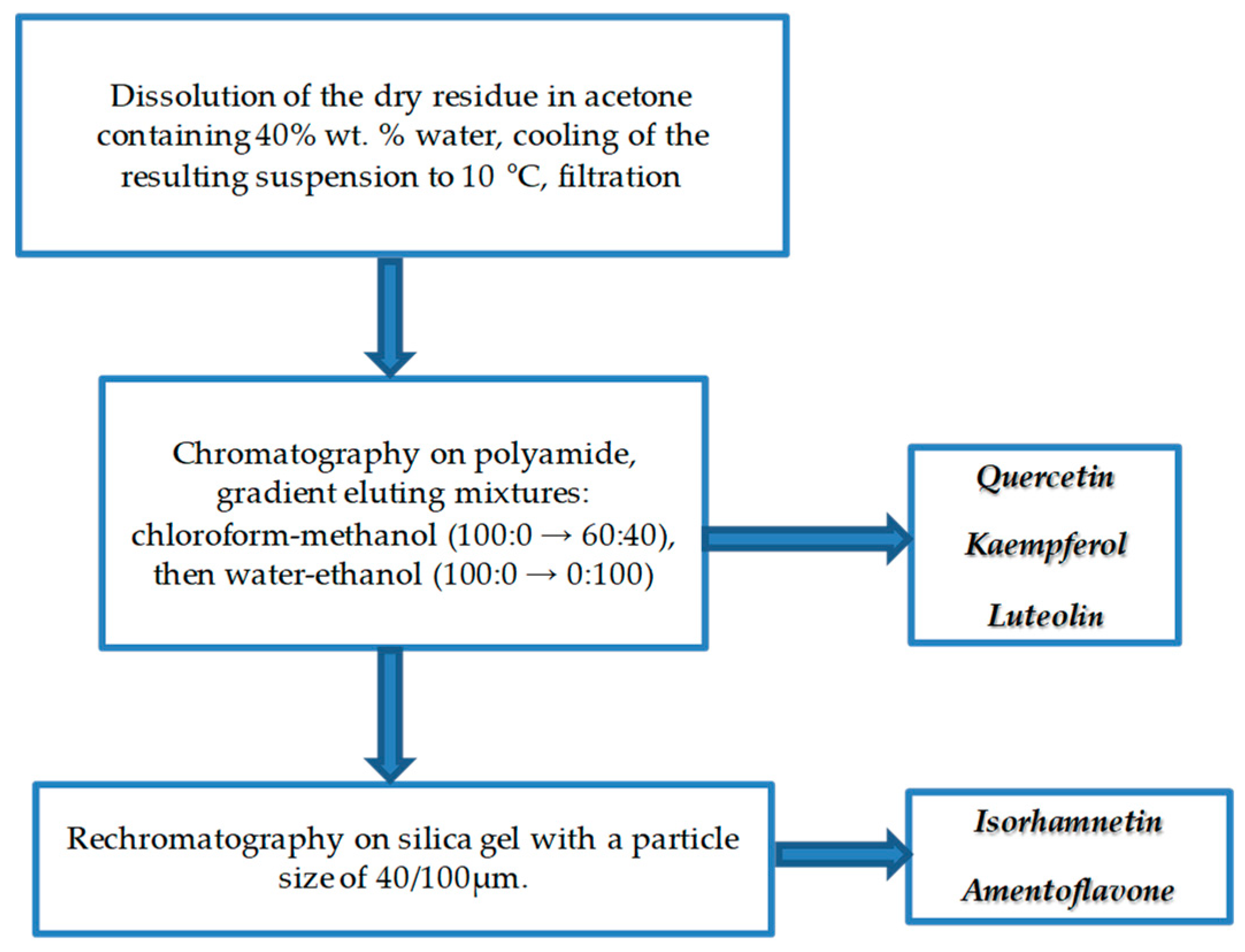
4.4. Method of Purification of Biologically Active Compounds
4.5. High Performance Chromatography Method
4.6. HRMS Spectrometry
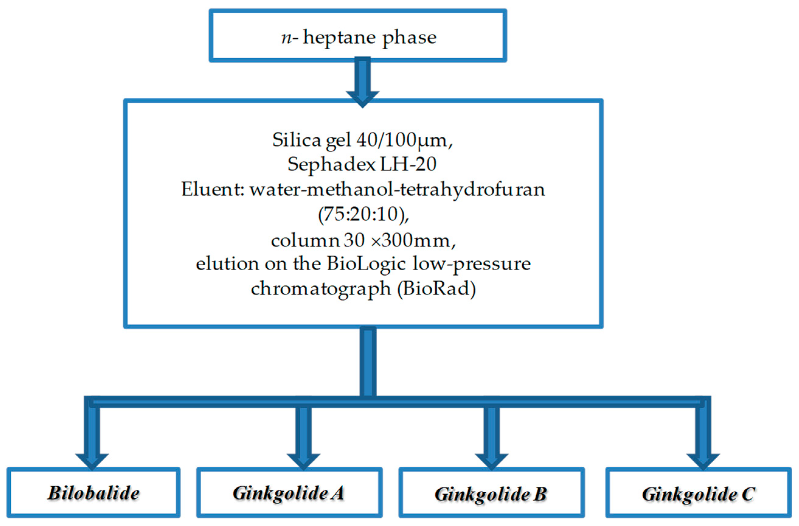
4.7. Low-Pressure Column Chromatography Method
4.8. Spectrophotometry of Samples
4.9. IR Spectrometry
4.10. Sample Preparation
4.11. Statistical Analysis Methods
5. Conclusions
Author Contributions
Funding
Institutional Review Board Statement
Informed Consent Statement
Data Availability Statement
Acknowledgments
Conflicts of Interest
Sample Availability
References
- Akram, M.; Rashid, A. Anti-coagulant activity of plants: Mini review. J. Thromb. Thrombolysis 2017, 44, 406–411. [Google Scholar] [CrossRef] [PubMed]
- Liu, Y.; Xin, H.; Zhang, Y.; Che, F. Leaves, seeds and exocarp of Ginkgo biloba L. (Ginkgoaceae): A Comprehensive Review of Traditional Uses, phytochemistry, pharmacology, resource utilization and toxicity. J. Ethnopharmacol. 2022, 298, 115645. [Google Scholar] [CrossRef] [PubMed]
- Abdel-Emama, R.A.; Abd-Eldayem, A.M. Systemic and topical Ginkgo biloba leaf extract (Egb-761) ameliorated rat paw inflammation in comparison to dexamethasone. Ethnopharmacology 2022, 282, 114619. [Google Scholar] [CrossRef] [PubMed]
- Sirotkin, A.V. Potential effets of ginkgo (Ginkgo biloba, L.) on female reproduction. Reprod. Biol. 2021, 21, 100568. [Google Scholar] [CrossRef]
- Shareena, G.; Kumar, D. Traversing through half a century research timeline on Ginkgo biloba, in transforming a botanical rarity into an active functional food ingredient. Biomed. Pharmacother. 2022, 153, 113299. [Google Scholar] [CrossRef]
- Milentyeva, I.S.; Le, V.M.; Kozlova, O.V.; Velichkovich, N.S.; Fedorova, A.M.; Loseva, A.I.; Yustratov, V.P. Secondary metabolites in in vitro cultures of Siberian medicinal plants: Content, antioxidant properties, and antimicrobial characteristics. Foods Raw Mater. 2021, 9, 153–163. [Google Scholar] [CrossRef]
- Yu, M.; Aoki, D.; Akita, T.; Fujiyasu, S.; Takada, S.; Matsushitam, Y.; Yoshida, M.; Fukushima, K. Distribution of lignans and lignan mono/diglucosides within Ginkgo biloba L. stem. Phytochemistry 2022, 196, 113102. [Google Scholar] [CrossRef]
- Zhou, W.; Yang, Z.; Huang, S.; Fang, Z.; Chen, B.; Ma, M. Rapid quantitative analysis of ginkgo flavonoids using paper spray mass spectrometry. J. Pharm. Biomed. Anal. 2019, 171, 158–163. [Google Scholar] [CrossRef]
- Liu, L.; Wang, Y.; Zhang, J.; Wang, S. Advances in the chemical constituents and chemical analysis of Ginkgo biloba leaf, extract, and phytopharmaceuticals. J. Pharm. Biomed. Anal. 2021, 193, 113704. [Google Scholar] [CrossRef]
- Mousavi, S.N.; Hosseinikia, M.; Yousefi Rad, E.; Saboori, S. Beneficial effects of Ginkgo biloba leaf extract on inflammatory markers: A systematic review and meta-analysis of the clinical trials. Phytother. Res. 2022, 36, 3459–3469. [Google Scholar] [CrossRef]
- Yang, X.; Xu, Q.; Le, L.; Zhou, T.; Yu, W.; Wang, G.; Fu, F.-F.; Cao, F. Comparative histology, transcriptome, and metabolite profiling unravel the browning mechanisms of calli derived from ginkgo (Ginkgo biloba L.). J. For. Res. 2022, in press. [Google Scholar] [CrossRef]
- Lu, Z.; Zhu, L.; Lu, J.; Shen, N.; Wang, L.; Lin, J. Rejuvenation increases leaf biomass and flavonoid accumulation in Ginkgo biloba. Hortic. Res. 2022, 9, uhab018. [Google Scholar] [CrossRef] [PubMed]
- Frommenwiler, D.A.; Booker, A.; Vila, R.; Heinrich, M.; Reich, E.; Cañigueral, S. Comprehensive HPTLC fingerprinting as a tool for a simplified analysis of purity of Ginkgo products. J. Ethnopharmacol. 2019, 243, 112084. [Google Scholar] [CrossRef] [PubMed]
- Singh, D. Astrocytic and microglial cells as the modulators of neuroinflammation in Alzheimer’s disease. J. Neuroinflamm. 2022, 19, 206. [Google Scholar] [CrossRef] [PubMed]
- Pinto, M.D.S.; Kwon, Y.-I.; Apostolidis, E.; Lajolo, F.M.; Genovese, M.I.; Shetty, K. Potential of Ginkgo biloba L. leaves in the management of hyperglycemia and hypertension using in vitro models. Bioresour. Technol. 2009, 100, 6599–6609. [Google Scholar] [CrossRef] [PubMed]
- Wang, H.; Shi, M.; Cao, F.; Su, E. Ginkgo biloba seed exocarp: A waste resource with abundant active substances and other components for potential applications. Food. Res. Int. 2022, 160, 111637. [Google Scholar] [CrossRef]
- Meo, F.; Cuciniello, R.; Margarucci, S.; Bergamo, P.; Petillo, O.; Peluso, G.; Filosa, S.; Crispi, S. Ginkgo biloba prevents oxidative stress-induced apoptosis blocking p53 activation in neuroblastoma cells. Antioxidants 2020, 9, 279. [Google Scholar] [CrossRef]
- Babich, O.; Sukhikh, S.; Pungin, A.; Ivanova, S.; Asyakina, L.; Prosekov, A. Modern Trends in the In Vitro Production and Use of Callus, Suspension Cells and Root Cultures of Medicinal Plants. Molecules 2020, 25, 5805. [Google Scholar] [CrossRef]
- Babich, O.; Sukhikh, S.; Prosekov, A.; Asyakina, L.; Ivanova, S. Medicinal Plants to Strengthen Immunity during a Pandemic. Pharmaceuticals 2020, 13, 313. [Google Scholar] [CrossRef]
- Raks, V.; Al-Suod, H.; Buszewski, B. Isolation, Separation, and Preconcentration of Biologically Active Compounds from Plant Matrices by Extraction Techniques. Chromatographia 2018, 81, 189–202. [Google Scholar] [CrossRef]
- Gong, Q.; Guo, Z.; Sun, Z.; Gong, J.; Wei, F. Graphene oxide-assisted ethanol reflux extraction of total flavonoids from Ginkgo biloba leaves: Study of kinetics and mechanism. Chem. Pap. 2020, 74, 971–984. [Google Scholar] [CrossRef]
- Tian, L.; Zhou, M.; Pan, X.; Xiao, G.; Liu, Y. Supercritical CO2 extraction and response surface optimization of ginkgolic acids from Ginkgo biloba exopleura. Korean J. Chem. Eng. 2015, 32, 1649–1654. [Google Scholar] [CrossRef]
- Mei, N.; Guo, X.; Ren, Z.; Kobayashi, D.; Wada, K.; Guo, L. Review of Ginkgo biloba-induced toxicity, from experimental studies to human case reports. J. Environ. Sci. Health C Environ. Carcinog. Ecotoxicol. Rev. 2017, 35, 1–28. [Google Scholar] [CrossRef] [PubMed]
- Gong, H.; Wu, C.-E.; Fan, G.-J.; Li, T.-T.; Wang, J.-H.; Wang, T. Determination of native contents of 4′-O-methylpyridoxine and its glucoside in raw and heated Ginkgo biloba seeds by high-performance liquid chromatography. J. Food Meas. Charact. 2020, 14, 917–924. [Google Scholar] [CrossRef]
- Wen, L.; Zhang, Z.; Sun, D.-W.; Sivagnanam, S.P.; Tiwari, B.K. Combination of emerging technologies for the extraction of bioactive compounds. Crit. Rev. Food Sci. Nutr. 2020, 60, 1826–1841. [Google Scholar] [CrossRef] [PubMed]
- Zhang, C.X.; Hu, X.L. Novel Atoxic Method of Flavonoid Extraction from Ginkgo biloba Leaves. Bull. Environ. Contam. Toxicol. 2003, 71, 662–667. [Google Scholar] [CrossRef] [PubMed]
- Khizrieva, S.S.; Borisenko, S.N.; Maksimenko, E.V.; Zharkova, G.V.; Borisenko, N.I.; Minkin, V.I. Evaluation of the Polyphenol Composition and Acetylcholinesterase Inhibitory Activity of Ginkgo biloba Leaf Extracts Produced in Subcritical Water. Russ. J. Phys. Chem. B 2022, 16, 1294–1300. [Google Scholar] [CrossRef]
- Su, E.; Yang, M.; Cao, J.; Lu, C.; Wang, J.; Cao, F. Deep eutectic solvents as green media for efficient extraction of terpene trilactones from Ginkgo biloba leaves. J. Liq. Chromatogr. Relat. Technol. 2017, 40, 385–391. [Google Scholar] [CrossRef]
- Dai, Y.; Row, K.H. Determination of Rutin from Ginkgo biloba L. Leaves by Ultrasound-Assisted Extraction with Natural Deep Eutectic Solvent-Based Cellulose Polymers and High-Performance Liquid Chromatography (HPLC). Anal. Lett. 2022, 55, 566–579. [Google Scholar] [CrossRef]
- Yang, C.; Xu, Y.-R.; Yao, W.-X. Extraction of pharmaceutical components from Ginkgo biloba leaves using supercritical carbon dioxide. J. Agric. Food Chem. 2002, 50, 846–849. [Google Scholar] [CrossRef]
- Wang, J.; Cao, F.; Su, E.; Wu, C.; Zhao, L.; Ying, R. Improving flavonoid extraction from Ginkgo biloba leaves by prefermentation processing. J. Agric. Food Chem. 2013, 61, 5783–5791. [Google Scholar] [CrossRef] [PubMed]
- Zhou, G.; Yao, X.; Tang, Y.; Yang, N.; Pang, H.; Mo, X.; Zhu, S.; Su, S.; Qian, D.; Jin, C.; et al. Two new nonacosanetriols from Ginkgo biloba sarcotesta. Chem. Phys. Lipids 2012, 165, 731–736. [Google Scholar] [CrossRef] [PubMed]
- Liu, X.-G.; Wu, S.-Q.; Li, P.; Yang, H. Advancement in the chemical analysis and quality control of flavonoid in Ginkgo biloba. J. Pharm. Biomed. Anal. 2015, 113, 212–225. [Google Scholar] [CrossRef] [PubMed]
- United State Pharmacopeia. Available online: https://www.uspnf.com/ (accessed on 31 January 2023).
- Karamova, N.S.; Khabibrakhmanova, V.R.; Abdul-Hafiz, I.Y.; Gumerova, S.K.; Kamalova, Y.N.; Kovalenko, S.A.; Ibrahim, O.K.M.; Orabi, M.A.-M.A. Composition of biologically active substances and anti-radical activity of extracts from five types plants of the ASPARAGACEAE family. Khimiya Rastit. Syr’ya 2021, 4, 277–289. (In Russian) [Google Scholar] [CrossRef]
- Zhao, L.J.; Liu, W.; Xiong, S.H.; Tang, J.; Lou, Z.H.; Xie, M.X.; Xia, B.H.; Lin, L.M.; Liao, D.F. Determination of Total Flavonoids Contents and Antioxidant Activity of Ginkgo biloba Leaf by Near-Infrared Reflectance Method. Int. J. Anal. Chem. 2018, 2018, 8195784. [Google Scholar] [CrossRef]
- Geng, Y.; Xiang, B. Simultaneous quantisation of flavonol aglycones in Ginkgo biloba leaf extracts applying moving window partial least squares regression models. J. Near Infrared Spectrosc. 2008, 16, 551–559. [Google Scholar] [CrossRef]
- Shi, J.-Y.; Zou, X.-B.; Zhao, J.-W. Determination of total flavonoids content in fresh Ginkgo biloba leaf with different colors using near infrared spectroscopy. Spectrochim. Acta A Mol. Biomol. Spectrosc 2012, 94, 271–276. [Google Scholar] [CrossRef]
- Abouheif, S.A.; Sallam, S.M.; Sohafy, S.M.; Kassem, F.F.; Shawky, E. Optimization of terpene lactones and ginkgolic acids extraction from Ginkgo biloba L. leaves by natural deep eutectic solvents using experimental design and HPTLC-MS analysis. Microchem. J. 2022, 176, 107246. [Google Scholar] [CrossRef]
- Chen, S.; Xing, X.-H.; Huang, J.-J.; Xu, M.-S. Enzyme-assisted extraction of flavonoids from Ginkgo biloba leaves: Improvement effect of flavonol transglycosylation catalyzed by Penicillium decumbens cellulose. Enzym. Microb. Technol. 2011, 48, 100–105. [Google Scholar] [CrossRef]
- Kalyniukova, A.; Holusa, J.; Musiolek, D.; Sedlakova-Kadukova, J.; Płotka-Wasylka, J.; Andruch, V. Application of deep eutectic solvents for separation and determination of bioactive compounds in medicinal plants. Ind. Crops Prod. 2021, 172, 114047. [Google Scholar] [CrossRef]
- Wang, T.; Wang, Q.; Guo, Q.; Li, P.; Yang, H. A hydrophobic deep eutectic solvents-based integrated method for efficient and green extraction and recovery of natural products from Rosmarinus officinalis leaves, Ginkgo biloba leaves and Salvia miltiorrhiza roots. Food Chem. 2021, 363, 130282. [Google Scholar] [CrossRef] [PubMed]
- Boateng, I.D. A critical review of current technologies used to reduce ginkgotoxin, ginkgotoxin-5′-glucoside, ginkgolic acid, allergic glycoprotein, and cyanide in Ginkgo biloba L. seed. Food Chem. 2022, 382, 132408. [Google Scholar] [CrossRef] [PubMed]
- Boateng, I.D.; Yang, X.-M. Ginkgo biloba L. seed; A comprehensive review of bioactives, toxicants, and processing effects. Ind. Crops Prod. 2022, 176, 114281. [Google Scholar] [CrossRef]
- Yua, C.; Chen, J.; Xiong, Y.; Li, X.; Daia, X.-Y.; Shi, C.-C. Optimization of multi-stage countercurrent extraction of antioxidants from Ginkgo biloba L. leaves. Food Bioprod. Process. 2012, 90, 95–101. [Google Scholar] [CrossRef]
- Jay, M.; Gonnet, J.-F.; Wollenweber, E.; Voirin, B. Sur l’analyse qualitative des aglycones flavoniques dans une optique chimiotaxinomique. Phytochemistry 1975, 14, 1605–1612. [Google Scholar] [CrossRef]
- Sukhikh, A.S.; Kuznetsov, P.V. The use of the universal purpose sorbent sefadex LG-20 in modern biomedical research. Med. Kuzbass 2009, 8, 3–12. (In Russian). Available online: https://www.elibrary.ru/download/elibrary_12967156_41987740.pdf (accessed on 5 February 2023).
- Zhang, J.; Hayat, K.; Zhang, X.; Tong, J.; Xia, S. Separation and purification of flavonoid from Ginkgo extract by Polyamide Resin. Sep. Sci. Technol. 2010, 45, 2413–2419. [Google Scholar] [CrossRef]
- Zhu, M.; Yun, Y.; Xiang, W. Purification of Ginkgo biloba flavonoids by UF membrane technology. Desalination Water Treat 2013, 51, 3847–3853. [Google Scholar] [CrossRef]
- Babich, O.O.; Zaushintsena, A.V.; Milentyeva, I.S.; Prosekov, A.Y.; Lukin, A.A. Method for Obtaining Root Culture In Vitro Potentilla alba L.—Flavonoid Producer. Patent No. 2714403 RU, 14 February 2020. [Google Scholar]
- Yang, M.; Cao, J.; Cao, F.; Lu, C.; Su, E. Efficient Extraction of Bioactive Flavonoids from Ginkgo biloba Leaves Using Deep Eutectic Solvent/Water Mixture as Green Media Chem. Biochem. Eng. Q. 2018, 32, 315–324. [Google Scholar] [CrossRef]
- Li, R.; Xia, Z.; Li, B.; Tian, Y.; Zhang, G.; Li, M.; Dong, J. Advances in Supercritical Carbon Dioxide Extraction of Bioactive Substances from Different Parts of Ginkgo biloba L. Molecules 2021, 26, 4011. [Google Scholar] [CrossRef]
- Agnolet, S.; Jaroszewski, J.W.; Verpoorte, R.; Staerk, D. 1H NMR-based metabolomics combined with HPLC-PDA-MS-SPE-NMR for investigation of standardized Ginkgo biloba. Metabolomics 2010, 6, 292–302. [Google Scholar] [CrossRef] [PubMed]
- Ping, Y.; Jing, Y.-L. A new sesquiterpene trilactone from the roots of Ginkgo biloba. Chin. Chem. Lett. 2009, 20, 1224–1226. [Google Scholar] [CrossRef]
- Parveen, I.; Wang, M.; Zhao, J.; Chittiboyina, A. Investigating sesquiterpene biosynthesis in Ginkgo biloba: Molecular cloning and functional characterization of (E,E)-farnesol and α-bisabolene synthases. Plant Mol. Biol. 2015, 89, 451–462. [Google Scholar] [CrossRef] [PubMed]
- Cimanga, K.; Bruyne, T.D.; Lasure, A.; Li, Q.; Pieters, L.; Claeys, M. Flavonoid O-Glycosides from the leaves of Morinda morindoides. Phytochemistry 1995, 38, 1301–1303. [Google Scholar] [CrossRef]
- Mabry, T.J.; Markham, K.R.; Thomas, M.B. Reagents and procedures for the ultraviolet spectral analysis of flavonoids. In The Systematic Identification of Flavonoids; Springer: Berlin/Heidelberg, Germany, 1970. [Google Scholar] [CrossRef]
- Mabry, T.J.; Markham, K.R.; Thomas, M.B. The ultraviolet spectra of flavones and flavonols. In The Systematic Identification of Flavonoids; Springer: Berlin/Heidelberg, Germany, 1970. [Google Scholar] [CrossRef]
- Silverstein, R.M.; Webster, F.X.; Kiemle, D.J.; Bryce, D.L. Spectrometric Identification of Organic Compounds; John Wiley & Sons: Hoboken, NJ, USA, 2015. [Google Scholar]
- Nariya, P.B.; Shukla, V.J.; Acharya, R.; Nariya, M.B. Isolation and Simultaneous Determination of Three Biologically Active Flavonoids from Some Indigenous Cordia Species by Thin-Layer Chromatography with UV Absorption Densitometry Method. J. Planar Chromatogr. 2017, 30, 264–270. [Google Scholar] [CrossRef]
- Haghi, G.; Hatami, A. Simultaneous quantification of flavonoids and phenolic acids in plant materials by a newly developed isocratic high-performance liquid chromatography approach. Agric. Food Chem. 2010, 58, 10812–10816. [Google Scholar] [CrossRef] [PubMed]


| Extraction Mode | Temperature, °C | Volume Fraction of Ethanol in the Extractant, % | Optical Density | ||
|---|---|---|---|---|---|
| Duration of Extraction, h | |||||
| № | 2 | 4 | 6 | ||
| 1 | 30 | 30 | 0.2300 ± 0.0025 | 0.2090 ± 0.0036 | 0.2230 ± 0.0030 |
| 2 | 30 | 50 | 0.1410 ± 0.0010 | 0.2450 ± 0.0035 | 0.2310 ± 0.0010 |
| 3 | 30 | 70 | 0.1830 ± 0.0022 | 0.2060 ± 0.0010 | 0.3100 ± 0.0015 |
| 4 | 50 | 30 | 0.2530 ± 0.0030 | 0.2530 ± 0.0030 | 0.2680 ± 0.0033 |
| 5 | 50 | 50 | 0.2200 ± 0.0015 | 0.2250 ± 0.0010 | 0.2860 ± 0.0022 |
| 6 | 50 | 70 | 0.2480 ± 0.0028 | 0.2790 ± 0.0019 | 0.3950 ± 0.0031 |
| 7 | 70 | 30 | 0.2100 ± 0.0010 | 0.2630 ± 0.0036 | 0.3020 ± 0.0041 |
| 8 | 70 | 50 | 0.2430 ± 0.0030 | 0.2850 ± 0.0020 | 0.2790 ± 0.0029 |
| 9 | 70 | 70 | 0.2450 ± 0.0034 | 0.2560 ± 0.0028 | 0.2590 ± 0.0010 |
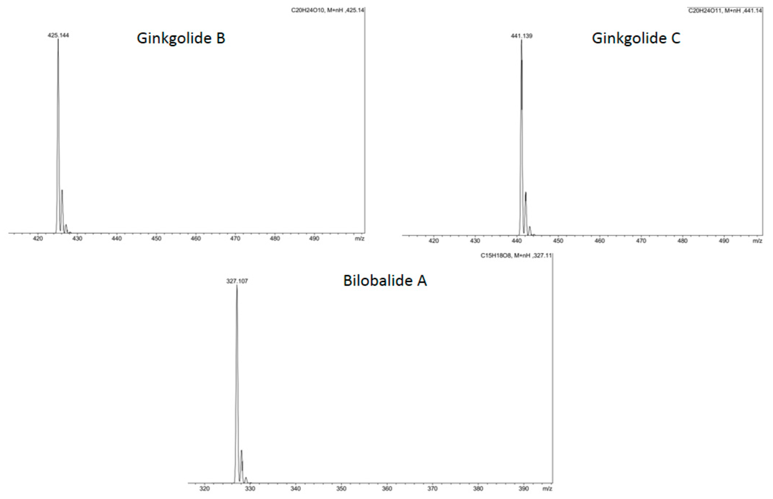
| Peak No. | Retention Time, min | Component Name | Quantitative Content, μg/mL |
|---|---|---|---|
| 1 | 4.76 ± 0.80 | Ginkgolide A | - |
| 2 | 5.16 ± 0.80 | Ginkgolide B | - |
| 3 | 6.60 ± 0.80 | Bilobalide A | 5.93 ± 0.27 |
| 4 | 6.92 ± 0.80 | Ginkgolide C | 5.23 ± 0.27 |
| 5 | 8.05 ± 0.80 | Quercetin | 11.40 ± 0.76 |
| 6 | 9.03 ± 0.80 | Ginkgetin | 0.64 ± 0.05 |
| 7 | 9.93 ± 0.80 | Isorhamnetin | 17.92 ± 0.93 |
| 8 | 13.72 ± 0.80 | Luteolin | 25.63 ± 0.86 |
| 9 | 17.49 ± 0.80 | Kaempferol | 4.63 ± 0.50 |
| 10 | 23.82 ± 0.80 | Amentoflavone | 7.91 ± 0.50 |
| Peak No. | Component Name | Substance Content in Extract *, % | Purity Degree after Purification According to HPLC *, % | Substance Yield *, Mg/Total Amount of Biologically Active Substances |
|---|---|---|---|---|
| 1 | Ginkgolide A | - | 95.3 | 14.0 |
| 2 | Ginkgolide B | - | 95.1 | 75.0 |
| 3 | Bilobalide A | 6.14 | 96.3 | 43.4 |
| 4 | Ginkgolide C | 5.42 | 95.6 | 48.3 |
| 5 | Quercetin | 11.81 | 99.8 | 93.4 |
| 6 | Ginkgetin | 0.66 | 97.1 | 3.7 |
| 7 | Isorhamnetin | 18.57 | 99.5 | 110.0 |
| 8 | Luteolin | 26.56 | 99.4 | 189.3 |
| 9 | Kaempferol | 4.79 | 98.7 | 38.4 |
| 10 | Amentoflavone | 8.19 | 96.3 | 50.3 |
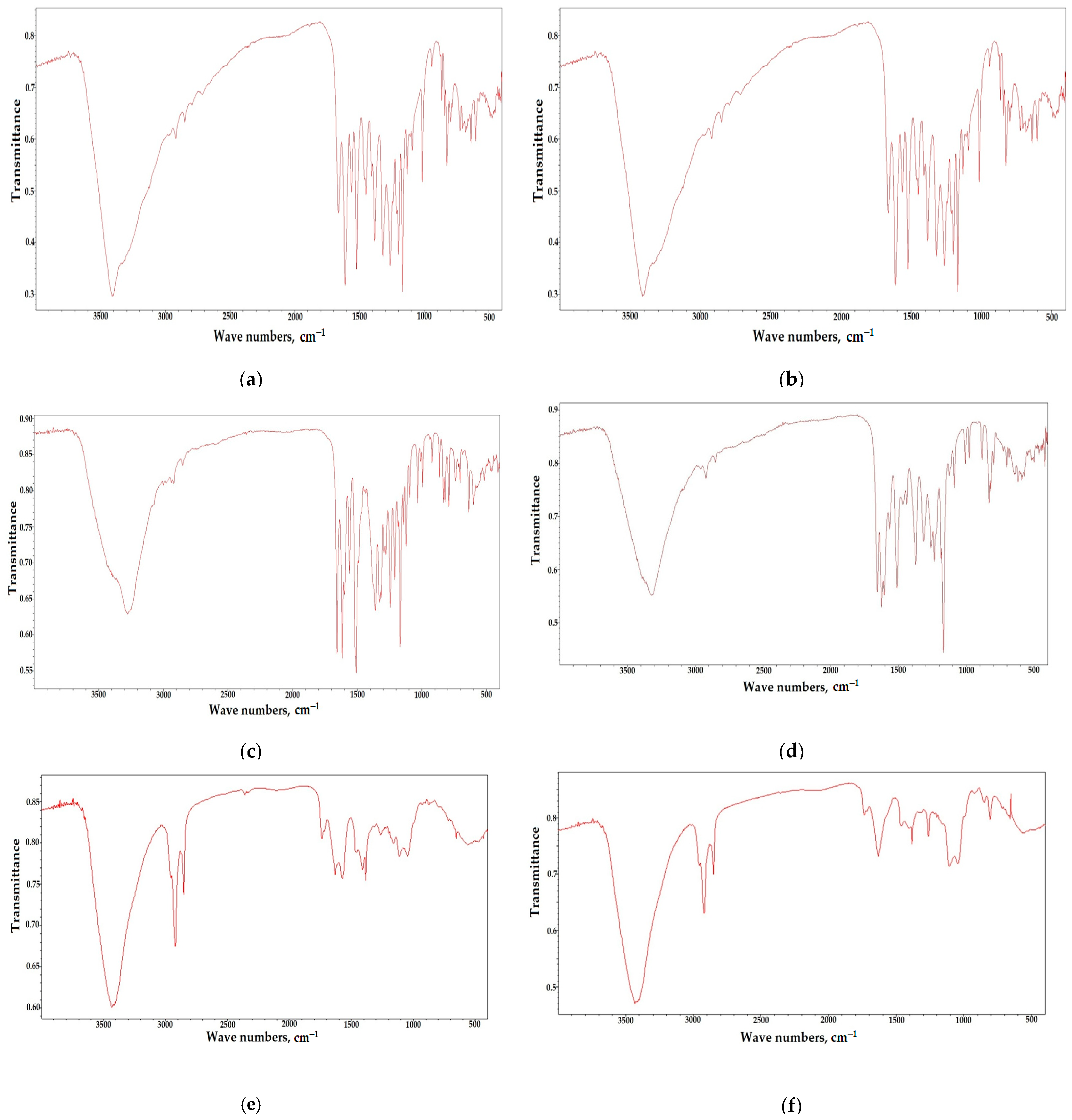
| Wave Number, cm−1 | Note |
|---|---|
| 3422 | ν (O-H) of the intramolecular H bonds |
| 2964 | ν (as) C-H в CH3 |
| 2922 | ν (as) CH2 |
| 2851 | ν (s) CH2 |
| 1735 | Lact. |
| 1631 | ν (C-O-C) |
| 1199 | ν (C–C( = O)–O) |
| 1157 | ν (C–O–C) |
| 1112 | ν (C–OH) |
| 1043 | ν (O-C-C) |
| 903 | tBut |
| Wave Number, cm−1 | Note |
|---|---|
| 3430 | ν (O-H) of the intramolecular H bonds |
| 2954 | ν(as) C-H в CH3 |
| 2921 | ν (as) CH2 |
| 2850 | ν (s) CH2 |
| 1737 | Lact. |
| 1718 | ν (C = O) |
| 1631 | ν (C-O-C) |
| 1571 | ν (C–O–C) |
| 1114 | ν (C–OH) |
| 1043 | ν (O-C-C) |
| Wave Number, cm−1 | Note |
|---|---|
| 3435 | ν (O-H) of the intramolecular H bonds |
| 2955 | ν (as) C-H в CH3 |
| 2921 | ν (as) CH2 |
| 2850 | ν (s) CH2 |
| 1737 | Lact. |
| 1716 | ν (C = O) |
| 1631 | ν (C-O-C) |
| 1107 | ν (-C( = O)-C |
| 1043 | ν (O-C-C) |
| 849 | ν (C-O-C) lact. |
| 805 | ν (-CH2-) |
| Wave Number, cm−1 | Note |
|---|---|
| 3405 | ν (O-H) of the intramolecular H bonds |
| 2969 | ν (as) C-H в CH3 |
| 2924 | ν (as) CH2 |
| 2853 | ν (s) CH2 |
| 1785 | Lact. |
| 1628 | ν (C = O) |
| 1380 | δ(O-H) |
| 1158 | ν (C–O–C) |
| 902 | δ( = C-H) |
Disclaimer/Publisher’s Note: The statements, opinions and data contained in all publications are solely those of the individual author(s) and contributor(s) and not of MDPI and/or the editor(s). MDPI and/or the editor(s) disclaim responsibility for any injury to people or property resulting from any ideas, methods, instructions or products referred to in the content. |
© 2023 by the authors. Licensee MDPI, Basel, Switzerland. This article is an open access article distributed under the terms and conditions of the Creative Commons Attribution (CC BY) license (https://creativecommons.org/licenses/by/4.0/).
Share and Cite
Le, V.; Sukhikh, A.; Larichev, T.; Ivanova, S.; Prosekov, A.; Dmitrieva, A. Isolation of the Main Biologically Active Substances and Phytochemical Analysis of Ginkgo biloba Callus Culture Extracts. Molecules 2023, 28, 1560. https://doi.org/10.3390/molecules28041560
Le V, Sukhikh A, Larichev T, Ivanova S, Prosekov A, Dmitrieva A. Isolation of the Main Biologically Active Substances and Phytochemical Analysis of Ginkgo biloba Callus Culture Extracts. Molecules. 2023; 28(4):1560. https://doi.org/10.3390/molecules28041560
Chicago/Turabian StyleLe, Violeta, Andrey Sukhikh, Timothy Larichev, Svetlana Ivanova, Alexander Prosekov, and Anastasia Dmitrieva. 2023. "Isolation of the Main Biologically Active Substances and Phytochemical Analysis of Ginkgo biloba Callus Culture Extracts" Molecules 28, no. 4: 1560. https://doi.org/10.3390/molecules28041560
APA StyleLe, V., Sukhikh, A., Larichev, T., Ivanova, S., Prosekov, A., & Dmitrieva, A. (2023). Isolation of the Main Biologically Active Substances and Phytochemical Analysis of Ginkgo biloba Callus Culture Extracts. Molecules, 28(4), 1560. https://doi.org/10.3390/molecules28041560