

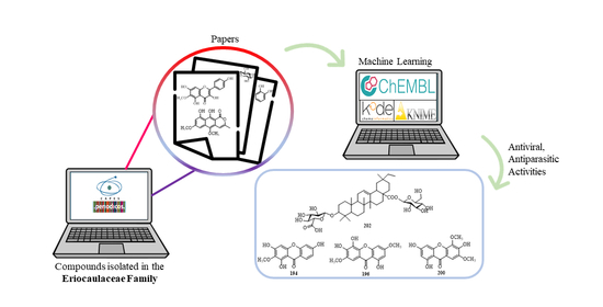
Review on Compounds Isolated from Eriocaulaceae Family and Evaluation of Biological Activities by Machine Learning
 ,
,  ,
,  ,
,  , and
, and
Abstract
1. Introduction
2. Results
2.1. Chemical Constituents of Eriocaulaceae
2.1.1. Flavonoids


| Compounds | MF | [M-H]− | Organ | Species | Reference |
|---|---|---|---|---|---|
| Quercetagetin (1) | C15H10O8 | 317 | Leaves | Eriocaulon septangulare | [29] |
| Leaves | E. brownianum | [29] | |||
| - | E. nilagirense | [29] | |||
| Leaves | E. decangulare | [29] | |||
| Leaves | E. sexangulare | [29] | |||
| Leaves | E. wightianum | [29] | |||
| Capitula | Paepalanthus polyanthus P. robustus P. ramosus | [35] | |||
| - | P. ramosus | [22] | |||
| Patuletin (2) | C16H12O8 | 331 | Leaves | E. brownianum | [29] |
| Leaves | E. truncatum | [29] | |||
| Capitula | P. planifolius | [32] | |||
| Capitula | P. polyanthus | [35] | |||
| Capitula | P. bromelioides P. latipes | [38] | |||
| Capitula | P. chlorocephalus P. argenteus var. argenteus | [52] | |||
| Capitula | P. macrocephalus | [22] | |||
| whole plant | E. buergerianum | [53] | |||
| whole plant | E. buergerianum | [30] | |||
| Capitula/ seeds | E. buergerianum E. cinereum E. faberi | [67] | |||
| - | P. macropodus | [22] | |||
| Quercetin (3) | C15H10O7 | 301 | Leaves | E. brownianum | [29] |
| Capitula | E. ligulatum | [55] | |||
| Aerial part | P. giganteus | [33] | |||
| Luteolin 7-O-glucoside (4) | C21H20O11 | 447 | Leaves | Leiothrix curvifolia var. mucronata | [31] |
| Luteolin 7-O-triglucoside (5) | - | - | Leaves | L. curvifolia L. curvifolia var. mucronata L. plantago L. sclerophylla L. vivípara | [31] |
| Luteolin 7-O-diarabinoside (6) | - | - | Leaves | L. spiralis | [31] |
| Nepetin (7) | C16H12O7 | 315 | Leaves | L. curvifolia var. curvifolia | [31] |
| Capitula | P. chlorocephalus | [52] | |||
| Leaves | P. chlorocephalus | [41] | |||
| Capitula | E. ligulatum | [47] | |||
| Nepetin 7-O-glucoside (8) | C22H22O12 | 477 | Leaves | L. curvifolia var. mucronata | [31] |
| Capitula | E. ligulatum | [47] | |||
| Nepetin 7-O-arabinoside (9) | C21H20O11 | 447 | Leaves | L. curvifolia var. curvifolia | [31] |
| Apigenin (10) | C15H10O5 | 269 | Capitula | P. hilairei | [32] |
| Capitula | L. curvifolia | [40] | |||
| Capitula | L. flavescens | [45] | |||
| Capitula | E. ligulatum | [55] | |||
| Capitula | Syngonanthus suberosus S. dealbatus E. ligulatum L. spiralis | [56] | |||
| Leaves | L. spiralis | [56] | |||
| Aerial part | E. buergerianum | [66] | |||
| Apigenin-4′-O-glucoside (11) | C21H20O10 | 431 | Capitula | P. hilairei | [32] |
| Luteolin (12) | C15H10O6 | 285 | Capitula | P. planifolius | [32] |
| Capitula | L. curvifolia | [40] | |||
| Aereal parts | S. bisulcatus | [49] | |||
| Capitula | L. flavescens | [45] | |||
| Capitula | E. ligulatum | [55] | |||
| Scapes | S. nintens | [57] | |||
| Scapes | S. macrolepis | [59] | |||
| Aerial part | S. nitens | [33] | |||
| Scapes | Syngonanthus spp. | [63] | |||
| 3-methoxyquercetin (13) | C16H12O7 | 315 | Capitula | P. bifrons | [32] |
| 3-methoxyquercetagetin (14) | C16H12O8 | 331 | Capitula | P. planifolius | [32] |
| Capitula | P. robustus | [32] | |||
| 3-methoxypatuletin (15) | C17H14O8 | 345 | Capitula | P. planifolius | [32] |
| Apigenin-7-O-galactoside (16) | C22H22O9 | 429 | Leaves | S. suberosus | [34] |
| Apigenin-7-O-glucoside (17) | C22H22O9 | 429 | Leaves | S. fuscescens S. eriopus | [34] |
| Capitula | S. suberosus S. dealbatus E. ligulatum L. spiralis | [56] | |||
| Leaves | L. spiralis | [56] | |||
| Capitula | E. australe | [62] | |||
| Aerial part | E. buergerianum | [66] | |||
| Luteolin-7-O-arabinoside (18) | C20H18O10 | 417 | Leaves | S. laricifolius S. nitens | [34] |
| Luteolin-7-O-galactoside (19) | C21H20O11 | 447 | Leaves | S. verticillatus S. elegans S. eriopus S. brasiliana S. xaranthemoides | [34] |
| Luteolin-7-O-xyloside (20) | C20H18O10 | 417 | Leaves | S. aff. mucugensis S. xaranthemoides | [34] |
| Luteolin-7-O-digalactoside (21) | - | - | Leaves | S. elegans S. elegantulus S. eriopus S. xaranthemoides | [34] |
| Luteolin-7-O-diglucoside (22) | - | - | Leaves | S. anthemidiflorus S. arenarius S. niveus S. suberosus S. brasiliana S. xaranthemoides | [34] |
| 6-hydroxyluteolin-7-O-galactoside (23) | C21H20O12 | 463 | Leaves | S. verticillatus | [34] |
| 6-hydroxyluteolin-7-O-diarabinoside (24) | - | - | Leaves | S. arenariusS. gracilisS. laricifolius | [34] |
| 6-hydroxyluteolin-7-O-digalactoside (25) | - | - | Leaves | S. verticillatus S. macrolepis S. nitens | [34] |
| 6-hydroxyluteolin-7-O-diglucoside (26) | - | - | Leaves | S. anthemidiflorus S. fuscescens S. helminthorryzus S. nitens | [34] |
| 6-methoxyquercetin-3-O-β-D-6′’-(p-coumaroyl)-glucopyranoside (27) | C31H28O15 | 639 | Capitula | P. polyanthus P. robustus P. denudatus | [35] |
| Capitula | P. denudatus | [52] | |||
| 6-methoxykaempferol-3-O-β-D-6′’-(p-coumaroyl)-glucopyranoside (28) | C31H28O14 | 623 | Capitula | P. hilairei P. robustus P. ramosus P. denudatus | [35] |
| Capitula | P. ramosus | [39] | |||
| Capitula | P. macrocephalus | [52] | |||
| Flowers | P. geniculatus | [61] | |||
| Capitula | P. chiquitensis | [64] | |||
| Quercetagetin-7-O-glucopyranoside (29) | C21H20O13 | 479 | Capitula | P. polyanthus P. hilairei P. ramosus | [35] |
| Capitula | P. bromelioides | [38] | |||
| Capitula | P. denudatus | [52] | |||
| Patuletin-3-O-glucopyranoside (30) | C22H22O13 | 493 | Capitula | P. polyanthus P. robustus | [35] |
| Capitula | P. macrocephalus P. denudatus | [52] | |||
| Aerial parts | P. polyanthus | [42] | |||
| Capitula | P. macropodus | [43] | |||
| whole plant | E. buergerianum | [53] | |||
| Flowers | P. geniculatus | [61] | |||
| Aerial parts | Tonina fluviatilis | [1] | |||
| Capitula/seeds | E. sexangulare E. buergerianum E. cinereum E. faberi | [67] | |||
| - | P. brachypy | [22] | |||
| Patuletin-3-O-rutinoside (31) | C28H32O17 | 639 | Capitula | P. polyanthus P. hilairei | [35] |
| Aerial parts | P. polyanthus | [42] | |||
| Capitula/seeds | E. buergerianum E. cinereum E. faberi | [67] | |||
| 6-methoxykaempferol (32) | C16H12O7 | 315 | Capitula | P. hilairei P. ramosus P. denudatus | [35] |
| Capitula | P. hilairei P. vellozioides | [38] | |||
| Capitula | P. chlorocephalus P. macrocephalus P. denudatus | [52] | |||
| - | P. brachypy P. vellozioides P. latipes | [22] | |||
| - | P. macropodus | [22] | |||
| 6-methoxykaempferol-3-O-glucopyranoside (33) | C22H22O12 | 477 | Capitula | P. hilairei P. robustus P. ramosus P. denudatus | [35] |
| Capitula | P. hilairei P. bromelioides | [38] | |||
| Capitula | P. ramosus | [39] | |||
| Capitula | P. denudatus | [52] | |||
| Flowers | P. geniculatus | [61] | |||
| Capitula | P. acanthophyllus | [65] | |||
| - | P. vellozioides P. latipes P. planifolius | [22] | |||
| 7-methoxyquercetagetin (34) | C16H12O8 | 331 | Leaves | P. latipes P. vellozioides | [24] |
| - | P. bromelioides | [22] | |||
| Scapes | P. latipes | [36] | |||
| Leaves | P. vellozioides P. latipes | [52] | |||
| 7-methoxyquercetagetin-3-O-β-D-glucopyranoside (35) | C22H22O13 | 493 | Leaves | P. latipes P. vellozioides | [24] |
| Leaves | P. vellozioides P. latipes | [52] | |||
| Aerial parts | T. fluviatilis | [1] | |||
| - | P. planifolius | [22] | |||
| 7-methoxyquercetagetin-4′-O-β-D-glucopyranoside (36) | C22H22O13 | 493 | Leaves | P. latipes | [24] |
| Leaves | P. vellozioides P. latipes | [52] | |||
| Scapes | P. latipes | [36] | |||
| 7-methoxyquercetagetin-3-O-cellobioside (37) | C28H32O18 | 655 | Leaves | P. latipes P. vellozioides | [24] |
| 7-methoxyquercetagetin-3-O-neohesperidoside (38) | C28H32O17 | 639 | Leaves | P. vellozioides | [24] |
| Aerial parts | P. polyanthus | [42] | |||
| 7-methoxyquercetagetin-3-O-[2-O-caffeoyl-β-D-glucopyranosyl-(1-2)-O-β-D-glucuronopyranoside (39) | C37H36O22 | 831 | Leaves | P. latipes | [24] |
| Patuletin-3-O-β-D-rutinoside (40) | C28H32O17 | 639 | Capitula | P. hilairei | [38] |
| Leaves | P. vellozioides P. latipes | [52] | |||
| whole plant | E. buergerianum | [53] | |||
| 5,7,4′-trihydroxy-6,3′-dimethoxyflavone (41) | C17H14O7 | 329 | Capitula | P. bromelioides | [38] |
| Capitula | P. argenteus var. argenteus | [52] | |||
| Leaves | P. argenteus var. argenteus | [41] | |||
| Capitula | E. australe | [62] | |||
| Aerial part | E. buergerianum | [66] | |||
| - | L. curvifolia | [22] | |||
| 5,7,4′-trihydroxy-6,3′-dimethoxyflavonol (42) | C17H14O8 | 345 | Capitula | P. bromelioides | [38] |
| 5,3′-dihydroxy-7-4′,5′-trimethoxyisoflavone (43) | C18H16O7 | 343 | Capitula | L. curvifolia | [40] |
| 3′,4′,5,6-tetrahydroxy-7-methoxyflavone (44) | C16H12O7 | 315 | Capitula | L. flavescens | [40] |
| Leaves | L. spiralis | [60] | |||
| 3′,4′,5-trihydroxy-7-methoxy-6-C-glucopyranosylflavone (45) | C22H22O11 | 461 | Capitula | L. flavescens | [40] |
| Scapes | S. nintens | [57] | |||
| Leaves | L. spiralis | [60] | |||
| Scapes | S. macrolepis | [59] | |||
| Scapes | Syngonanthus spp. | [63] | |||
| Aerial part | E. buergerianum | [66] | |||
| 4′,5-dihydroxy-3′,6-dimethoxy-7-O-β-D-glucopyranosylflavone (46) | C23H24O11 | 475 | Capitula | L. flavescens | [40] |
| Capitula | P. argenteus var. argenteus | [52] | |||
| Leaves | P. argenteus var. argenteus | [41] | |||
| whole plant | E. buergerianum | [53] | |||
| Capitula | E. australe | [62] | |||
| 3′,4′,5,7-tetrahydroxy-6-C-glucopyranosylflavone (47) | C21H20O11 | 447 | Capitula | L. flavescens | [40] |
| Scapes | S. nintens | [57] | |||
| Leaves | L. spiralis | [60] | |||
| Scapes | S. macrolepis | [59] | |||
| - | S. bisulcatus | [22] | |||
| 3′,4′,5,6,7,8-hexahydroxyflavone (48) | C15H10O8 | 317 | Capitula | L. flavescens | [40] |
| 5,6,7,8,3′,4′-hexahydroxyflavonol (49) | C15H10O9 | 333 | Aerial parts | P. polyanthus | [42] |
| Capitula | P. macropodus | [43] | |||
| 6-methoxyquercetin-3-O-(6′’-E-feruloyl)-β-D-glucopyranoside (50) | C32H30O16 | 669 | Aerial parts | P. polyanthus | [42] |
| whole plant | E. buergerianum | [53] | |||
| 6-methoxyquercetagetin-3-O-β-D-glucopyranosyl-(1→4)-glucopyranoside (51) | C28H32O18 | 655 | Capitula | P. macropodus | [43] |
| (2S)-3′,4′-methylenedioxy-5,7-dimethoxyflavan (52) | C18H18O5 | 313 | Capitula | E. buergerianum | [44] |
| Hispidulin-7-(6-E-p-coumaroyl)-β-D-glucopyranoside (53) | C31H28O13 | 607 | Capitula | E. buergerianum | [44] |
| Capitula | E. australe | [62] | |||
| Aerial part | E. buergerianum | [66] | |||
| Hispidulin (54) | C16H12O6 | 299 | Capitula | E. buergerianum | [44] |
| Capitula | E. ligulatum | [47] | |||
| Capitula | L. flavescens | [45] | |||
| whole plant | E. buergerianum | [53] | |||
| Capitula | E. ligulatum | [55] | |||
| Capitula | E. australe | [62] | |||
| Aerial part | E. buergerianum | [66] | |||
| Capitula/seeds | E. sexangulare E. buergerianum E. cinereum E. faberi | [67] | |||
| Hispidulin 7-O-glucoside (55) | C22H22O11 | 461 | Capitula | E. buergerianum | [44] |
| Capitula | E. ligulatum | [47] | |||
| Capitula | E. ligulatum | [51] | |||
| whole plant | E. buergerianum | [53] | |||
| Capitula | E. australe | [62] | |||
| Aerial part | E. buergerianum | [66] | |||
| Capitula/seeds | E. sexangulare E. buergerianum E. cinereum E. faberi | [67] | |||
| 3′,4′,5,6,7-pentahydroxyflavone (56) | C15H10O7 | 301 | Capitula | P. microphyllus | [46] |
| Scapes | S. nintens | [57] | |||
| Scapes | S. macrolepis | [59] | |||
| Scapes | Syngonanthus spp. | [63] | |||
| - | P. robustus P. planifolius P. flavescens | [22] | |||
| 3′,4′,5,6-tetrahydroxy-7-O-β-D-glucopyranosilflavone (57) | C21H20O12 | 463 | Capitula | P. microphyllus | [46] |
| Aereal parts | S. bisulcatus | [49] | |||
| - | P. planifolius | [22] | |||
| - | P. flavescens | [22] | |||
| 6-methoxyluteolin-7-O-β-D-allopyranoside (58) | C22H22O12 | 477 | Capitula | E. ligulatum | [47] |
| Capitula | E. ligulatum | [48] | |||
| 6-methoxyapigenin-7-O-β-D-allopyranoside (59) | C22H22O11 | 461 | Capitula | E. ligulatum | [47] |
| Capitula | E. ligulatum | [48] | |||
| Capitula | E. ligulatum | [51] | |||
| Capitula | E. ligulatum | [55] | |||
| 6,4′-dimethoxyapigenin-7-O-β-D-glucopyranoside (60) | C23H24O11 | 475 | Capitula | E. ligulatum | [47] |
| 5,7,8,3′,4′-pentahydroxy-6-methoxyquercetin (61) | C16H12O9 | 347 | Capitula | E. ligulatum | [47] |
| 5-hydroxy-7,4′-dimethoxy-6-C-β-D-glucopyranosylflavone (62) | C23H24O10 | 459 | Aereal parts | S. bisulcatus | [49] |
| isovitexin (5,7,4′-trihydroxy-6-C-β-D-glucopyranosylflavone) (63) | C21H20O10 | 431 | Aereal parts | S. bisulcatus | [49] |
| lutonarin (5,3′,4′-trihydroxy-6-C-7-O-β-D-glucopyranosylflavone) (64) | C27H30O16 | 609 | Aereal parts | S. bisulcatus | [49] |
| Xeractinol (65) | C21H22O12 | 465 | Leaves | P. argenteus var. argenteus | [50] |
| 6,4′-dimethoxyquercetin-3-O-β-D-6′’-[3,4,5-trihydroxy(E)-cinnamoyl]- glucopyranoside (66) | C32H32O17 | 687 | Capitula | E. ligulatum | [51] |
| 7-methoxyquercetagetin-3-O-glucopyranosyl-rhamnoside (67) | - | - | Leaves | P. vellozioides P. latipes | [52] |
| Flowers | P. geniculatus | [61] | |||
| 7-methoxyquercetagetin-3-O-[ caffeoyl]-glucopyranosyl-glucopyranoside (68) | - | - | Leaves | P. vellozioides P. latipes | [52] |
| 6,3′-dimethoxyquercetin-7-O-β-D-glucopyranoside (69) | C23H24O13 | 507 | Capitula | P. macrocephalus | [52] |
| - | P. bromelioides | [22] | |||
| Apigenin-6-C-8-C-glucopyranoside (70) | C27H30O15 | 593 | - | P. planifolius | [22] |
| 7,3′-dihydroxy-5,4′,5′-trimethoxyisoflavone (71) | C18H16O7 | 343 | whole plant | E. buergerianum | [53] |
| Capitula/seeds | E. buergerianum E. cinereum E. faberi | [67] | |||
| Patuletin-3-O-[2-O-E-feruloyl-β-D-glucopyranosyl-(1→6)-β-D-glucopyranoside] (72) | C38H40O21 | 831 | whole plant | E. buergerianum | [53] |
| Patuletin-3-O-[β-D-glucopyranosyl-(1→6)-2-O-E-caffeoyl-β-D-glucopyranosyl-(1→6)-β-D-glucopyranoside] (73) | C43H48O26 | 979 | whole plant | E. buergerianum | [53] |
| Capitula/seeds | E. buergerianum E. cinereum E. faberi | [67] | |||
| 5,7,3′-trihydroxy-6,4′,5′-trimethoxyisoflavone (74) | C18H16O7 | 343 | whole plant | E. buergerianum | [53] |
| Gerontoisoflavone A (75) | C16H12O6 | 299 | whole plant | E. buergerianum | [53] |
| 6-methoxyquercetin-7-O-β-D-glucopyranosyl-(1→6)-β-D-glucopyranoside (76) | C28H32O18 | 655 | Capitula | E. ligulatum | [55] |
| Capitula | P. chiquitensis | [64] | |||
| 6-methoxyluteolin-7-O-β-D-glucopyranosyl-(1→6)-β-D-glucopyranoside (77) | C28H32O17 | 639 | Capitula | E. ligulatum | [55] |
| 6-methoxyquercetin-7-O-(6”‘-vanilloyl)-β-D-glucopyranosyl-(1→6)-β-D-glucopyranoside (78) | C35H38O20 | 777 | Capitula | E. ligulatum | [55] |
| 6,4′-dimethoxyquercetin-7-O-β-D-glucopyranosyl-(1→6)-β-D-glucopyranoside (79) | C29H34O18 | 669 | Capitula | E. ligulatum | [55] |
| 6-methoxyquercetin-7-O-β-D-glucopyranoside (80) | C22H22O13 | 493 | Capitula | E. ligulatum | [55] |
| Scapes | P. chiquitensis | [64] | |||
| 6-methoxyquercetin-O-diglycosylrhamnoside (81) | - | - | Capitula | E. ligulatum | [56] |
| Rutin (82) | C27H30O16 | 609 | Capitula | S. suberosus S. dealbatus E. ligulatum L. spiralis | [56] |
| Leaves | L. spiralis | [56] | |||
| 5,7,4′,5′-tetrahydroxy-3′-methoxy-6-C- β-D-glucopyranosylflavone (83) | C22H22O12 | 477 | Scapes | S. nintens | [57] |
| 5,4′,5′-trihydroxy-7,3′-dimethoxy-6-C- β-D-glucopyranosylflavone (84) | C23H24O12 | 491 | Scapes | S. nintens | [57] |
| 3′,4′,5-trihydroxy-7-methoxy-6-C-glucopyranosylflavanone (85) | C22H24O11 | 463 | Scapes | S. nintens | [57] |
| 5,4′,5′-trihydroxy-7,3′-dimethoxy-8-C- β-D-glucopyranosylflavone (86) | C23H24O12 | 491 | Scapes | S. nintens | [57] |
| 3′,4′,5-trihydroxy-7-methoxy-8-C-glucopyranosylflavone (87) | C22H22O11 | 461 | Scapes | S. nintens | [57] |
| Leaves | L. spiralis | [60] | |||
| Scapes | Syngonanthus spp. | [63] | |||
| 4′,5-dihydroxy-3′,7-dimethoxy-6-C-glucopyranosylflavone (88) | C23H24O11 | 475 | Scapes | S. nintens | [57] |
| Scapes | S. macrolepis | [59] | |||
| Scapes | Syngonanthus spp. | [63] | |||
| 3′,4′,5-trihydroxy-7-methoxy-8-C-glucopyranosylflavanone (89) | C22H24O11 | 463 | Scapes | S. nintens | [57] |
| 4′,5,7-trihydroxy-8-C-glucopyranosylflavanone (90) | C21H22O10 | 433 | Scapes | S. nintens | [57] |
| 4′-methoxyluteolin-6-C-β-D-glucopyranoside (91) | C22H22O11 | 461 | Leaves | L. spiralis | [60] |
| 6-methoxykaempferol-3-O-(2,3-di-O-acetyl)-α-L-rhamnopyranosyl-(1→6)-β-D-glucopyranoside (92) | C32H36O18 | 707 | Flowers | P. geniculatus | [61] |
| 6-methoxykaempferol-3-O-(2-O-acetyl)-α-L-rhamnopyranosyl-(1→6)-β-D-glucopyranoside (93) | C30H34O17 | 665 | Flowers | P. geniculatus | [61] |
| 6-methoxykaempferol-3-O-(4-O-acetyl)-α-L-rhamnopyranosyl-(1→6)-β-D-glucopyranoside (94) | C30H34O17 | 665 | Flowers | P. geniculatus | [61] |
| 6-methoxykaempferol-3-O-α-L-rhamnopyranosyl-(1→6)-β-D-glucopyranoside (95) | C28H32O16 | 623 | Flowers | P. geniculatus | [61] |
| 6-methoxyquercetin-3-O-(2-O-acetyl)-α-L-rhamnopyranosyl-(1→6)-β-D-glucopyranoside (96) | C30H34O18 | 681 | Flowers | P. geniculatus | [61] |
| 6-methoxyquercetin-3-O-(4-O-acetyl)-α-L-rhamnopyranosyl-(1→6)-β-D-glucopyranoside (97) | C30H34O18 | 681 | Flowers | P. geniculatus | [61] |
| 6-methoxyquercetin-3-O-(2,4-di-O-acetyl)-α-L-rhamnopyranosyl-(1→6)-β-D-glucopyranoside (98) | C32H36O19 | 723 | Flowers | P. geniculatus | [61] |
| 6-methoxykaempferol-3-O-α-L-rhamnopyranosyl-(1→2)-β-D-glucopyranoside (99) | C28H32O16 | 623 | Flowers | P. geniculatus | [61] |
| 6-methoxykaempferol-3-O-(6-E-feruloyl)-β-D-glucopyranoside (100) | C32H30O15 | 653 | Flowers | P. geniculatus | [61] |
| Geniculatin (101) | C62H56O28 | 1247 | Flowers | P. geniculatus | [61] |
| Eriocaulin A (102) | C17H16O5 | 299 | Capitula | E. australe | [62] |
| 3′,4′-methylenedioxyorobol (103) | C16H10O6 | 297 | Capitula | E. australe | [62] |
| Iristectorigenin A (104) | C17H14O7 | 329 | Capitula | E. australe | [62] |
| Capitula/seeds | E. sexangulare E. buergerianum E. cinereum E. faberi | [67] | |||
| Irigenin (105) | C18H16O8 | 359 | Capitula | E. australe | [62] |
| Capitula/seeds | E. buergerianum E. cinereum E. faberi | [67] | |||
| Eriocauloside A (106) | C31H28O12 | 591 | Capitula | E. australe | [62] |
| Eriocauloside B (107) | C32H30O14 | 637 | Capitula | E. australe | [62] |
| Eriocauloside C (108) | C30H28O14 | 611 | Capitula | E. australe | [62] |
| Hispidulin-7-O-β-D-(6-O-feruloyl)-glucopyranoside (109) | C32H30O14 | 637 | Capitula | E. australe | [62] |
| Flavanonol-di-O-hexose (110) | - | 627 | Scapes | P. chiquitensis | [64] |
| Quercetin-3-O-di-hexose (111) | C27H30O17 | 625 | Capitula | P. chiquitensis | [64] |
| Capitula/seeds | E. cinereum | [67] | |||
| 6-hydroxyquercetin-3-O-di-hexose (112) | C27H30O18 | 641 | Scapes | P. chiquitensis | [64] |
| 4′-methoxyapigenin-7-O-(3-galloyl)-α-D-arabinopyranosyl-(2→1)-apiofuranosyl-(3→1)-α-D-arabinopyranoside (113) | C38H40O21 | 831 | Scapes | P. chiquitensis | [64] |
| 6,3′-dimethoxyquercetin-7-O-β-D-glucopyranosyl-(6→1)-O-β-D-glucopyranoside (114) | C29H34O18 | 669 | Capitula | P. chiquitensis | [64] |
| 6-hydroxy-7,3,4-trimethoxyflavanonol (115) | C18H18O8 | 361 | Scapes | P. chiquitensis | [64] |
| 6-metoxykaempferol-3-O-hexose-O-pentose (116) | - | 577 | Scapes | P. chiquitensis | [64] |
| 6-hydroxy-7-methoxyquercetin-3-O-pentose (117) | C21H20O12 | 463 | Scapes | P. chiquitensis | [64] |
| 7-methoxyquercetin-O-hexose (118) | C22H24O15 | 477 | Capitula | P. chiquitensis | [64] |
| 6-hydroxy-7-4-dimethoxyquercetin-3-O-hexose (119) | C23H24O13 | 507 | Capitula | P. chiquitensis | [64] |
| 6,3′-dimethoxyquercetin-3-O-β-D-6′’-(p-coumaroyl)-glucopyranoside (120) | C32H30O15 | 653 | Capitula/scapes | P. chiquitensis | [64] |
| 6,7-dimethoxyquercetin-3-O-β-D-glucopyranoside (121) | C23H24O13 | 507 | Aerial parts | T. fluviatilis | [1] |
| 6-methoxykaempferol-3-O-(6”-p-coumaroyl)-β-D-glucopyranosyl-7-O-β-D-glucopyranoside (122) | C37H38O19 | 785 | Capitula | P. acanthophyllus | [65] |
| 6-methoxykaempferol-3-7-di-O-β-D-glucopyranoside (123) | C28H32O17 | 639 | Capitula | P. acanthophyllus | [65] |
| Kaempferol-7-O-β-D-6′’-(p-coumaroyl)-glucopyranoside (124) | C30H26O12 | 577 | Aerial part | E. buergerianum | [66] |
| Patuletin-3-O-hexosyl-hexosyl-hexoside (125) | C34H42O23 | 817 | Capitula/seeds | E. buergerianumE. cinereumE. faberi | [67] |
| Quercetagetin-3-O-hexoside (126) | C21H20O13 | 479 | Capitula/seeds | E. sexangulare | [67] |
| Quercetin-3-O-hexosyl-pentoside (127) | C26H30O17 | 613 | Capitula/seeds | E. cinereum | [67] |
| 7-methoxy-hesperetin (128) | C17H16O6 | 315 | Capitula/seeds | - | [67] |
| 5,4′-dihydroxy-6,3′-dimethoxyflavone (129) | C17H14O6 | 313 | Capitula/seeds | E. sexangulare E. buergerianum E. cinereum | [67] |
| Gerontoisoflavone A (130) | C17H14O6 | 313 | Capitula/seeds | E. cinereum E. faberi | [67] |
2.1.2. Naphthopyranones


| Compounds | MF | [M−H]− | Organ | Species | Reference |
|---|---|---|---|---|---|
| Paepalantine (131) | C16H14O6 | 301 | Capitula | Paepalanthus bromelioides | [69] |
| Capitula | P. bromelioides | [72] | |||
| Capitula | P. bromelioides | [73] | |||
| Capitula | P. bromelioides | [78] | |||
| Capitula | P. bromelioides | [86] | |||
| Capitula | P. bromelioides | [87] | |||
| Capitula | P. vellozioides | [70] | |||
| Capitula | P. vellozioides | [71] | |||
| Capitula/ Scape | P. giganteus | [33] | |||
| - | P. speciosus | [22] | |||
| - | P. microphyllus | [22] | |||
| Capitula | P. acanthophyllus | [65] | |||
| Paepalantine-9-O-β-D-glucopyranoside (132) | C22H24O11 | 463 | Capitula | P. bromelioides | [74] |
| Capitula | P. bromelioides | [78] | |||
| Capitula | P. bromelioides | [38] | |||
| Capitula | P. acanthophyllus | [65] | |||
| Capitula | P. planifolius | [75] | |||
| Capitula | P. hilairei | [38] | |||
| Capitula | P. hilairei | [35] | |||
| Capitula | P. vellozioides | [38] | |||
| Capitula | P. latipes | [38] | |||
| Capitula | P. robustus | [35] | |||
| Capitula | P. ramosus | [35] | |||
| Capitula | P. denudatus | [35] | |||
| Capitula | P. denudatus | [52] | |||
| Capitula | P. macrocephalus | [52] | |||
| Capitula | P. chiquitensis | [64] | |||
| Capitula | Eriocaulon ligulatum | [47] | |||
| - | P. speciosus | [22] | |||
| - | P. microphyllus | [22] | |||
| Aerial part | P. microphyllus | [82] | |||
| Paepalantine-9-O-β-D-allopyranosyl-(1→6)-glucopyranoside (133) | C28H34O16 | 625 | Capitula | P. bromelioides | [74] |
| Capitula | P. bromelioides | [78] | |||
| Capitula | P. bromelioides | [38] | |||
| Aerial part | P. bromelioides | [37] | |||
| Capitula | P. hilairei | [38] | |||
| Capitula | P. hilairei | [35] | |||
| Capitula | P. vellozioides | [38] | |||
| Capitula | P. latipes | [38] | |||
| Capitula | P. robustus | [35] | |||
| Capitula | P. ramosus | [35] | |||
| Capitula | P. denudatus | [35] | |||
| Capitula | P. denudatus | [52] | |||
| Capitula | P. macrocephalus | [52] | |||
| Capitula | P. macropodus | [43] | |||
| Capitula | E. ligulatum | [47] | |||
| - | P. speciosus | [22] | |||
| - | P. microphyllus | [22] | |||
| 3,4-dihydro-10-hydroxy-7-methoxy-3-(R)-methyl-1H-3,4-dihydronaphtho-[2,3c]-pyran-1-one-9-O-β-D-glucopyranoside (134) | C21H24O10 | 435 | Leaves | P. vellozioides | [23] |
| Leaves | P. latipes | [23] | |||
| Aerial parts | Actinocephalus divaricatus | [79] | |||
| Whole plant | E. buergerianum | [53] | |||
| 3,4-dihydro-10-hydroxy-7-methoxy-3-(R)-methyl-1H-3,4-dihydronaphtho-[2,3c]-pyran-1-one-9-O-β-D-glucopyranosyl-(1→6)-glucopyranoside (135) | C27H34O15 | 597 | Leaves | P. vellozioides | [23] |
| Leaves | P. latipes | [23] | |||
| Whole plant | E. buergerianum | [53] | |||
| Capitula | P. planifolius | [75] | |||
| 3,4-dihydro-10-dihydroxy-7-methoxy-3-(R)-methyl-1H-3,4-dihydronaphtho-[2,3c]-pyran-1-one-9-O-β-D-allopyranosyl-(1→6)-glucopyranoside (136) | C27H34O15 | 597 | Leaves | P. vellozioides | [23] |
| Leaves | P. latipes | [23] | |||
| Aerial parts | A. divaricatus | [79] | |||
| Whole plant | E. buergerianum | [53] | |||
| Leaves | P. planifolius | [80] | |||
| Planifolin (137) | C31H26O10 | 557 | Capitula | P. planifolius | [80] |
| Paepalantine-9-O-β-D-glucopyranosyl-(1→6)-β-D-glucopyranoside (138) | C28H34O16 | 625 | Aerial part | P. microphyllus | [82] |
| Aerial part | P. bromelioides | [37] | |||
| Paepalantine-9-O-a-L-arabinopyranosyl-(1→6)-β-D-glucopyranoside (139) | C27H32O15 | 595 | Aerial part | P. microphyllus | [82] |
| Paepalantine-9-O-a-L-rhamnopyranosyl-(1→6)-β-D-glucopyranoside (140) | C28H34O15 | 609 | Aerial part | P. microphyllus | [82] |
| 8,8′-paepalantine dimer (141) | C32H28O12 | 603 | Capitula | P. bromelioides | [83] |
| Capitula | P. bromelioides | [86] | |||
| Capitula | P. bromelioides | [87] | |||
| Capitula | P. planifolius | [75] | |||
| Vioxanthin (142) | C30H26O10 | 545 | Collar | P. falcifolius | [85] |
| Collar | P. albovaginatus | [85] | |||
| Collar | P. argenteus | [85] | |||
| Collar | P. cuspidatus | [85] | |||
| Collar | P. ramosus | [85] | |||
| Capitula | P. bromelioides | [86] | |||
| Capitula | P. bromelioides | [87] | |||
| Capitula | P. planifolius | [75] | |||
| Rhizomes | P.diffissus | [88] | |||
| Eriocauline (143) | C30H22O8 | 509 | Capitula | E. ligulatum | [51] |
| 9,10-dihydroxy-7-methoxy-3-(R)-methyl-1H-naphtho[2,3c]pyran-1-one-9-O-β-D-allopyranosyl-(1→6)-glucopyranoside (144) | C27H32O15 | 595 | Capitula | E. ligulatum | [38] |
| 9,10-dihydroxy7-methoxy-3-(R)-methyl-1H-3,4-dihydronaphtho[2,3c]pyran-1-one-9-O-β-D-glucopyranoside (145) | C21H24O10 | 435 | Capitula | E. ligulatum | [38] |
| Capitula | P. planifolius | [75] | |||
| Capitula/seeds | E. buergerianum | [67] | |||
| Capitula/seeds | E. cinereum | [67] | |||
| 9,10-dihydroxy-7-methoxy-3-(R)-methyl-1Hnaphtho[2,3c]pyran-1-one-9-O-β-D-glucopyranoside (146) | C21H22O10 | 433 | Capitula | E. ligulatum | [38] |
| Whole plant | E. buergerianum | [53] | |||
| Capitula | P. denudatus | [52] | |||
| Capitula | P. chiquitensis | [64] | |||
| Scapes | P. chiquitensis | [64] | |||
| Capitula/seeds | E. sexangulare | [67] | |||
| - | P. speciosus | [22] | |||
| 5-desmethoxypaepalantine (147) | C15H12O5 | 271 | Capitula | P. macrocephalus | [52] |
| Aerial part | E. buergerianum | [66] | |||
| 5-desmethoxypaepalantine-9-O-β-D-glucopyranosyl-(1→6)-glucopyranosideo (148) | C27H32O15 | 595 | Capitula | P. macrocephalus | [52] |
| Aerial part | E. buergerianum | [66] | |||
| Capitula/seeds | E. sexangulare | [67] | |||
| Capitula/seeds | E. cinereum | [67] | |||
| Capitula/seeds | E. faberi | [67] | |||
| Paepalantine-9-O-α-D-glucopyranosyl-(1→6)-α-D-glucopyranoside (149) | C28H34O16 | 625 | Flowers | P. geniculatus | [61] |
| Paepalantine-9-O-β-D-allopyranosyl-(1→6)-α-D-glucopyranoside (150) | C28H34O16 | 625 | Flowers | P. geniculatus | [61] |
| 7,9,10-trihydroxy-5-methoxy-3-methyl-1H-naphtho[2,3-c]pyran-1-one-9-O-β-D-glucopyranosyl-(1→6)-β-D-glucopyranoside (151) | C27H32O16 | 611 | Flowers | P. geniculatus | [61] |
| Aerial parts | A. divaricatus | [79] | |||
| 7,9,10-trihydroxy-5-methoxy-3-methyl-1H-naphtho[2,3-c]pyran-1-one-9-O-β-D-glucopyranoside (152) | C21H22O11 | 449 | Flowers | P. geniculatus | [61] |
| Aerial parts | A. divaricatus | [79] | |||
| (+)-Semi-vioxanthin (153) | C15H14O5 | 273 | Rhizomes | P.diffissus | [88] |
| Capitula | P. planifolius | [75] | |||
| Capitula/seeds | E. buergerianum | [67] | |||
| Capitula/seeds | E. cinereum | [67] | |||
| Capitula/seeds | E. faberi | [67] | |||
| 5-10-Ddihydroxy-7-methoxy-3-methyl-1Hnaphtho[2,3c]pyran-1-one-9-O-α-L-rhamnopyranosyl-(1→6)-O-β-D-glucopyranoside (154) | C27H32O15 | 595 | Capitula | P. chiquitensis | [64] |
| Scapes | P. chiquitensis | [64] | |||
| 10-Hydroxy-5,7-dimethoxy-3-methyl-1H-naphtho[2,3c]pyran1-one-9-O-β-D-allopyranosyl-(1→6)-O-β-D-glucopyranoside (155) | C28H34O16 | 625 | Capitula | P. chiquitensis | [64] |
| Aerial parts | A. divaricatus | [79] | |||
| 1H-naphtho[2,3-c]pyran-1-one,3,4-dihydro-9,10-dihydroxy-5,7-dimethoxy-3-methyl (156) | Capitula | P. planifolius | [75] | ||
| Planifoliusin A (157) | C31H26O11 | 573 | Capitula | P. planifolius | [75] |
| Planifoliusin B (158) | C31H28O11 | 575 | Capitula | P. planifolius | [75] |
| Planifoliusin C (159) | C32H30O12 | 605 | Capitula | P. planifolius | [75] |
| Planifoliusin D (160) | C32H24O10 | 543 | Capitula | P. planifolius | [75] |
| Planifoliusin E (161) | C32H28O12 | 603 | Capitula | P. planifolius | [75] |
| Planifoliusin F (162) | C30H22O10 | 541 | Capitula | P. planifolius | [75] |
| Planifoliusin G (163) | C31H24O11 | 571 | Capitula | P. planifolius | [75] |
| Semivioxanthin-9-O-hexosyl-hexoside (164) | C27H34O15 | 597 | Capitula/seeds | E. sexangulare | [67] |
| Capitula/seeds | E. buergerianum | [67] | |||
| Capitula/seeds | E. cinereum | [67] | |||
| Capitula/seeds | E. faberi | [67] | |||
| (R)-Semivioxanthin-9-O-β-D-allosyl-(1→6)-glucoside (165) | C27H34O15 | 597 | Capitula/seeds | E. buergerianum | [67] |
| Capitula/seeds | E. cinereum | [67] | |||
| Capitula/seeds | E. faberi | [67] | |||
| (R)-Semivioxanthin-9-O-β-D-glucosyl-(1→6)-glucoside (166) | C27H34O15 | 597 | Capitula/seeds | E. buergerianum | [67] |
| Capitula/seeds | E. cinereum | [67] | |||
| Capitula/seeds | E. faberi | [67] |
2.1.3. Compounds Isolated from Eriocaulaceae Fungi


| Compounds | MF | [M−H]− | Organ | Species | Reference |
|---|---|---|---|---|---|
| (+)-(6R*,7S*,8R*)-6,7,8-trihydroxy- 2,2-dimethyl-5,6,7,8-tetrahydro-croman-4-one (167) | C11H16O5 | 227 | Capítulo/ Anthostomella brabeji | Paepalanthus planifolius | [92] |
| 6-hydroxy-2,2-dimethyl-5,6,7,8- tetrahydro-7,8-epoxychroman-4-one (168) | C11H14O4 | 209 | Capítulo/ Anthostomella brabeji | P. planifolius | [92] |
| Sicaine (169) | C11H10O2 | 173 | Capítulo/ Anthostomella brabeji | P. planifolius | [92] |
| Eutypinol (170) | C12H12O2 | 187 | Capítulo/ Anthostomella brabeji | P. planifolius | [92] |
| 2-(4-butylpicolinamide) acetic acid (171) | C12H16N2O3 | 235 | Parte aérea/ Fusarium fujikuroi | P. chiquitensis | [93] |
| Fusaric acid (172) | C10H13NO2 | 178 | Partes aéreas/ Fusarium fujikuroi | P. chiquitensis | [93] |
| Indoleacetic acid (173) | C10H9NO2 | 174 | Partes aéreas/ Fusarium fujikuroi | P. chiquitensis | [93] |
| Terpestacine (174) | C25H38O4 | 401 | Parte aérea/ Fusarium fujikuroi | P. chiquitensis | [93] |
| Sporulosaldeine A (175) | C12H14O5 | 237 | Folhas/ Paraphaeosphaeria sp. F03 | P. planifolius | [94] |
| Sporulosaldeine B (176) | C12H12O5 | 235 | Folhas/ Paraphaeosphaeria sp. F03 | P. planifolius | [94] |
| Sporulosaldeine C (177) | C12H14O4 | 221 | Folhas/ Paraphaeosphaeria sp. F03 | P. planifolius | [94] |
| Sporulosaldeine D (178) | C12H12O4 | 219 | Folhas/ Paraphaeosphaeria sp. F03 | P. planifolius | [94] |
| Sporulosaldeine E (179) | C12H14O4 | 221 | Folhas/ Paraphaeosphaeria sp. F03 | P. planifolius | [94] |
| Sporulosaldeine F (180) | C13H14O5 | 249 | Folhas/ Paraphaeosphaeria sp. F03 | P. planifolius | [94] |
| Triticones E (181) | C14H19NO6 | 296 | Capitula/ Curvularia lunata | P. chiquitensis | [95] |
| Triticones F (182) | C14H19NO6 | 296 | Capitula/ Curvularia lunata | P. chiquitensis | [95] |
| (1S*,2Z,3Z,3′S,6R*)-6,6′-dihydroxy-1′-methoxy-2-ropylidene-3′,4′-dihydro-2′H-spiro[cyclohexane-1,3′-cyclopenta[β]pyrrol]-3-ene-2′,5,5′(1′H)-trione (183) | C16H17NO6 | 318 | Capitula/ Curvularia lunata | P. chiquitensis | [95] |
| (1S*,2Z,3Z,3′S,6S*)-6,6′-dihydroxy-1′-methoxy-2-ropylidene-3′,4′-dihydro-2′H-spiro[cyclohexane-1,3′-cyclopenta[β]pyrrol]-3-ene-2′,5,5′(1′H)-trione (184) | C16H17NO6 | 318 | Capitula/ Curvularia lunata | P. chiquitensis | [95] |
| 5-O-methylcurvulinic acid (185) | C11H12O5 | 223 | Capitula/ Curvularia lunata | P. chiquitensis | [95] |
| Curvulinic acid (186) | C10H10O5 | 209 | Capitula/ Curvularia lunata | P. chiquitensis | [95] |
| Curvulin (187) | C12H14O5 | 237 | Capitula/ Curvularia lunata | P. chiquitensis | [95] |
2.1.4. Xanthones


| Compounds | MF | [M−H]− | Organ | Species | Reference |
|---|---|---|---|---|---|
| 8-carboxymethyl-1,6-dihydroxy-3,5-dimethoxyxanthone (188) | C17H14O8 | 345 | Capitula | Leiothrix curvifolia | [40] |
| 8-carboxymethyl-1,5,6-trihydroxy-3-methoxyxanthone (189) | C16H12O8 | 331 | Capitula | L. curvifolia | [40] |
| 8-carboxymethyl-1,3,5,6-tetrahydroxyxanthone (190) | C15H10O8 | 317 | Capitula | L. flavescens | [40] |
| 1,3,6-trihydroxy-2,5,7-trimethoxyxanthone (191) | C16H14O8 | 333 | whole plant | Eriocaulon buergerianum | [53] |
| Capitula, seeds | E. faberi | [67] | |||
| 1,3,6,8-tetrahydroxy-2,7-dimethoxyxanthone (192) | C15H12O8 | 319 | whole plant | E. buergerianum | [53] |
| vines | E. buergerianum | [54] | |||
| 1,3,6,8-tetrahydroxy-2-methoxyxanthone (193) | C14H10O7 | 289 | whole plant | E. buergerianum | [53] |
| flowers | Syngonanthus nitens | [57] | |||
| - | - | [67] | |||
| 1,3,6-trihydroxy-2-methoxyxanthone (194) | C14H10O6 | 273 | flowers | S. nitens | [57] |
| flowers | Syngonanthus ssp. | [63] | |||
| 1,3,6-trihydroxy-2,5-dimethoxyxanthone (195) | C15H12O7 | 303 | flowers | S. nitens | [57] |
| flowers | Syngonanthus ssp. | [63] | |||
| Capitula, seeds | E. buergerianum | [67] | |||
| Capitula, seeds | E. cinereum | [67] | |||
| Capitula, seeds | E. faberi | [67] | |||
| 3,4,8-trihydroxy-2,6-dimethoxyxanthone (196) | C15H12O7 | 303 | flowers | S. nitens | [57] |
| 1,3,6,8-tetrahydroxy-2,5-dimethoxyxanthone (197) | C15H12O8 | 319 | flowers | S. nitens | [57] |
| flowers | Syngonanthus ssp. | [63] | |||
| 1,3,6,8-tetrahydroxy-5-methoxyxanthone (198) | C14H10O7 | 289 | flowers | Syngonanthus ssp. | [63] |
| 1,5,7-trihydroxy-3,6-dimethoxyxanthone (199) | C15H12O7 | 303 | Capitula | S. nitens | [33] |
| flowers | Syngonanthus ssp. | [63] | |||
| 1,3,6-trihydroxy-5,7-dimethoxyxanthone (200) | C15H12O7 | 303 | Capitula, seeds | E. cinereum | [67] |
| Capitula, seeds | E. faberi | [67] | |||
| 1,3,6-trihydroxy-2,7,8-trimethoxyxanthone (201) | C16H14O8 | 333 | vines | E. buergerianum | [54] |
2.1.5. Saponins

| Compounds | MF | [M−H]− | Organ | Species | Reference |
|---|---|---|---|---|---|
| 3-O-glucuronide oleanolic acid-28-O-hexose (202) | C42H66O14 | 793 | Scapes | Actinocephalus divaricatus | [79] |
| 3-O-glucuronide-29-hydroxyoleanolic acid-28-O-hexose esther (203) | C42H66015 | 809 | Scapes | A. divaricatus | [79] |
| 3-O-[pentose-(1→3)-O-glucuronide]-30-norolean-12,20(29)-dien-28-O-hexose ester (204) | C46H70O18 | 909 | Scapes, leaves | A. divaricatus | [79] |
| 3-O-glucuronopyranosyl-30-norolean-12,20(29)-dien-28-O-glucopyranosyl ester (205) | C41H62015 | 793 | Scapes, leaves | A. divaricatus | [79] |
| 3-O-[pentose-(1→4)-glucuronide] oleanolic acid-28-O-hexose (206) | C47H74O18 | 925 | Scapes, leaves | A. divaricatus | [79] |
| 3-O-glucuronide oleanolic acid (207) | C36H56O9 | 631 | Scapes, leaves | A. divaricatus | [79] |
| 3-O-pentose-(1→3)-O-glucuronide-30-norolean-12,20(29)-dien-28-oic acid (208) | C40H60O13 | 747 | Scapes, leaves | A. divaricatus | [79] |
| 30-norolean-12,20(29)-dien-28-O-hexose ester (209) | C35H54O8 | 601 | Scapes | A. divaricatus | [79] |
2.1.6. Steroids

| Compounds | MF | [M−H]− | Organ | Species | Reference |
|---|---|---|---|---|---|
| Stigmasta-7,22-dien-3β,4β-diol (210) | C29H48O2 | 427 | whole plant | Eriocaulon sieboldianum | [100] |
| stigmasterol 3-O-β-D-glucopyranoside (211) | C35H58O6 | 573 | whole plant | E. sieboldianum | [100] |
| β-sitosterol (212) | C29H50O | 413 | whole plant | E. sieboldianum | [100] |
2.1.7. Quinones

| Compounds | MF | [M−H]− | Organ | Species | Reference |
|---|---|---|---|---|---|
| 5-methoxy-3,4-Dehydroxanthomegnin (213) | C16H12O7 | 315 | Capitula | Paepalanthus latipes | [101] |
| Emodin (214) | C15H10O5 | 269 | whole plant | Eriocaulon buergerianum | [53] |
| (R)-Semixanthomegnin (215) | C15H12O6 | 287 | Capitula/seeds | E. buergerianum | [67] |
2.1.8. Phenolic Acid Derivatives


| Compounds | MF | [M−H]− | Organ | Species | Reference |
|---|---|---|---|---|---|
| Vanillic acid (216) | C8H8O4 | 167 | whole plant | Eriocaulon buergerianum | [53] |
| Capitula, seeds | E. buergerianum | [67] | |||
| Ferulic acid (217) | C10H10O4 | 193 | whole plant | E. buergerianum | [53] |
| Protocatechuic acid (218) | C7H6O4 | 153 | whole plant | E. buergerianum | [53] |
| Capitula, seeds | E. buergerianum | [67] | |||
| Caffeic acid (219) | C9H8O4 | 180 | Capitula, seeds | E. buergerianum | [67] |
| 3-di-E-caffeoylglycerol (220) | C21H20O9 | 415 | Capitula | Paepalanthus microphyllus | [67] |
| 1,3-O-diferuloilglicerol (221) | C23H24O9 | 443 | Capitula | Leiothrix flavescens | [67] |
2.1.9. Tocopherol

2.2. Ligand-Based Virtual Screening
3. Materials and Methods
3.1. Database
3.2. Dataset
3.3. DRAGON 7.0 Descriptors
3.4. Random Forest Model
4. Conclusions
Author Contributions
Funding
Institutional Review Board Statement
Informed Consent Statement
Data Availability Statement
Conflicts of Interest
References
- Amorim, M.R.; Rinaldo, D.; Amaral, F.P.; Vilegas, W.; Magenta, M.A.G.; Vieira, G.M., Jr.; Santos, L.C. HPLC-DAD based method for the quantification of flavonoids in the hydroethanolic extract of Tonina fluviatilis Aubl. (Eriocaulaceae) and their radical scavenging activity. Quim. Nova 2014, 37, 1122–1127. [Google Scholar] [CrossRef]
- Giulietti, A.M.; Amaral, M.C.E.; Bittrich, V. Phylogenetic Analysis of Inter and Infrageneric Relationships of Leiothrix Ruhland (Eriocaulaceae). Kew Bull. 1995, 50, 55–71. [Google Scholar] [CrossRef]
- Giulietti, A.M.; Andrade, M.J.G.; Scatena, V.L.; Trovó, M.; Coan, A.I.; Sano, P.T.; Santos, F.A.R.; Borges, R.L.B.; van den Berg, C. Molecular Phylogeny, Morphology and Their Implications for the Taxonomy of Eriocaulaceae. Rodriguésia 2012, 63, 1–19. [Google Scholar] [CrossRef]
- Freitas, H.M.V.; Trovó, M. Eriocaulaceae da Região Serrana do Estado do Rio de Janeiro, Brasil. Rodriguesia 2017, 68, 749–757. [Google Scholar] [CrossRef]
- Sano, P.T.; Andrino, C.O.; Chagas, E.C.O.; Costa, F.N.; Echternacht, L.; Hensold, N.; Oliveira, A.L.R.; Parra, L.R.; Ramos, R.; Sauthier, L.J.; et al. Eriocaulaceae. Available online: http://floradobrasil.jbrj.gov.br/reflora/floradobrasil/FB110 (accessed on 18 January 2022).
- Chagas, E.C.O.; Oliveira, A.L.R.; Giulietti, A.M. Eriocaulon. Available online: http://floradobrasil.jbrj.gov.br/reflora/floradobrasil/FB7516 (accessed on 18 January 2022).
- Giulietti, A.M.; Hensold, N. Padrões de Distribuição Geográfica dos Gêneros de Eriocaulaceae. Acta Bot. Bras. 1990, 4, 133–158. [Google Scholar] [CrossRef]
- Oliveira, A.L.R.; Bove, C.P. Eriocaulon albosetaceum: A New Species of Eriocaulaceae from the Brazilian Cerrado. J. Plant Taxon. Geogr. 2019, 74, 15–21. [Google Scholar] [CrossRef]
- Costa, F.N.; Andrino, C.O.; Sano, P.T.; Trovó, M.; Echternacht, L. Paepalanthus (Eriocaulaceae) in the Central Espinhaço Range in Minas Gerais, Brazil: Checklist, Endemism, and Nomenclatural Changes. Phytotaxa 2018, 367, 133–144. [Google Scholar] [CrossRef]
- Forzza, R.C.; Leitman, P.M.; Costa, A.; Carvalho, A.A., Jr.; Peixoto, A.L.; Walter, B.M.T.; Bicudo, C.; Zappi, D.; Costa, D.P.; Lleras, E.; et al. Catálogo de Plantas e Fungos do Brasil—Volume 1; Instituto de Pesquisa Jardim Botânico do Rio de Janeiro: Rio de Janeiro, Brazil, 2010; ISBN 9788560035083. [Google Scholar]
- Andrino, C.O.; Sano, P.T.; Costa, F.N.; Echternacht, L.; Sauthier, L.J.; Hensold, N.; Ramos, R.; Tissot-Squalli, M.; Trovó, M. Paepalanthus. Available online: http://floradobrasil.jbrj.gov.br/reflora/floradobrasil/FB7558 (accessed on 18 January 2022).
- Newman, D.J.; Cragg, G.M. Natural Products as Sources of New Drugs over the Nearly Four Decades from 01/1981 to 09/2019. J. Nat. Prod. 2020, 83, 770–803. [Google Scholar] [CrossRef]
- Barros, R.P.C.; Scotti, L.; Scotti, M.T. Exploring Secondary Metabolites Database of Apocynaceae, Menispermaceae, and Annonaceae to Select Potential Anti-HCV Compounds. Curr. Top. Med. Chem. 2019, 19, 900–913. [Google Scholar] [CrossRef] [PubMed]
- Mao, J.; Akhtar, J.; Zhang, X.; Sun, L.; Guan, S.; Li, X.; Chen, G.; Liu, J.; Jeon, H.; Kim, M.S.; et al. Comprehensive Strategies of Machine-Learning-Basedquantitative Structure-Activity Relationship Models. iScience 2021, 24, 103052. [Google Scholar] [CrossRef] [PubMed]
- Du, Q.-S.; Huang, R.-B.; Chou, K.-C. Recent Advances in QSAR and Their Applications in Predicting the Activities of Chemical Molecules, Peptides and Proteins for Drug Design. Curr. Protein Pept. Sci. 2008, 9, 248–259. [Google Scholar] [CrossRef] [PubMed]
- Quadri, T.W.; Olasunkanmi, L.O.; Fayemi, O.E.; Akpan, E.D.; Verma, C.; Sherif, E.S.M.; Khaled, K.F.; Ebenso, E.E. Quantitative Structure Activity Relationship and Artificial Neural Network as Vital Tools in Predicting Coordination Capabilities of Organic Compounds with Metal Surface: A Review. Coord. Chem. Rev. 2021, 446, 2–27. [Google Scholar] [CrossRef]
- Hotez, P.J.; Bottazzi, M.E.; Franco-Paredes, C.; Ault, S.K.; Periago, M.R. The Neglected Tropical Diseases of Latin America and the Caribbean: A Review of Disease Burden and Distribution and a Roadmap for Control and Elimination. PLoS Negl. Trop. Dis. 2008, 2, e300. [Google Scholar] [CrossRef] [PubMed]
- Meireles, C.B.; Maia, L.C.; Soares, G.C.; Teodoro, I.P.P.; do Gadelha, M.S.V.; da Silva, C.G.L.; de Lima, M.A.P. Atypical Presentations of Cutaneous Leishmaniasis: A Systematic Review. Acta Trop. 2017, 172, 240–254. [Google Scholar] [CrossRef] [PubMed]
- Acharya, A.S.; Kaur, R.; Goel, A.D. Neglected Tropical Diseases—Challenges and Opportunities in India. Indian J. Med. Spec. 2017, 8, 102–108. [Google Scholar] [CrossRef]
- WHO. Schistosomiasis. Available online: https://www.who.int/en/news-room/fact-sheets/detail/schistosomiasis (accessed on 9 February 2022).
- WHO. Schistosomiasis: Progress Report 2001–2011 and Strategic Plan 2012–2020; WHO: Geneva, Switzerland, 2013.
- Dokkedal, A.L.; Santos, L.C.; Sano, P.T.; Vilegas, W. Chemistry in Eriocaulaceae. Z. Nat. 2008, 63, 169–175. [Google Scholar] [CrossRef]
- Vilegas, W.; Dokkeddal, A.L.; Piacente, S.; Rastrelli, L.; Pizza, C. New Naphthopyranone Glycosides from Paepalanthus vellozioides and Paepalanthus latipes. J. Nat. Prod. 1999, 62, 746–749. [Google Scholar] [CrossRef]
- Vilegas, W.; Nehme, C.J.; Dokkeddal, A.L.; Piacente, S.; Rastrelli, L.; Pizza, C. Quercetagetin 7-Methyl Ether Glycosides from Paepalanthus vellozioides and Paepalanthus latipes. Phytochemistry 1999, 51, 403–409. [Google Scholar] [CrossRef]
- Simões, C.M.O.; Schenkel, E.P.; Mello, J.C.P.; Mentz, L.A.; Petrovick, P.R. Farmacognosia: Do Produto Natural ao Medicamento; Artmed: Porto Alegre, Brazil, 2017. [Google Scholar]
- Shi, X.; Niu, L.; Zhao, L.; Wang, B.; Jin, Y.; Li, X. The Antiallergic Activity of Flavonoids Extracted from Citri reticulatae pericarpium. J. Food Process. Preserv. 2018, 42, e13588. [Google Scholar] [CrossRef]
- Geethalakshmi, R.; Sarada, D.V.L. In Vitro and In Silico Antimicrobial Activity of Sterol and Flavonoid Isolated from Trianthema decandra L. Microb. Pathog. 2018, 121, 77–86. [Google Scholar] [CrossRef]
- Khan, H.; Perviz, S.; Sureda, A.; Nabavi, S.M.; Tejada, S. Current Standing of Plant Derived Flavonoids as an Antidepressant. Food Chem. Toxicol. 2018, 119, 176–188. [Google Scholar] [CrossRef] [PubMed]
- Bate-Smith, E.C.; Harbornet, J.B. Quercetagetin and Patuletin in Eriocaulon. Phytochemistry 1969, 8, 1035–1037. [Google Scholar] [CrossRef]
- Zhu, W.; Chunmei, L.V.; Wang, J.; Gao, Q.; Zhu, H.; Wen, H. Patuletin Induces Apoptosis of Human Breast Cancer SK-BR-3 Cell Line via Inhibiting Fatty Acid Synthase Gene Expression and Activity. Oncol. Lett. 2017, 14, 7449–7454. [Google Scholar] [CrossRef]
- Dokkedal, A.L.; Salatino, A. Flavonoids of Brazilian Species of Leiothrix (Eriocaulaceae). Biochem. Syst. Ecol. 1992, 20, 31–32. [Google Scholar] [CrossRef]
- Mayworm, M.A.S.; Salatino, A. Flavonoides de Quatro Espécies de Paepalanthus Ruhl. (Eriocaulaceae). Acta Bot. Bras. 1993, 7, 129–133. [Google Scholar] [CrossRef]
- Cardoso, C.A.L.; Zanutto, F.V.; Varanda, E.A.; Sano, P.T.; Vilegas, W.; Santos, L.C. Quantification of Flavonoids, Naphthopyranones and Xanthones in Eriocaulaceae Species by LC-PDA. Am. J. Anal. Chem. 2012, 03, 138–146. [Google Scholar] [CrossRef]
- Ricci, C.V.; Patricio, M.C.B.; Salatino, M.L.F.; Salatino, A.; Giulietti, A.M. Flavonoids of Syngonanthus Ruhl. (Eriocaulaceae): Taxonomic Implications. Biochem. Syst. End Ecol. 1996, 24, 577–583. [Google Scholar] [CrossRef]
- Andrade, F.D.P.; Santos, L.C.; Dokkedal, A.L.; Vilegas, W. Acyl Glucosylated Flavonols from Paepalanthus Species. Phytochemistry 1999, 51, 411–415. [Google Scholar] [CrossRef]
- Moreira, R.R.D.; Santos, L.E.; Varella, S.D.; Varanda, E.A.; Vilegas, W. Avaliação Da Atividade Mutagênica Do Extrato Etanólico Bruto de Paepalanthus latipes (Eriocaulaceae) e Dos Compostos Flavonoídicos 7-Metoxilados Relacionados. Rev. Bras. Farmacogn. 2002, 12, 11–19. [Google Scholar] [CrossRef]
- Moreira, R.R.D.; Martins, G.Z.; Pietro, R.C.L.R.; Sato, D.N.; Pavan, F.R.; Leite, S.R.A.; Vilegas, W.; Leite, C.Q.F. Paepalanthus spp.: Antimycobacterial Activity of Extracts, Methoxylated Flavonoids and Naphthopyranone Fractions. Rev. Bras. Farmacogn. 2013, 23, 268–272. [Google Scholar] [CrossRef]
- Santos, L.C.; Andrade, F.D.P.; Vasconcelos, E.C.; Coelho, R.G.; Dokkedal, A.L.; Garcia, A.C.L.; Sano, P.T.; Vilegas, W. Separation of Flavonoids and Naphthopyranones from Four Brazilian Paepalanthus Species by Droplet Countercurrent Chromatography. Rev. Bras. Plantas Med. 1999, 2, 43–47. [Google Scholar]
- Santos, L.C.; Dachtler, M.; Andrade, F.D.P.; Albert, K.; Vilegas, W. Application of HPLC-NMR Coupling Using C30 Phase in the Separation and Identification of Flavonoids of Taxonomic Relevance. Fresenius J. Anal. Chem. 2000, 368, 540–542. [Google Scholar] [CrossRef]
- Santos, L.C.; Piacente, S.; Riccardis, F.; Eletto, A.M.; Pizza, C.; Vilegas, W. Xanthones and Flavonoids from Leiothrix curvifolia and Leiothrix flavescens. Phytochemistry 2001, 56, 853–856. [Google Scholar] [CrossRef]
- Dokkedal, A.L.; Sano, P.T.; Vilegas, W. Chemistry in Paepalanthus and Taxonomic Implications. Biochem. Syst. Ecol. 2004, 32, 503–504. [Google Scholar] [CrossRef]
- Santos, L.C.; Piacente, S.; Pizza, C.; Toro, R.; Sano, P.T.; Vilegas, W. 6-Methoxyquercetin-3-O-(6-E-Feruloyl)-β-D-Glucopyranoside from Paepalanthus polyanthus (Eriocaulaceae). Biochem. Syst. Ecol. 2002, 30, 451–456. [Google Scholar] [CrossRef]
- Andrade, F.; Rastrelli, L.; Pizza, C.; Sano, P.T.; Vilegas, W. Flavonol Glycosides and a Naphthopyranone Glycoside from Paepalanthus macropodus (Eriocaulaceae). Biochem. Syst. Ecol. 2002, 30, 275–277. [Google Scholar] [CrossRef]
- Ho, J.; Chen, C. Flavonoids from the Aquatic Plant Eriocaulon buergerianum. Phytochemistry 2002, 61, 405–408. [Google Scholar] [CrossRef]
- Silva, M.A. Estudo Químico e Biológico de Plantas da Família Eriocaulaceae. Ph.D. Thesis, Universidade Estadual Paulista “Júlio de Mesquita Filho” Instituto, São Paulo, Brazil, 2008. [Google Scholar]
- Santos, L.C.; Sannomiya, M.; Piacente, S.; Pìzza, C.; Sano, P.T.; Vilegas, W. Chemical Profile of the Polar Extract of Paepalanthus microphyllus (Guill.) Kunth (Eriocaulaceae). Rev. Bras. Ciências Farm. 2004, 40, 433–436. [Google Scholar] [CrossRef]
- Santos, L.C.; Rodrigues, C.M.; Silva, M.A.; Coelho, R.G.; Sannomiya, M.; Vilegas, W. Chemical Profile of Eriocaulon ligulatum (Vell.) L.B. Smith (Eriocaulaceae). Biochem. Syst. Ecol. 2005, 33, 1159–1166. [Google Scholar] [CrossRef]
- Santos, L.; Silva, M.A.; Rodrigues, C.M.; Rodrigues, J.; Rinaldo, D.; Sannomiya, M.; Vilegas, W. Fast Preparative Separation of Flavones from Capitula of Eriocaulon ligulatum (Vell.) L.B.Smith (Eriocaulaceae) by High-Speed Countercurrent Chromatography (HSCCC). Rev. Ciências Farm. Básica Apl. 2005, 26, 101–103. [Google Scholar]
- Coelho, R.G.; Batista, L.M.; Santos, L.C.; Brito, A.M.D.; Vilegas, W. Phytochemical Study and Antiulcerogenic Activity of Syngonanthus bisulcatus (Eriocaulaceae). Rev. Bras. Ciências Farm. 2006, 42, 413–417. [Google Scholar] [CrossRef]
- Dokkedal, A.L.; Lavarda, F.; Campaner, L.; Santos, L.C.; Vilegas, W. Xeractinol-A New Flavanonol C-Glucoside from Paepalanthus argenteus var. argenteus (Bongard) Hensold (Eriocaulaceae). J. Braz. Chem. Soc. 2007, 18, 437–439. [Google Scholar] [CrossRef]
- Silva, M.A.; Oliveira, A.P.S.; Sannomiya, M.; Sano, P.T.; Varanda, E.A.; Vilegas, W.S.L.C. Flavonoids and a Naphthopyranone from Eriocaulon ligulatum and Their Mutagenic Activity. Chem. Pharm. Bull. 2007, 55, 1635–1639. [Google Scholar] [CrossRef]
- Bosqueiro, A.L.D. Estudo Fitoquímico e Implicação Taxonômica Em Paepalanthus Mart. (Eriocaulaceae). Ph.D. Thesis, Universidade Estadual Paulista júlio de Mesquita Filho, Araraquara, Brazil, 2000. [Google Scholar]
- Fang, J.J.; Ye, G.; Chen, W.L.; Zhao, W.M. Antibacterial Phenolic Components from Eriocaulon buergerianum. Phytochemistry 2008, 69, 1279–1286. [Google Scholar] [CrossRef] [PubMed]
- Liang, Y.; Luo, D.; Gao, X.; Wu, H. Novel Fatty Acid Synthase Inhibitors Isolated from Eriocaulon buergerianum. Am. J. Pharm. 2018, 37, 1529–1563. [Google Scholar]
- Santos, L.C.; Silva, M.A.; Rodrigues, C.M.; Carbone, V.; Napolitano, A.; Bassarello, C.; Mari, A.; Piacente, S.; Pizza, C.; Vilegas, W. Characterization of Flavonoid and Naphthopyranone Derivatives from Eriocaulon ligulatum Using Liquid Chromatography Tandem Mass Spectrometry. Nat. Prod. Commun. 2009, 4, 1651–1656. [Google Scholar] [CrossRef]
- Silva, M.; Cardoso, C.A.; Vilegas, W.; Santos, L. High-Performance Liquid Chromatographic Quantification of Flavonoids in Eriocaulaceae Species and Their Antimicrobial Activity. Molecules 2009, 14, 4644–4654. [Google Scholar] [CrossRef]
- Pacifico, M.; Napolitano, A.; Masullo, M.; Hilario, F.; Vilegas, W.; Piacente, S.; Santos, L.C. Metabolite Fingerprint of “Capim Dourado” (Syngonanthus nitens), a Basis of Brazilian Handcrafts. Ind. Crops Prod. 2011, 33, 488–496. [Google Scholar] [CrossRef]
- Barroso, R.P.; Berlim, L.S.; Ito, A.S.; Costa-Filho, A.J. In Vitro Antioxidant Properties of Golden Grass (Syngonanthus nitens) by Electron Paramagnetic Resonance. Food Sci. Nutr. 2019, 7, 1353–1360. [Google Scholar] [CrossRef]
- Batista, L.M.; Almeida, A.B.A.; Morais Lima, G.R.; Sousa Falcão, H.; pietro Magri, L.; Luiz-Ferreira, A.; Santos, L.C.; Hiruma-Lima, C.A.; Vilegas, W.; Souza Brito, A.R.M. Gastroprotective Effects (in Rodents) of a Flavonoid Rich Fraction Obtained from Syngonanthus macrolepsis. J. Pharm. Pharmacol. 2014, 66, 445–452. [Google Scholar] [CrossRef]
- De Araújo, M.G.F.; Hilário, F.; Nogueira, L.G.; Vilegas, W.; dos Santos, L.C.; Bauab, T.M. Chemical Constituents of the Methanolic Extract of Leaves of Leiothrix spiralis Ruhland and Their Antimicrobial Activity. Molecules 2011, 16, 10479–10490. [Google Scholar] [CrossRef]
- Amaral, F.P.; Napolitano, A.; Masullo, M.; Santos, L.C.; Festa, M.; Vilegas, W.; Pizza, C.; Piacente, S. HPLC-ESIMSn Profiling, Isolation, Structural Elucidation, and Evaluation of the Antioxidant Potential of Phenolics from Paepalanthus geniculatus. J. Nat. Prod. 2012, 75, 547–556. [Google Scholar] [CrossRef] [PubMed]
- Xu, Q.; Xie, H.; Wu, P.; Wei, X. Flavonoids from the Capitula of Eriocaulon australe. Food Chem. 2013, 139, 149–154. [Google Scholar] [CrossRef] [PubMed]
- Oliveira, A.P.S.; Sousa, J.F.; Silva, M.A.; Hilário, F.; Resende, F.A.; Camargo, M.S.; Vilegas, W.; Santos, L.C.; Varanda, E.A. Estrogenic and Chemopreventive Activities of Xanthones and Flavones of Syngonanthus (Eriocaulaceae). Steroids 2013, 78, 1053–1063. [Google Scholar] [CrossRef]
- Zanutto, F.V.; Boldrin, P.K.; Varanda, E.A.; Souza, S.F.; Sano, P.T.; Vilegas, W.; Santos, L.C. Characterization of Flavonoids and Naphthopyranones in Methanol Extracts of Paepalanthus chiquitensis Herzog by HPLC-ESI-IT-MSn and Their Mutagenic Activity. Molecules 2013, 18, 244–262. [Google Scholar] [CrossRef] [PubMed]
- Ignácio, F.G. Estudo Químico e Biológico do Extrato Metanólico dos Capítulos de Paepalanthus acanthophyllus Ruhland (Eriocaulaceae). Master’s Thesis, Universidade Estadual Paulista, Araraquara, Brazil, 2016. [Google Scholar]
- Lee, I.J.; Chung, C.P.; Chang, S.J.; Lin, Y.L. Morphological and Chemical Analyses of Eriocauli flos Sold in Taiwan Markets. J. Food Drug Anal. 2017, 25, 939–945. [Google Scholar] [CrossRef] [PubMed]
- Qiao, X.; Ye, G.; Liu, C.F.; Zhang, Z.X.; Tu, Q.; Dong, J.; Li, Y.Q.; Guo, D.A.; Ye, M. Chemical Analysis of Eriocaulon buergerianum and Adulterating Species by High-Performance Liquid Chromatography with Diode Array Detection and Electrospray Ionization Tandem Mass Spectrometry. J. Pharm. Biomed. Anal. 2012, 57, 133–142. [Google Scholar] [CrossRef]
- Donner, C.D. Naphthopyranones-Isolation, Bioactivity, Biosynthesis and Synthesis. Nat. Prod. Rep. 2015, 32, 578–604. [Google Scholar] [CrossRef]
- Vilegas, W.; Roque, N.F.; Salatino, A.; Giesbrecht, A.M.; Davino, S. Isocoumarin from Paepalanthus bromeloioides. Phytochemistry 1990, 29, 2299–2301. [Google Scholar] [CrossRef]
- Varanda, E.A.; Raddi, M.S.G.; Dias, F.L.; Araújo, M.C.P.; Gibran, S.C.A.; Takahashi, C.S.; Vilegas, W. Mutagenic and Cytotoxic Activity of an Isocoumarin (Paepalantine) Isolated From Paepalanthus vellozioides. Teratog Carcinog Mutagen 1997, 17, 85–95. [Google Scholar] [CrossRef]
- Tavares, D.C.; Varanda, E.A.; Andrade, F.D.P.; Vilegas, W.; Takahashi, C.S. Evaluation of the Genotoxic Potential of the Isocoumarin Paepalantine in in Vivo and in Vitro Mammalian Systems. J. Ethnopharmacol. 1999, 68, 115–120. [Google Scholar] [CrossRef]
- Stasi, L.C.; Camuesco, D.; Nieto, A.; Vilegas, W.; Zarzuelo, A.; Galvez, J. Intestinal Anti-Inflammatory Activity of Paepalantine, an Isocoumarin Isolated from the Capitula of Paepalanthus bromelioides, in the Trinitrobenzenesulphonic Acid Model of Rat Colitis. Planta Med. 2004, 70, 315–320. [Google Scholar] [CrossRef] [PubMed]
- Damasceno, J.P.L.; Giuberti, C.S.; Gonçalves, R.C.R.; Kitagawa, R.R. Preformulation Study and Influence of DMSO and Propylene Glycol on the Antioxidant Action of Isocoumarin Paepalantine Isolated from Paepalanthus bromelioides. Rev. Bras. Farmacogn. 2015, 25, 395–400. [Google Scholar] [CrossRef]
- Vilegas, W.; Santos, L.C.; Alécio, A.C.; Pizza, C.; Piacente, S.; Pauw, E.; Sano, P.T. Naphthopyranone Glycosides from Paepalanthus bromelioides. Phytochemistry 1998, 49, 207–210. [Google Scholar] [CrossRef]
- Amorim, M.R.; Hilário, F.; Sano, P.T.; Bauab, T.M.; Santos, L.C. Antimicrobial Activity of Paepalanthus planifolius and Its Major Components against Selected Human Pathogens. J. Braz. Chem. Soc. 2018, 29, 766–774. [Google Scholar] [CrossRef]
- Devienne, K.F.; Stella, M.; Raddi, G.; Varanda, E.A.; Vilegas, W. In Vitro Cytotoxicity of Some Natural and Semi-Synthetic Isocoumarins from Paepalanthus bromelioides. Z. Nat. 2002, 57, 85–88. [Google Scholar] [CrossRef]
- Devienne, K.F.; Raddi, M.S.G.; Coelho, R.G.; Vilegas, W. Structure-Antimicrobial Activity of Some Natural Isocoumarins and Their Analogues. Phytomedicine 2005, 12, 378–381. [Google Scholar] [CrossRef]
- Leitão, G.G.; Leitão, S.G.; Vilegas, W. Quick Preparative Separation of Natural Naphthopyranones with Antioxidant Activity by High-Speed Counter-Current Chromatography. Z. Nat. 2002, 57, 1051–1055. [Google Scholar] [CrossRef]
- Zanatta, A.C.; Mari, A.; Masullo, M.; Carlos, I.Z.; Vilegas, W.; Piacente, S.; Santos, L.C. Chemical Metabolome Assay by High-Resolution Orbitrap Mass Spectrometry and Assessment of Associated Antitumoral Activity of Actinocephalus divaricatus. Rapid Commun. Mass Spectrom. 2018, 32, 241–250. [Google Scholar] [CrossRef]
- Santos, L.G.; Piacente, S.; Pizza, C.; Albert, K.; Dachtler, M.; Vilegas, W. Planifolin, a New Naphthopyranone Dimer and Flavonoids from Paepalanthus planifolius. J. Nat. Prod. 2001, 64, 122–124. [Google Scholar] [CrossRef]
- Varanda, E.A.; Varella, S.D.; Rampazo, R.A.; Kitagawa, R.R.; Raddi, M.S.G.; Vilegas, W.; Santos, L.C. Mutagenic and Cytotoxic Effect of Planifolin: A Naphthopyranone Dimer Isolated from Paepalanthus planifolius. Toxicol. In Vitro 2006, 20, 664–668. [Google Scholar] [CrossRef] [PubMed]
- Piacente, S.; Santos, L.C.; Mahmood, N.; Zampelli, A.; Pizza, C.; Vilegas, W. Naphthopyranone Glycosides from Paepalanthus microphyllus. J. Nat. Prod. 2001, 64, 680–682. [Google Scholar] [CrossRef] [PubMed]
- Coelho, R.G.; Vilegas, W.; Devienne, K.F.; Stella, M.; Raddi, G. A New Cytotoxic Naphthopyrone Dimer from Paepalanthus bromelioides. Fitoterapia 2000, 71, 497500. [Google Scholar] [CrossRef]
- Varanda, E.A.; Devienne, K.F.; Raddi, M.S.G.; Furuya, E.M.; Vilegas, W. Mutagenicity of Paepalantine Dimer and Glycoside Derivatives from Paepalanthus bromelioides. Toxicol. In Vitro 2004, 18, 109–114. [Google Scholar] [CrossRef] [PubMed]
- Provost, J.; Garcia, M. First Report on the Isolation of Vioxanthin from a Plant Source: Occurrence in Paepalanthus Species. Planta Med. 1990, 56, 647. [Google Scholar] [CrossRef]
- Cálgaro-Helena, A.F.; Devienne, K.F.; Rodrigues, T.; Dorta, D.J.; Raddi, M.S.G.; Vilegas, W.; Uyemura, S.A.; Santos, A.C.; Curti, C. Effects of Isocoumarins Isolated from Paepalanthus bromelioides on Mitochondria: Uncoupling, and Induction/Inhibition of Mitochondrial Permeability Transition. Chem. Biol. Interact. 2006, 161, 155–164. [Google Scholar] [CrossRef]
- Devienne, K.F.; Cálgaro-Helena, A.F.; Dorta, D.J.; Prado, I.M.R.; Raddi, M.S.G.; Vilegas, W.; Uyemura, S.A.; Santos, A.C.; Curti, C. Antioxidant Activity of Isocoumarins Isolated from Paepalanthus bromelioides on Mitochondria. Phytochemistry 2007, 68, 1075–1080. [Google Scholar] [CrossRef]
- Alvarado, J.G.; Abad-Reyes, J.A.; Montealegre, R.; Amaro-Luis, J.M. Naphthopyranones from Rhizomes of Paepalanthus diffissus. Av. Química 2013, 8, 131–138. [Google Scholar]
- Hassani, M.A.; Durán, P.; Hacquard, S. Microbial Interactions within the Plant Holobiont. Microbiome 2018, 6, 58. [Google Scholar] [CrossRef]
- Khalil, A.M.A.; Hassan, S.E.D.; Alsharif, S.M.; Eid, A.M.; Ewais, E.E.D.; Azab, E.; Gobouri, A.A.; Elkelish, A.; Fouda, A. Isolation and Characterization of Fungal Endophytes Isolated from Medicinal Plant Ephedra pachyclada as Plant Growth-Promoting. Biomolecules 2021, 11, 140. [Google Scholar] [CrossRef]
- Zhao, J.; Shan, T.; Mou, Y.; Zhou, L. Plant-Derived Bioactive Compounds Produced by Endophytic Fungi. Mini-Rev. Med. Chem. 2011, 11, 159–168. [Google Scholar] [CrossRef] [PubMed]
- Amorim, M.R.; Somensi, A.; Araujo, A.R.; Bonifácio, B.V.; Bauab, T.M.; Santos, L.C. Compounds of Anthostomella brabeji, an Endophytic Fungus Isolated from Paepalanthus planifolius (Eriocaulaceae). J. Braz. Chem. Soc. 2016, 27, 1048–1054. [Google Scholar] [CrossRef]
- Hilário, F.; Chapla, V.M.; Araujo, A.R.; Sano, P.T.; Bauab, T.M.; Santos, L.C. Antimicrobial Screening of Endophytic Fungi Isolated from the Aerial Parts of Paepalanthus chiquitensis (Eriocaulaceae) Led to the Isolation of Secondary Metabolites Produced by Fusarium fujikuroi. J. Braz. Chem. Soc. 2017, 28, 1389–1395. [Google Scholar] [CrossRef]
- Amorim, M.R.; Hilário, F.; Santos, F.M.; Batista, J.M.; Bauab, T.M.; Araújo, A.R.; Carlos, I.Z.; Vilegas, W.; Santos, L.C. New Benzaldehyde and Benzopyran Compounds from the Endophytic Fungus Paraphaeosphaeria sp. F03 and Their Antimicrobial and Cytotoxic Activities. Planta Med. 2019, 85, 957–964. [Google Scholar] [CrossRef] [PubMed]
- Hilario, F.; Polinário, G.; Amorim, M.R.; Batista, V.S.; Nascimento Júnior, N.M.; Araújo, A.R.; Bauab, T.M.; Santos, L.C. Spirocyclic Lactams and Curvulinic Acid Derivatives from the Endophytic Fungus Curvularia lunata and Their Antibacterial and Antifungal Activities. Fitoterapia 2020, 141, 104466. [Google Scholar] [CrossRef] [PubMed]
- Niaz, K.; Khan, F. Analysis of Polyphenolics; Elsevier Inc.: Amsterdam, The Netherlands, 2020; ISBN 9780128164556. [Google Scholar]
- Santos, L.C.; Piacente, S.; Montoro, P.; Pizza, C.; Vilegas, W. Atividade Antioxidante de Xantonas Isoladas de Espécies de Leiothrix (Eriocaulaceae). Rev. Bras. Farmacogn. 2003, 13, 67–74. [Google Scholar] [CrossRef]
- Aziz, M.M.A.; Ashour, A.S.; Melad, A.S.G. A Review on Saponins from Medicinal Plants: Chemistry, Isolation, and Determination. J. Nanomed. Res. 2019, 7, 282–288. [Google Scholar] [CrossRef]
- Cole, T.J.; Short, K.L.; Hooper, S.B. The Science of Steroids. Semin. Fetal Neonatal Med. 2019, 24, 170–175. [Google Scholar] [CrossRef]
- Song, M.-C.; Yang, H.-J.; Park, S.-K.; Choi, H.-K.; Baek, N.-I. A New Sterol from Whole Plants of Eriocaulon sieboldianum. Bull. Korean Chem. Soc. 2008, 29, 669–671. [Google Scholar]
- Kitagawa, R.R.; Raddi, M.S.G.; Santos, L.C.; Vilegas, W. A New Cytotoxic Naphthoquinone from Paepalanthus latipes. Chem. Pharm. Bull. 2004, 52, 1487–1488. [Google Scholar] [CrossRef][Green Version]
- Kitagawa, R.R.; Vilegas, W.; Carlos, I.Z.; Raddi, M.S.G. Antitumor and Immunomodulatory Effects of the Naphthoquinone 5-Methoxy-3,4-Dehydroxanthomegnin. Rev. Bras. Farmacogn. 2011, 21, 1084–1088. [Google Scholar] [CrossRef]
- Kitagawa, R.R.; Bonacorsi, C.; Fonseca, M.L.; Vilegas, W.; Raddi, M.S.G. Anti-Helicobacter pylori Activity and Oxidative Burst Inhibition by the Naphthoquinone 5-Methoxy-3,4-Dehydroxanthomegnin from Paepalanthus latipes. Braz. J. Pharmacogn. 2011, 22, 53–59. [Google Scholar] [CrossRef]
- Kitagawa, R.R.; Vilegas, W.; Varanda, E.A.; Raddi, M.S.G. Evaluation of Mutagenicity and Metabolism-Mediated Cytotoxicity of the Naphthoquinone 5-Methoxy-3,4-Dehydroxanthomegnin from Paepalanthus latipes. Rev. Bras. Farmacogn. 2015, 25, 16–21. [Google Scholar] [CrossRef]
- Kumar, N.; Goel, N. Phenolic Acids: Natural Versatile Molecules with Promising Therapeutic Applications. Biotechnol. Rep. 2019, 24, e00370. [Google Scholar] [CrossRef] [PubMed]
- Silva, M.A.; Vilegas, W.; Santos, L.C. Compostos Fenólicos e Atividade Antioxidante de Leiothrix flavescens (Bong.) Ruhland (Eriocaulaceae). Rev. Ciências Farm. Básica Apl. 2007, 28, 319–324. [Google Scholar]
- Cherkasov, A.; Muratov, E.N.; Fourches, D.; Varnek, A.; Baskin, I.I.; Cronin, M.; Dearden, J.; Gramatica, P.; Martin, Y.C.; Todeschini, R.; et al. QSAR Modeling: Where Have You Been? Where Are You Going to? J. Med. Chem. 2014, 57, 4977–5010. [Google Scholar] [CrossRef]
- Fourches, D.; Pu, D.; Tassa, C.; Weissleder, R.; Shaw, S.Y.; Mumper, R.J.; Tropsha, A. Quantitative Nanostructure—Activity Relationship Modeling. ACS Nano 2010, 4, 5703–5712. [Google Scholar] [CrossRef]
- Gaulton, A.; Bellis, L.J.; Bento, A.P.; Chambers, J.; Davies, M.; Hersey, A.; Light, Y.; McGlinchey, S.; Michalovich, D.; Al-Lazikani, B.; et al. ChEMBL: A Large-Scale Bioactivity Database for Drug Discovery. Nucleic Acids Res. 2012, 40, 101–107. [Google Scholar] [CrossRef] [PubMed]
- Willighagen, E.L.; Waagmeester, A.; Spjuth, O.; Ansell, P.; Williams, A.J.; Tkachenko, V.; Hastings, J.; Chen, B.; Wild, D.J. The ChEMBL Database as Linked Open Data. J. Cheminform. 2013, 5, 23. [Google Scholar] [CrossRef]
- Davies, M.; Nowotka, M.; Papadatos, G.; Dedman, N.; Gaulton, A.; Atkinson, F.; Bellis, L.; Overington, J.P. ChEMBL Web Services: Streamlining Access to Drug Discovery Data and Utilities. Nucleic Acids Res. 2015, 43, W612–W620. [Google Scholar] [CrossRef]
- ChemAxon Marvin. Available online: https://chemaxon.com/marvin (accessed on 20 January 2022).
- Fourches, D.; Muratov, E.; Tropsha, A. Trust, but Verify II: A Practical Guide to Chemogenomics Data Curation. J. Chem. Inf. Model. 2016, 56, 1243–1252. [Google Scholar] [CrossRef] [PubMed]
- Fourches, D.; Muratov, E.; Tropsha, A. Curation of Chemogenomics Data. Nat. Chem. Biol. 2015, 11, 535. [Google Scholar] [CrossRef] [PubMed]
- Fourches, D.; Muratov, E.; Tropsha, A. Trust, but Verify: On the Importance of Chemical Structure Curation in Cheminformatics and QSAR Modeling Research. J. Chem. Inf. Model. 2010, 50, 1189–1204. [Google Scholar] [CrossRef] [PubMed]
- Mauri, A.; Consonni, V.; Pavan, M.; Todeschini, R. DRAGON Software: An Easy Approach to Molecular Descriptor Calculations. MATCH Commun. Math. Comput. Chem. 2006, 56, 237–248. [Google Scholar]
- Berthold, M.R.; Cebron, N.; Dill, F.; Gabriel, T.R.; Meinl, T.; Ohl, P.; Thiel, K.; Wiswedel, B. KNIME-The Konstanz Information Miner Version 2.0 and Beyond. SIGKDD Explor. 2009, 11, 26–31. [Google Scholar] [CrossRef]
- Matthews, B.W. Comparison of the Predicted and Observed Secondary Structure of T4 Phage Lysozyme. Biochim. Biophys. Acta 1975, 405, 442–451. [Google Scholar] [CrossRef]
- Aptula, A.O.; Roberts, D.W. Mechanistic Applicability Domains for Nonanimal-Based Prediction of Toxicological End Points: General Principles and Application to Reactive Toxicity. Chem. Res. Toxicol. 2006, 19, 1097–1105. [Google Scholar] [CrossRef]
- Scotti, M.T.; Scotti, L.; Ishiki, H.M.; Peron, L.M.; de Rezende, L.; do Amaral, A.T. Variable-Selection Approaches to Generate QSAR Models for a Set of Antichagasic Semicarbazones and Analogues. Chemom. Intell. Lab. Syst. 2016, 154, 137–149. [Google Scholar] [CrossRef]
| Models | Parasitic Stage | Sensitivity | Specificity | Accuracy | PPV | NPV | MCC | ROC Curve Area under the Curve |
|---|---|---|---|---|---|---|---|---|
| Aedes aegypti | Larva | 0.95 | 0.75 | 0.85 | 0.82 | 0.92 | 0.72 | 0.97 |
| Leishmania amazonensis | Amastigote | 0.6 | 0.65 | 0.63 | 0.6 | 0.65 | 0.35 | 0.71 |
| Promastigote | 0.7 | 0.88 | 0.81 | 0.66 | 0.8 | 0.61 | 0.89 | |
| Leishmania infantum | Amastigote | 0.81 | 0.9 | 0.85 | 0.81 | 0.9 | 0.71 | 0.94 |
| Promastigote | 0.77 | 0.78 | 0.78 | 0.75 | 0.8 | 0.56 | 0.89 | |
| Trypanosoma cruzi | Trypomastigote | 0.79 | 0.83 | 0.81 | 0.83 | 0.8 | 0.62 | 0.86 |
| Amastigote | 0.81 | 0.78 | 0.79 | 0.79 | 0.79 | 0.59 | 0.89 | |
| Epimastigote | 0.78 | 0.70 | 0.72 | 0.76 | 0.70 | 0.44 | 0.82 | |
| Schistosoma mansoni | Adult | 0.96 | 0.86 | 0.91 | 0.83 | 0.97 | 0.81 | 0.98 |
| Models | Parasitic Stage | Applicability Domain | Active Prediction | Probability to Be Active |
|---|---|---|---|---|
| Aedes aegypti | Larva | 166 | 23 | 51–87% |
| Leishmania amazonensis | Amastigote | 131 | 57 | 52–81% |
| Promastigote | 140 | 81 | 51–88% | |
| Leishmania infantum | Amastigote | 200 | 8 | 51–66% |
| Promastigote | 148 | 122 | 51–96% | |
| Trypanosoma cruzi | Trypomastigote | 183 | - | - |
| Amastigote | 158 | 2 | 54%, 68% | |
| Epimastigote | 162 | 99 | 51–74% | |
| Schistosoma mansoni | Adult | 96 | 71 | 51–87% |
| Models | Parasitic Stage | Molecules | Probability to Be Active |
|---|---|---|---|
| Aedes aegypti | Larva | 206 196 158 194 205 | 87% 82% 80% 78% 63% |
| Leishmania amazonensis | Amastigote | 25 195 30 24 52 | 81% 81% 80% 80% 79% |
| Promastigote | 196 195 194 42 59 | 88% 84% 83% 75% 75% | |
| Leishmania infantum | Amastigote | 200 153 202 34 63 | 66% 58% 57% 54% 53% |
| Promastigote | 202 124 123 200 125 | 96% 92% 92% 91% 91% | |
| Trypanosoma cruzi | Trypomastigote | - | - |
| Amastigote | 194 196 | 69% 54% | |
| Epimastigote | 204 182 166 44 177 | 74% 70% 69% 68% 68% | |
| Schistosoma mansoni | Adult | 122 205 143 128 145 | 87% 83% 82% 82% 81% |
| Molecule | Parasitic Stage | Probability to Be Active |
|---|---|---|
| 194 | Leishmania amazonensis (amastigote) Leishmania amazonensis (promastigote) Leishmania infantum (promastigote) Trypanosoma cruzi (amastigote) Schistosoma mansoni Aedes aegypti (larva) | 77% 83% 81% 69% 62% 78% |
| 196 | L. amazonensis (promastigote) L. infantum (promastigote) T. cruzi (amastigote) T. cruzi (epimastigote) S. mansoni Ae. aegypti (larva) | 88% 81% 55% 54% 55% 82% |
| 200 | L. amazonensis (amastigote) L. infantum (amastigote) L. infantum (promastigote) Ae. Aegypti | 59% 57% 91% 54% |
| 202 | L. amazonensis (amastigote) L. infantum (amastigote) L. infantum (promastigote) Ae. Aegypti | 64% 57% 96% 57% |
| Database | Parasitic Stage | Total Molecules | Active | Inactive | ChEMBL ID |
|---|---|---|---|---|---|
| Aedes aegypti | Larva | 173 | pIC50 ≥ 4.15 | pIC50 < 4.15 | CHEMBL613468 |
| Leishmania amazonensis | Amastigote | 226 | pIC50 ≥ 5 | pIC50 < 5 | CHEMBL612877 |
| Promastigote | 576 | pIC50 ≥ 4.8 | pIC50 < 4.8 | ||
| Leishmania infantum | Amastigote | 372 | pIC50 ≥ 5.5 | pIC50 < 5.5 | CHEMBL612848 |
| Promastigote | 388 | pIC50 ≥ 4.7 | pIC50 < 4.7 | ||
| Trypanosoma cruzi | Trypomastigote | 1694 | pIC50 ≥ 5 | pIC50 < 5 | CHEMBL368 |
| Amastigote | 1157 | pIC50 ≥ 4.71 | pIC50 < 471 | ||
| Epimastigote | 1884 | pIC50 ≥ 4.7 | pIC50 < 4.7 | ||
| Schistosoma mansoni | Adult | 360 | pIC50 ≥ 6 | pIC50 < 6 | CHEMBL3563 |
Publisher’s Note: MDPI stays neutral with regard to jurisdictional claims in published maps and institutional affiliations. |
© 2022 by the authors. Licensee MDPI, Basel, Switzerland. This article is an open access article distributed under the terms and conditions of the Creative Commons Attribution (CC BY) license (https://creativecommons.org/licenses/by/4.0/).
Share and Cite
Moreira, L.L.P.F.; de Menezes, R.P.B.; Scotti, L.; Scotti, M.T.; Lacerda Júnior, V.; Borges, W.d.S. Review on Compounds Isolated from Eriocaulaceae Family and Evaluation of Biological Activities by Machine Learning. Molecules 2022, 27, 7186. https://doi.org/10.3390/molecules27217186
Moreira LLPF, de Menezes RPB, Scotti L, Scotti MT, Lacerda Júnior V, Borges WdS. Review on Compounds Isolated from Eriocaulaceae Family and Evaluation of Biological Activities by Machine Learning. Molecules. 2022; 27(21):7186. https://doi.org/10.3390/molecules27217186
Chicago/Turabian StyleMoreira, Laysa Lanes Pereira Ferreira, Renata Priscila Barros de Menezes, Luciana Scotti, Marcus Tullius Scotti, Valdemar Lacerda Júnior, and Warley de Souza Borges. 2022. "Review on Compounds Isolated from Eriocaulaceae Family and Evaluation of Biological Activities by Machine Learning" Molecules 27, no. 21: 7186. https://doi.org/10.3390/molecules27217186
APA StyleMoreira, L. L. P. F., de Menezes, R. P. B., Scotti, L., Scotti, M. T., Lacerda Júnior, V., & Borges, W. d. S. (2022). Review on Compounds Isolated from Eriocaulaceae Family and Evaluation of Biological Activities by Machine Learning. Molecules, 27(21), 7186. https://doi.org/10.3390/molecules27217186