
Optimization of an Analytical Protocol for the Extraction of Microplastics from Seafood Samples with Different Levels of Fat

 ,
,  ,
,  , and
, and
Abstract
:1. Introduction
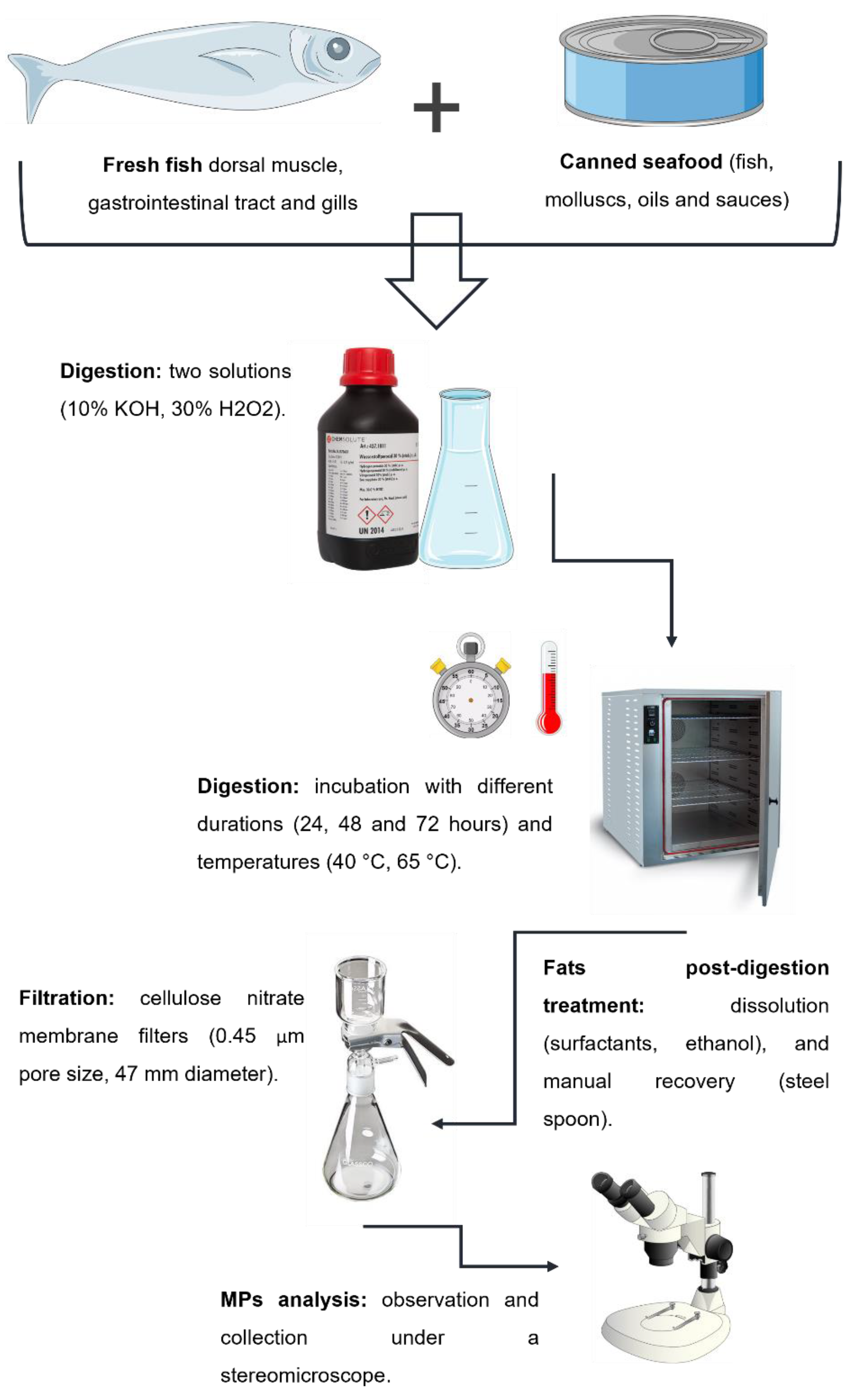
2. Methodology
2.1. Laboratorial Procedures
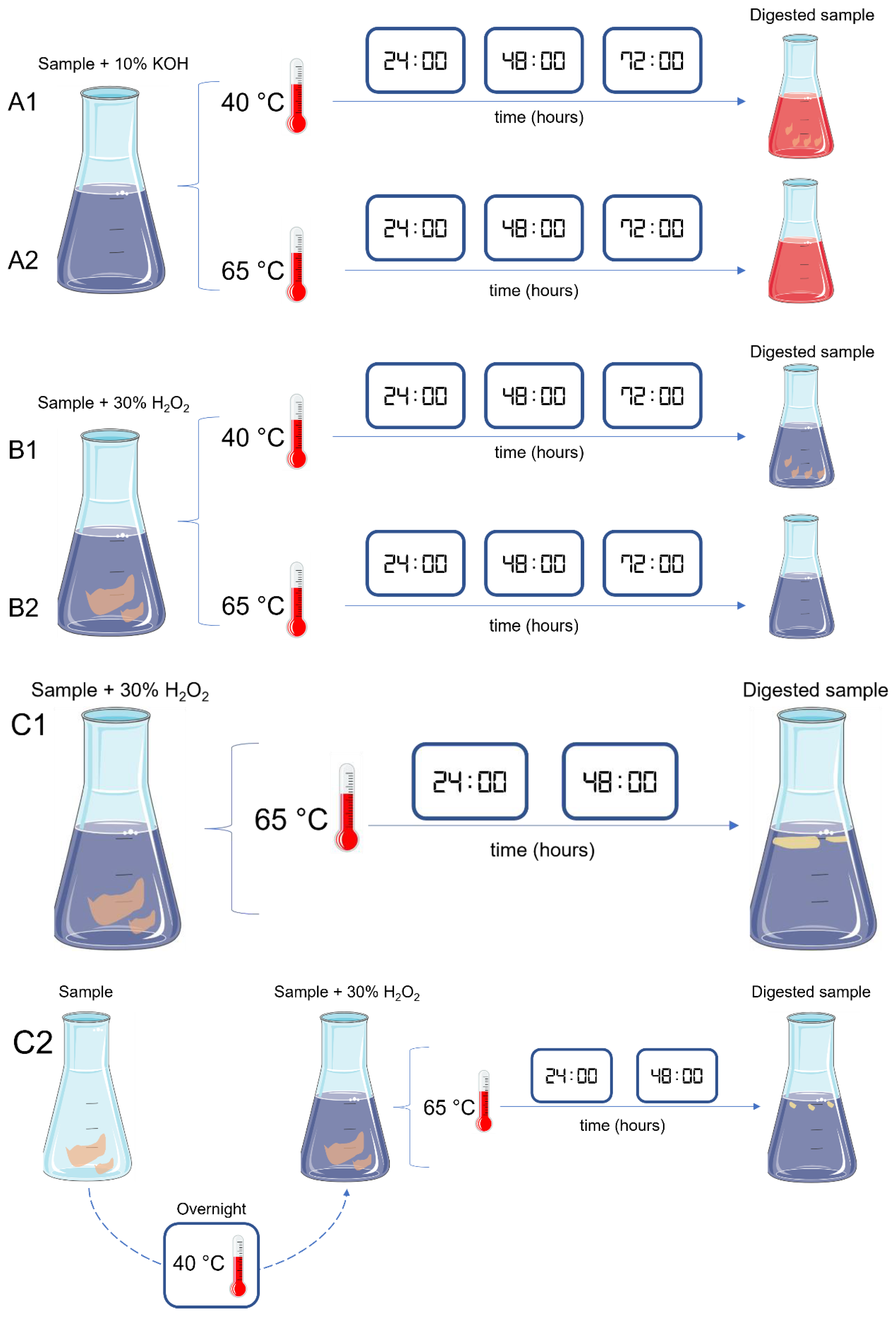
2.1.1. Sample Digestion
2.1.2. Fat Post-Digestion Treatment
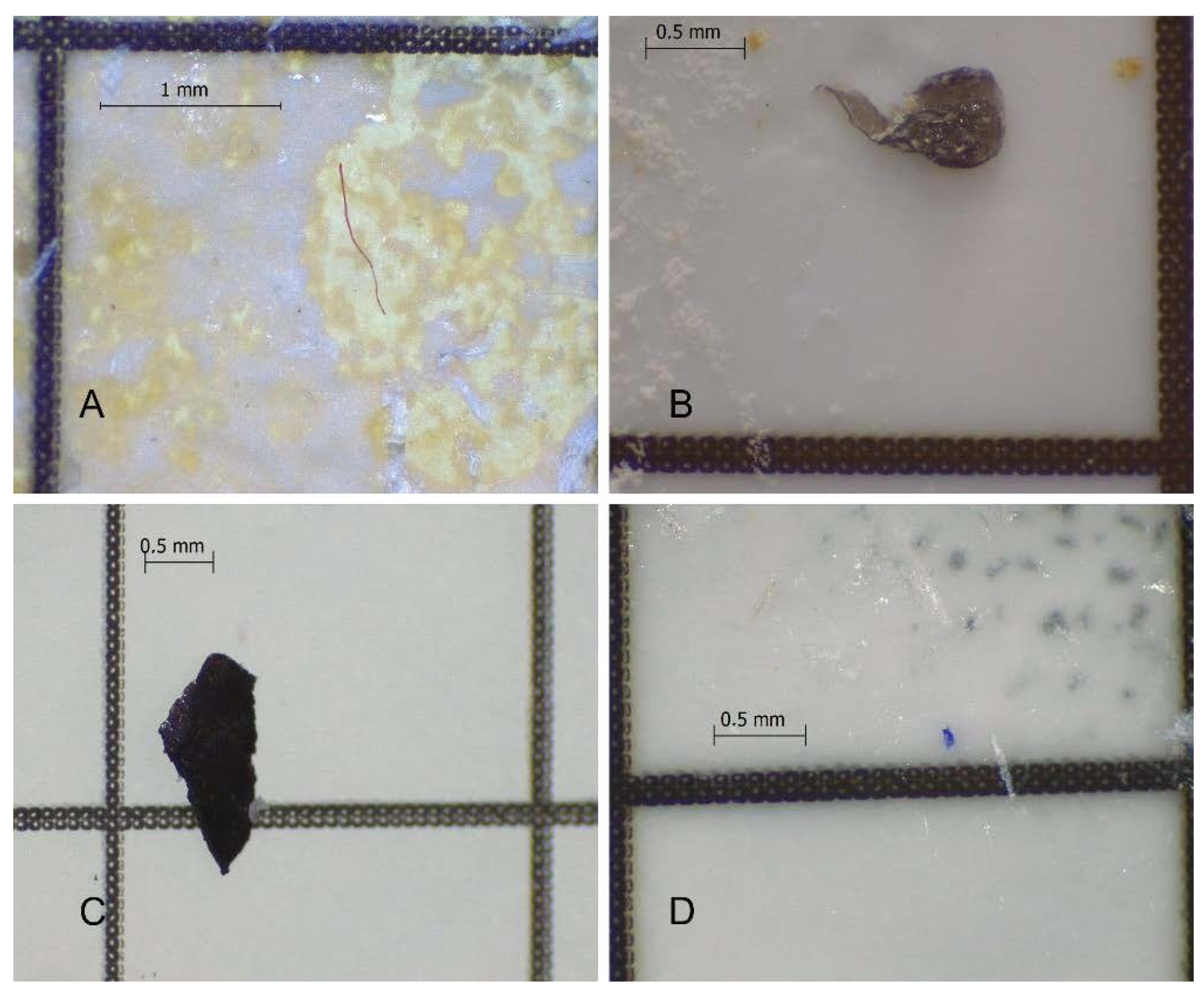
2.1.3. Filtration and Analysis of MPs
2.2. Microplastics Integrity Test
2.3. Contamination Control
3. Results
3.1. Sample Digestion

| Digestion Rate (%) | |||||
|---|---|---|---|---|---|
| Test | Sample | 24 H | 48 H | 72 H | Observations |
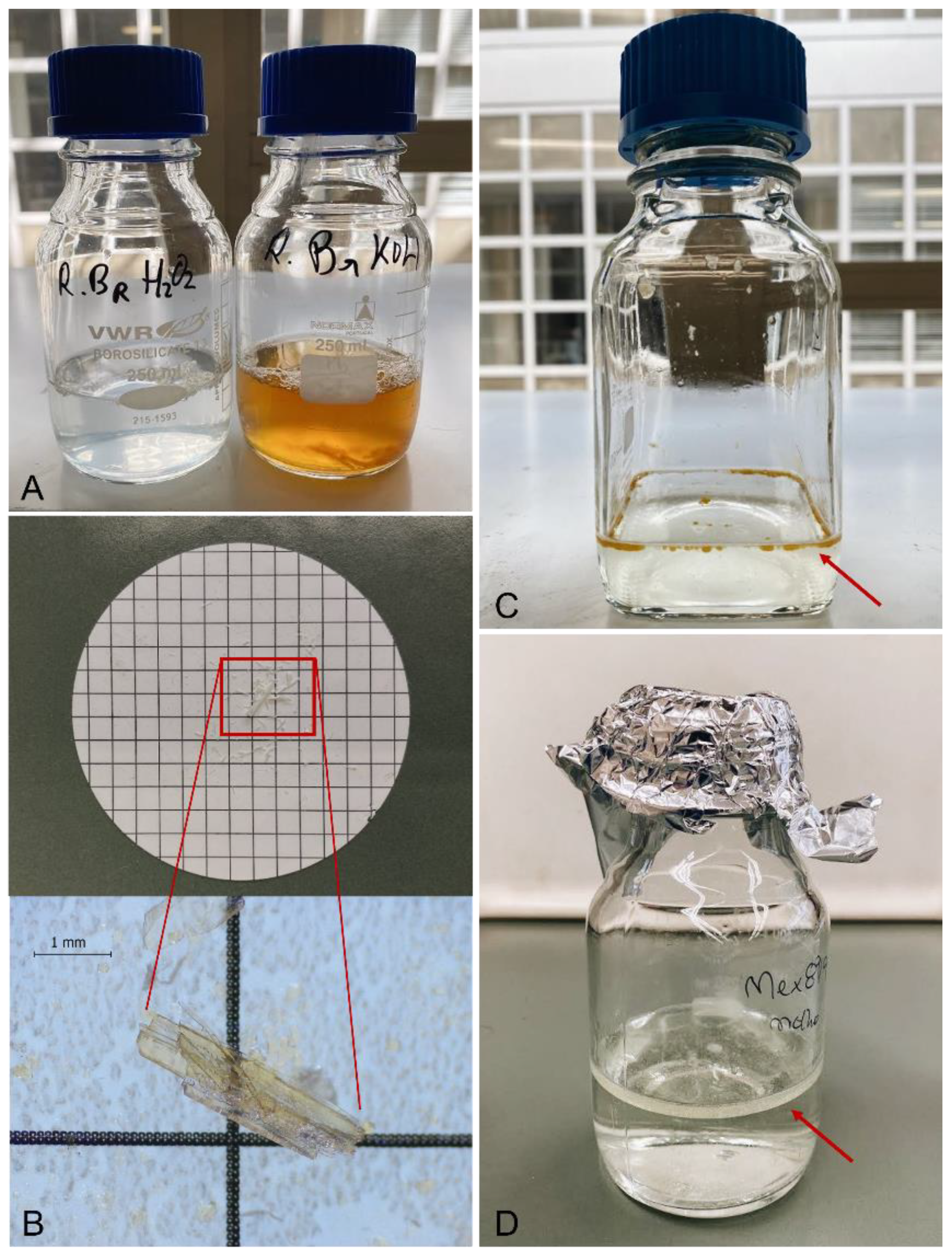
| A1 | Fresh low-fat fish muscle (n = 2) | 75 | 95 | 95 | Incomplete digestion; orange/reddish and viscous digestion liquid (Figure 4A); * bone remains (Figure 4B) |
| Fresh low-fat fish GIT (n = 2) | 50 | 75 | 95 | ||
| Fresh low-fat fish Gill (n = 2) | 50 | 75 | 95 * | ||
| A2 | Fresh low-fat fish muscle (n = 2) | 95 | 95 | 95 | Incomplete digestion; orange/reddish and viscous liquid; * bone remains |
| Fresh low-fat fish GIT (n = 2) | 75 | 95 | 95 | ||
| Fresh low-fat fish Gill (n = 2) | 75 | 95 | 95 * | ||
| B1 | Fresh low-fat fish muscle (n = 2) | 50 | 100 | 100 | Complete digestion; translucent and fluid liquid (Figure 4A); |
| Fresh low-fat fish GIT (n = 2) | 50 | 100 | 100 | ||
| Fresh low-fat fish Gill (n = 2) | 50 | 95 | 95 | Incomplete digestion: bone remains; translucent and fluid liquid | |
| B2 | Fresh low-fat fish muscle (n = 2) | 50 | 100 | 100 | Complete digestion; translucent and fluid liquid |
| Fresh low-fat fish GIT (n = 2) | 50 | 100 | 100 | ||
| Fresh low-fat fish Gill (n = 2) | 50 | 95 * | 95 * | Incomplete digestion: * bone remains; translucent and fluid liquid | |
| C1 | Fresh high-fat fish muscle (n = 4) | 50 | 100 | - | Complete digestion; fat droplets (Figure 4C) |
| Fresh high-fat fish GIT (n = 4) | 50 | 100 | - | ||
| Fresh high-fat fish Gill (n = 4) | 50 | 95 | - | Incomplete digestion: bone remains | |
| Canned fish (n = 4) | 50 | 95 | - | Incomplete digestion: bone remains; fat droplets | |
| Canned mollusc (n = 4) | 50 | 100 | - | Complete digestion; fat droplets | |
| Canned oil (n = 4) | 100 | 100 | - | Complete digestion; layer of fat (Figure 4D) | |
| Canned sauce (n = 4) | 100 | 100 | - | ||
| C2 | Fresh high-fat fish muscle (n = 4) | 50 | 100 | - | Complete digestion; smaller fat droplets |
| Fresh high-fat fish GIT (n = 4) | 50 | 100 | - | ||
| Fresh high-fat fish Gill (n = 4) | 50 | 95 | - | Incomplete digestion: bone remains | |
| Canned fish (n = 4) | 50 | 95 | - | Incomplete digestion: bone remains; smaller fat droplets | |
| Canned mollusc (n = 4) | 50 | 100 | - | Complete sample digestion; smaller fat droplets | |
| Canned oil (n = 4) | 100 | 100 | - | Complete sample digestion; smaller layer of fat | |
| Canned sauce (n = 4) | 100 | 100 | - | ||
3.2. Fat Post-Digestion Treatment
4. Microplastics Integrity Test
5. Conclusions
Author Contributions
Funding
Institutional Review Board Statement
Informed Consent Statement
Acknowledgments
Conflicts of Interest
Sample Availability
References
- Plastics Europe. Plastics–The Facts 2021. In An Analysis of European Plastics Production, Demand and Waste Data; Plastics Europe: Brussels, Belgium, 2019; p. 34. [Google Scholar]
- Arthur, C.; Baker, J.; Bamford, H. Proceedings of the International Research Workshop on the Occurrence, Effects and Fate of Microplastic Marine Debris, Tacoma, WA, USA, 9–11 September 2008; NOAA Marine Debris Division: Silver Spring, MD, USA; pp. 9–11.
- Botterell, Z.L.R.; Beaumont, N.; Dorrington, T.; Steinke, M.; Thompson, R.C.; Lindeque, P.K. Bioavailability and effects of microplastics on marine zooplankton: A review. Environ. Pollut. 2019, 245, 98–110. [Google Scholar] [CrossRef]
- Barboza, L.G.A.; Lopes, C.; Oliveira, P.; Bessa, F.; Otero, V.; Henriques, B.; Raimundo, J.; Caetano, M.; Vale, C.; Guilhermino, L. Microplastics in wild fish from North East Atlantic Ocean and its potential for causing neurotoxic effects, lipid oxidative damage, and human health risks associated with ingestion exposure. Mar. Pollut. Bull. 2020, 717, 134625. [Google Scholar] [CrossRef] [PubMed]
- Filgueiras, A.V.; Preciado, I.; Carton, A.; Gago, J. Microplastic ingestion by pelagic and benthic fish and diet composition: A case study in the NW Iberian shelf. Mar. Pollut. Bull. 2020, 160, 111623. [Google Scholar] [CrossRef]
- Gong, Y.; Wang, Y.; Chen, L.; Li, Y.; Chen, X.; Liu, B. Microplastics in different tissues of a pelagic squid (Dosidicus gigas) in the northern Humboldt Current ecosystem. Mar. Pollut. Bull. 2021, 169, 112509. [Google Scholar] [CrossRef]
- Nalbone, L.; Cincotta, F.; Giarratana, F.; Ziino, G.; Panebianco, A. Microplastics in fresh and processed mussels sampled from fish shops and large retail chains in Italy. Food Control 2021, 125, 108003. [Google Scholar] [CrossRef]
- Karami, A.; Golieskardi, A.; Choo, C.K.; Larat, V.; Karbalaei, S.; Salamatinia, B. Microplastic and mesoplastic contamination in canned sardines and sprats. Mar. Pollut. Bull. 2018, 612, 1380–1386. [Google Scholar] [CrossRef] [PubMed]
- Akhbarizadeh, R.; Dobaradaran, S.; Nabipour, I.; Tajbakhsh, S.; Darabi, A.H.; Spitz, J. Abundance, composition, and potential intake of microplastics in canned fish. Mar. Pollut. Bull. 2020, 160, 111633. [Google Scholar] [CrossRef]
- Hussien, N.A.; Mohammadein, A.; Tantawy, E.M.; Khattab, Y.; Malki, J.S.A. Investigating microplastics and potentially toxic elements contamination in canned Tuna, Salmon, and Sardine fishes from Taif markets, KSA. Open Life Sci. 2021, 16, 827–837. [Google Scholar] [CrossRef]
- Masiá, P.; Ardura, A.; Garcia-Vazquez, E. Microplastics in seafood: Relative input of Mytilus galloprovincialis and table salt in mussel dishes. Food Res. Int. 2022, 153, 110973. [Google Scholar] [CrossRef] [PubMed]
- Pan, Z.; Liu, Q.; Jiang, R.; Li, W.; Sun, X.; Lin, H.; Jiang, S.; Huang, H. Microplastic pollution and ecological risk assessment in an estuarine environment: The Dongshan Bay of China. Chemosphere 2021, 262, 127876. [Google Scholar] [CrossRef] [PubMed]
- Thilsted, S.H.; Thorne-Lyman, A.; Webb, P.; Bogard, J.R.; Subasinghe, R.; Phillips, M.J.; Allison, E.H. Sustaining healthy diets: The role of capture fisheries and aquaculture for improving nutrition in the post-2015 era. Food Policy 2016, 61, 126–131. [Google Scholar] [CrossRef]
- Hicks, C.C.; Cohen, P.J.; Graham, N.A.J.; Nash, K.L.; Allison, E.H.; D’Lima, C.; Mills, D.J.; Roscher, M.; Thilsted, S.H.; Thorne-Lyman, A.L.; et al. Harnessing global fisheries to tackle micronutrient deficiencies. Nature 2019, 574, 95–98. [Google Scholar] [CrossRef] [PubMed]
- Béné, C.; Barange, M.; Subasinghe, R.; Pinstrup-Andersen, P.; Merino, G.; Hemre, G.I.; Williams, M. Feeding 9 billion by 2050-Putting fish back on the menu. Food Secur. 2015, 7, 261–274. [Google Scholar] [CrossRef]
- Naylor, R.L.; Hardy, R.W.; Buschmann, A.H.; Bush, S.R.; Cao, L.; Klinger, D.H.; Little, D.C.; Lubchenco, J.; Shumway, S.E.; Troell, M. A 20-year retrospective review of global aquaculture. Nature 2021, 591, 551–563. [Google Scholar] [CrossRef] [PubMed]
- Mazorra-Manzano, M.A.; Ramírez-Suárez, J.C.; Moreno-Hernández, J.M.; Pacheco-Aguilar, R. Seafood proteins. In Proteins in Food Processing, 2nd ed.; Yada, R.Y., Ed.; Woodhead Publishing: Sawston, UK, 2018; pp. 445–475. [Google Scholar]
- Tsironi, T.; Houhoula, D.; Taoukis, P. Hurdle technology for fish preservation. Aquac. Fish 2022, 5, 65–71. [Google Scholar] [CrossRef]
- Moody, M.W. FISH | Processing. Encyclopedia of Food Sciences and Nutrition, 2nd ed.; Caballero, B., Ed.; Academic Press: Oxford, UK, 2003; pp. 2453–2457. [Google Scholar]
- Naseri, M.; Rezaei, M. Lipid Changes during Long-Term Storage of Canned Sprat. J. Aquat. Food Prod. Technol. 2012, 21, 48–58. [Google Scholar] [CrossRef]
- Naseri, M.; Rezaei, M.; Moieni, S.; Hosseini, H.; Eskandari, S. Effects of different filling media on the oxidation and lipid quality of canned silver carp (Hypophthalmichthys molitrix). Int. J. Food Sci. Technol. 2011, 46, 1149–1156. [Google Scholar] [CrossRef]
- FAO Fisheries and Aquaculture Department. FAO (2021) Aquaculture Regional Reviews; FAO: Rome, Italy, 2021. [Google Scholar]
- National Oceanic and Atmospheric Administration. Laboratory Methods for the Analysis of Microplastics in the Marine Environment: Recommendations for Quantifying Synthetic Particles in Waters and Sediments; National Oceanic and Atmospheric Administration: Silver Spring, MD, USA, 2015.
- Rodrigues, S.M.; Almeida, C.M.R.; Ramos, S. Adaptation of a laboratory protocol to quantity microplastics contamination in estuarine waters. MethodsX 2019, 6, 740–749. [Google Scholar] [CrossRef]
- Rivoira, L.; Castiglioni, M.; Rodrigues, S.M.; Freitas, V.; Bruzzoniti, M.C.; Ramos, S.; Almeida, C.M.R. Microplastic in marine environment: Reworking and optimisation of two analytical protocols for the extraction of microplastics from sediments and oysters. MethodsX 2021, 7, 101116. [Google Scholar] [CrossRef]
- Dehaut, A.; Cassone, A.L.; Frere, L.; Hermabessiere, L.; Himber, C.; Rinnert, G.; Rivière, G.; Lambert, C.; Soudant, P.; Huvet, A.; et al. Microplastics in seafood: Benchmark protocol for their extraction and characterization. Environ. Pollut. 2016, 215, 223–233. [Google Scholar] [CrossRef]
- Karami, A.; Golieskardi, A.; Choo, C.K.; Romano, N.; Ho, Y.B.; Salamatinia, B. A high-performance protocol for extraction of microplastics in fish. Sci. Total Environ. 2017, 578, 485–494. [Google Scholar] [CrossRef] [PubMed]
- Dawson, A.L.; Motti, C.A.; Kroon, F.J. Solving a Sticky Situation: Microplastic Analysis of Lipid-Rich Tissue. Front. Environ. Sci. 2020, 8, 563565. [Google Scholar] [CrossRef]
- Díaz-Basantes, M.F.; Nacimba-Aguirre, D.; Conesa, J.A.; Fullana, A. Presence of microplastics in commercial canned tuna. Food Chem. 2022, 385, 132721. [Google Scholar] [CrossRef] [PubMed]


| Test | Sample Pre-Treatment | Solution | Volume 1 (mL) | Duration (Hours) | Temperature (°C) | Sample 2 |
|---|---|---|---|---|---|---|
| A1 | - | 10% KOH | 50–70 | 72 | 40 | Fresh low-fat fish (muscle, GIT and gills) |
| A2 | - | 10% KOH | 50–70 | 72 | 65 | |
| B1 | - | 30% H2O2 | 50–70 | 72 | 40 | Fresh low-fat fish (muscle, GIT and gills) |
| B2 | - | 30% H2O2 | 50–70 | 72 | 65 | |
| C1 | - | 30% H2O2 | 50–70 | 48 | 65 | Fresh high-fat fish (muscle, GIT and gills); canned fish, canned mollusc, canned oil, canned sauce |
| C2 | Overnight 40 °C | 30% H2O2 | 50–70 | 48 | 65 |
| Test | Fat Treatment | Volume | Sample |
|---|---|---|---|
| D | Triton® X-100 | 2% of sample volume | Fresh high-fat fish (muscle and GIT); canned oil, canned sauce |
| E | TWEEN® 80 | 2% of sample volume | Fresh high-fat fish (muscle and GIT); canned oil, canned sauce |
| F | Commercial dish detergent | 2% of sample volume | Fresh high-fat fish (muscle and GIT); canned oil, canned sauce |
| G | 96% EtOH | Same as sample volume (1:1 v/v) | Fresh high-fat fish (muscle and GIT); canned oil, canned sauce |
| H | Removed manually | - | Fresh high-fat fish muscle and GIT; canned oil, canned sauce |
| Test | Sample | Fat Removal Rate (%) | Notes |
|---|---|---|---|
| D | Fresh high-fat fish muscle (n = 2) | 50 | Incomplete fat dissolution with formation of bubbles; difficult visualization |
| Fresh high-fat fish GIT (n = 2) | 50 | ||
| Canned oil (n = 1) | 25 | ||
| Canned sauce (n = 1) | 25 | ||
| E | Fresh high-fat fish muscle (n = 2) | 50 | Incomplete fat dissolution with formation of bubbles; difficult visualization |
| Fresh high-fat fish GIT (n = 2) | 50 | ||
| Canned oil (n = 1) | 25 | ||
| Canned sauce (n = 1) | 25 | ||
| F | Fresh high-fat fish muscle (n = 2) | 50 | Incomplete fat dissolution with formation of foam and bubbles; impossible visualization |
| Fresh high-fat fish GIT (n = 2) | 50 | ||
| Canned oil (n = 1) | 25 | ||
| Canned sauce (n = 1) | 25 | ||
| G | Fresh high-fat fish muscle (n = 2) | 75 | Incomplete fat dissolution; slow subsequent filtration |
| Fresh high-fat fish GIT (n = 2) | 75 | ||
| Canned oil (n = 1) | 25 | Incomplete fat dissolution; impossible subsequent filtration | |
| Canned sauce (n = 1) | 25 | ||
| H | Fresh high-fat fish muscle (n = 2) | 100 | Complete fat removal |
| Fresh high-fat fish GIT (n = 2) | 100 | ||
| Canned oil (n = 1) | 100 | ||
| Canned sauce (n = 1) | 100 |
| MPs Polymer | Initial Mass (g) | Final Mass (g) | Recovery Rate (%) | Observations |
|---|---|---|---|---|
| PE-LD | 0.00217 | 0.00213 | 98.2 | Color changed |
| PET | 0.00232 | 0.00225 | 96.9 | Without changes |
| PE | 0.00203 | 0.00195 | 97.5 | Without changes |
| AC | 0.00200 | 0.00204 | 102.0 | Without changes |
| PS | 0.00220 | 0.00211 | 95.9 | Without changes |
| Lycra | 0.00200 | 0.00178 | 89.0 | Without changes |
Publisher’s Note: MDPI stays neutral with regard to jurisdictional claims in published maps and institutional affiliations. |
© 2022 by the authors. Licensee MDPI, Basel, Switzerland. This article is an open access article distributed under the terms and conditions of the Creative Commons Attribution (CC BY) license (https://creativecommons.org/licenses/by/4.0/).
Share and Cite
Silva, D.M.; Almeida, C.M.R.; Guardiola, F.; Rodrigues, S.M.; Ramos, S. Optimization of an Analytical Protocol for the Extraction of Microplastics from Seafood Samples with Different Levels of Fat. Molecules 2022, 27, 5172. https://doi.org/10.3390/molecules27165172
Silva DM, Almeida CMR, Guardiola F, Rodrigues SM, Ramos S. Optimization of an Analytical Protocol for the Extraction of Microplastics from Seafood Samples with Different Levels of Fat. Molecules. 2022; 27(16):5172. https://doi.org/10.3390/molecules27165172
Chicago/Turabian StyleSilva, Diogo M., C. Marisa R. Almeida, Francisco Guardiola, Sabrina M. Rodrigues, and Sandra Ramos. 2022. "Optimization of an Analytical Protocol for the Extraction of Microplastics from Seafood Samples with Different Levels of Fat" Molecules 27, no. 16: 5172. https://doi.org/10.3390/molecules27165172
APA StyleSilva, D. M., Almeida, C. M. R., Guardiola, F., Rodrigues, S. M., & Ramos, S. (2022). Optimization of an Analytical Protocol for the Extraction of Microplastics from Seafood Samples with Different Levels of Fat. Molecules, 27(16), 5172. https://doi.org/10.3390/molecules27165172