Article

Thiosemicarbazide-Substituted Coumarins as Selective Inhibitors of the Tumor Associated Human Carbonic Anhydrases IX and XII

1
Department of Chemistry, Faculty of Science and Art, Balikesir University, 10145 Balikesir, Turkey
2
NEUROFARBA Department, Sezione di Scienze Farmaceutiche e Nutraceutiche, University of Florence, Via Ugo Schiff 6, Sesto Fiorentino, 50019 Florence, Italy
3
Computer-Aided Drug Discovery Laboratory, Department of Pharmacology, Faculty of Pharmacy, Bezmialem Vakif University, 34093 Istanbul, Turkey
*
Authors to whom correspondence should be addressed.
Molecules 2022, 27(14), 4610; https://doi.org/10.3390/molecules27144610
Submission received: 20 June 2022 / Revised: 14 July 2022 / Accepted: 17 July 2022 / Published: 19 July 2022
(This article belongs to the Special Issue Enzyme Inhibitors: Design, Synthesis and Biological Evaluation)

Abstract

:
A novel series of thiosemicarbazide-substituted coumarins was synthesized and the inhibitory effects against four physiologically relevant carbonic anhydrase isoforms I, II, IX and XII showed selective activities on the tumor-associated IX and XII isozymes. Molecular modeling studies on selected compounds 14a and 22a were performed. The binding modes of such compounds were determined assuming their enzymatically active structures (i.e., cinnamic acid) in the thermodynamically favored, and not previously explored, E geometry. Molecular modelling suggests multiple interactions within the enzymatic cavity and may explain the high potency and selectivity reported for the hCAs IX and XII.

1. Introduction

Extensive original contributions either on coumarin- and thiosemicarbazide-containing structures are reported every year within the field of Medicinal Chemistry and other disciplines not necessarily related to each other [1,2,3,4,5,6,7,8,9]. The unique features of such moieties (i.e., electronic/chemical) along with the straightforward synthetic access are the main reason for them being considered privileged scaffolds endowed with versatile biomedical applications [1,4,7,8,9]. In this context, researchers have largely contributed to the field by demonstrating that the coumarin ring effectively inhibits the α-carbonic anhydrase (CA; EC 4.2.1.1) enzymes and acts as a prodrug by making use of the lactone ring [10]. Such a discovery is of particular value since large amounts of coumarin compounds are derived from natural sources (i.e., in majority from plants) and thus de facto legitimized direct retrieval of CA inhibitors (i.e., CAIs) from the “natural chemical repository”. A variety of biological activities have been attributed to the thiosemicarbazide moiety (i.e., anti-cancer, anti-microbial, anti-viral, antioxidant properties), however a clear definition of its role in biology still remains blurred, mainly regarding the multiple and overlapping biological events triggered [7,8,9]. In this context we sought to investigate the inhibitory effects of coumarin-based CAIs functionalized with variegate thiosemicarbazide-containing tails on the physio/pathologically relevant hCA isoforms I, II, IX and XII. By means of this study we intend to present the first line of knowledge useful to understand the main structural determinants which regulate inhibition potency and selectivity of hCAs.

2. Results and Discussion

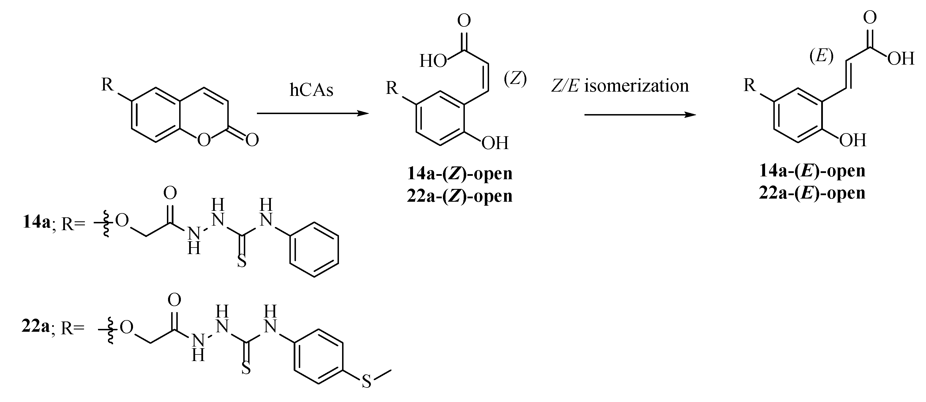
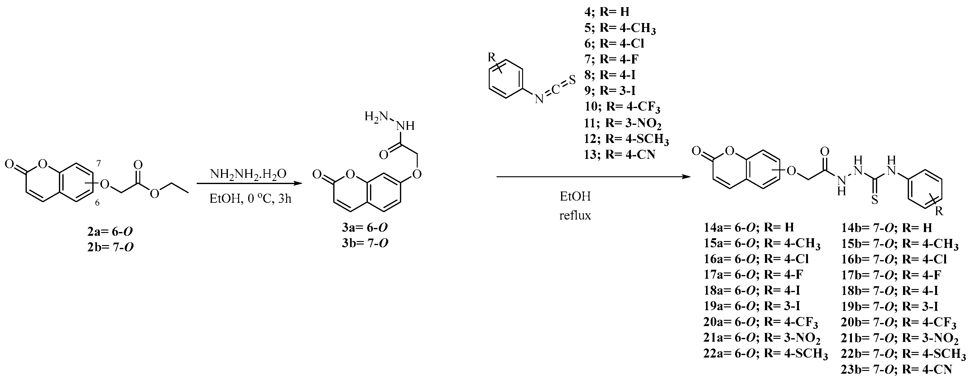
2.1. Synthesis of Coumarins 14a,b–22a,b and 23b

The target compounds were synthesized according to the procedures reported in Scheme 1.
Intermediates 2a,b and 3a,b were prepared according to previously reported experimental procedures [6,7] which were subjected to addition reactions on freshly prepared aryl isothiocyanates 413 [11] to afford the final 6- and 7-substituted coumarins 14a,b22a,b and 23b. All final compounds were purified by silica gel column chromatography or crystallization from the appropriate solvents and were fully characterized by means of 1H-NMR, 13C-NMR and mass spectra.

2.2. CA In Vitro Inhibition Assay

The synthesized compounds 14a,b22a,b and 23b were investigated in vitro for their inhibition potencies against the four physiologically relevant hCAs I, II, IX and XII, by means of the stopped flow CO2 hydrase assay [12]. Commercially available coumarin (COU) was used as a reference drug (Table 1).
As expected, and in agreement with the literature data [6], all the compounds were ineffective inhibitors of the house-keeping hCAs I and II with Ki values > 10,000 nM. Conversely well-defined structure–activity relationships (SARs) referring to the compound series on the tumor-associated hCAs IX and XII were obtained.
(i) As for the isozyme IX and within the 6-substituted coumarin series, the phenyl thioureido derivative 14a was quite an effective inhibitor, having a Ki value of 8.8 nM. Introduction of the tolyl moiety to afford 15a heavily spoiled the potency up to 26.6-fold (Ki of 234 nM). Substitution of the methyl moiety in 15a with halogens instead (i.e., F, Cl and I) restored the inhibition potencies to a medium-low nanomolar range. Specifically, the 4-chloro derivative 16a showed a Ki value of 48 nM, followed by the 4-fluoro (i.e., Ki of 17a 39 nM) and the 4-iodo which were similar to the unsubstituted derivative 14a (i.e., Kis of 9.6 and 8.8 nM for 18a and 14a respectively). Different substitutions at the same position of the phenyl ring, such as the -CF3 and the -SCH3, heavily affected the Ki values which resulted in increases of 9.6- and 5.9-fold for 20a and 22a, respectively (i.e., Kis of 92 and 57 nM 20a and 22a). Shift of the iodo in 18a to the adjacent 3-position, as in compound 19a, did not affect the inhibition potency against the hCA IX isozyme (Kis of 9.6 nM for either 18a and 19a). The presence of the nitro group at 3-position as in compound 21a determined a Ki value of 71 nM. Discrete Ki value differences were observed when the 7-coumarin substituted series was compared. For instance, the unsubstituted derivative 14b was 5.2-fold less effective than its regioisomer 14a (i.e., Kis of 8.8 and 46 nM for 14a and 14b respectively). The introduction of the methyl moiety in 14b to afford 15b determined a kinetic trend similar to the 6-substituted coumarin series, although only a slight increase of the Ki value was reported (i.e., 1.7-fold). Interestingly, the inhibition potency for the 4-substituted halogen derivatives 16b18b was the opposite when compared to the 6-substituted counterparts 16a18a (Kis of 55, 64 and 721 nM for 16b, 17b and 18b, respectively). No appreciable differences were observed between the regioisomeric pairs 19a and 19b (i.e., Kis of 9.6 and 9.4 nM for 19a and 19b respectively) and the 3-nitro derivative 21b (Ki of 9.1 nM). Close matching differences were observed between the 4-CF3 and 4-SCH3 pairs, being 20b and 22b 1.3- and 1.7-fold more potent than their counterparts 20a and 22a, respectively. Finally, the 4-cyano derivative 23b was the least effective among the entire series with a Ki value of 437.5 nM.
(ii) Overall, the compounds reported in this study were less effective inhibitors against the second tumor associated hCA XII isoform. The phenyl unsubstituted pairs 14a and 14b were equally potent inhibitors, with Kis of 739 and 743.5 nM, respectively. Similarly, the tolyl-containing compounds 15a and 15b showed very close inhibition potencies (i.e., Kis of 709 and 684 nM for 15a and 15b, respectively). The halogen-containing derivatives 16a18a exerted inhibition of the hCA XII at high nanomolar concentrations with kinetic trends identical to the isoform IX (Table 1). Of note is the 4-iodo derivative 18a which was equally potent on both the tumor-associated isoforms IX/XII (i.e., Kis of 9.6 and 8.4 nM for 18a on hCA IX and XII, respectively). Strong regioisomeric effects were observed when the 4-iodophenyl tail in 18a was replaced with the 3-iodophenyl moiety to afford 19a (i.e., Kis of 8.4 and 79 nM for 18a and 19a, respectively). Replacement of the 3-iodo (19a) with a nitro group (21a) drastically increased the Ki value (i.e., 328.5 nM). The presence at 4-position of the -SCH3 group drastically enhanced the inhibition potency for both the 6- and 7-substituted coumarin pairs 22a and 22b, which showed Kis of 4 and 4.6 nM, respectively. The kinetic trends of 16b18b were identical to their 16a18a counterparts, with the chloro derivative 16b the least effective (Ki of 866 nM), followed by the fluoro- and iodo-containing compounds 17b and 18b with Kis of 745 and 7.5 nM, respectively. In analogy to 18a/19a, a switch of the halogen to 3-position within the 18b/19b pair spoiled the inhibition potency against the hCA XII isoforms (i.e., Kis of 7.5 and 573 nM for 18b and 19b respectively). Interestingly, the 4-CF3-phenyl derivatives 20a and 20b showed a kinetic trend on the hCA XII comparable to hCA IX isozyme. Enhanced regioisomeric effects were observed for the hCA XII, with 20b and 21b up to 5.4- and 38.2-fold more effective than 20a and 21a, respectively. Finally, also for the hCA XII isozyme the 4-CN derivative 23b was a high nanomolar inhibitor (i.e., Ki of 473 nM).

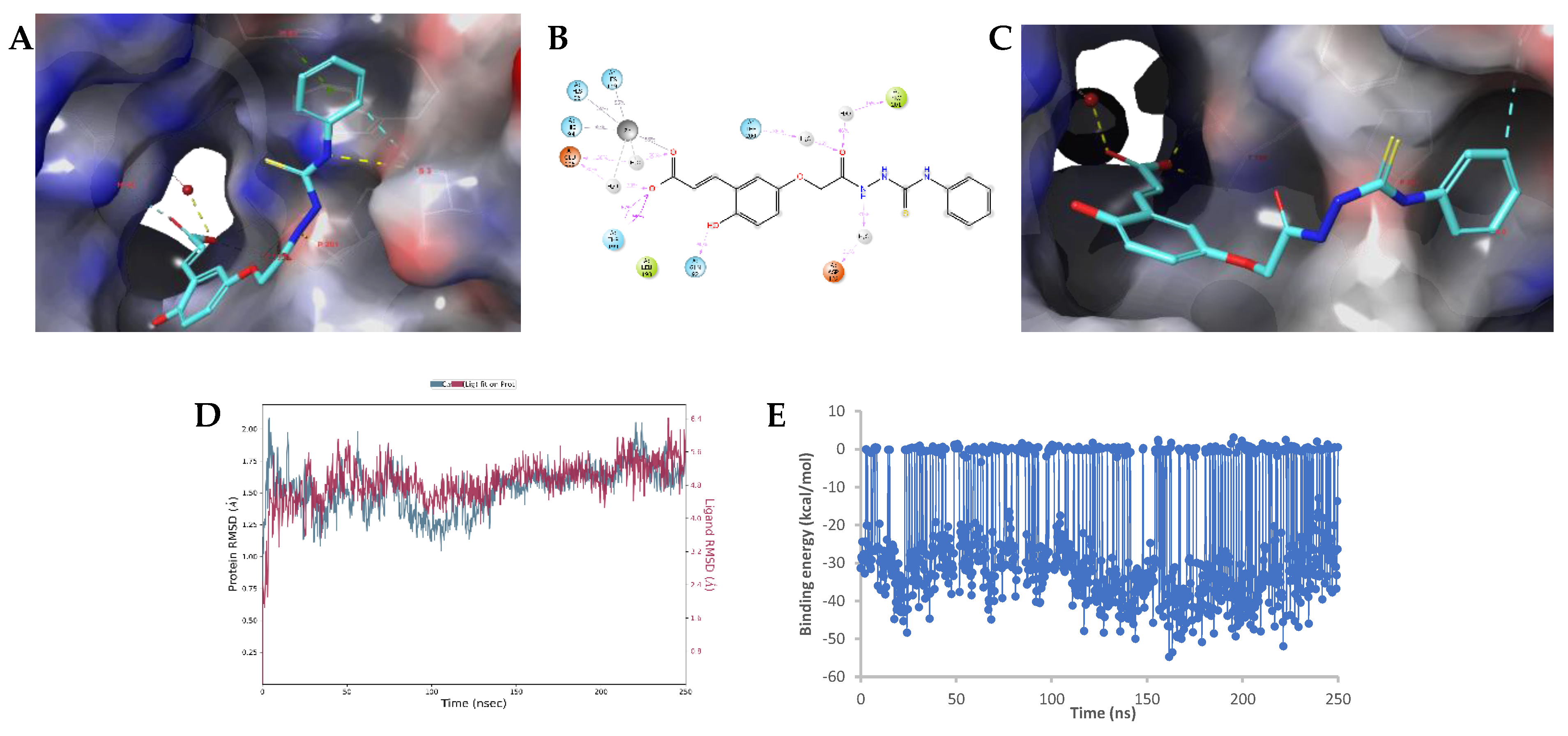
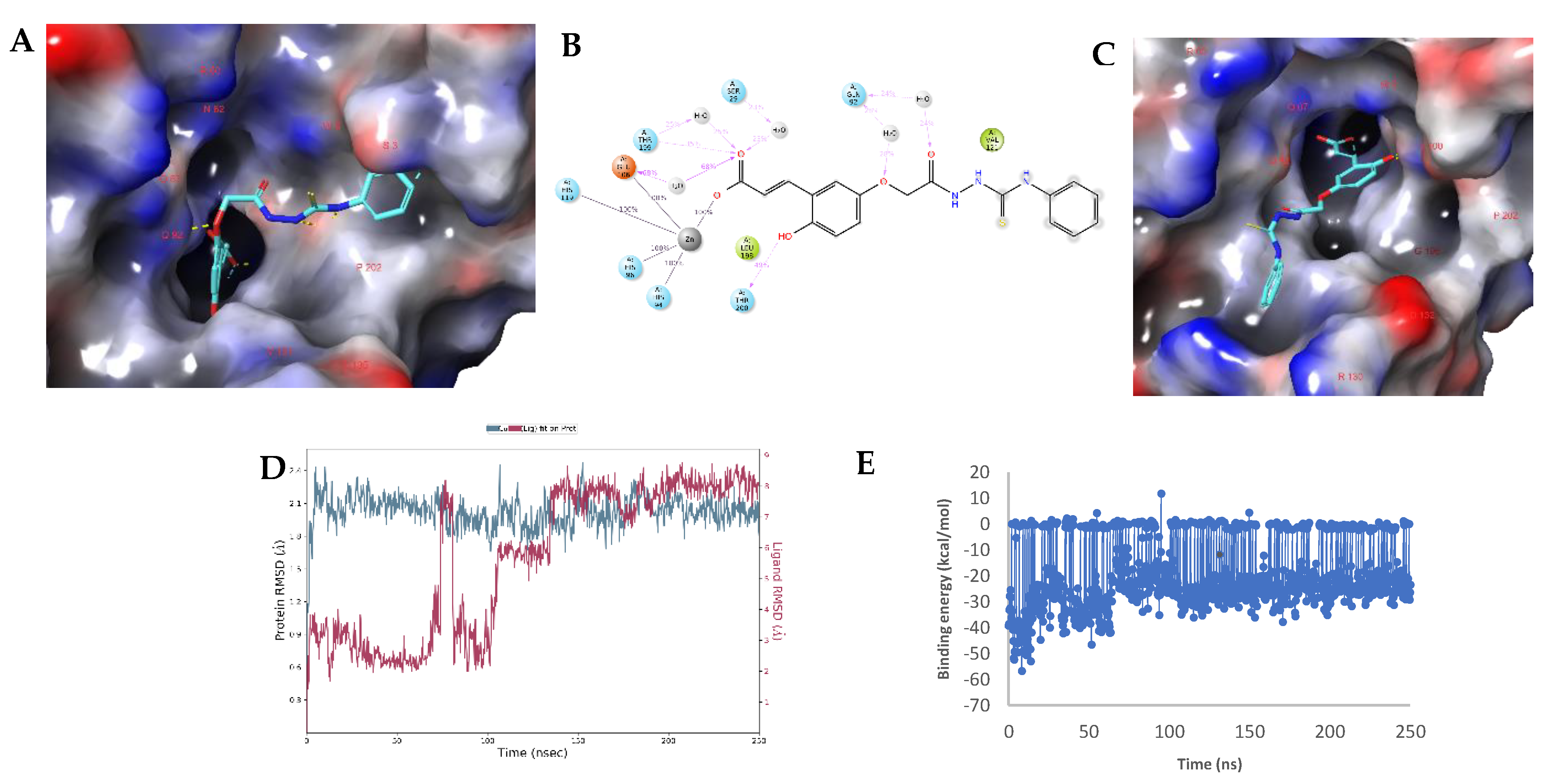
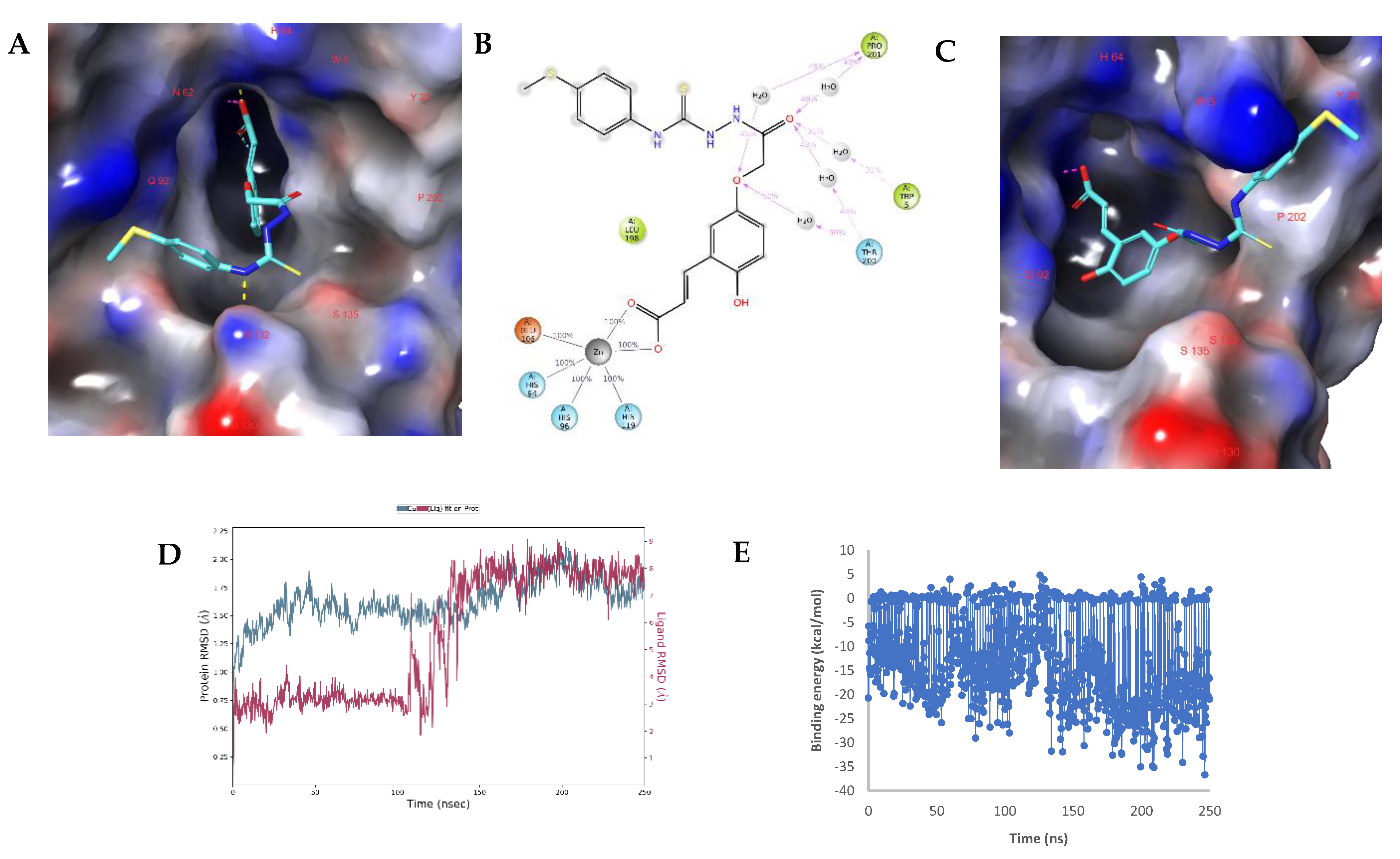
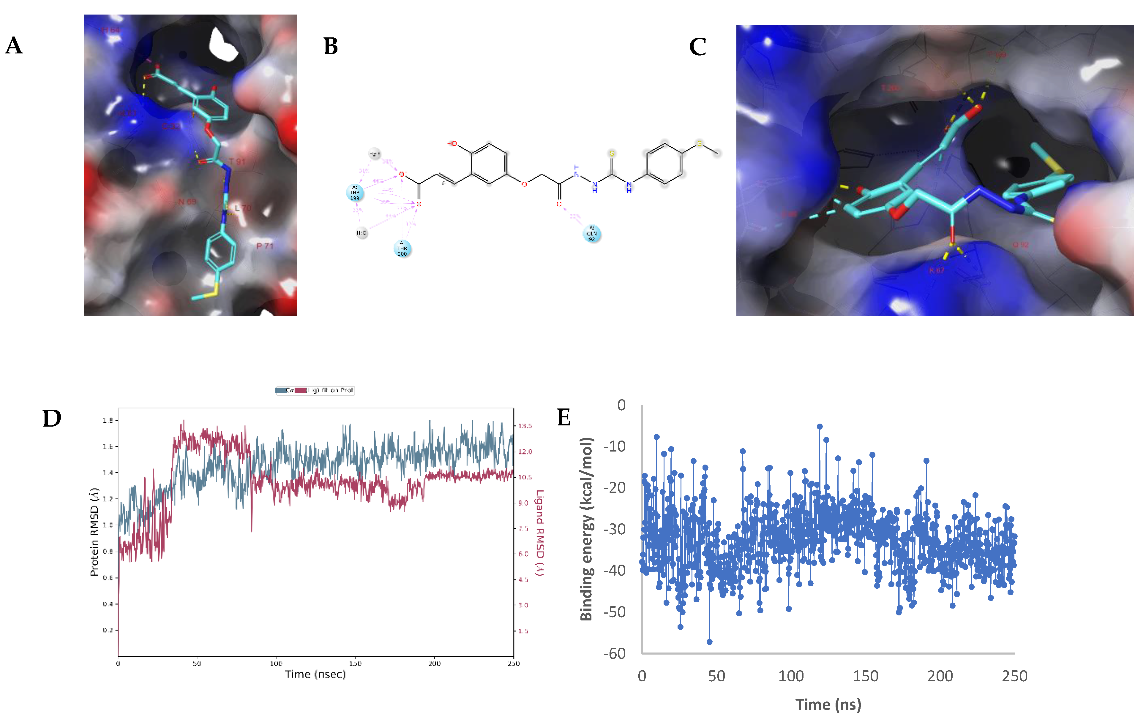
2.3. Molecular Modelling Studies

We performed molecular modelling studies in order to decipher the molecular features underlying the in vitro Ki value differences occurring among the compounds set. Specifically, we turned our attention to 14a, which was the strongest hCA IX inhibitor within the series (Ki of 8.8 nM) and was 84-fold more potent than the hCA XII (Ki of 739 nM). In contrast, compound 22a was highly effective on the hCA XII (Ki of 4.1 nM) and 14-fold less effective on the IX isoform (Ki of 57 nM). Considering the mechanism of action of the coumarin moiety towards CAs [10], we assumed the coumarin warheads in either 14a and 22a were the open/hydrolyzed form which underwent f an isomerization to the thermodynamically more stable E isomer (i.e., hereafter referred as 14a-(E)-open and 22a-(E)-open, Figure 1) and thus docked into the hCA IX and XII crystal structure cavity sites.
The obtained ligand–enzyme complexes were subjected to 250 ns molecular dynamics (MD) simulations which confirmed the interaction of ligands within the hCA IX/XII isoforms.

Modelling Studies of hCA IX– Hydrolyzed 14a-Open

Simulation on the hCA IX in complex with 14a-(E)-open indicated stable interactions occurring between the ligand carboxylic moiety to the zinc-bound water molecule and by means of a second water molecule to Thr199 and to the Zn2+ ion (Figure 2). During MD simulations both waters could interact with the Zn2+ as well as Glu106. A direct hydrogen bond was observed with Gln92 (40%) and water-mediated hydrogen interactions were observed with Asp132 (31%), Thr200 (36%) and Pro201 (46%). The RMSD values for the protein revealed a stable protein structure, while the ligand was observed to be more dynamic, which is mainly due to the movement of the terminal phenyl group (Figure 2A,C). The MM-GBSA binding energy fluctuated between approximately −55 and 3 kcal/mol during the simulation with an average of −24.19 kcal/mol and a standard deviation of 16.1 kcal/mol.
Docking studies indicated that compound 14a-(E)-open may also interact directly with the hCA IX metal ion. A hydrogen bond with the backbone NH group of Thr199 (Figure 3) was also formed. In addition, hydrogen bonds were observed with the side chain of Gln92 and the backbone of Pro201. During a 250 ns MD simulation, the RMSD values of the protein Cα atoms were in the 1.8–2.4 Å range, while the ligand was much higher to reach approximately 8 Å. Major ligand–protein interactions during the simulation occurred with Zn2+ (100%), Thr199 (35%) and Thr200 (49%). Many hydrogen bonds via bridging water molecules as well as hydrophobic interactions were also observed. The snapshot at 250 ns clearly shows that 14a-(E)-open adopted a different orientation within the active site, while the interaction with the metal remained. The MM-GBSA binding energy fluctuated between approximately −57 and 12 kcal/mol during the simulation with an average of −20.28 kcal/mol and a standard deviation of 12.97 kcal/mol.

2.4. Modelling Studies of hCA XII–Hydrolyzed 22a-Open

To our surprise, the 22a-(E)-open revealed interactions within the hCA XII active site either with the metal ion (Figure 4) or the active site entrance amino acids (Figure 5). A pose in which the substrate carboxylic moiety interacted with the zinc-bound water was obtained, but was unstable during a 250 ns MD simulation.
As above, 22a-(E)-open interacted with the catalytic Zn2+ of the hCA XII during the entire duration of the MD simulation (Figure 5). The docked pose also revealed a hydrogen bond interaction that was established with the side chain of Ser132, which however was not stable during simulations as the ligand adopted different orientations within the enzymatic site to form water-bridged hydrogen bonds as well as hydrophobic interactions. The MM-GBSA binding energy fluctuated between approximately −36 and 5 kcal/mol during the simulation with an average of −12.76 kcal/mol and a standard deviation of 9.44 kcal/mol. As depicted in Figure 5, 22a-(E)-open also interacted with the entrance amino acids of the hCA XII active cleft. In the docked pose, the carboxylic acid moiety was hydrogen bonded with His64 and Lys67 and additional interactions were with Leu70, Thr91 and Gln92 (Figure 5). During the MD simulation the ligand’s RMSD increased towards approximately 10 Å, while the protein Cα RMSD values remained low. This indicated large re-orientation of the ligand. Specifically, the carboxylic acid moved away from His64 and Lys67 and formed water-mediated and direct hydrogen bonds with Thr199 and direct hydrogen bonds with Thr200 instead. In addition, a hydrogen bond was formed between the ligand carbonyl group and Gln92. The MM-GBSA binding energy fluctuated between approximately −57.2 and 5.2 kcal/mol during the simulation with an average of −32.8 kcal/mol and a standard deviation of 7.0 kcal/mol.
Overall, the isoform selectivity of 14 and 22 on hCA IX and XII, respectively, was assessed by means of modelling studies applied to their enzymatic active forms 14a-(E)-open and 22a-(E)-open. The obtained data indicated that 14a-(E)-open was able to interact within the hCA IX active site by means of its carboxylic acid to the zinc-bound water molecule (−24.19 ± 16.10 kcal/mol) or directly to the Zn2+ ion (−20.28 ± 12.97 kcal/mol). As for hCA XII-22a-(E)-open, the main interactions detected were between the carboxylic moiety and the Zn2+ ion (−12.76 ± 9.44 kcal/mol) or the entrance amino acids (−32.80 ± 7.0 kcal/mol). The different methods of the ligands in targeting hCA’s active sites and the dynamic bindings make it difficult to directly relate the calculated MM-GBSA binding energy to the measured Ki values, which are also likely dependent on the free energy changes resulting from displacing water molecules within the active sites [13,14,15,16].

3. Materials and Methods

3.1. Chemistry

All anhydrous solvents and reagents used in this study were purchased from Alfa Aesar, TCI, and Sigma-Aldrich. The synthetic reactions involving air- or moisture-sensitive chemicals were carried out under a nitrogen atmosphere using dried glassware and syringe techniques in order to transfer the solutions. Melting points were determined in open capillaries in an electrical melting point apparatus and are uncorrected. Nuclear magnetic resonance (1H-, 13C-, and 19F-NMR) spectra were recorded using a Bruker Avance III 400 MHz spectrometer using DMSO-d6 as solvent. The chemical shifts are reported in parts per million (ppm), and the coupling constants (J) are expressed in Hertz (Hz). The splitting patterns are designated as s, singlet; d, doublet; t, triplet; q, quartet; m, multiplet; brs, broad singlet; dd, doublet of doublets. The correct assignment of exchangeable protons (i.e., OH and NH) was carried out by means of the addition of D2O. The high-resolution mass spectrometry (HRMS) analysis was performed with a Thermo Finnigan LTQ Orbitrap mass spectrometer coupled with an electrospray ionization source (ESI).

3.2. General Procedure for Synthesis of 14a,b22a,b and 23b

A solution of coumarin 3ab (0.1g, 0.43 mmol, 1.0 equv) in EtOH (20 mL) was treated with phenylisothiocyanate 4–13 (0.43 mmol, 1.0 equv) then the reaction was refluxed overnight (ON). The reaction mixture was cooled to room temperature and the formed precipitate was filtered-off, washed with Et2O (3 × 5 mL) dried under vacuum to obtain a residue which was recrystallized from EtOH/H2O to afford the desired products 14a,b22a,b and 23b as solids.
N-Phenyl-2-(2-((2-oxo-2H-chromen-6-yl)oxy)acetyl)hydrazine-1-carbothioamide 14a. 76% yield; m.p. 173–175 °C; 1H-NMR (400 MHz, DMSO-d6) δ 4.72 (2H, s), 6.54 (1H, d, J 9.6), 7.21 (1H, t, J 7.1), 7.37 (7H, m), 8.02 (1H, d, J 9.6), 9.71 (2H, m, exchange with D2O, NHCSNH), 10.37 (1H, s, exchange with D2O, NHCO); 13C-NMR (100 MHz, DMSO-d6) δ 67.6, 112.7, 117.6, 118.3, 120.0, 120.9, 126.0, 129.0, 129.8, 139.9, 144.8, 149.1, 154.9, 160.9, 168.1, 181.9; m/z calcd. for C18H15N3O4S (369.4), found: (ESI negative) 368.0 [M − H]; ESI-HRMS (m/z) calculated for [M + H]+ ion species C18H16N3O4S = 370.0783, found 370.0780.
N-Phenyl-2-(2-((2-oxo-2H-chromen-7-yl)oxy)acetyl)hydrazine-1-carbothioamide 14b. 80% yield; m.p. 155–156 °C; 1H-NMR (400 MHz, DMSO-d6) δ 4.81 (2H, s), 6.35 (1H, d, J 9.5), 7.08 (2H, m), 7.21 (1H, t, J 7.6), 7.38 (2H, t, J 7.6), 7.47 (2H, d, J 7.6), 7.70 (1H, d, J 8.5), 8.04 (1H, d, J 9.5), 9.71 (2H, m, exchange with D2O, NHCSNH), 10.37(1H, s, exchange with D2O, NHCO); 13C-NMR (100 MHz, DMSO-d6) δ 67.3, 102.5, 113.7, 113.8, 118.9, 126.1, 126.8, 129.0, 130.4, 139.9, 145.1, 156.1, 161.1, 161.7, 167.8, 181.9; m/z calcd. for C18H15N3O4S (369.4), found: (ESI negative) 368.0 [M − H]; ESI-HRMS (m/z) calculated for [M + H]+ ion species C18H16N3O4S = 370.0783, found 370.0784. Experimental values in agreement with reported data [7].
N-(p-Tolyl)-2-(2-((2-oxo-2H-chromen-6-yl)oxy)acetyl)hydrazine-1-carbothioamide 15a. 73% yield; m.p. 167–169 °C; 1H-NMR (400 MHz, DMSO-d6) δ 2.32 (3H, s), 4.72 (2H, s), 6.54 (1H, d, J 9.6), 7.17 (2H, d, J 8), 7.35 (5H, m), 8.01 (1H, d, J 9.6), 9.64 (2H, m, exchange with D2O, NHCSNH), 10.34 (1H, s, exchange with D2O, NHCO); 13C-NMR (100 MHz, DMSO-d6) δ 21.6, 67.7, 112.9, 117.7, 118.5, 120.1, 121.2, 129.6, 130.4, 135.5, 137.4, 145.0, 149.3, 155.1, 161.2, 168.4, 182.4; m/z calcd. for C19H17N3O4S (383.4), found: (ESI negative) 382.0 [M − H]; ESI-HRMS (m/z) calculated for [M + H]+ ion species C19H18N3O4S = 384.0940, found 384.0942.
N-(p-Tolyl)-2-(2-((2-oxo-2H-chromen-7-yl)oxy)acetyl)hydrazine-1-carbothioamide 15b. 80% yield; m.p. 147–149 °C (dec); 1H-NMR (400 MHz, DMSO-d6) δ 2.32 (3H, s), 4.80 (2H, s), 6.35 (1H, d, J 9.5), 7.08 (2H, m), 7.18 (2H, d, J 8.0), 7.33 (2H, d, J 8.0), 7.69 (1H, d, J 8.4), 8.04 (1H, d, J 9.5), 9.63 (1H, s, exchange with D2O, NHCSNH), 9.66 (1H, s, exchange with D2O, NHCSNH), 10.32 (1H, s, exchange with D2O, NHCO); 13C-NMR (100 MHz, DMSO-d6) δ 21.4, 67.3, 102.5, 113.7, 113.7, 113.8, 126.7, 129.5, 130.4, 135.3, 137.3, 145.1, 156.1, 161.1, 161.7, 167.8, 182.0; m/z calcd. for C19H17N3O4S (383.4), found: (ESI negative) 382.0 [M − H]; ESI-HRMS (m/z) calculated for [M + H]+ ion species C19H18N3O4S = 384.0940, found 384.0943.
N-(4-Chlorophenyl)-2-(2-((2-oxo-2H-chromen-6-yl)oxy)acetyl)hydrazine-1-carbothioamide 16a. 64% yield; m.p. 163–165 °C; 1H-NMR (400 MHz, DMSO-d6) δ 4.73 (2H, s), 6.54 (1H, d, J 9.6), 7.35 (2H, m), 7.43 (3H, m), 7.50 (2H, d, J 8.0), 8.02 (1H, d, J 9.6), 9.81 (2H, m, exchange with D2O, NHCSNH), 10.38 (1H, s, exchange with D2O, NHCO); 13C-NMR (100 MHz, DMSO-d6) δ 67.6, 112.7, 117.6, 118.3, 120.0, 120.9, 128.9, 129.5, 132.3, 138.9, 144.8, 149.1, 154.9, 160.9, 168.2, 182.0; m/z calcd. for C18H14ClN3O4S (403.8), found: (ESI negative) 402.0 [M − H]; ESI-HRMS (m/z) calculated for [M + H]+ ion species C18H15ClN3O4S = 404.0394, found 404.0397.
N-(4-Chlorophenyl)-2-(2-((2-oxo-2H-chromen-7-yl)oxy)acetyl)hydrazine-1-carbothioamide 16b. 67% yield; m.p. 202–203 °C; 1H-NMR (400 MHz, DMSO-d6) δ 4.81 (2H, s), 6.35 (1H, d, J 9.5), 7.08 (2H, m), 7.44 (2H, d, J 8.4), 7.52 (2H, d, J 8.4), 7.70 (1H, d, J 8.5), 8.04 (1H, d, J 9.5), 9.82 (2H, m, exchange with D2O, NHCSNH), 10.39 (1H, s, exchange with D2O, NHCO); 13C-NMR (100 MHz, DMSO-d6) δ 67.2, 102.5, 113.7, 113.8, 114.0, 119.9, 128.9, 129.8, 130.3, 138.9, 145.1, 156.1, 161.1, 161.6, 167.8, 182.0; m/z calcd. for C18H14ClN3O4S (403.8), found: (ESI negative) 402.0 [M − H]; ESI-HRMS (m/z) calculated for [M + H]+ ion species C18H15ClN3O4S = 404.0394, found 404.0399. Experimental values in agreement with reported data [17].
N-(4-Fluorophenyl)-2-(2-((2-oxo-2H-chromen-6-yl)oxy)acetyl)hydrazine-1-carbothioamide 17a. 60% yield; m.p. 181–183 °C; 1H-NMR (400 MHz, DMSO-d6) δ 4.72 (2H, s), 6.54 (1H, d, J 9.6), 7.21 (2H, m), 7.33–7.45 (5H, m), 8.02 (1H, d, J 9.6), 9.73 (2H, m, exchange with D2O, NHCSNH), 10.37(1H, s, exchange with D2O, NHCO); 13C-NMR (100 MHz, DMSO-d6) δ 67.6, 112.8, 115.7 (d, 2J 22.4), 117.6, 118.3, 120.0, 121.0, 128.9 (m), 136.3 (d, 4J 3), 144.8, 149.1, 155.0, 160.5 (d, 1J 239.7), 161.0, 168.17, 182.3; 19F-NMR (376 MHz, DMSO-d6) δ −117.1 (1F, s); m/z calcd. for C18H14FN3O4S (387.4), found: (ESI negative) 386.0 [M − H]; ESI-HRMS (m/z) calculated for [M + H]+ ion species C18H15FN3O4S = 388.0689, found 388.0685.
N-(4-Fluorophenyl)-2-(2-((2-oxo-2H-chromen-7-yl)oxy)acetyl)hydrazine-1-carbothioamide 17b. 85% yield; m.p. 119–121 °C (dec); 1H-NMR (400 MHz, DMSO-d6) δ 4.81 (2H, s), 6.35 (1H, d, J 9.5), 7.06–7.10 (2H, m), 7.21 (2H, m), 7.46 (2H, m), 7.70 (1H, d, J 8.4), 8.04 (1H, d, J 9.5), 9.74 (2H, m, exchange with D2O, NHCSNH), 10.36 (1H, s, exchange with D2O, NHCO); 13C-NMR (100 MHz, DMSO-d6) δ 67.6, 102.5, 113.7, 113.8, 113.8, 115.7 (d, 2J 22.4), 128.9 (d, 3J 9), 130.4, 136.2 (d, 4J 3), 145.2, 156.1, 160.5 (d, 1J 240.5) 161.2, 161.7, 167.8, 182.3; 19F-NMR (376 MHz, DMSO-d6) δ −117.1 (1F, s); m/z calcd. for C18H14FN3O4S (387.4), found: (ESI negative) 386.0 [M − H]; ESI-HRMS (m/z) calculated for [M + H]+ ion species C18H15FN3O4S = 388.0689, found 388.0687. Experimental values in agreement with reported data [17].
N-(4-Iodophenyl)-2-(2-((2-oxo-2H-chromen-6-yl)oxy)acetyl)hydrazine-1-carbothioamide 18a. 79% yield; m.p. 160–162 °C; 1H-NMR (400 MHz, DMSO-d6) δ 4.72 (2H, s), 6.54 (1H, d, J 9.6), 7.34 (4H, m), 7.42 (1H, d, J 8.9), 7.72(2H, d, J 8.4), 8.02 (1H, d, J 9.6), 9.71 (1H, s, exchange with D2O, NHCSNH), 9.80 (1H, s, exchange with D2O, NHCSNH), 10.37 (1H, s, exchange with D2O, NHCO); 13C-NMR (100 MHz, DMSO-d6) δ 67.6, 90.7, 112.7, 117.6, 118.3, 120.0, 120.9, 128.8, 137.7, 139.8, 144.8, 149.1, 154.9, 160.9, 168.1, 181.8; m/z calcd. for C18H14IN3O4S (495.3), found: (ESI negative) 494.0 [M − H]; ESI-HRMS (m/z) calculated for [M + H]+ ion species C18H15IN3O4S = 494.9750, found 494.9753.
N-(4-Iodophenyl)-2-(2-((2-oxo-2H-chromen-7-yl)oxy)acetyl)hydrazine-1-carbothioamide 18b. 90% yield; m.p. 164–166 °C; 1H-NMR (400 MHz, DMSO-d6) δ 4.81 (2H, s), 6.35 (1H, d, J 9.5), 7.08 (2H, m), 7.34 (2H, d, J 8.2), 7.71 (3H, m), 8.04 (1H, d, J 9.5), 9.81 (2H, m, exchange with D2O, NHCSNH), 10.37 (1H, s, exchange with D2O, NHCO); 13C-NMR (100 MHz, DMSO-d6) δ 67.2, 90.8, 102.5, 113.7, 113.7, 128.7, 130.1, 130.3, 137.7, 139.8, 145.1, 156.0, 161.0, 161.6, 167.7, 181.8; m/z calcd. for C18H14IN3O4S (495.3), found: (ESI negative) 494.0 [M − H]; ESI-HRMS (m/z) calculated for [M + H]+ ion species C18H15IN3O4S = 494.9750, found 494.9748.
N-(3-Iodophenyl)-2-(2-((2-oxo-2H-chromen-6-yl)oxy)acetyl)hydrazine-1-carbothioamide 19a. 80% yield; m.p. 144–146 °C; 1H-NMR (400 MHz, DMSO-d6) δ 4.73 (2H, s), 6.54 (1H, d, J 9.6), 7.18 (1H, t, J 8), 7.34 (1H, dd, J 8.9, 2.7), 7.37 (1H, m), 7.42 (1H, d, J 8.9), 7.56 (2H, m), 7.85 (1H, s), 8.02 (1H, d, J 9.6), 9.74(1H, s, exchange with D2O, NHCSNH), 9.85 (1H, s, exchange with D2O, NHCSNH), 10.38 (1H, s, exchange with D2O, NHCO); 13C-NMR (100 MHz, DMSO-d6) δ 67.6, 94.1, 112.7, 117.6, 118.3, 120.0, 120.9, 125.9, 130.9, 134.5, 141.4, 144.7, 149.1, 154.9, 160.9, 168.2, 181.7; m/z calcd. for C18H14IN3O4S (495.3), found: (ESI negative) 494.0 [M − H]; ESI-HRMS (m/z) calculated for [M + H]+ ion species C18H15IN3O4S = 494.9750, found 494.9752.
N-(3-Iodophenyl)-2-(2-((2-oxo-2H-chromen-7-yl)oxy)acetyl)hydrazine-1-carbothioamide 19b. 88% yield; m.p. 168–169 °C; 1H-NMR (400 MHz, DMSO-d6) δ 4.80 (2H, s), 6.35 (1H, d, J 9.5), 7.07 (2H, m), 7.19 (1H, t, J 8), 7.56 (2H, d, J 8), 7.70 (1H, d, J 9), 7.87 (1H, s), 8.04 (1H, d, J 9.5), 9.76 (1H, s, exchange with D2O, NHCSNH), 9.86 (1H, s, exchange with D2O, NHCSNH) 10.39 (1H, s, exchange with D2O, NHCO); 13C-NMR (100 MHz, DMSO-d6) δ 67.3, 94.1, 102.5, 113.7, 113.7, 113.8, 126.0, 130.3, 130.9, 134.5, 141.3, 145.1, 156.0, 161.0, 161.6, 167.8, 181.7; m/z calcd. for C18H14IN3O4S (495.3), found: (ESI negative) 494.0 [M − H]; ESI-HRMS (m/z) calculated for [M + H]+ ion species C18H15IN3O4S = 494.9750, found 494.9757.
N-(4-(Trifluoromethyl)phenyl)-2-(2-((2-oxo-2H-chromen-6-yl)oxy)acetyl)hydrazine-1-carbothioamide20a. 71% yield; m.p. 182–184 °C; 1H-NMR (400 MHz, DMSO-d6) δ 4.74 (2H, s), 6.54 (1H, d, J 9.6), 7.33–7.38 (2H, m), 7.42 (1H, d, J 9), 7.75 (4H, m), 8.02 (1H, d, J 9.6), 9.96 (2H, m, exchange with D2O, NHCSNH), 10.43 (1H, s, exchange with D2O, NHCO); 13C-NMR (100 MHz, DMSO-d6) δ 67.7, 112.8, 117.6, 118.3, 120.0, 121.0, 125.2 (q, 1J 268), 126.1 (q, 2J 32), 143.8, 144.8, 149.2, 154.9, 161.0, 168.4, 182.1; 19F-NMR (376 MHz, DMSO-d6) δ − 60.4 (3F, s); m/z calcd. for C19H14F3N3O4S (437.4), found: (ESI negative) 436.0 [M − H]; ESI-HRMS (m/z) calculated for [M + H]+ ion species C18H15F3N3O4S = 438.0657, found 437.0653.
N-(4-(Trifluoromethyl)phenyl)-2-(2-((2-oxo-2H-chromen-7-yl)oxy)acetyl)hydrazine-1-carbothioamide20b. 81% yield; m.p. 207–209 °C; 1H-NMR (400 MHz, DMSO-d6) δ 4.83 (2H, s), 6.36 (1H, d, J 9.5), 7.07–7.12 (2H, m), 7.70 (1H, d, J 8.6), 7.74 (2H, m), 7.81 (2H, m), 8.04 (1H, d, J 9.5), 9.96 (2H, m, exchange with D2O, NHCSNH), 10.43 (1H, s, exchange with D2O, NHCO); 13C-NMR (100 MHz, DMSO-d6) δ 67.2, 102.5, 113.7, 113.8, 125.2 (q, 1J 270), 126.1 (q, 2J 32), 126.6 (q, 3J 4), 130.4, 143.7 (1, 4J 1), 145.1, 156.1, 161.1, 161.7, 168.0, 181.9; 19F-NMR (376 MHz, DMSO-d6) δ − 60.4 (3F, s); m/z calcd. for C19H14F3N3O4S (437.4), found: (ESI negative) 436.0 [M − H]; ESI-HRMS (m/z) calculated for [M + H]+ ion species C18H15F3N3O4S = 438.0657, found 437.0652.
N-(3-Nitrophenyl)-2-(2-((2-oxo-2H-chromen-6-yl)oxy)acetyl)hydrazine-1-carbothioamide 21a. 69% yield; m.p. 210–212 °C; 1H-NMR (400 MHz, DMSO-d6) δ 4.75 (2H, s), 6.54 (1H, d, J 9.6), 7.39 (3H, m), 7.67 (1H, t, J 8.2), 8.03 (3H, m), 8.47 (1H, s), 10.04 (2H, m, exchange with D2O, NHCSNH), 10.46 (1H, s, exchange with D2O, NHCO); 13C-NMR (100 MHz, DMSO-d6) δ 67.6, 112.7, 117.6, 118.3, 120.0, 120.5, 120.9, 130.2, 132.5, 141.2, 144.8, 148.1, 149.1, 154.8, 160.9, 168.3, 181.9; m/z calcd. for C14H14N4O6S (414.4), found: (ESI negative) 413.0 [M − H]; ESI-HRMS (m/z) calculated for [M + H]+ ion species C14H15N4O6S = 415.0634, found 415.0631.
N-(3-Nitrophenyl)-2-(2-((2-oxo-2H-chromen-7-yl)oxy)acetyl)hydrazine-1-carbothioamide 21b. 62% yield; m.p. 214–215 °C; 1H-NMR (400 MHz, DMSO-d6) δ 4.83 (2H, s), 6.36 (1H, d, J 9.5), 7.09 (2H, m), 7.68 (2H, m), 8.04 (3H, m), 8.51 (1H, s), 10.06 (2H, m, exchange with D2O, NHCSNH), 10.46 (1H, s, exchange with D2O, NHCO); 13C-NMR (100 MHz, DMSO-d6) δ 67.3, 102.5, 113.8, 113.8, 120.6, 129.6, 130.3, 130.4, 132.4, 132.6, 141.2, 145.2, 148.2, 156.1, 161.1, 161.6, 168.1, 181.9; m/z calcd. for C14H14N4O6S (414.4), found: (ESI negative) 413.0 [M − H]; ESI-HRMS (m/z) calculated for [M + H]+ ion species C14H15N4O6S = 415.0634, found 415.0637.
N-(4-(Methylthio)phenyl)-2-(2-((2-oxo-2H-chromen-6-yl)oxy)acetyl)hydrazine-1-carbothioamide 22a. 82% yield; m.p. 148–150 °C, 1H-NMR (400 MHz, DMSO-d6) δ 2.57 (3H, s), 4.75 (2H,s), 6.57 (1H, d, J 9.4), 7.30 (2H, d, J 7.8), 7.41 (5H, m), 8.05 (1H, d, J 9.4), 9.73 (2H, m, exchange with D2O, NHCSNH), 10.38 (1H, s, exchange with D2O, NHCO); 13C-NMR (100 MHz, DMSO-d6) δ 16.0, 67.6, 112.7, 117.6, 118.2, 120.0, 120.9, 126.7, 135.3, 137.1, 144.8, 149.1, 154.9, 160.9, 168.1, 181.9; m/z calcd. for C19H17N3O4S2 (415.5), found: (ESI negative) 414.0 [M − H]; ESI-HRMS (m/z) calculated for [M + H]+ ion species C14H18N3O4S2 = 416.0660, found 416.0658.
N-(4-(Methylthio)phenyl)-2-(2-((2-oxo-2H-chromen-7-yl)oxy)acetyl)hydrazine-1-carbothioamide 22b. 94% yield; m.p. 163–164 °C; 1H-NMR (400 MHz, DMSO-d6) δ 2.50 (3H, s), 4.81 (2H, s), 6.35 (1H, d, J 9.5), 7.08 (2H, m), 7.28 (2H, d, J 8.6), 7.42 (2H, d, J 8.6), 7.70 (1H, d, J 8.5), 8.04 (1H, d, J 9.5), 9.71 (2H, m, exchange with D2O, NHCSNH), 10.35 (1H, s, exchange with D2O, NHCO); 13C-NMR (100 MHz, DMSO-d6) δ 16.0, 67.3, 102.6, 113.8, 113.8, 113.9, 126.8, 129.6, 130.4, 132.5, 137.1, 145.2, 156.1, 161.2, 161.7, 167.9, 182.2; m/z calcd. for C19H17N3O4S2 (415.5), found: (ESI negative) 414.0 [M − H]; ESI-HRMS (m/z) calculated for [M + H]+ ion species C14H18N3O4S2 = 416.0660, found 416.0663.
N-(4-Cyanophenyl)-2-(2-((2-oxo-2H-chromen-7-yl)oxy)acetyl)hydrazine-1-carbothioamide 23b. 80% yield; m.p. 224–226 °C; 1H-NMR (400 MHz, DMSO-d6) δ 4.83 (2H, s), 6.36 (1H, d, J 9.5), 7.08 (2H, m), 7.70 (1H, d, J 8.5), 7.84 (4H, m), 8.04 (1H, d, J 9.5), 10.04 (2H, m, exchange with D2O, NHCSNH), 10.45 (1H, s, exchange with D2O, NHCO); 13C-NMR (100 MHz, DMSO-d6) δ 67.3, 102.3, 102.5, 107.8, 113.8, 113.8, 119.8, 126.3, 130.4, 133.2, 144.5, 145.2, 156.1, 161.1, 161.6, 168.0, 181.6; m/z calcd. for C19H14N4O4S (394.4), found: (ESI negative) 393.0 [M − H]; ESI-HRMS (m/z) calculated for [M + H]+ ion species C19H15N4O4S = 395.0736, found 395.0738.

3.3. In Vitro Carbonic Anhydrase Inhibition

An Applied Photophysics stopped-flow instrument was used to assay the CA catalyzed CO2 hydration activity [12]. Phenol red (at a concentration of 0.2 mM) was used as an indicator, working at the absorbance maximum of 557 nm, with 20 mM Hepes (pH 7.4) as a buffer, and 20 mM Na2SO4 (to maintain constant ionic strength), following the initial rates of the CA-catalyzed CO2 hydration reaction for a period of 10–100 s. The CO2 concentrations ranged from 1.7 to 17 mM for the determination of the kinetic parameters and inhibition constants. Enzyme concentrations ranged between 5–12 nM. For each inhibitor, at least six traces of the initial 5–10% of the reaction were used to determine the initial velocity. The uncatalyzed rates were determined in the same manner and subtracted from the total observed rates. Stock solutions of the inhibitor (0.1 mM) were prepared in distilled–deionized water and dilutions up to 0.01 nM were done thereafter with the assay buffer. Inhibitor and enzyme solutions were preincubated together for 6 hrs at room temperature prior to the assay, to allow for the formation of the E–I complex. The inhibition constants were obtained by non-linear least-squares methods using PRISM 3 and the Cheng–Prusoff equation as reported earlier, and represent the mean from at least three different determinations. All CA isoforms were recombinant proteins obtained in-house, as reported earlier [18,19,20,21].

3.4. Preparation of Protein Structures

The crystal structures of hCA IX (PDB 3iai) and hCA XII (PDB 1jd0) co-crystallized with acetazolamide were obtained from the RCSB Protein Data Bank. Afterwards, the crystal structures of hCA II in complex with open-coumarin analogs were downloaded as well, i.e., in complex with 3-(4-methoxyphenyl)but-2-enoic acid (directly bound to the Zn2+ ion; PDB 5eh8), in complex with the open-coumarin 3-(2,4-dichlorophenyl)prop-2-enoic acid (interacts with the zinc-bound water molecule; PDB 5ehw), and in complex with (2E)-3-(2-hydroxyphenyl)acrylic acid (interacts with entrance amino acids of active site; PDB 5bnl). The structures were superposed on hCA IX and XII and the coordinates of the open-coumarin analog and the zinc-bound water molecule were copied into both hCA IX and XII. Subsequently, the structures were prepared using the protein preparation tool of Schrödinger (v2022-1, Schrödinger, Inc., New York, NY, USA). All water (except the zinc-bound water) and buffer molecules were omitted. Subunit A was retained and all other subunits, if present, were omitted. Subsequently, hydrogen atoms were added, and the system was minimized using the OPLS4 forcefield.

3.5. Docking Studies

Ligands 14a and 22a were prepared in the open and closed coumarin form using the LigPrep tool of Schrödinger and minimized with the OPLS4 forcefield. Subsequently, the ligands in the open-coumarin form were docked into the binding sites of the prepared protein structures. The binding sites were assigned as all residues within 5 Å of the co-crystallized ligands. Docking was performed using the Glide tool of Schrödinger with the SP settings. The pose generation was guided by the core of the co-crystallized open-coumarin analog. The closed-coumarin forms of the ligands were docked in the same way, however no core-guided docking was used. The three highest scoring poses were obtained for each ligand and the poses were subsequently minimized using the Prime tool and MM-GBSA forcefield. To this end, the docked ligand and all residues within 5 Å (with the exception of Zn2+, His94, His96 and His119) were unrestrained. High scoring compounds that formed binding interactions (hydrogen bonds, electrostatic interactions and hydrophobic interactions) and showed complementarity in shape and (a)polarity were selected for molecular dynamics (MD) simulations.

3.6. Molecular Dynamics Simulations

The ligand–enzyme complexes obtained with the docking procedure were subjected to a 250 ns MD simulation using Desmond. The complex was first placed in an orthorhombic box (at least 10 Å between complex and boundary) and then filled with Tip5P water molecules and 0.15 M NaCl. The amount of Na+ or Cl ions were adjusted to create a neutral system. Afterwards, all heavy atoms were restrained, and the system was minimized for 100 ps using the OPLS4 forcefield. Finally, the system was simulated for 250 ns under isothermic (Nose–Hoover chain, 1ps relaxation time) and isobaric (Martyna–Tobial–Klein, 2 ps relaxation time, isotropic coupling) conditions without restraints. Snapshots were saved every 250 ps. Finally, the percentage occurrence of the ligand–protein binding interactions as well as the MM-GBSA binding energy were calculated.

4. Conclusions

In conclusion, we reported a series of 6- and 7-substituted coumarins bearing variegated aryl thiosemicarbazide tails and we investigated their in vitro inhibition activity against the physiologically relevant hCA isoforms I, II, IX and XII. All compounds were effective inhibitors against the tumor-related isoforms IX and XII with Ki values spanning between the medium-low nanomolar range. The housekeeping hCAs I and II did not show appreciable inhibition, with the Kis > 10,000 nM. The binding modes of such compounds were explored by means of modelling experiments carried out on selected compounds assumed in their open form and in the thermodynamically most favored geometry (i.e., 14a-(E)-open and 22a-(E)-open). Molecular modelling suggests multiple interactions within the enzymatic cavity and may explain the high potency and selectivity reported for the hCAs IX and XII.

Author Contributions

Conceptualization, A.G., F.C. and C.T.S.; methodology, M.B. and S.S.; software, A.A. (Atilla Akdemir) and A.A. (Andrea Angeli); writing—review and editing, A.G., F.C., A.A. (Atilla Akdemir), A.A. (Andrea Angeli) and C.T.S. All authors have read and agreed to the published version of the manuscript.

Funding

This research received no external funding.

Institutional Review Board Statement

Not applicable.

Informed Consent Statement

Not applicable.

Data Availability Statement

Not applicable.

Acknowledgments

Arzu Gumus is grateful to TUBITAK (The Scientific and Technological Research Council of Turkey) for the award of 2219 International Post Doctoral Research Fellowship Programme.

Conflicts of Interest

The authors declare no conflict of interest.

References

  1. Bonelli, J.; Ortega-Forte, E.; Rovira, A.; Bosch, M.; Torres, O.; Cuscó, C.; Rocas, J.; Ruiz, J.; Marchán, V. Improving photodynamic therapy anticancer activity of a mitochondria-targeted coumarin photosensitizer using a polyurethane-polyurea hybrid nanocarrier. Biomacromolecules 2022, 23, 7. [Google Scholar] [CrossRef] [PubMed]
  2. Yin, Z.; Shi, W.; Wu, X.-F. Transition-Metal-Catalyzed carbonylative multifunctionalization of alkynes. J. Org. Chem. 2022. [Google Scholar] [CrossRef] [PubMed]
  3. Akkol, E.K.; Genç, Y.; Karpuz, B.; Sobarzo-Sánchez, E.; Capasso, R. Coumarins and coumarin-related compounds in pharmacotherapy of cancer. Cancers 2020, 12, 1959. [Google Scholar] [CrossRef] [PubMed]
  4. Hassan, A.A.; Shawky, A.M. Thiosemicarbazides in heterocyclization. J. Heterocycl. Chem. 2011, 3, 495–516. [Google Scholar] [CrossRef]
  5. Magdy, G.; Al-Enna, A.A.; Belal, F.; El-Domany, R.A.; Abdel-Megied, A.M. Application of sulfur and nitrogen doped carbon quantum dots as sensitive fluorescent nanosensors for the determination of saxagliptin and gliclazide. R. Soc. Open Sci. 2022, 6, 220285. [Google Scholar] [CrossRef]
  6. Peperidou, A.; Bua, S.; Bozdag, M.; Hadjipavlou-Litina, D.; Supuran, C.T. Novel 6- and 7-substituted coumarins with inhibitory action against lipoxygenase and tumor-associated carbonic anhydrase IX. Molecules 2018, 23, 153. [Google Scholar] [CrossRef] [Green Version]
  7. Al-Amiery, A.A.; Musa, A.Y.; Kadhum, A.A.H.; Mohamad, A.B. The use of umbelliferone in the synthesis of new heterocyclic compounds. Molecules 2011, 16, 6833–6843. [Google Scholar] [CrossRef]
  8. Janowska, S.; Khylyuk, D.; Andrzejczuk, S.; Wujec, M. Design, synthesis, antibacterial evaluations and in silico studies of novel thiosemicarbazides and 1,3,4-thiadiazoles. Molecules 2022, 10, 3161. [Google Scholar] [CrossRef]
  9. Icharam Narkhede, H.; Shridhar Dhake, A.; Rikhabchand Surana, A. Synthesis and screening of thiosemicarbazide-dithiocarbamate conjugates for antioxidant and anticancer activities. Bioorg. Chem. 2022, 124, 105832. [Google Scholar] [CrossRef]
  10. Maresca, A.; Temperini, C.; Vu, H.; Pham, N.B.; Poulsen, S.A.; Scozzafava, A.; Quinn, R.J.; Supuran, C.T. Non-zinc mediated inhibition of carbonic anhydrases: Coumarins are a new class of suicide inhibitors. J. Am. Chem. Soc. 2009, 8, 3057–3062. [Google Scholar] [CrossRef] [Green Version]
  11. Cecchi, A.; Ciani, L.; Winum, J.-Y.; Montero, J.L.; Scozzafava, A.; Ristori, A.; Supuran, C.T. Carbonic anhydrase inhibitors: Design of spin-labeled sulfonamides incorporating TEMPO moieties as probes for cytosolic or transmembrane isozymes. Bioorg. Med. Chem. Lett. 2008, 12, 3475–3480. [Google Scholar] [CrossRef]
  12. Khalifah, R.G. The carbon dioxide hydration activity of carbonic anhydrase. I. Stop flow kinetic studies on the native human isoenzymes B and C. J. Biol. Chem. 1971, 246, 2561. [Google Scholar] [CrossRef]
  13. Lazaridis, T. Inhomogeneous fluid approach to solvation thermodynamics. 1. Theory. J. Phys. Chem. B 1998, 102, 3531–3541. [Google Scholar] [CrossRef]
  14. Lazaridis, T. Solvent reorganization energy and entropy in hydrophobic hydration. J. Phys. Chem. B 2000, 104, 4964–4979. [Google Scholar] [CrossRef]
  15. Abel, R.; Young, T.; Farid, R.; Berne, B.J.; Friesner, R.A. Role of the active-site solvent in the thermodynamics of Factor Xa ligand binding. J. Am. Chem. Soc. 2008, 9, 2817–2831. [Google Scholar] [CrossRef] [Green Version]
  16. Young, T.; Abel, R.; Kim, B.; Berne, B.J.; Friesner, R.A. Motifs for molecular recognition exploiting hydrophobic enclosure in protein-ligand binding. Proc. Natl. Acad. Sci. USA 2007, 3, 808–813. [Google Scholar] [CrossRef] [Green Version]
  17. Raman, K.; Singh, H.K.; Salzman, S.K.; Parmar, S.S. Substituted thiosemicarbazides and corresponding cyclized 1,3,4-oxadiazoles and their anti-inflammatory activity. J. Pharm. Sci. 1993, 2, 167–169. [Google Scholar] [CrossRef]
  18. Karioti, A.; Carta, F.; Supuran, C.T. Phenols and polyphenols as carbonic anhydrase inhibitors. Molecules 2016, 12, 1649. [Google Scholar] [CrossRef] [Green Version]
  19. Carta, F.; Maresca, A.; Scozzafava, A.; Supuran, C.T. 5- and 6-Membered (thio)lactones are prodrug type carbonic anhydrase inhibitors. Bioorg. Med. Chem. Lett. 2012, 1, 267–270. [Google Scholar] [CrossRef]
  20. Carta, F.; Vullo, D.; Maresca, A.; Scozzafava, A.; Supuran, C.T. New chemotypes acting as isozyme-selective carbonic anhydrase inhibitors with low affinity for the offtarget cytosolic isoform II. Bioorg. Med. Chem. Lett. 2012, 6, 2182–2185. [Google Scholar] [CrossRef]
  21. Carta, F.; Vullo, D.; Maresca, A.; Scozzafava, A.; Supuran, C.T. Mono-/dihydroxybenzoic acid esters and phenol pyridinium derivatives as inhibitors of the mammalian carbonic anhydrase isoforms I, II, VII, IX, XII and XIV. Bioorg. Med. Chem. 2013, 6, 1564–1569. [Google Scholar] [CrossRef] [Green Version]
Scheme 1. Synthesis of coumarins 14a,b22a,b and 23b.
Molecules 27 04610 sch001
Figure 1. Hydrolysis of the coumarin scaffolds and Z/E geometrical isomerization.
Molecules 27 04610 g001
Figure 2. (A) The docked pose 14a-(E)-open. (B) Ligand–protein binding interactions observed for 30% or more of the duration of the unrestricted MD simulation. (C) Ligand pose after a 250 MD simulation. (D) RMSD value of the protein Cα-atoms and the ligand heavy atoms. (E) MM-GBSA binding energy. In the 2D plots, anionic residues are indicated in red, cationic residues are indicated in purple, hydrophobic residues and surfaces are indicated in green and hydrophilic residues and surfaces are indicated in light blue. Hydrogen bonds are indicated in purple (2D plots) or yellow (A,C) dashed lines.
Molecules 27 04610 g002
Figure 3. Modelling results of the hCA IX–14a-(E)-open interacting with Zn2+ ion. (A) Docked pose compound 14a-(E)-open. (B) Ligand–protein binding interactions observed for 30% or more of the duration of the unrestricted MD simulation. (C) Ligand pose after a 250 MD simulation. (D) RMSD value of the protein Cα-atoms and the ligand heavy atoms. (E) MM-GBSA binding energy. In the 2D plots, anionic residues are indicated in red, cationic residues are indicated in purple, hydrophobic residues and surfaces are indicated in green and hydrophilic residues and surfaces are indicated in light blue. Hydrogen bonds are indicated in purple (2D plots) or yellow (A,C) dashed lines.
Molecules 27 04610 g003
Figure 4. Molecular modelling results of the hCA XII–22a-(E)-open interaction. (A) Docked pose of 22a-(E)-open. (B) The ligand–protein binding interactions observed for 30% or more of the duration of the unrestricted MD simulation. (C) Ligand pose after a 250 MD simulation. (D) RMSD value of the protein Cα-atoms and the ligand heavy atoms. (E) Binding energy. In the 2D plots, anionic residues are indicated in red, cationic residues are indicated in purple, hydrophobic residues and surfaces are indicated in green and hydrophilic residues and surfaces are indicated in light blue. Hydrogen bonds are indicated in purple (2D plots) or yellow (A,C) dashed lines.
Molecules 27 04610 g004
Figure 5. Molecular modelling results of the hCA XII–22a-(E)-open. (A) Docked pose compound 22a-(E)-open. (B) Ligand–protein binding interactions observed for 30% or more of the duration of the unrestricted MD simulation. (C) Ligand pose after a 250 MD simulation. (D) RMSD value of the protein Cα-atoms and the ligand heavy atoms. (E) Binding energy. In the 2D plots, anionic residues are indicated in red, cationic residues are indicated in purple, hydrophobic residues and surfaces are indicated in green and hydrophilic residues and surfaces are indicated in light blue. Hydrogen bonds are indicated in purple (2D plots) or yellow (A,C) dashed lines.
Molecules 27 04610 g005
Table 1. Inhibition data of hCA isoforms I, II, IX and XII with compounds 14a,b22a,b and 23b and coumarin (COU) by the stopped flow CO2 hydrase assay [12].
CompoundhCA I (Ki; nM *)hCA II (Ki; nM *))hCA IX (Ki; nM *))hCA XII (Ki; nM *))
14a>10,000>10,0008.8739
14b>10,000>10,00046743.5
15a>10,000>10,000234709
15b>10,000>10,00078684
16a>10,000>10,00048608
16b>10,000>10,00055866
17a>10,000>10,00039494
17b>10,000>10,00064745
18a>10,000>10,0009.68.4
18b>10,000>10,0007217.5
19a>10,000>10,0009.679
19b>10,000>10,0009.4573
20a>10,000>10,00092306
20b>10,000>10,0007056.6
21a>10,000>10,00071328.5
21b>10,000>10,0009.18.6
22a>10,000>10,000574
22b>10,000>10,000334.6
23b>10,000>10,000437.5473
COU [10]31009200>10,000>10,000
* Mean from 3 different assays, by the stopped flow technique (errors were in the range of ±5–10% of the reported values).
Publisher’s Note: MDPI stays neutral with regard to jurisdictional claims in published maps and institutional affiliations.

