
Review on Bee Products as Potential Protective and Therapeutic Agents in Male Reproductive Impairment


Abstract
1. Introduction
2. Factors Involved in Male Reproductive Impairment
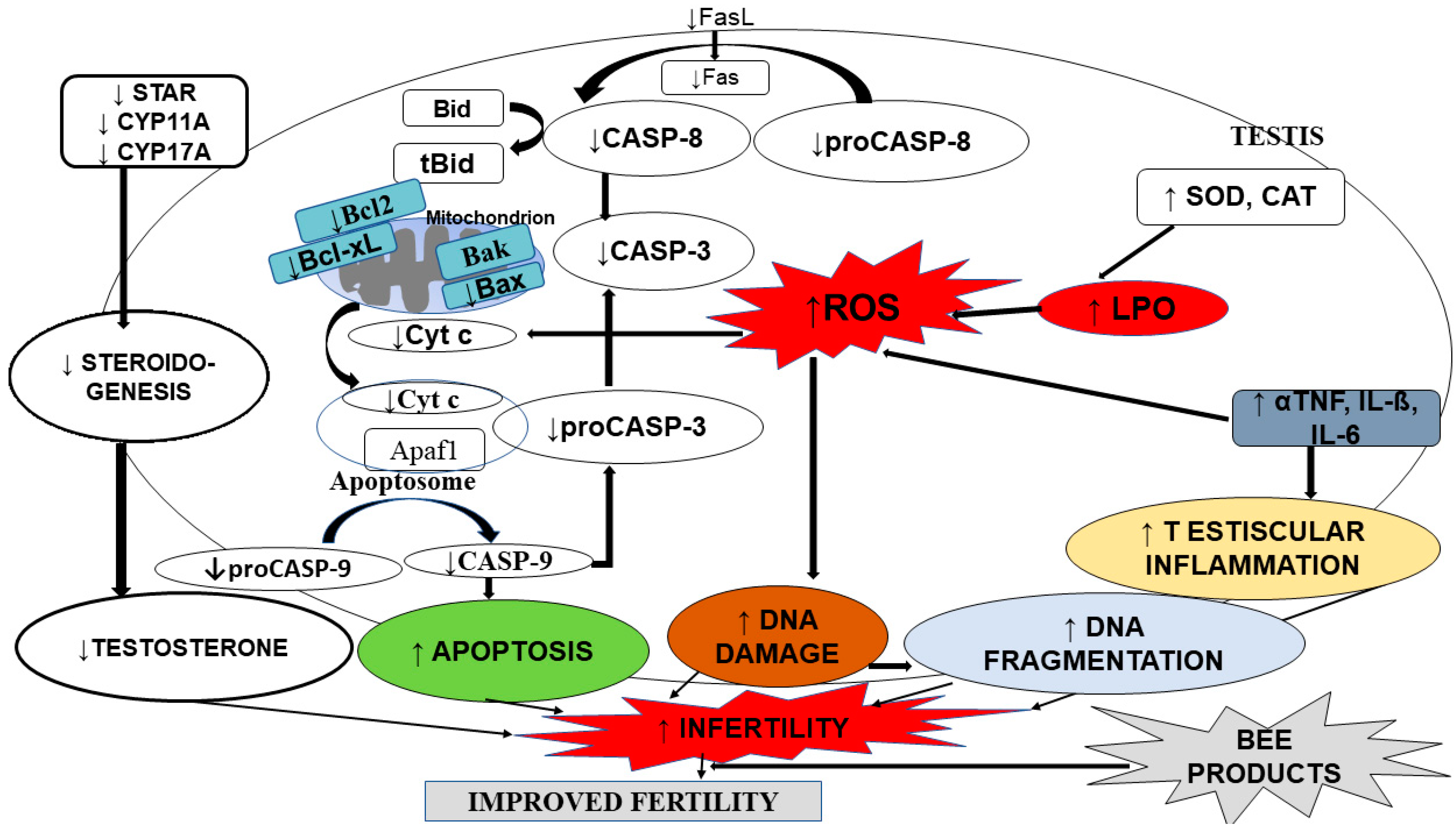
2.1. Testicular Steroidogenesis Dysfunction
2.2. Testicular Apoptosis
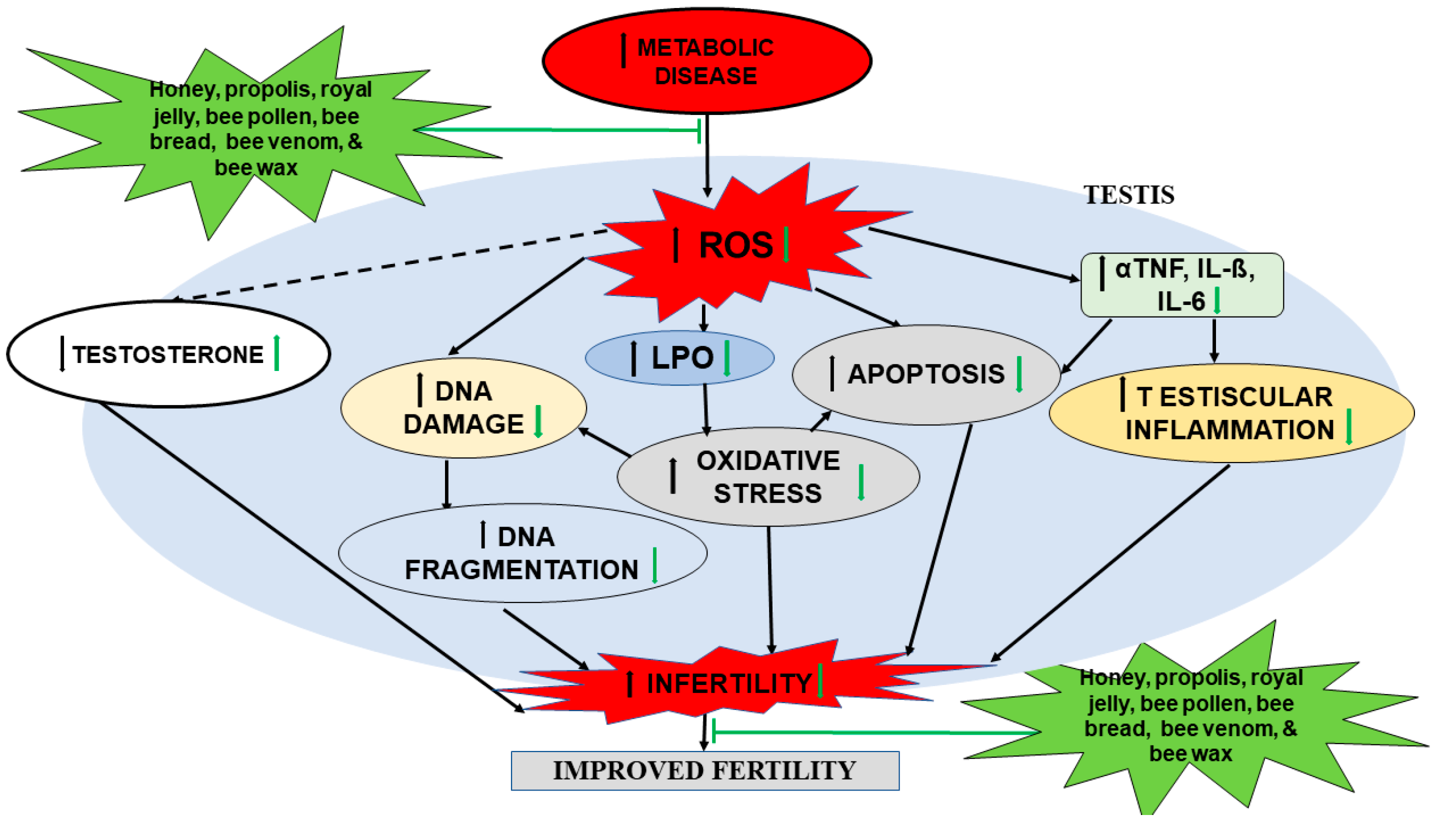
2.3. Testicular Reactive Oxygen Species and Oxidation Stress
2.4. Testicular Inflammation
3. Composition of Bee Products
4. Role of Bee Products in Male Reproductive Impairment
4.1. Effects of Bee Pollen on Male Reproductive Parameters

| s/n | Bee Products | Dose/Duration of Treatment | Substance Used to Induce Stress | Animal Model Used | Route of Administration | Standard Drug | Effect on Reproductive Function Parameters | Possible Molecular Mechanisms | References |
|---|---|---|---|---|---|---|---|---|---|
| 1. | Bee pollen (Egypt) | 100 mg/kg bw/day for 4 weeks | Streptozotocin (STZ)-injection (single dose) | Rats | i.p | - | ↑ Testis weight, testosterone, LH, FSH, sperm count, motility and viability, ↓ MDA, ↑ (SOD, GR, GPx, GST, CAT, and GSH) | Act by scavenging toxic and mutagenic electrophiles and free radicals/modification of antioxidant pathways due to presence of flavonoids | [12] |
| 2. | Bee pollen (India) | 100 mg/kg bw | Rifampicin 100 mg/kg bw/day and isoniazid 50 mg/kg bw/day | Rats | Oral | - | ↓ MDA, ↑ (SOD, GR, GPx, GST, CAT, and GSH) | Presence of bioactive elements (caffeic acid phenethyl ester, myricetin, kaempherol, isoquercetin, and flavonoids) convert the reactive free radicals to inactive products | [71] |
| 3. | Bee pollen (Algeria) | 100 mg/kg bw for 15 days | 30 mg mg/kg bw of lead acetate | Rats | Oral | - | ↑ Spermatogenesis and ↓ Sertoli cells destruction | Acts by lowering lipid, anti-inflammatory, and protective effect against testis cell injury due to potentiated synthesis of proteins | [72] |
| 4. | Bee pollen (Turkey) | 60 mg/per animal (30-day) | Rats | Oral | ↑ Testosterone level and sperm counts | Beneficial effects | [70] |
4.2. Effects of Bee Venom on Male Reproductive Parameters
| s/n | Bee Products | Dose/Duration of Treatment | Substance used to Induce Stress | Animal Model Used | Route of Administration | Standard Drug | Effect on Reproductive Function Parameters | Possible Molecular Mechanisms | References |
|---|---|---|---|---|---|---|---|---|---|
| 1. | Bee Venom (Egypt) | 0.1 (G1), 0.2 (G2) and 0.3 (G3) mg/rabbit twice weekly over 20 wks | High temperature | Rabbits | Intravenous injection | - | ↑ TAC, GST, GSH, IgA, IgM, Testosterone, spermatogenesis and fertility | These effects could be attributed to pituitary gland stimulation to release the adrenocorticotropic hormone, which causes release of the sex hormones such as testosterone in blood circulation, which has significant effects on spermatogenesis and fertility | [73] |
| 2. | Bee Venom (Iraq) | 155 stings | hydrogen peroxide | Mice | Stings | - | Protection and maintenance of some sexual efficiency parameters | Cortisol inhibits Sertoli cells from releasing activin-B, which normally stimulates spermatogonia to induce mitosis to form spermatocytes | [10] |
| 3. | Bee venom (Romania) | 700 μg BV/kg | Rats | Injection | - | ↓ Testicular weight and Sertoli cells, ↑diameter in seminiferous tubules | Mellitin interacts with the proteins in tight junctions between the adjacent Sertoli cells | [13] | |
| 4. | Bee wax (USA) | 15 mg bees wax pellet containing 3.0 mg | Mice | Injection | - | Differential testicular response to photoperiod | Post-pineal mechanism | [74] |
4.3. Effects of Honey on Male Reproductive Parameters
| s/n | Bee Products | Dose/Duration of Treatment | Substance used to Induce stress | Animal Model Used | Route of Administration | Standard Drug | Effect on Reproductive Function Parameters | Possible Molecular Mechanisms | References |
|---|---|---|---|---|---|---|---|---|---|
| 1. | Honey (Nigeria) | 100, 200, and 400 mg/kg | - | Rat | Oral | 2.5, 5, and 7.5 mg/kg of testosterone i.p | ↑ Sperm count | Chrysin (5,7-dihydroxyflavone) blocked the conversion of androgens into oestrogens with a consequent increase in testosterone | [75] |
| 2. | Honey (Egypt) | 0.05 mL (4 weeks) | 5 mL/kg of 0.3% CCL 4 daily subcutaneously (4 Weeks) | Mice | Oral | - | ↓ Degenerative changes of seminiferous tubules and ↑ plasma levels of testosterone significantly | Via reduction of the elevated levels of free radicals and increase in the antioxidant defense system | [78] |
| 3. | Honey (Malaysia gelam honey) | 1.0 mL/100 g (60 days) | - | Rats | Oral | - | ↑ Sperm count and number of sperm with normal morphology | Acts as a physiologic modulator of spermatogenic cells proliferation, which influence the cell cycle of the seminiferous epithelium thus, ↑ spermatogenesis | [14] |
| 4. | Honey (Malaysia) | 1.2 g/kg bw/daily | Cigarette 8 min 3 times/day | Rats | Oral | - | ↑ Intromission and ejaculation, mating, and fertility indexes | Acts as a physiologic modulator of spermatogenic cells proliferation, which influence the cell cycle of the seminiferous epithelium and thus increase spermatogenesis | [82] |
| 5. | Honey (Malaysia) | 1.2 g kg−1 bw daily (21 days) | Prenatal restraint stress (three times per day) from day 11 of pregnancy until delivery | Rats | Oral | - | ↑ Testis and epididymis weights as well as improved the percentages of abnormal spermatozoa and sperm motility | Acts partly by its counteraction on oxidative stress within penile tissues via its antioxidant property | [81] |
| 6. | Honey (Malaysian honey) | 0.2, 1.2, and 2.4 g kg−1 (4 weeks) | - | Rats | Oral | - | ↑ Epididymal sperm count without affecting spermatid count and reproductive hormones | Due to its one or more constituents that could protect germ cells against oxidative stress. This might have further enhanced spermiogenesis | [83] |
| 7. | Honey (Nigeria) | 1, 2, and 2.5 mL of honey daily for 21 days | - | Rats | Oral | 0.3 mL FSH drug for 6 days | Improves the sperm quality and spermatogenesis rate and no sign of degeneration or cellular loss in the testicular histoarchitecture | Suggestive of zinc accumulating in the testis during early spermatogenesis, and important in DNA synthesis and the regulation of spematogonial proliferation | [85] |
| 8. | Honey (Nigeria) | 1 mL of honey per 100 g of bw (65 days) | - | Rat | Oral | Manix capsules (6220 mg/100 mL of drug solution) | ↑ Sperm count, sperm motility, and improves sperm morphology | ↓ Lipid peroxidation and oxidative stress on the sperm cells by reactive oxygen species such as super oxide, hydrogen peroxide | [87] |
| 9. | Honey (Nigeria) | (100 mg/kg bw) (35 days) | Nicotine (1.0 mg/kg bwt) | Rats | Oral | - | ↑ Sperm motility, viability, morphology, counts, FSH, LH, and testosterone | Mediated by its counteraction on oxidative stress | [88] |
| 10. | Honey supplements (Iran) | 70 g (8 weeks) | 8 weeks of intensive cycling training | Humans | Oral | - | ↓ Seminal interleukin (IL)- 1 b, IL-6, IL-8, tumor necrosis factor (TNF)-α, ROS, MDA, ↑ Levels of seminal SOD and catalase | ↓ Seminal plasma cytokines and oxidative stress biomarkers as well as increasing seminal antioxidant levels | [76] |
| 11. | Honey (Palestinian Honey) | 5% honey for 20 days | - | Rats | Oral | - | Induces spermatogenesis in rats by ↑ epididymal sperm count, relative weight of the epididymis, SDH activity, and ↓ LDH activity | Needs further experiments to establish mechanism | [89] |
| 12. | Honey (Saudi Arabia) | 20 mg/kg body weight/day) for 4 weeks | Octylphenol (0.1 and 1.0 mg kg_1 bw) | Rats | Oral | - | Ameliorates toxic effects and ↓ histopathological stress toxicity | Further studies required | [9] |
| 13. | Honey (Taulang) (Malaysia) | 0.2, 1.2, or 2.4 g/kg/day of honey for 28 days | - | Rats | Oral | - | ↑ Sperm counts significantly. | Further studies required | [84] |
| 14. | Honey bee and pollen grains (Saudi Arabia) | (1 g/kg) 2 weeks | Cyclophosphamide (10 mg/kg) i.p | Mice | Oral | - | ↓ Sperm abnormality, chromosomal aberrations, ameliorates GSH and MDA | Presence of CAPE as protective agent against chemotherapy-induced oxidative stress | [90] |
| 15. | Honey bee Drone milk (Hungary) | 110 mg/kg/day | - | Castrated Rats | Oral | - | ↑ Relative weights of the androgen-dependent organs and the plasma testosterone level in castrated rats and tissue mRNA and protein level of SLAP | Scavenging of free radicals by polyphenols before free radicals can interact with DNA | [91] |
| 16. | Honey (Iran) | 10% of honey | - | Mice | IVF | - | Enhances sperm motility and pregnancy rate of female mice | Antioxidant activity | [79] |
| 17. | Honey (Gelam) (Malaysia) | 1.0 mL/100 g bw | Nicotine (N) group were intraperitoneally (i.p.) injected with 5.0 mg/kg | Rats (4–5 weeks old) | Intra peritoneal | ↑ Fertility of juvenile male rats by increasing sperm motility and number of morphologically normal sperm | Further study required | [77] |
4.4. Effects of Propolis on Male Reproductive Parameters
| s/n | Bee Products | Dose/Duration of Treatment | Substance Used to Induce Stress | Animal Model Used | Route of administration | Standard Drug | Effect on Reproductive Function Parameters | Possible Molecular Mechanisms | References |
|---|---|---|---|---|---|---|---|---|---|
| 1. | Propolis (Iraq) | 200 mg/kg bw (4 weeks) | Acrylamide (150 mg/kg BW) | Rats | Oral | - | ↓ Sperm concentration, sperm motility, rate of viability, normal sperms, weights of testes, epididymis, prostate gland, seminal vesicles, serum testosterone, FSH, LH levels with significant ↑ sperm abnormalities | Anti-oxidative effectiveness of propolis mainly via its flavonoids and phenolic content | [92] |
| 2. | Propolis (Egypt) | 50 mg/kg bw extract (70 days) | Chlorpyrifos (9 mg/kg) (insecticide) | Oral | - | ↓ LPO level, normalized CAT, SOD, GPx, and GST activities, ↑ GSH content in testicular tissue | Protective effect can be due to scavenging MDA molecules by propolis active ingredients or inhibition of mitochondrial and cytosolic lipoperoxidation chain reactions | [93] | |
| 3. | Propolis (Egypt) | Propolis extract (200 mg kg 1; p.o.) for 3 weeks | Doxorubicin 18 mg kg 1 total cumulative dose of Dox i.p. | Rats | Intraperitoneal | - | ↓ Testicular oxidative stress, inflammatory and apoptotic markers | Tumor necrosis factor-related apoptosis inducing ligand via phenolic compounds | [15] |
| 4. | Propolis (Egypt) | 50 mg propolis/kg bw/day | Aluminium chloride 34 mg AlCl3/kg bw (70 days) | Rats | Oral | - | ↓ Dead and abnormal sperm and TBARS, and ↑ testosterone, GSH, 17-ketosteroid reductase, CAT, and GST | Antioxidant property of propolis | [94] |
| 5. | Propolis (Turkey) | 100 mg/kg/day (oral gavage) (15 days) | Methotrexate (20 mg/kg) | Rats | Oral | - | ↓ Malondialdehyde, xanthine oxidase levels, and HSP-70 expression and improves testicular morphology and JTBS | Scavenging free radicals and thereby protection against lipid peroxidation | [95] |
| 6. | Propolis (Balikesir, Turkey) | Propolis (200 mg/kg/days, gavage) and pollen (100 mg/kg/days | L-NAME (40 mg/kg, i.p.) for induction of hypertension | Rats | Oral | - | ↓ Levels of TOS, NF-κB, and MDA | Inhibiting the functioning of inflammatory pathways | [96] |
| 7. | Propolis (Chilean propolis) | - | benzo[a]pyrene, hydrogen peroxide (H2O2) and hydrogen peroxide in combination with adenosine 5 V-diphosphate (ADP) and ferrous sulfate (FeSO4) | Human spermatozoa | In vitro | - | Protects sperm membrane from the deleterious action of oxidative attack, reducing TBARS formation and LDH release | Exhibited a strong antioxidant activity | [97] |
| 8. | Propolis (Czech Republic) | (1 uL) 10 participants | Human spermatozoa (0.1 mL of fresh ejaculate) | In vitro | - | Maintains sperm motility and improves the total mitochondrial respiratory efficiency | Antioxidant property | [98] | |
| 9. | Propolis (Egypt) | 50 mg/kg bw/day | - | Rats | Oral | Intraperitoneal injection of genta micin (5 mg/kg bw/day) | Improves structure of seminiferous tubules and ↑ daily sperm production | ↓ Level of free radicals and lactate dehydrogenase | [99] |
| 10. | Propolis (Egypt) | 100, 200, and 300 mg/kg bw/day, respectively for two weeks (one week before and after mating) for five consecutive times | - | New Zealand White (NZW) rabbit | - | Improves all studied traits | Substantial levels of antioxidant nutrients, including vitamins, minerals, phenolic constituents, and enzymes | [100] | |
| 11. | Propolis (green brazallian propolis) | 3, 6, and 10 mg/kg/day (56 days) | - | Rats | Oral | - | ↑ Sperm production and greater epithelium height of the epididymis initial segment and no induction of oxidative stress | Mechanism still under investigation | [101] |
| 12. | Propolis (Egypt) | 50 mg kg/bw (4 weeks) | Paclitaxel 5 mg/kg/bw | Rats | Oral | - | ↑ Sperm count, motility, viability, and sperm morphology | Scavenging the free radicals and enhancing the antioxidant activities | [8] |
| 13. | Propolis (India) | 400 mg/kg bw (5 days a week for 4 weeks) | Mitomycin C (2, 4, and 8 mg/kg bodyweight, single dose) (i.p) | Mice | Oral | - | ↓ Oxidative stress and DNA damage, ↑ testicular testosterone and inhibin B | Strong antioxidant activity | [103] |
| 14. | Propolis + Bee pollen (Turkey) | Propolis (200 mg/kg/day) and pollen (100 mg/kg/day) the last 14 of 28 days | N(ω)-nitro-L-arginine methyl ester (L-NAME) (40 mg/kg, i.p.) | Rats | Oral | ↓ TOS, NF-κB, MDA, TAS levels, PON1, and CAT activities in the testis tissue | Protective effect of antioxidant mechanisms against oxidative mechanisms on the reproductive system | [96] | |
| 15. | Propolis (Malaysia) | Propolis (300 mg/kg bw for 4 weeks | streptozotocin (60 mg/kg bw | Rats | Oral | Metformin (300 mg/kg/day | ↑ Testosterone level, steroidogenic and sperm parameters | ↑ In penile cGMP and serum testosterone levels due to presence of phenols | [102] |
4.5. Effects of Royal Jelly on Male Reproductive Parameters
| s/n | Bee Products | Dose/Duration of Treatment | Substance Used to Induce Stress | Animal Model Used | Route of Administration | Standard Drug | Effect on Reproductive Function Parameters | Possible Molecular Mechanisms | References |
|---|---|---|---|---|---|---|---|---|---|
| 1. | Royal jelly (Iraq) | 1 g/kg bw (1 month) | hydrogen peroxide (0.5%) in drinking water | Oral | - | ↑ Testicular weight and the body of epididymis, sperm count, testosterone hormone and glutathione levels; ↓ sperm deformity percentage, while there were no significant differences in the prostate weight, seminal vesicles, the percentage of live sperm, MDA level, and body weight | Central effect of royal jelly because it contains acetylcholine | [105] | |
| 2. | Royal jelly (Iraq) | 100 mg/kg (5, 10, and 15 days | 20, 40, and 60 m/kg cyclosporine A for 5, 10 and 15 days (i.p) | Rats | Oral | - | ↓ Toxic effect | Antitumor, antioxidant | [106] |
| 3. | Royal jelly (Egypt) | 200, 400, or 800 mg royal jelly (RJ)/kg body weight once a week (6 weeks) | - | Rabbits | Oral | - | ↑ Testosterone level, ejaculated volume, seminal plasma fructose, improves sperm motility, sperm total output, ↓ abnormal sperm, and dead sperm | Presence of vitamin C and amino acids have increased spermatic concentration | [107] |
| 4. | Royal jelly (Turkey) | 50 and 100 mg/kg (10 days) | Cisplatin (single dose of 7 mg/kg i.p) | Rats | Oral | - | ↓ MDA level and ↑ SOD, catalase, and glutathione peroxidase activities and weights of testes, epididymides, seminal vesicles, and prostate along with epididymal sperm concentration and motility | Antioxidant property | [117] |
| 5. | Royal jelly (Japan) | 50 μg/g diet or 500 μg/g diet for 12 weeks | - | Hamsters | Oral (food) | - | ↑ Intensity of spermatogenesis and testosterone levels | Inhibited the age-associated decline and testosterone-secreting cells | [112] |
| 6. | Royal jelly (Turkey) | (400 mg/kg daily for 4 weeks) | a single intraperitoneal injection of STZ (60 mg/kg) | Rats | Oral | - | ↓ Caspase-3-positive cells in testicular apoptosis | Estrogenic effect | [114] |
| 7. | Royal jelly (Chinese) | 50, 100, or 150 mg of Chinese royal jelly (RJ)/kg twice per week, respectively, over a 20-week period | temperatures ranging from 23 to 36 °C | Rabbits | Oral | - | ↑ Sperm concentration, total sperm output, sperm motility, live sperm, and normal sperm | Amino acids and vitamins may play a role | [108] |
| 8. | Royal jelly (Egypt) | 100 g of Egyptian bee honey mixed with 3 g of royal jelly and 1 teaspoon of bee bread | Asthenozoospermia | Humans | Intravaginal | - | ↑ Pregnancy rate due to ↑ in sperm capacitation | Antioxidant and scavenging activities against free oxygen species | [104] |
| 9. | Royal jelly (Egypt) | 0.4% royal jelly + heparin | - | Buffalo (Bubalus Bubalis) | IVF | - | Induces sperm acrosome reaction but also is effective for in vitro fertilizing capacity of the cryopreserved buffalo spermatozoa | Contain motility stimulants such as adenosine and adenosine monophosphate ((AMP) N (1)-oxide) | [7] |
| 10. | Royal jelly (Iran) | 100 mg/kg bw | Streptozotocin (STZ) 60 mg/kg body weight (BW) i.p | Rats | Oral | - | ↑ Testicular weight, sperm count, motility, viability, and serum testosterone levels and ↑ sperm deformity, DNA integrity, chromatin quality, and tissue MDA levels | Antioxidant activity due to the presence of vitamins E and C | [109] |
| 11. | Royal jelly (Japan) | 300 mg (6 months) | - | Human voluntiers | Oral | - | Accelerates conversion from DHEA-S to testosterone | Antioxidant activity | [113] |
| 13. | Royal jelly (Iran) | 100 mg/kg daily (48 days) | Bleomycin group (BLG) received BL (10 mg/kg twice a week) with i.p for 48 days | Rats | Oral | - | Improves bleomycin-induced toxicity on sperm parameters, testosterone, and MDA concentrations | Antioxidant activity | [110] |
| 14. | Royal jelly (Iran) | (0, 50, 100, and 150 mg/kg bw) | Taxol 7.5 mg/kg body weight (bw), weekly | Rats | Oral | - | ↑ Sperm and significant upregulation of transcription factor E2f1 mRNA | Antioxidant activity | [111] |
4.6. Effects of Bee Bread on Male Reproductive Parameters
| s/n | Bee Products | Dose/Duration of Treatment | Substance Used to Induce Stress | Animal Model Used | Route of Administration | Standard Drug | Effect on Reproductive Function Parameters | Possible Molecular Mechanisms | References |
|---|---|---|---|---|---|---|---|---|---|
| 1. | Bee bread (Malaysia) | 0.5 g/kg/day bw (12 weeks) | High-fat diet | Rats | Oral | Orlistat | Upregulated testicular antioxidant enzymes, downregulated inflammation and apoptosis, and increased PCNA immunoexpression, as well as improving lactate transport | Antioxidant, anti-inflammatory, and antiapoptotic properties | [118,119] |
5. Conclusions and Future Directions
Author Contributions
Funding
Institutional Review Board Statement
Informed Consent Statement
Data Availability Statement
Acknowledgments
Conflicts of Interest
References
- Kaškonienė, V.; Katilevičiūtė, A.; Kaškonas, P.; Maruška, A. The impact of solid-state fermentation on bee pollen phenolic compounds and radical scavenging capacity. Chem. Pap. 2018, 72, 2115–2120. [Google Scholar] [CrossRef]
- Ahuja, V.; Ahuja, A. Apitherapy-A sweet approach to dental diseases. Part II: Propolis. J. Adv. Oral Res. 2011, 2, 1–8. [Google Scholar] [CrossRef]
- Çelik, K.; Aşgun, H.F. Apitherapy: Health and Healing from the Bees; Tudás Alapítvány: Hódmezővásárhely, Hungary, 2020. [Google Scholar]
- Gupta, R.K.; Stangaciu, S. Apitherapy: Holistic healing through the honeybee and bee products in countries with poor healthcare system. In Beekeeping for Poverty Alleviation and Livelihood Security; Springer: Berlin/Heidelberg, Germany, 2014; pp. 413–446. [Google Scholar]
- Selamaglu, Z. Apitherapy and biomedical application. Appl. Nat. Sci. 2019 2019, 25. [Google Scholar]
- Ratiu, I.A.; Al-Suod, H.; Bukowska, M.; Ligor, M.; Buszewski, B. Correlation study of honey regarding their physicochemical properties and sugars and cyclitols content. Molecules 2020, 25, 34. [Google Scholar] [CrossRef] [PubMed]
- Abd-Allah, S.M. Effect of royal jelly on the fertilizing ability of buffalo spermatozoa in vitro. J. Buffalo Sci. 2012, 1, 1–4. [Google Scholar] [CrossRef]
- Abd-Elrazek, A.M.; El-dash, H.A.; Said, N.I. The role of propolis against paclitaxel—Induced oligospermia, sperm abnormality, oxidative stress and DNA damage in testes of male rats. Andrologia 2020, 52, e13394. [Google Scholar] [CrossRef]
- Abu-Zinadah, O.; Alsaggaf, S.; Shaikh Omar, A.; Hussein, H. Effect of honey on testicular functions in rats exposed to octylphenol. Life Sci. J. Acta Zhengzhou Univ. Overseas Ed. 2013, 10, 979–984. [Google Scholar]
- Al-Sayigh, M.A.; Al-Mallah, K.H.; Abdul-Rasoul, E.M.; Al-Sadi, H.I. Effect of Bee Venom on Sexual Efficiency in Normal and Hydrogen Peroxide Treated Adult Male Rats; International Animal Science Congress of Turkish and Relatives Communities: Isparta, Turkey, 2012. [Google Scholar]
- Budin, S.B.; Jubaidi, F.F.; Azam, S.N.F.M.N.; Yusof, N.L.M.; Taib, I.S.; Mohamed, J. Kelulut honey supplementation prevents sperm and testicular oxidative damage in streptozotocin-induced diabetic rats. J. Teknol. 2017, 79. [Google Scholar] [CrossRef]
- Mohamed, N.A.; Ahmed, O.M.; Hozayen, W.G.; Ahmed, M.A. Ameliorative effects of bee pollen and date palm pollen on the glycemic state and male sexual dysfunctions in streptozotocin-Induced diabetic wistar rats. Biomed. Pharmacother. 2018, 97, 9–18. [Google Scholar] [CrossRef]
- Florea, A.; Puică, C.; Hamed, S.; Tilinca, M.; Matei, H. Histopathological and ultrastructural changes experimentally induced by bee venom in seminiferous epithelium via structural-functional alteration of Sertoli cells. Micron 2017, 102, 1–14. [Google Scholar] [CrossRef] [PubMed]
- Majid, A.; Durriyah Sharifah, M.; Kamaruddin, M. Effects of Gelam Honey on Sperm Quality and Testis of Rat. Sains Malays. 2011, 40, 1243–1246. [Google Scholar]
- Rizk, S.M.; Zaki, H.F.; Mina, M.A. Propolis attenuates doxorubicin-induced testicular toxicity in rats. Food Chem. Toxicol. 2014, 67, 176–186. [Google Scholar] [CrossRef]
- Agarwal, A.; Virk, G.; Ong, C.; du Plessis, S.S. Effect of oxidative stress on male reproduction. World J. Men’s Health 2014, 32, 1–17. [Google Scholar] [CrossRef]
- Sanderson, J.T. The steroid hormone biosynthesis pathway as a target for endocrine-disrupting chemicals. Toxicol. Sci. 2006, 94, 3–21. [Google Scholar] [CrossRef] [PubMed]
- Ilacqua, A.; Francomano, D.; Aversa, A. The Physiology of the Testis. Princ. Endocrinol. Horm. Action 2018, 455–491. [Google Scholar]
- O’Donnell, L.; Stanton, P.; de Kretser, D.M. Endocrinology of the Male Reproductive System and Spermatogenesis; MDText. com, Inc.: South Dartmouth, MA, USA, 2017. [Google Scholar]
- Santillo, A.; Giacco, A.; Falvo, S.; Di Giacomo Russo, F.; Senese, R.; Di Fiore, M.M.; Chieffi Baccari, G.; Lanni, A.; de Lange, P. Mild Exercise Rescues Steroidogenesis and Spermatogenesis in Rats Submitted to Food Withdrawal. Front. Endocrinol. 2020, 11, 302. [Google Scholar] [CrossRef]
- Wang, Y.; Chen, F.; Ye, L.; Zirkin, B.; Chen, H. Steroidogenesis in Leydig cells: Effects of aging and environmental factors. Reproduction 2017, 154, R111–R122. [Google Scholar] [CrossRef]
- Savchuk, I.; Morvan, M.; Antignac, J.; Kurek, M.; Le Bizec, B.; Söder, O.; Svechnikov, K. Ontogenesis of human fetal testicular steroidogenesis at early gestational age. Steroids 2019, 141, 96–103. [Google Scholar] [CrossRef] [PubMed]
- Belli, S.; Santi, D.; Leoni, E.; Dall’Olio, E.; Fanelli, F.; Mezzullo, M.; Pelusi, C.; Roli, L.; Tagliavini, S.; Trenti, T. Human chorionic gonadotropin stimulation gives evidence of differences in testicular steroidogenesis in Klinefelter syndrome, as assessed by liquid chromatography-tandem mass spectrometry. Eur. J. Endocrinol. 2016, 174, 801–811. [Google Scholar] [CrossRef]
- Eze, U.; Lewis, S.; Connolly, L.; Gong, Y. Mycotoxins as potential cause of human infertility—A review of evidence from animal and cellular models. In Proceedings of the III All Africa Horticultural Congress 1225, Ibadan, Nigeria, 7–12 August 2016; pp. 513–525. [Google Scholar]
- Jana, K.; Samanta, P.K.; Manna, I.; Ghosh, P.; Singh, N.; Khetan, R.P.; Ray, B.R. Protective effect of sodium selenite and zinc sulfate on intensive swimming-induced testicular gamatogenic and steroidogenic disorders in mature male rats. Appl. Physiol. Nutr. Metab. 2008, 33, 903–914. [Google Scholar] [CrossRef]
- Zou, P.; Wang, X.; Yang, W.; Liu, C.; Chen, Q.; Yang, H.; Zhou, N.; Zeng, Y.; Chen, H.; Zhang, G. Mechanisms of stress-induced spermatogenesis impairment in male rats following unpredictable chronic mild stress (uCMS). Int. J. Mol. Sci. 2019, 20, 4470. [Google Scholar] [CrossRef]
- Galluzzi, L.; Bravo-San Pedro, J.; Vitale, I.; Aaronson, S.; Abrams, J.; Adam, D.; Alnemri, E.; Altucci, L.; Andrews, D.; Annicchiarico-Petruzzelli, M. Essential versus accessory aspects of cell death: Recommendations of the NCCD 2015. Cell Death Differ. 2015, 22, 58. [Google Scholar] [CrossRef]
- Elkon, K.B.; Oberst, A. Apoptosis and Inflammatory Forms of Cell Death. In Dubois’ Lupus Erythematosus and Related Syndromes; Elsevier: Amsterdam, The Netherlands, 2019; pp. 237–247. [Google Scholar]
- Rodriguez, A.; Matzuk, M.M.; Pangas, S.A. Growth Factors and Reproduction. In Yen and Jaffe’s Reproductive Endocrinology, 8th ed.; Elsevier: Amsterdam, The Netherlands, 2019; pp. 132–148. [Google Scholar]
- Rotgers, E.; Nurmio, M.; Pietilä, E.; Cisneros-Montalvo, S.; Toppari, J. E2F1 controls germ cell apoptosis during the first wave of spermatogenesis. Andrology 2015, 3, 1000–1014. [Google Scholar] [CrossRef] [PubMed]
- Czabotar, P.E.; Lessene, G.; Strasser, A.; Adams, J.M. Control of apoptosis by the BCL-2 protein family: Implications for physiology and therapy. Nat. Rev. Mol. Cell Biol. 2014, 15, 49. [Google Scholar] [CrossRef] [PubMed]
- Zhao, Y.; Tan, Y.; Dai, J.; Li, B.; Guo, L.; Cui, J.; Wang, G.; Shi, X.; Zhang, X.; Mellen, N. Exacerbation of diabetes-induced testicular apoptosis by zinc deficiency is most likely associated with oxidative stress, p38 MAPK activation, and p53 activation in mice. Toxicol. Lett. 2011, 200, 100–106. [Google Scholar] [CrossRef]
- Sakkas, D.; El-Fakahany, H.M. Apoptosis in Ejaculated Spermatozoa and in the Normal and Pathological Testes: Abortive Apoptosis and Sperm Chromatin Damage. In A Clinician’s Guide to Sperm DNA and Chromatin Damage; Springer: Berlin/Heidelberg, Germany, 2018; pp. 197–218. [Google Scholar]
- Köroğlu, K.M.; Çevik, Ö.; Şener, G.; Ercan, F. Apocynin alleviates cisplatin-induced testicular cytotoxicity by regulating oxidative stress and apoptosis in rats. Andrologia 2019, 51, e13227. [Google Scholar] [CrossRef]
- Halawa, A.A.; El-Adl, M.A.; Hamed, M.F.; Balboula, A.Z.; Elmetwally, M.A. Lipopolysaccharide Prompts Oxidative Stress and Apoptosis in Rats’ Testicular Tissue. J. Vet. Healthc. 2018, 1, 20. [Google Scholar] [CrossRef]
- Kaur, S.; Saluja, M.; Bansal, M. Bisphenol A induced oxidative stress and apoptosis in mice testes: Modulation by selenium. Andrologia 2018, 50, e12834. [Google Scholar] [CrossRef] [PubMed]
- Rinaldi, M.; Micali, A.; Marini, H.; Adamo, E.B.; Puzzolo, D.; Pisani, A.; Trichilo, V.; Altavilla, D.; Squadrito, F.; Minutoli, L. Cadmium, organ toxicity and therapeutic approaches: A review on brain, kidney and testis damage. Curr. Med. Chem. 2017, 24, 3879–3893. [Google Scholar] [CrossRef] [PubMed]
- Facchini, G.; Rossetti, S.; Cavaliere, C.; D’Aniello, C.; Di Franco, R.; Iovane, G.; Grimaldi, G.; Piscitelli, R.; Muto, P.; Botti, G. Exploring the molecular aspects associated with testicular germ cell tumors: A review. Oncotarget 2018, 9, 1365. [Google Scholar] [CrossRef]
- Venkatesan, R.S.; Sadiq, A.M.M. Effect of morin-5′-sulfonic acid sodium salt on the expression of apoptosis related proteins caspase 3, Bax and Bcl 2 due to the mercury induced oxidative stress in albino rats. Biomed. Pharmacother. 2017, 85, 202–208. [Google Scholar] [CrossRef]
- Wagner, H.; Cheng, J.W.; Ko, E.Y. Role of reactive oxygen species in male infertility: An updated review of literature. Arab J. Urol. 2018, 16, 35–43. [Google Scholar] [CrossRef] [PubMed]
- Bhattacharya, S. Reactive oxygen species and cellular defense system. In Free Radicals in Human Health and Disease; Springer: Berlin/Heidelberg, Germany, 2015; pp. 17–29. [Google Scholar]
- Bardaweel, S.K.; Gul, M.; Alzweiri, M.; Ishaqat, A.; ALSalamat, R.M. Reactive Oxygen Species: The Dual Role in Physiological and Pathological Conditions of the Human Body. Eurasian J. Med. 2018, 50, 193–201. [Google Scholar] [CrossRef]
- Meitern, R. Redox Physiology of Wild Birds: Validation and Application of Techniques for Detecting Oxidative Stress. Ph.D. Thesis, University of Tartu, Tartu, Estonia, 8 December 2016. [Google Scholar]
- Ramalho-Santos, J.; Amaral, S.; Oliveira, P.J. Diabetes and the impairment of reproductive function: Possible role of mitochondria and reactive oxygen species. Curr. Diabetes Rev. 2008, 4, 46–54. [Google Scholar] [CrossRef] [PubMed]
- Suleiman, J.B.; Nna, V.U.; Zakaria, Z.; Othman, Z.A.; Bakar, A.B.A.; Mohamed, M. Obesity-induced testicular oxidative stress, inflammation and apoptosis: Protective and therapeutic effects of orlistat. Reprod. Toxicol. 2020, 95, 113–122. [Google Scholar] [CrossRef]
- Oguntibeju, O.O.; Aboua, Y.; Kachepe, P. Possible therapeutic effects of vindoline on testicular and epididymal function in diabetes-induced oxidative stress male Wistar rats. Heliyon 2020, 6, e03817. [Google Scholar] [CrossRef] [PubMed]
- Aydın, A.; Küçükgergin, C.; Çoban, J.; Doğan-Ekici, I.; Doğru-Abbasoğlu, S.; Uysal, M.; Koçak-Toker, N. Carnosine prevents testicular oxidative stress and advanced glycation end product formation in D-galactose-induced aged rats. Andrologia 2018, 50, e12939. [Google Scholar] [CrossRef]
- Hasanein, P.; Fazeli, F.; Parviz, M.; Roghani, M. Ferulic acid prevents lead-induced testicular oxidative stress and suppressed spermatogenesis in rats. Andrologia 2018, 50, e12798. [Google Scholar] [CrossRef] [PubMed]
- Marouani, N.; Hallegue, D.; Sakly, M.; Benkhalifa, M.; Rhouma, K.B.; Tebourbi, O. p, p′-DDT induces testicular oxidative stress-induced apoptosis in adult rats. Reprod. Biol. Endocrinol. 2017, 15, 40. [Google Scholar] [CrossRef]
- Li, N.; Wang, T.; Han, D. Structural, cellular and molecular aspects of immune privilege in the testis. Front. Immunol. 2012, 3, 152. [Google Scholar] [CrossRef]
- Opal, S.M.; Depalo, V.A. Anti-inflammatory cytokines. Chest 2000, 117, 1162–1172. [Google Scholar] [CrossRef] [PubMed]
- Fijak, M.; Meinhardt, A. The testis in immune privilege. Immunol. Rev. 2006, 213, 66–81. [Google Scholar] [CrossRef]
- Schuppe, H.-C.; Meinhardt, A. Immune privilege and inflammation of the testis. In Immunology of Gametes and Embryo Implantation; Karger Publishers: Basel, Switzerland, 2005; Volume 88, pp. 1–14. [Google Scholar]
- Hedger, M.P. Immunophysiology and pathology of inflammation in the testis and epididymis. J. Androl. 2011, 32, 625–640. [Google Scholar] [CrossRef]
- Almeer, R.S.; Albasher, G.; Kassab, R.B.; Ibrahim, S.R.; Alotibi, F.; Alarifi, S.; Ali, D.; Alkahtani, S.; Moneim, A.E.A. Ziziphus spina-christi leaf extract attenuates mercury chloride-induced testicular dysfunction in rats. Environ. Sci. Pollut. Res. 2020, 27, 3401–3412. [Google Scholar] [CrossRef]
- Ghosh, S.; Chowdhury, S.; Das, A.K.; Sil, P.C. Taurine ameliorates oxidative stress induced inflammation and ER stress mediated testicular damage in STZ-induced diabetic Wistar rats. Food Chem. Toxicol. 2019, 124, 64–80. [Google Scholar] [CrossRef] [PubMed]
- Atta, M.S.; Almadaly, E.A.; El-Far, A.H.; Saleh, R.M.; Assar, D.H.; Al Jaouni, S.K.; Mousa, S.A. Thymoquinone defeats diabetes-induced testicular damage in rats targeting antioxidant, inflammatory and aromatase expression. Int. J. Mol. Sci. 2017, 18, 919. [Google Scholar] [CrossRef]
- Yildirim, O.G.; Sumlu, E.; Aslan, E.; Koca, H.B.; Pektas, M.B.; Sadi, G.; Akar, F. High-fructose in drinking water initiates activation of inflammatory cytokines and testicular degeneration in rat. Toxicol. Mech. Methods 2019, 29, 224–232. [Google Scholar] [CrossRef] [PubMed]
- Lv, Z.-M.; Ling, M.-Y.; Chen, C. Comparative proteomics reveals protective effect of resveratrol on a high-fat diet-induced damage to mice testis. Syst. Biol. Reprod. Med. 2020, 66, 37–49. [Google Scholar] [CrossRef]
- Suleiman, J.B. Role of Heterotrigona Itama Bee Bread on Reproductive System in Male Rats Fed with High-Fat Diet. Ph.D. Thesis, Pusat Pengajian Sains Perubatan, Universiti Sains Malaysia, Penang, Malaysia, 2020. [Google Scholar]
- Ahmed, S.; Sulaiman, S.A.; Baig, A.A.; Ibrahim, M.; Liaqat, S.; Fatima, S.; Jabeen, S.; Shamim, N.; Othman, N.H. Honey as a Potential Natural Antioxidant Medicine: An Insight into Its Molecular Mechanisms of Action. Oxidative Med. Cell. Longev. 2018, 2018, 8367846. [Google Scholar] [CrossRef]
- Ahuja, A.; Ahuja, V. Apitherapy–A sweet approach to dental diseases-Part I: Honey. J. Adv. Dent. Res. I 2010, 1, 81–86. [Google Scholar]
- Ligor, M.; Bukowska, M.; Ratiu, I.-A.; Gadzała-Kopciuch, R.; Buszewski, B. Determination of Neonicotinoids in Honey Samples Originated from Poland and Other World Countries. Molecules 2020, 25, 5817. [Google Scholar] [CrossRef]
- Huang, S.; Zhang, C.-P.; Wang, K.; Li, G.Q.; Hu, F.-L. Recent advances in the chemical composition of propolis. Molecules 2014, 19, 19610–19632. [Google Scholar] [CrossRef]
- Kunrath, C.A.; Savoldi, D.C.; Mileski, J.P.F.; Novello, C.R.; Alfaro, A.d.T.; Marchi, J.F.; Tonial, I.B. Application and evaluation of propolis, the natural antioxidant in Italian-type salami. Braz. J. Food Technol. 2017, 20. [Google Scholar] [CrossRef]
- Bogdanov, S. Royal jelly, bee brood: Composition, health, medicine: A review. Lipids 2011, 3, 8–19. [Google Scholar]
- Komosinska-Vassev, K.; Olczyk, P.; Kaźmierczak, J.; Mencner, L.; Olczyk, K. Bee pollen: Chemical composition and therapeutic application. Evid. Based Complement. Altern. Med. 2015, 2015, 297425. [Google Scholar] [CrossRef] [PubMed]
- Açikgöz, Z.; Yücel, B. Using facilities of apilarnil (bee drone larvae) in poultry nutrition. God. LXI Broj 66 2016, LXI, 12. [Google Scholar]
- Pak, S.C. Chemical composition of bee venom. In Bee Products-Chemical and Biological Properties; Springer: Berlin/Heidelberg, Germany, 2017; pp. 279–285. [Google Scholar]
- Selmanoğlu, G.; Hayretdağ, S.; Kolankaya, D.; Özkök-Tüylü, A.; Sorkun, K. The effect of pollen on some reproductive parameters of male rats. Pestic. I Fitomed. 2009, 24, 59–63. [Google Scholar] [CrossRef]
- Bharti, U.; Kumar, N.R.; Kaur, J. Bee Pollen attenuates Rifampicin and Isoniazid in Combination induced Oxidative Stress in Testis of SD Rats. Res. J. Pharm. Technol. 2018, 11, 1159–1163. [Google Scholar] [CrossRef]
- Bouazza, S.; Demmouche, A.; Toumi-Benali, F.; Zouba, M.; Bahri, M.R.; Agher, N.; Merakchi, N.; El Ahmar, M. Effect of bee pollen extract on lead-induced toxicity in rat testis. South Asian J. Exp. Biol. 2018, 8, 91–102. [Google Scholar]
- El-Hanoun, A.; El-Komy, A.; El-Sabrout, K.; Abdella, M. Effect of bee venom on reproductive performance and immune response of male rabbits. Physiol. Behav. 2020, 223, 112987. [Google Scholar] [CrossRef]
- Blank, J.; Freeman, D. Differential reproductive response to short photoperiod in deer mice: Role of melatonin. J. Comp. Physiol. A 1991, 169, 501–506. [Google Scholar] [CrossRef] [PubMed]
- Salman, T.M.; Alagbonsi, I.A.; Olayaki, L.A.; Biliaminu, S.A.; Salahdeen, H.M.; Olowu, O.A. Honey increases sperm count in male albino rats by enhancing testosterone production. Biokemistri 2013, 25, 39–44. [Google Scholar]
- Tartibian, B.; Maleki, B.H. The effects of honey supplementation on seminal plasma cytokines, oxidative stress biomarkers, and antioxidants during 8 weeks of intensive cycling training. J. Androl. 2012, 33, 449–461. [Google Scholar] [CrossRef] [PubMed]
- Sharifah, D. Effects of nicotine and Gelam honey on testis parameters and sperm qualities of juvenile rats. Sci. Res. Essays 2011, 6, 5471–5474. [Google Scholar]
- EL-Ghait, A.T.A.; Ahmed, O.G. The Actions of Honey on Adult Male Mice Testes Exposed to Carbon Tetra-Chloride (Ccl4): Histological and Physiological Studies. AAMJ 2004, 2. [Google Scholar]
- Hadi, I.H. Effect of Honey on Sperm Characteristics and Pregnancy Rate in Mice. Bull. Iraq Nat. Hist. Mus. 2017, 14, 223–233. [Google Scholar] [CrossRef]
- Mohammed, W.H. Hormonal and Histological Study on the Effect of Honey on Mice Male. Eng. Technol. J. 2014, 32, 862–868. [Google Scholar]
- Haron, M.N.; Mohamed, M. Effect of honey on the reproductive system of male rat offspring exposed to prenatal restraint stress. Andrologia 2016, 48, 525–531. [Google Scholar] [CrossRef]
- Mohamed, M.; Sulaiman, S.A.; Sirajudeen, K.N.S. Protective effect of honey against cigarette smoke induced-impaired sexual behavior and fertility of male rats. Toxicol. Ind. Health 2013, 29, 264–271. [Google Scholar] [CrossRef]
- Mohamed, M.; Sulaiman, S.; Jaafar, H.; Sirajudeen, K. Effect of different doses of Malaysian honey on reproductive parameters in adult male rats. Andrologia 2012, 44, 182–186. [Google Scholar] [CrossRef]
- Islam, M.; Zul Izhar, M.; Yatiban, M. A Pilot Study to Compare the Effect of Honey on Spermatogenesis In Rats Exposed to Cigarette Smoke. Malays. J. Med. Sci. 2007, 14, 126. [Google Scholar]
- Kadir, E.R.; Ojulari, L.S.; Ibrahim, A.; Ekundayo, O.J.; Jaji-Sulaimon, R.; Jimoh-Abdulghaffaar, H.O. Testicular morphology and seminal fluid parameters of adult Wistar rats following honey administration. Trop. J. Pharm. Res. 2018, 17, 1331–1335. [Google Scholar] [CrossRef]
- Dare, W.; Igbigbi, P.; Avwioro, O. The effect of chronic honey intake on sperm parameters and fertility potential in adult male wistar rats. World Appl. Sci. J. 2013, 22, 657–661. [Google Scholar]
- Igbokwe, V.; Samuel, O. Pure Honey Potent Fertility Booster: Activities of Honey on Sperm. IOSR J. Dent. Med. Sci. 2013, 9, 43–47. [Google Scholar]
- Kolawole, T.; Oyeyemi, W.; Adigwe, C.; Leko, B.; Udeh, C.; Dapper, D. Honey Attenuates the Detrimental Effects of Nicotine on Testicular Functions in Nicotine Treated Wistar Rats. Niger. J. Physiol. Sci. 2015, 30, 10–16. [Google Scholar]
- Abdul-Ghani, A.-S.; Dabdoub, N.; Muhammad, R.; Abdul-Ghani, R.; Qazzaz, M. Effect of Palestinian honey on spermatogenesis in rats. J. Med. Food 2008, 11, 799–802. [Google Scholar] [CrossRef]
- Zoheir, K.M.A.; Harisa, G.I.; Abo-Salem, O.M.; Ahmad, S.F. Honey bee is a potential antioxidant against cyclophosphamide-induced genotoxicity in albino male mice. Pak. J. Pharm. Sci 2015, 28, 973–981. [Google Scholar] [PubMed]
- Seres, A.; Ducza, E.; Báthori, M.; Hunyadi, A.; Béni, Z.; Dékány, M.; Hajagos-Tóth, J.; Verli, J.; Gáspár, R. Androgenic effect of honeybee drone milk in castrated rats: Roles of methyl palmitate and methyl oleate. J. Ethnopharmacol. 2014, 153, 446–453. [Google Scholar] [CrossRef]
- Ghazi, A.; Ulaiwi, H.K.; Jary, S. The role of local propolis extract against harmful effects of acrylamide on some male reproductive parameters in rats. AL-Qadisiyah J. Vet. Med. Sci. 2013, 12, 87–95. [Google Scholar] [CrossRef]
- Attia, A.A.; ElMazoudy, R.H.; El-Shenawy, N.S. Antioxidant role of propolis extract against oxidative damage of testicular tissue induced by insecticide chlorpyrifos in rats. Pestic. Biochem. Physiol. 2012, 103, 87–93. [Google Scholar] [CrossRef]
- Yousef, M.I.; Salama, A.F. Propolis protection from reproductive toxicity caused by aluminium chloride in male rats. Food Chem. Toxicol. 2009, 47, 1168–1175. [Google Scholar] [CrossRef] [PubMed]
- Sönmez, M.F.; Çilenk, K.T.; Karabulut, D.; Ünalmış, S.; Deligönül, E.; Öztürk, İ.; Kaymak, E. Protective effects of propolis on methotrexate-induced testis injury in rat. Biomed. Pharmacother. 2016, 79, 44–51. [Google Scholar] [CrossRef] [PubMed]
- Gulhan, M.F. Therapeutic potentials of propolis and pollen on biochemical changes in reproductive function of L-NAME induced hypertensive male rats. Clin. Exp. Hypertens. 2018, 41, 1–7. [Google Scholar] [CrossRef] [PubMed]
- Russo, A.; Troncoso, N.; Sanchez, F.; Garbarino, J.; Vanella, A. Propolis protects human spermatozoa from DNA damage caused by benzo [a] pyrene and exogenous reactive oxygen species. Life Sci. 2006, 78, 1401–1406. [Google Scholar] [CrossRef]
- Cedikova, M.; Miklikova, M.; Stachova, L.; Grundmanova, M.; Tuma, Z.; Vetvicka, V.; Zech, N.; Kralickova, M.; Kuncova, J. Effects of the czech propolis on sperm mitochondrial function. Evid. Based Complement. Altern. Med. 2014, 2014, 248768. [Google Scholar] [CrossRef] [PubMed]
- Fetouh, F.A.; Azab, A.E.S. Ameliorating effects of curcumin and propolis against the reproductive toxicity of gentamicin in adult male guinea pigs: Quantitative analysis and morphological study. Am. J. Life Sci. 2014, 2, 138–149. [Google Scholar] [CrossRef]
- Kamel, K.; El-Hanoun, A.; El-Sbeiy, M.; Gad, H. Effect of bee propolis extract (bee glue) on some productive. reproductive and physiological traits of rabbits does and their progenys. In Proceedings of the International Conference on Rabbits Production in Hot Climes, Hurghada, Egypt, 4–7 December 2007; pp. 403–415. [Google Scholar]
- Capucho, C.; Sette, R.; de Souza Predes, F.; de Castro Monteiro, J.; Pigoso, A.A.; Barbieri, R.; Dolder, M.A.H.; Severi-Aguiar, G.D. Green Brazilian propolis effects on sperm count and epididymis morphology and oxidative stress. Food Chem. Toxicol. 2012, 50, 3956–3962. [Google Scholar] [CrossRef]
- Nna, V.U.; Bakar, A.B.A.; Ahmad, A.; Umar, U.Z.; Suleiman, J.B.; Zakaria, Z.; Othman, Z.A.; Mohamed, M. Malaysian propolis and metformin mitigate subfertility in streptozotocin-induced diabetic male rats by targeting steroidogenesis, testicular lactate transport, spermatogenesis and mating behaviour. Andrology 2020, 8, 731–746. [Google Scholar] [CrossRef]
- Kumari, S.; Nayak, G.; Lukose, S.T.; Kalthur, S.G.; Bhat, N.; Hegde, A.R.; Mutalik, S.; Kalthur, G.; Adiga, S.K. Indian propolis ameliorates the mitomycin C-induced testicular toxicity by reducing DNA damage and elevating the antioxidant activity. Biomed. Pharmacother. 2017, 95, 252–263. [Google Scholar] [CrossRef]
- Abdelhafiz, A.T.; Muhamad, J.A. Midcycle pericoital intravaginal bee honey and royal jelly for male factor infertility. Int. J. Gynecol. Obstet. 2008, 101, 146–149. [Google Scholar] [CrossRef]
- Hassan, A. Effect of royal jelly on sexual efficiency in adult male rats. Iraqi J. Vet. Sci. 2009, 23. [Google Scholar]
- Gawish, A.M.; ElFiky, S.; Therase, M.; AbdElraaof, A.; Khalil, W.; Mohamed, K.A. Sperm abnormality toxicity due to cyclosporine A and the ameliorative effect of royal jelly in male rats. J. Basic Appl. Zool. 2016, 76, 60–73. [Google Scholar] [CrossRef]
- Elnagar, S.A. Royal jelly counteracts bucks’ “summer infertility”. Anim. Reprod. Sci. 2010, 121, 174–180. [Google Scholar] [CrossRef] [PubMed]
- El-Hanoun, A.; Elkomy, A.; Fares, W.; Shahien, E. Impact of royal jelly to improve reproductive performance of male rabbits under hot summer conditions. World Rabbit Sci. 2014, 22, 241–248. [Google Scholar] [CrossRef]
- Ghanbari, E.; Nejati, V.; Najafi, G.; Khazaei, M.; Babaei, M. Study on the effect of royal jelly on reproductive parameters in streptozotocin-induced diabetic rats. Int. J. Fertil. Steril. 2015, 9, 113. [Google Scholar] [PubMed]
- Amirshahi, T.; Najafi, G.; Nejati, V. Protective effect of royal jelly on fertility and biochemical parameters in bleomycin- induced male rats. Iran. J. Reprod. Med. 2014, 12, 209. [Google Scholar]
- Delkhoshe-Kasmaie, F.; Malekinejad, H.; Khoramjouy, M.; Rezaei-Golmisheh, A.; Janbaze-Acyabar, H. Royal jelly protects from taxol-induced testicular damages via improvement of antioxidant status and up-regulation of E2f1. Syst. Biol. Reprod. Med. 2014, 60, 80–88. [Google Scholar] [CrossRef]
- Kohguchi, M.; Inoue, S.-i.; Ushio, S.; Iwaki, K.; Ikeda, M.; Kurimoto, M. Effect of royal jelly diet on the testicular function of hamsters. Food Sci. Technol. Res. 2007, 10, 420–423. [Google Scholar] [CrossRef]
- Morita, H.; Ikeda, T.; Kajita, K.; Fujioka, K.; Mori, I.; Okada, H.; Uno, Y.; Ishizuka, T. Effect of royal jelly ingestion for six months on healthy volunteers. Nutr. J. 2012, 11, 77. [Google Scholar] [CrossRef]
- Karaca, T.; Demirtaş, S.; Karaboğa, İ.; Ayvaz, S. Protective effects of royal jelly against testicular damage in streptozotocin-induced diabetic rats. Turk. J. Med. Sci. 2015, 45, 27–32. [Google Scholar] [CrossRef]
- Ghanbari, E.; Nejati, V.; Khazaei, M. Antioxidant and protective effects of Royal jelly on histopathological changes in testis of diabetic rats. Int. J. Reprod. BioMed. 2016, 14, 519. [Google Scholar] [CrossRef] [PubMed]
- Tohamy, H.G.; El-Karim, D.R.G.; El-Sayed, Y.S. Attenuation potentials of royal jelly against hydroxyurea-induced infertility through inhibiting oxidation and release of pro-inflammatory cytokines in male rats. Environ. Sci. Pollut. Res. 2019, 26, 21524–21534. [Google Scholar] [CrossRef] [PubMed]
- Silici, S.; Ekmekcioglu, O.; Eraslan, G.; Demirtas, A. Antioxidative effect of royal jelly in cisplatin-induced testes damage. Urology 2009, 74, 545–551. [Google Scholar] [CrossRef] [PubMed]
- Suleiman, J.B.; Nna, V.U.; Zakaria, Z.; Othman, Z.A.; Eleazu, C.O.; Bakar, A.B.A.; Ahmad, A.; Usman, U.Z.; Rahman, W.F.W.A.; Mohamed, M. Protective effects of bee bread on testicular oxidative stress, NF-κB-mediated inflammation, apoptosis and lactate transport decline in obese male rats. Biomed. Pharmacother. 2020, 131, 110781. [Google Scholar] [CrossRef]
- Suleiman, J.B.; Bakar, A.B.A.; Mohamed, M. Malaysian Bee Bread Attenuates Apoptosis and Improves Cell Proliferation in Testis of High-Fat Diet-Induced Obese Rats. Int. J. Hum. Health Sci. 2019, 44. [Google Scholar] [CrossRef]


Publisher’s Note: MDPI stays neutral with regard to jurisdictional claims in published maps and institutional affiliations. |
© 2021 by the authors. Licensee MDPI, Basel, Switzerland. This article is an open access article distributed under the terms and conditions of the Creative Commons Attribution (CC BY) license (https://creativecommons.org/licenses/by/4.0/).
Share and Cite
Suleiman, J.B.; Bakar, A.B.A.; Mohamed, M. Review on Bee Products as Potential Protective and Therapeutic Agents in Male Reproductive Impairment. Molecules 2021, 26, 3421. https://doi.org/10.3390/molecules26113421
Suleiman JB, Bakar ABA, Mohamed M. Review on Bee Products as Potential Protective and Therapeutic Agents in Male Reproductive Impairment. Molecules. 2021; 26(11):3421. https://doi.org/10.3390/molecules26113421
Chicago/Turabian StyleSuleiman, Joseph Bagi, Ainul Bahiyah Abu Bakar, and Mahaneem Mohamed. 2021. "Review on Bee Products as Potential Protective and Therapeutic Agents in Male Reproductive Impairment" Molecules 26, no. 11: 3421. https://doi.org/10.3390/molecules26113421
APA StyleSuleiman, J. B., Bakar, A. B. A., & Mohamed, M. (2021). Review on Bee Products as Potential Protective and Therapeutic Agents in Male Reproductive Impairment. Molecules, 26(11), 3421. https://doi.org/10.3390/molecules26113421