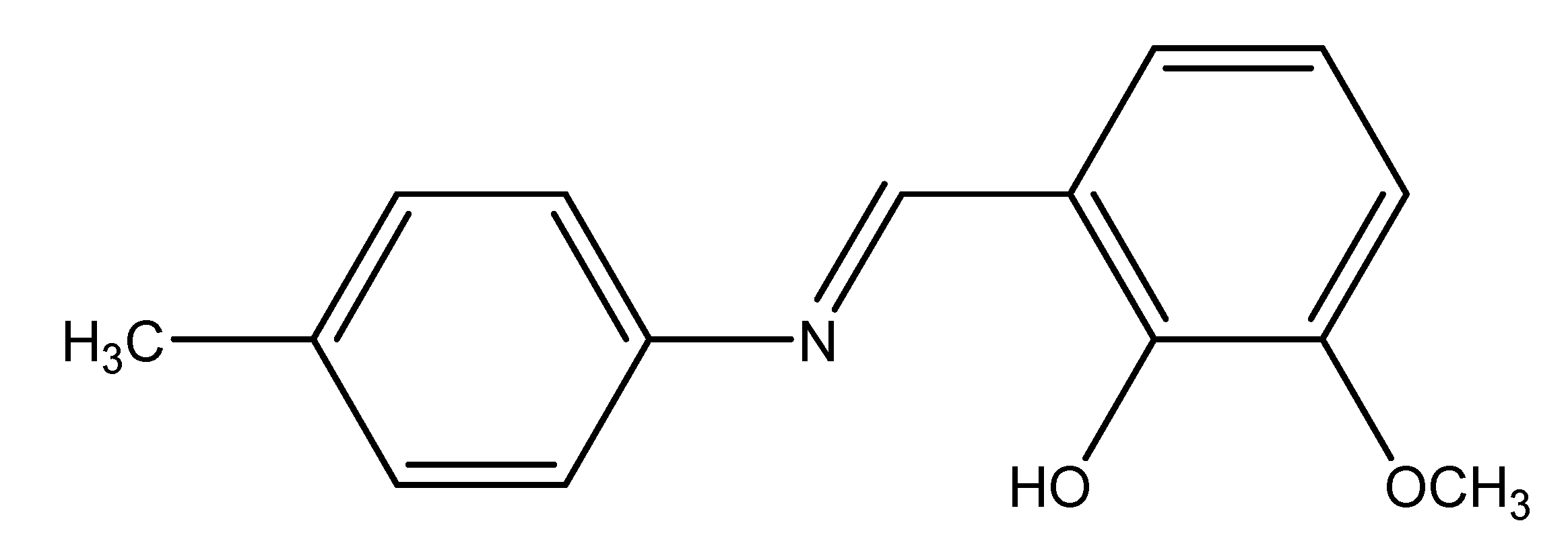
Synthesis, Crystal Structure, and Kinetics of the Thermal Decomposition of the Nickel(II) Complex of the Schiff Base 2-[(4 Methylphenylimino)methyl]-6-methoxyphenol

Abstract
:
1. Introduction
2. Results and Discussion
2.1. IR spectra

| Compound | v OH | v C=N | v C-O | v M-O |
|---|---|---|---|---|
| HL | 3468 (w) | 1614 (s) | 1257 (s) | — |
| [Ni2(H2O)L4]·5H2O | 3444 (m) | 1619 (s) | 1238 (s) | 509 (w) |
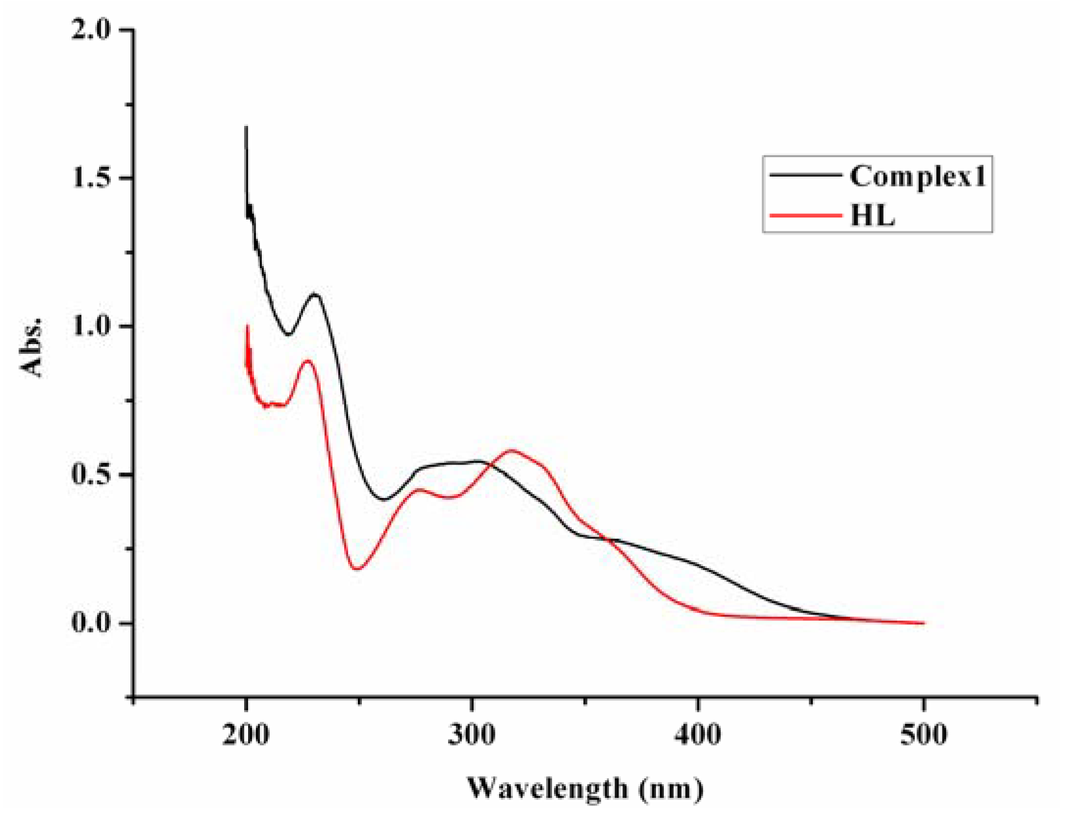
2.2. UV/Vis spectra

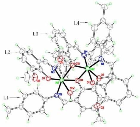
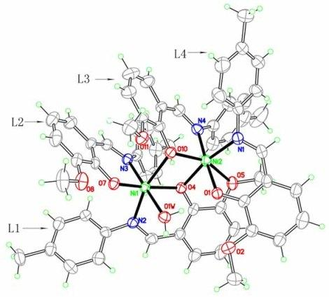
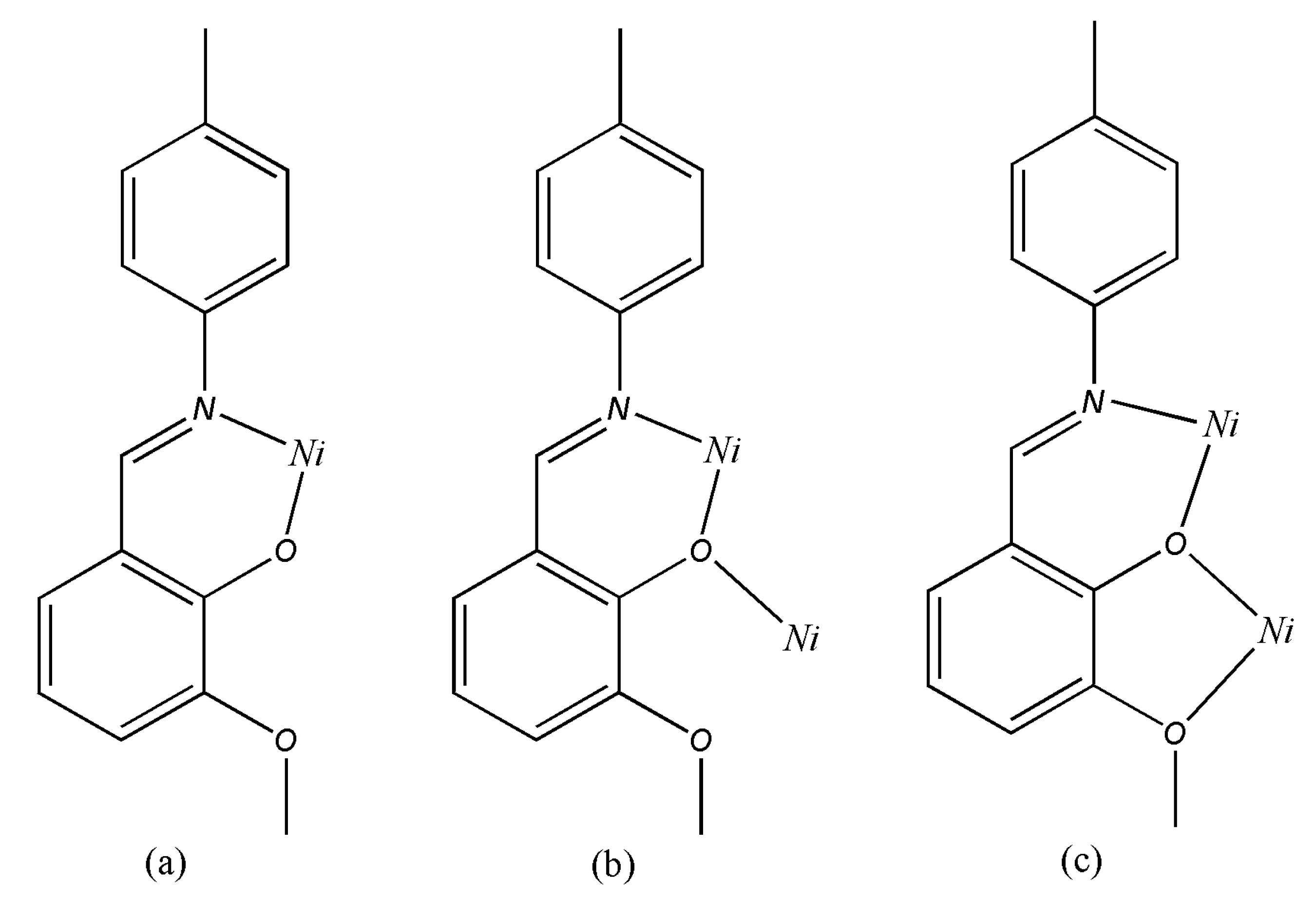
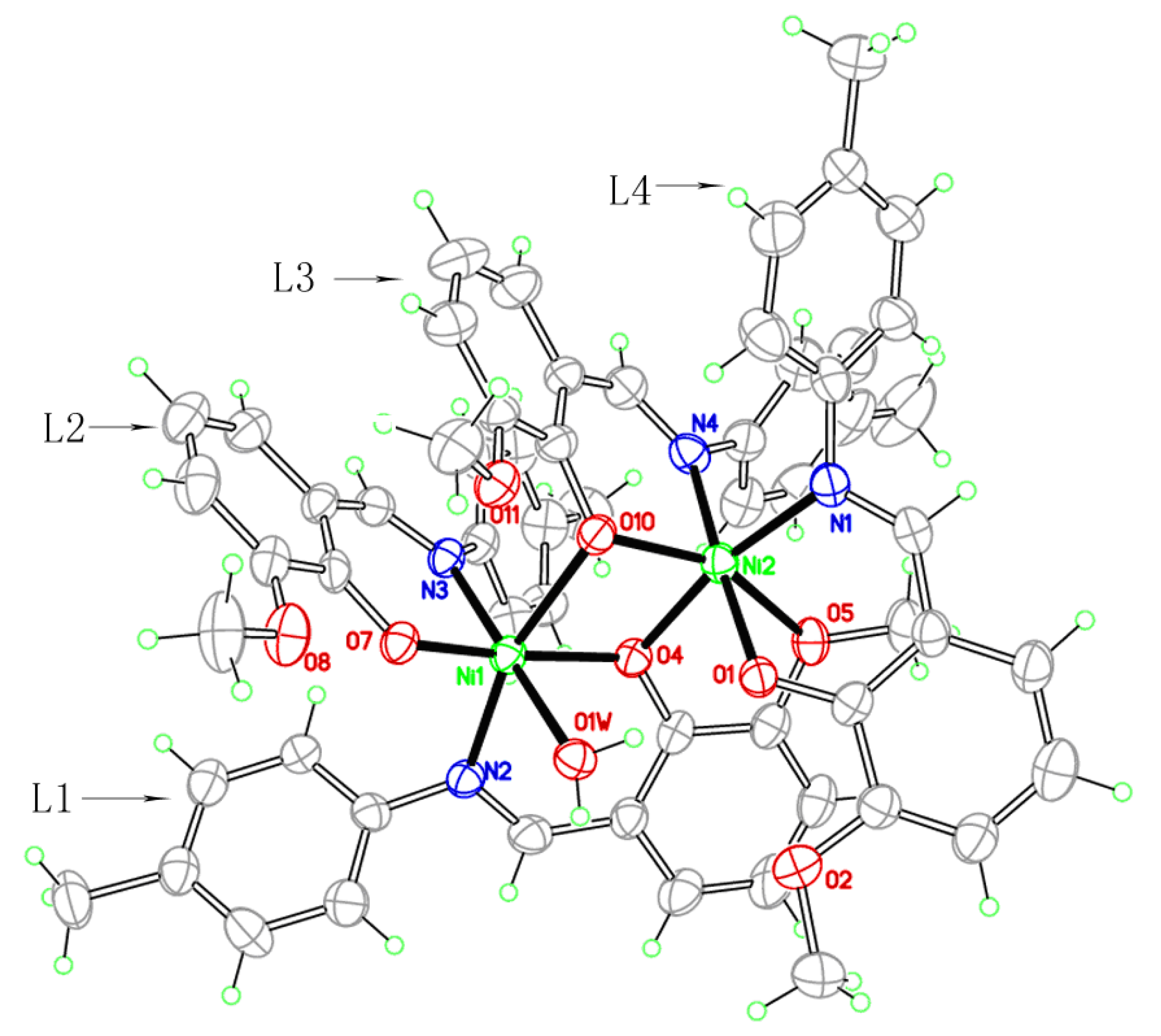
2.3. Crystal structure

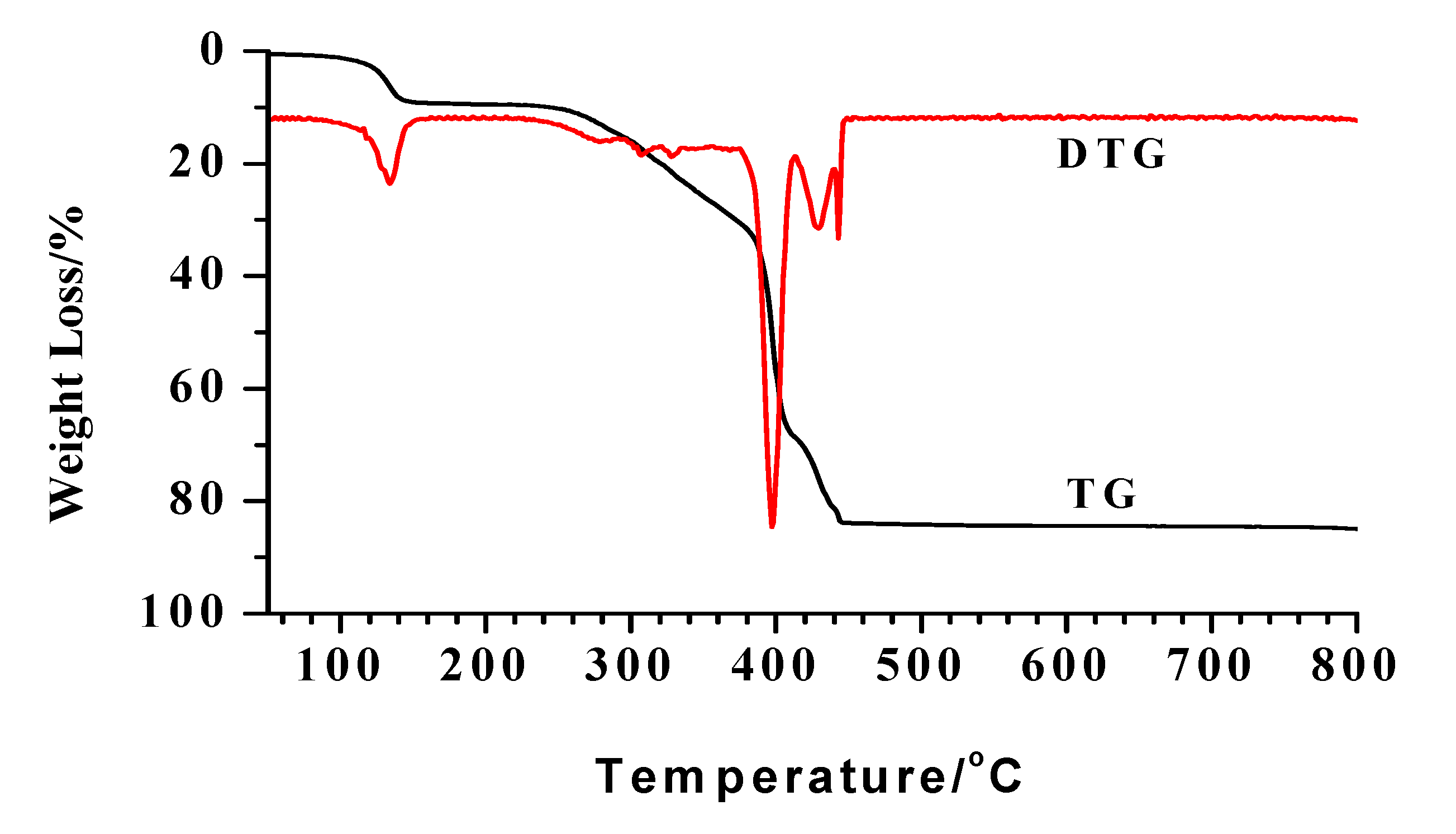
2.4. Thermal analysis
2.5. Non-isothermal kinetics for Steps 1 and 2
| Symbol | Integral kinetic function | Differential coefficient kinetic function | Mechanism |
|---|---|---|---|
| D1 | α2 | 1/(2α) | One-dimensional diffusion |
| D2 | α+(1-α)ln(1-α) | [-ln(1-α)]-1 | Two-dimensional diffusion |
| 1D3 | 1-(2/3)α-(1-α)2/3 | 3/2[(1-α)-1/3-1]-1 | Three-dimensional diffusion (cylindrical symmetry) |
| 2D3 | [1-(1-α)1/3]2 | 3/2(1-α)2/3[1-(1-α)1/3]-1 | Three-dimensional diffusion (spherical symmetry) |
| 3D3 | [(1+α)1/3-1]2 | 3/2(1+α)2/3[(1+α)1/3-1]-1 | Three-dimensional diffusion |
| 4D3 | [1/(1-α)1/3-1]1/2 | 3/2(1-α)4/3[1/(1-α)1/3-1]-1 | Three-dimensional diffusion |
| A1 | -ln(1-α) | (1-α) | Random nucleation and nuclei growth (n=1) |
| A1.5 | [-ln(1-α)]2/3 | 3/2(1-α)[-ln(1-α)]1/3 | Random nucleation and nuclei growth (n=1.5) |
| A2 | [-ln(1-α)]1/2 | 2(1-α)[-ln(1-α)]1/2 | Random nucleation and nuclei growth (n=2) |
| A3 | [-ln(1-α)]1/3 | 3(1-α) [-ln(1-α)]2/3 | Random nucleation and nuclei growth (n=3) |
| A4 | [-ln(1-α)]1/4 | 4(1-α) [-ln(1-α)]3/4 | Random nucleation and nuclei growth (n=4) |
| R2 | 1-(1-α)1/2 | 2(1-α)1/2 | Contracting sphere (cylindrical symmetry) |
| R3 | 1-(1-α)1/3 | 3(1-α)2/3 | Contractingsphere (spherical symmetry) |
| P1 | α | 1 | Exponential nucleation |
| P2 | α1/2 | 2α1/2 | Exponential nucleation |
| P3 | α1/3 | 3α2/3 | Exponential nucleation |
| P4 | α1/4 | 4α3/4 | Exponential nucleation |
| C2 | (1-α)-1-1 | (1-α)2 | Chemical reaction |
| C1.5 | (1-α)-1/2 | 2(1-α)3/2 | Chemical reaction |
| Step 1 | Step 2 | ||||
|---|---|---|---|---|---|
| T/K | α | dα/dt | T/K | α | dα/dt |
| 363.15 | 0.0204 | 2.1941 | 493.15 | 0.0158 | 2.5213 |
| 373.15 | 0.0548 | 1.8824 | 503.15 | 0.0416 | 1.3356 |
| 383.15 | 0.1163 | 1.5496 | 513.15 | 0.0930 | 1.8930 |
| 393.15 | 0.2237 | 1.6131 | 523.15 | 0.1888 | 1.6043 |
| 403.15 | 0.4937 | 1.8258 | 533.15 | 0.3547 | 1.3785 |
| 413.15 | 0.9043 | 0.6285 | 543.15 | 0.6250 | 1.2225 |
| Achar method | Coats–Redfern method | |||||
|---|---|---|---|---|---|---|
| No. | E/kJ mol-1 | A/s-1 | r | E/kJ mol-1 | A/s-1 | r |
| 1 | 207.59 | 2.18E+06 | 0.9548 | 171.38 | 3.37E+20 | 0.9989 |
| 2 | 265.51 | 4.98E-03 | 0.9914 | 181.63 | 5.40E+21 | 0.9990 |
| 3 | 290.10 | 8.78E+02 | 0.9947 | 185.89 | 5.06E+21 | 0.9986 |
| 4 | 336.58 | 8.53E-12 | 0.9905 | 194.64 | 9.66E+22 | 0.9970 |
| 5 | 162.82 | 4.90E-14 | 0.9032 | 161.29 | 1.20E+18 | 0.9980 |
| 6 | 475.99 | 2.52E+00 | 0.9573 | 224.61 | 2.31E+27 | 0.9867 |
| 7 | 74.73 | 1.27E+02 | 0.7679 | 101.24 | 3.26E+11 | 0.9926 |
| 8 | -35.34 | 1.16E-05 | 0.5499 | 65.41 | 3.97E+06 | 0.9922 |
| 9 | 36.58 | 2.58E-16 | 0.6520 | 47.49 | 1.25E+04 | 0.9918 |
| 10 | -145.40 | 6.08E-20 | 0.9530 | 29.57 | 3.38E+01 | 0.9907 |
| 11 | -172.92 | 8.57E-22 | 0.9683 | 20.61 | 1.55E+00 | 0.9894 |
| 12 | 5.02 | 4.07E+17 | 0.1679 | 90.98 | 4.87E+09 | 0.9981 |
| 13 | 28.25 | 4.70E+11 | 0.6219 | 94.19 | 9.74E+09 | 0.9969 |
| 14 | -64.69 | 1.16E-05 | 0.7463 | 82.55 | 5.37E+08 | 0.9988 |
| 15 | -200.83 | 2.58E-16 | 0.9640 | 38.14 | 4.51E+02 | 0.9986 |
| 16 | -246.21 | 6.08E-20 | 0.9761 | 23.34 | 3.37E+00 | 0.9983 |
| 17 | -268.90 | 8.57E-22 | 0.9799 | 15.94 | 2.54E-01 | 0.9979 |
| 18 | 214.14 | 4.07E+17 | 0.7969 | 127.51 | 2.47E+15 | 0.9673 |
| 19 | 144.43 | 4.70E+11 | 0.7922 | 16.21 | 6.03E-01 | 0.6840 |
| Achar method | Coats–Redfern method | |||||
|---|---|---|---|---|---|---|
| No. | E/kJ mol-1 | A/s-1 | r | E/kJ mol-1 | A/s-1 | r |
| 1 | 325.14 | 1.12E+21 | 0.9875 | 317.17 | 3.75E+28 | 0.9974 |
| 2 | 366.94 | 3.70E+23 | 0.9927 | 328.16 | 2.72E+29 | 0.9985 |
| 3 | 382.33 | 2.02E+24 | 0.9933 | 332.19 | 1.61E+29 | 0.9988 |
| 4 | 412.37 | 9.59E+25 | 0.9932 | 340.31 | 1.16E+30 | 0.9992 |
| 5 | 276.76 | 6.57E+16 | 0.9760 | 302.95 | 1.28E+26 | 0.9958 |
| 6 | 502.52 | 1.16E+32 | 0.9872 | 365.92 | 5.83E+32 | 0.9994 |
| 7 | 35.18 | 4.55E+02 | 0.4475 | 172.10 | 4.57E+14 | 0.9995 |
| 8 | -105.45 | 3.05E-07 | 0.8205 | 111.86 | 4.83E+08 | 0.9995 |
| 9 | 2.73 | 1.46E+00 | 0.0447 | 81.75 | 4.50E+05 | 0.9994 |
| 10 | -246.09 | 1.53E-16 | 0.9545 | 51.63 | 3.63E+02 | 0.9994 |
| 11 | -281.25 | 6.48E-19 | 0.9638 | 36.57 | 9.18E+00 | 0.9993 |
| 12 | -9.90 | 2.07E-01 | 0.1732 | 162.85 | 2.34E+13 | 0.9988 |
| 13 | 5.13 | 1.42E+00 | 0.0849 | 165.85 | 3.27E+13 | 0.9992 |
| 14 | -54.97 | 3.76E-04 | 0.7347 | 154.28 | 5.67E+12 | 0.9972 |
| 15 | -245.03 | 1.54E-16 | 0.9743 | 72.84 | 4.72E+04 | 0.9968 |
| 16 | -308.38 | 9.63E-21 | 0.9820 | 45.69 | 7.71E+01 | 0.9963 |
| 17 | -340.06 | 6.99E-23 | 0.9844 | 32.12 | 2.77E+00 | 0.9958 |
| 18 | 125.33 | 5.51E+08 | 0.7525 | 192.70 | 7.18E+16 | 0.9979 |
| 19 | 80.25 | 2.50E+05 | 0.6703 | 10.60 | 1.22E-02 | 0.7115 |
3. Experimental
3.1. Materials and general methods
3.2. Syntheses

3.3. Crystal structure determination
| Empirical formula | C60H68N4Ni2O14 |
|---|---|
| Formula weight | 1186.60 |
| T/ K | 296(2) |
| Crystal system | monoclinic |
| Space group | P 21/c |
| a (Å) | 13.2837(5) |
| b (Å) | 27.3886(10) |
| c (Å) | 17.5415(6) |
| α (°) | 90 |
| β (°) | 108.429(2) |
| γ (°) | 90 |
| V (Å3) | 6054.7(4) |
| Z | 4 |
| Density (g/cm3) | 1.302 |
| Μ (mm-1) | 0.687 |
| F (000) | 2496 |
| Absorption correction | none |
| Data/restrains/parameters | 10668 / 18 / 748 |
| θ range /° | 1.43 to 25.00 |
| Limiting indices | -15<=h<=15, -32<=k<=31, -20<=l<=20 |
| Reflections collected/ unique | 81485 / 10668 |
| Rint | 0.0950 |
| GOOF on F2 | 1.032 |
| R and wR (I > 2σ(I)) | R = 0.0873, wR = 0.2594 |
| R indices (all data) | R = 0.1287, wR = 0.3012 |
| (Δρ)max, (Δρ)min (e·Å-3) | 2.174 and -0.465 |
| Bond | (Å) | Bond | (Å) | Bond | (Å) |
|---|---|---|---|---|---|
| Ni(1)-O(4) | 1.9734(10) | C(7)-N(1) | 1.2942(19) | C(18)-O(5) | 1.3658(19) |
| Ni(1)-O(7) | 1.9410(10) | C(9)-N(1) | 1.4138(19) | C(23)-O(5) | 1.450(2) |
| Ni(1)-O(10) | 2.3389(10) | C(22)-N(2) | 1.310(2) | C(32)-O(7) | 1.3041(16) |
| Ni(1)-O(1W) | 2.2364(8) | C(24)-N(2) | 1.410(2) | C(33)-O(8) | 1.3614(15) |
| Ni(1)-N(2) | 2.0680(11) | C(39)-N(3) | 1.4309(18) | C(38)-O(8) | 1.436(2) |
| Ni(1)-N(3) | 2.0898(10) | C(37)-N(3) | 1.287(2) | C(47)-O(10) | 1.3431(16) |
| Ni(2)-O(1) | 2.0253(8) | C(52)-N(4) | 1.321(2) | C(48)-O(11) | 1.3759(15) |
| Ni(2)-O(4) | 2.0602(10) | C(54)-N(4) | 1.428(2) | C(53)-O(11) | 1.4333(19) |
| Ni(2)-O(5) | 2.2876(9) | C(2)-O(1) | 1.3079(18) | ||
| Ni(2)-O(10) | 2.0048(10) | C(3)-O(2) | 1.351(2) | ||
| Ni(2)-N(1) | 2.0977(13) | C(8)-O(2) | 1.451(2) | ||
| Ni(2)-N(4) | 2.0545(10) | C(17)-O(4) | 1.3049(16) | ||
| Angle | (°) | Angle | (°) | Angle | (°) |
| O(4)-Ni(1)-O(10) | 77.63(4) | N(3)-Ni(1)-O(4) | 96.47(4) | O(10)-Ni(2)-O(4) | 83.91(4) |
| O(4)-Ni(1)-O(1W) | 86.08(4) | N(3)-Ni(1)-O(7) | 91.81(4) | N(1)-Ni(2)-O(1) | 83.81(4) |
| O(7)-Ni(1)-O(4) | 165.05(4) | N(3)-Ni(1)-O(10) | 94.16(4) | N(1)-Ni(2)-O(4) | 169.18(4) |
| O(7)-Ni(1)-O(10) | 89.40(4) | N(3)-Ni(1)-O(1W) | 176.05(4) | N(1)-Ni(2)-O(10) | 103.13(4) |
| O(7)-Ni(1)-O(1W) | 85.06(4) | N(2)-Ni(1)-N(3) | 98.66(4) | N(1)-Ni(2)-O(5) | 100.19(4) |
| O(10)-Ni(1)-O(1W) | 83.41(3) | O(1)-Ni(2)-O(4) | 87.80(4) | N(4)-Ni(2)-O(1) | 176.94(5) |
| N(2)-Ni(1)-O(4) | 92.08(4) | O(1)-Ni(2)-O(5) | 84.21(3) | N(4)-Ni(2)-O(4) | 93.60(4) |
| N(2)-Ni(1)-O(7) | 98.97(4) | O(4)-Ni(2)-O(5) | 72.08(4) | N(4)-Ni(2)-O(5) | 93.62(4) |
| N(2)-Ni(1)-O(10) | 164.41(4) | O(5)-Ni(2)-O(10) | 155.78(4) | N(4)-Ni(2)-O(10) | 91.00(4) |
| N(2)-Ni(1)-O(1W) | 84.23(4) | O(10)-Ni(2)-O(1) | 91.85(4) | N(4)-Ni(2)-N(1) | 94.47(5) |
4. Supplementary Material
References and Notes
- Eddaoudi, M.; Moler, D.B.; Li, H.; Chen, B.; Reineke, T.M.; O'Keeffe, M.; Yaghi, O.M. Modular chemistry: Secondary building units as a basis for the design of highly porous and robust metal-organic carboxylate frameworks. Acc. Chem. Res. 2001, 34, 319–330. [Google Scholar] [CrossRef]
- Fontes, A.P.S.; Bandarage, R.; Farrell, N.; Qu, Y.; Rauter, H.; Kelland, L.R. Synthesis, Characterization, and Cytotoxicity of Trifunctional Dinuclear Platinum Complexes: Comparison of Effects of Geometry and Polyfunctionality on Biological Activity. J. Med. Chem. 2000, 43, 3189–3192. [Google Scholar] [CrossRef]
- Huang, Y.S. Chemistry of Spicery oil; Shanghai Science and Technology Press: Shanghai, China, 1959; p. 333. (in Chinese) [Google Scholar]
- Zhu, R.H.; Xue, Q.C. Handbook of Synthesis Practical Spicery; Light Industry Press: Beijing, China, 1986; p. 167. (in Chinese) [Google Scholar]
- Praefcke, K.; Bilgin, B.; Pickardt, J.; Borowski, M. A novel platinum methylene complex. J. Organomet. Chem. 1999, 592, 155–161. [Google Scholar] [CrossRef]
- Elmali, A.; Elerman, Y.; Zeyrek, C.T.; Svoboda, I. Crystal Structure and Magnetic Properties of a Dinuclear Iron(III) Doubly Oxygen Bridged Schiff Base Complex. Z. Naturforsch. B Chem. Sci. 2003, 58, 433–437. [Google Scholar]
- Yeap, G.Y.; Ha, S.T.; Ishizawa, N.; Suda, K.; Boey, P.L.; Mahmood, W.A.K. Synthesis, crystal structure and spectroscopic study of para substituted 2-hydroxy-3-methoxybenzalideneanilines. J. Mol. Struct. 2003, 658, 87–99. [Google Scholar] [CrossRef]
- Eran, B.B.; Singer, D.; Pickardt, J.; Praefcke, K. Thiocyanato-bridged platinum heterocycles: structure and properties of disc-like metallomesogens. J. Organomet. Chem. 2001, 620, 249–255. [Google Scholar] [CrossRef]
- Holm, R.H.; Evert, G.W.; Chakravorty, A. Metal Complexes of Schiff Bases and β–Ketoamines. Prog. Inorg. Chem. 1966, 7, 83–214. [Google Scholar]
- Dharmaraj, N.; Viswanathamurthi, P.; Natarajan, K. Ruthenium(II) complexes containing bidentate Schiff bases and their antifungal activity. Transition Met. Chem. 2001, 26, 105–109. [Google Scholar] [CrossRef]
- Yalpani, M.; Hall, L.D. Some chemical and analytical aspects of polysaccharide modifications. 3. Formation of branched-chain, soluble chitosan derivatives. Macromolecules 1984, 17, 272–281. [Google Scholar] [CrossRef]
- Miyasaka, H.; Ieda, H.; Matsumoto, N.; Crescenzi, R.; Floriani, C. Assembling Bi-, Tri- and Pentanuclear Complexes into Extended Structures Using a Desolvation Reaction: Synthesis, Structure, and Magnetic Properties of Manganese(III)−Schiff-Base−Hexacyanoferrate Polymeric Compounds and Their Derived Extended Structures. Inorg. Chem. 1998, 37, 255–263. [Google Scholar]
- Kato, M., Muto. Factors affecting the magnetic properties of dimeric copper(II) complexes. Coord. Chem. Rev. 1988, 92, 45–83. [Google Scholar] [CrossRef]
- Iglesias, R.; Marcos, M.; Serano, J.L.; Sierra, T. Ferroelectric Behavior of Chiral Bis(salicylideneaniline) Copper(II), Vanadium(IV), and Palladium(II) Liquid Crystals. Chem. Mater. 1996, 8, 2611–2617. [Google Scholar] [CrossRef]
- Zhao, G.L.; Feng, Y.L.; Wen, Y.H. Syntheses, crystal structures and kinetic mechanisms of thermal decomposition of rare earth complexes with Schiff base derived from o-vanillin and p-toluidine. J. Rare Earths 2006, 24, 268–275. [Google Scholar] [CrossRef]
- Li, H.Q.; Xian, H.D.; Liu, J.F.; Zhao, G.L. Catena-Poly[[di-μ-chlorido-bis{[6-methoxy-2-(4-methylphenyliminiomethyl)phenolato-κ2O,O']cadmium(II)}]-di-μ2-thiocyanato-κ2N:S;κ2S:N]. Acta Crystallogr., Sect. E: Struct. Rep. Online 2008, 64, m1593–m1594. [Google Scholar] [CrossRef]
- Yu, Y.Y.; Zhao, G.L.; Wen, Y.H. Syntheses, Characterizations, Crystal Structures and Antibacterial Activities of Two Zinc(II) Complexes with a Schiff Base Derived from o-Vanillin and p-Toluidine. Chinese J. Struct. Chem. 2007, 26, 1395–1402. [Google Scholar]
- Nakamoto, K. Infrared and Raman spectra of Inorganic and Coordination Compounds, 5th ed.; Wiley &Sons: New York, NY, USA, 1986; pp. 232–234. [Google Scholar]
- Dey, M.; Rao, C.P.; Saarenketo, P.K.; Rissanen, K. Mono-, di- and tri-nuclear Ni(II) complexes of N-, O-donor ligands: structural diversity and reactivity. Inorg.Chem.Commun. 2002, 5, 924–928. [Google Scholar] [CrossRef]
- Roesky, H.W.; Andruh, M. The interplay of coordinative, hydrogen bonding and π–π stacking interactions in sustaining supramolecular solid-state architectures. A study case of bis(4-pyridyl)- and bis(4-pyridyl-N-oxide) tectons. Coord. Chem. Rev. 2003, 236, 91–119. [Google Scholar] [CrossRef]
- Achar, B.N.; Brindley, G.W.; Sharp, J.H. Kinetics and mechanism of dehydroxylation process. III. Applications and limitations of dynamic methods. Proc. Int. Clay. Conf. Jerusalem 1966, 1, 67–73. [Google Scholar]
- Sharp, J.H.; Wendworth, S.A. Kinetic analysis of thermogravimetric data. Anal. Chem. 1969, 41, 2060–2062. [Google Scholar] [CrossRef]
- Coats, A.W.; Redfern, J.P. Kinetic Parameters from Thermogravimetric Data. Nature 1964, 201, 68–69. [Google Scholar] [CrossRef]
- Sheldrick, G.M. SHELXS97 and SHELXL97; University of Göttingen: Göttingen, Germany, 1997. [Google Scholar]
- Sample Availability: Samples of the compound is available from the authors.
© 2009 by the authors; licensee Molecular Diversity Preservation International, Basel, Switzerland. This article is an open access article distributed under the terms and conditions of the Creative Commons Attribution license ( http://creativecommons.org/licenses/by/3.0/).
Share and Cite
Wang, Y.-F.; Liu, J.-F.; Xian, H.-D.; Zhao, G.-L. Synthesis, Crystal Structure, and Kinetics of the Thermal Decomposition of the Nickel(II) Complex of the Schiff Base 2-[(4 Methylphenylimino)methyl]-6-methoxyphenol. Molecules 2009, 14, 2582-2593. https://doi.org/10.3390/molecules14072582
Wang Y-F, Liu J-F, Xian H-D, Zhao G-L. Synthesis, Crystal Structure, and Kinetics of the Thermal Decomposition of the Nickel(II) Complex of the Schiff Base 2-[(4 Methylphenylimino)methyl]-6-methoxyphenol. Molecules. 2009; 14(7):2582-2593. https://doi.org/10.3390/molecules14072582
Chicago/Turabian StyleWang, Yan-Fang, Jian-Feng Liu, Hui-Duo Xian, and Guo-Liang Zhao. 2009. "Synthesis, Crystal Structure, and Kinetics of the Thermal Decomposition of the Nickel(II) Complex of the Schiff Base 2-[(4 Methylphenylimino)methyl]-6-methoxyphenol" Molecules 14, no. 7: 2582-2593. https://doi.org/10.3390/molecules14072582
APA StyleWang, Y.-F., Liu, J.-F., Xian, H.-D., & Zhao, G.-L. (2009). Synthesis, Crystal Structure, and Kinetics of the Thermal Decomposition of the Nickel(II) Complex of the Schiff Base 2-[(4 Methylphenylimino)methyl]-6-methoxyphenol. Molecules, 14(7), 2582-2593. https://doi.org/10.3390/molecules14072582