Journal Description
Journal of Otorhinolaryngology, Hearing and Balance Medicine
is an international, scientific, peer-reviewed, open access journal of otorhinolaryngology, hearing and balance medical studies, published semiannually online by MDPI.
- Open Access— free for readers, with article processing charges (APC) paid by authors or their institutions.
- Rapid Publication: manuscripts are peer-reviewed and a first decision is provided to authors approximately 26.2 days after submission; acceptance to publication is undertaken in 4.6 days (median values for papers published in this journal in the first half of 2026).
- Recognition of Reviewers: reviewers who provide timely, thorough peer-review reports receive vouchers entitling them to a discount on the APC of their next publication in any MDPI journal, in appreciation of the work done.
Latest Articles
Thyroid-Presenting Plasmablastic Lymphoma Mimicking Anaplastic Thyroid Carcinoma
J. Otorhinolaryngol. Hear. Balanc. Med. 2026, 7(2), 32; https://doi.org/10.3390/ohbm7020032 - 20 Aug 2026
Abstract
Background/Objectives: Primary thyroid lymphoma accounts for approximately 0.2–2% of thyroid malignancies. Plasmablastic lymphoma (PBL), an aggressive large B-cell neoplasm with plasma-cell differentiation and frequent loss of conventional B-cell markers, is a rare thyroid presentation. We report a thyroid PBL presenting as an
[...] Read more.
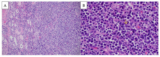
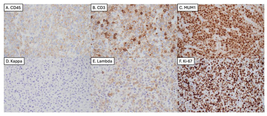
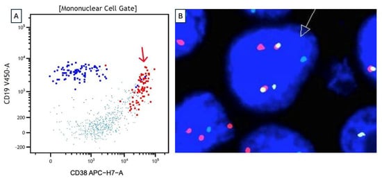
Background/Objectives: Primary thyroid lymphoma accounts for approximately 0.2–2% of thyroid malignancies. Plasmablastic lymphoma (PBL), an aggressive large B-cell neoplasm with plasma-cell differentiation and frequent loss of conventional B-cell markers, is a rare thyroid presentation. We report a thyroid PBL presenting as an acute surgical airway emergency in an immunocompetent patient and highlight the diagnostic and management pitfalls that distinguish this from anaplastic thyroid carcinoma. Case Presentation: A 75-year-old man without any significant past medical history presented with rapidly progressive right-sided neck swelling, dysphagia, inspiratory stridor, and respiratory failure. Imaging demonstrated a large, thyroid-centered mass with tracheal involvement, initially raising concern for anaplastic thyroid carcinoma. Histopathology revealed a diffuse infiltrate of large, atypical cells with immunoblastic and plasmablastic morphology. The neoplastic cells were CD20- and CD138-negative but strongly MUM1-positive, with lambda light-chain restriction, bright CD38 by flow cytometry, a Ki-67 proliferation index exceeding 95%, aberrant cytoplasmic CD3 expression, and a MYC::IGH rearrangement, supporting a diagnosis of PBL. Staging identified extranodal perinephric disease and mesenteric lymphadenopathy, consistent with disseminated extranodal Ann Arbor stage IV disease. The patient underwent systemic treatment and initially had an excellent response; however, one month after the last treatment cycle they presented to the hospital with a mass consistent with recurrence. Discussion: Rapid growth, fixation, and tracheal invasion strongly suggest anaplastic thyroid carcinoma in routine clinical practice. However, plasmablastic lymphomas can present similarly and require fundamentally different treatment. In this case, loss of conventional B-cell markers, CD138 negativity, and aberrant cytoplasmic CD3 expression created substantial diagnostic challenges. Light-chain restriction, plasma-cell-associated markers, flow cytometry, and MYC cytogenetics were vitally important. Conclusions: Thyroid-presenting PBL is exceptionally rare and may closely mimic anaplastic thyroid carcinoma, including presentation with life-threatening airway compromise and tracheal involvement. This case highlights several diagnostic pitfalls: CD138 negativity despite plasma-cell differentiation, and aberrant cytoplasmic CD3 expression. Prompt airway stabilization, adequate tissue acquisition, broad immunophenotyping, light-chain assessment, flow cytometry, EBV/HHV8/ALK testing, and MYC cytogenetics are essential for accurate diagnosis and lymphoma-directed treatment.
Full article
(This article belongs to the Section Head and Neck Surgery)
►
Show Figures
Open AccessReview
Contemporary Management of Patulous Eustachian Tube: A Narrative Review of Current Evidence and Future Directions
by
Laila Aldokhail, Enar Alotaibi, Mayar Alsaqr, Saleh Alabood, Anwar Alshehri and Essa Bakry
J. Otorhinolaryngol. Hear. Balanc. Med. 2026, 7(2), 31; https://doi.org/10.3390/ohbm7020031 - 14 Aug 2026
Abstract
Patulous Eustachian Tube (PET) is a rare otologic disorder, which is caused by abnormal patency of the Eustachian tube. Rapid weight loss is a recognized predisposing factor for PET potentially because of the reduction in peritubal adipose tissue that contributes to normal tubal
[...] Read more.
Patulous Eustachian Tube (PET) is a rare otologic disorder, which is caused by abnormal patency of the Eustachian tube. Rapid weight loss is a recognized predisposing factor for PET potentially because of the reduction in peritubal adipose tissue that contributes to normal tubal closure. Although there is increasing awareness of this association, the best treatment of PET is still debated with several conservative, medical and surgical operations reported in the literature. This narrative review summarizes the available evidence regarding conservative, medical, minimally invasive, and surgical management of PET, with particular consideration of PET associated with rapid weight loss. Relevant literature was identified through searches of PubMed/MEDLINE, Embase, Web of Science, and the Cochrane Library, with attention to studies using standardized diagnostic criteria and outcome measures. Conservative approaches, including hydration, nasal saline instillation, and behavioral modifications such as cessation of habitual sniffing, have been associated with symptomatic improvement and considered as initial options because of their accessibility and low procedural burden. Medical therapies, such as topical irritants, estrogen-based preparations, and mucosal-thickening agents, may provide temporary symptom relief, although their long-term effects require further evaluation. Among interventional strategies, silicone (Kobayashi) plug insertion has demonstrated encouraging symptom improvement and reductions in symptom scores in selected patients with refractory PET. Minimally invasive techniques, including autologous fat injection, cartilage augmentation, and soft tissue bulking procedures, have also demonstrated encouraging outcomes. Trans-tympanic interventions, including ventilation-tube insertion and catheter-based techniques, provided additional options with variable reported outcomes. Management of PET remains heterogeneous and largely individualized. Conservative and medical therapies are considered reasonable first-line options, while surgical and minimally invasive approaches may be considered for selected patients with persistent symptoms.
Full article
(This article belongs to the Section Otology and Neurotology)
Open AccessArticle
Outcomes of Audiologist-Delivered Tinnitus Management in Singapore Public Health Service: A Retrospective Service Evaluation
by
Gary Jek Chong Lee, Jasmine Jek Peng Liew and Andrew Wei Li Teng
J. Otorhinolaryngol. Hear. Balanc. Med. 2026, 7(2), 30; https://doi.org/10.3390/ohbm7020030 - 7 Aug 2026
Abstract
Background/Objectives: Tinnitus can have an adverse impact on some patients’ lives, affecting the daily functioning for a proportion of patients. In recent years, audiologists in Singapore who are specialised in tinnitus and hyperacusis rehabilitation have increasingly been providing tinnitus and/or hyperacusis management. The
[...] Read more.
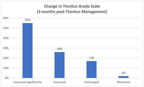
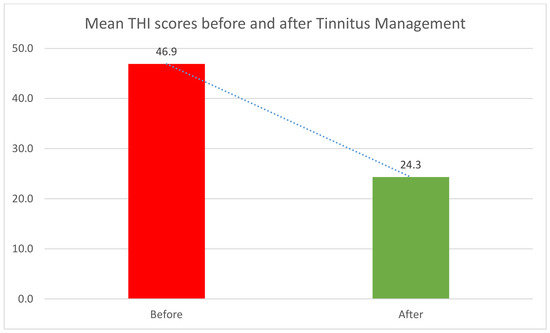
Background/Objectives: Tinnitus can have an adverse impact on some patients’ lives, affecting the daily functioning for a proportion of patients. In recent years, audiologists in Singapore who are specialised in tinnitus and hyperacusis rehabilitation have increasingly been providing tinnitus and/or hyperacusis management. The aim of this study was to evaluate the outcome of audiologist-delivered tinnitus management at a public hospital in Singapore. Methods: Retrospective data from patients seen between 2016 and 2019 were extracted from the hospital’s electronic medical record system. Outcomes were analysed using pre-treatment and three-month post-treatment Tinnitus Handicap Inventory (THI) scores among patients referred to the audiologist-led tinnitus clinic. Results: A total of 201 patients aged 18 to 86 years received tinnitus management. Among patients who returned for follow-up, 93% showed a reduction in THI score three months after treatment. Mean THI scores decreased significantly from 46.9 to 24.3, with a corrected effect size of 1.16, indicating a large treatment effect. Among patients with moderate or greater tinnitus handicap at baseline, 81% improved by at least one severity grade. Conclusions: This is one of the first studies to report outcomes of an audiologist-led tinnitus management service in Singapore. The findings are consistent with a potential role for structured, audiologist-led tinnitus services in Asian healthcare settings, although controlled studies are needed to confirm treatment effectiveness. General practitioners, otolaryngologists and other healthcare providers can play an important role in recognising tinnitus-related distress, offering early reassurance and directing patients to appropriate audiological and supportive care.
Full article
(This article belongs to the Section Otology and Neurotology)
►▼
Show Figures

Figure 1
Open AccessCase Report
Management of Adult Subglottic Foreign Body: A Case Report
by
Giada Cavallaro, Domenico Cifarelli, Margherita Laguardia, Guglielmo Larotonda, Camilla Gallipoli, Giuseppe De Cillis and Giacinto Asprella-Libonati
J. Otorhinolaryngol. Hear. Balanc. Med. 2026, 7(2), 29; https://doi.org/10.3390/ohbm7020029 - 3 Aug 2026
Abstract
Objectives: To describe a rare case of delayed diagnosis of a subglottic foreign body (FB) in an adult, resulting in subglottic stenosis, and to evaluate the effectiveness of Suspension Microlaryngoscopy (SML) as a minimally invasive technique for its management. Case presentation: An adult
[...] Read more.
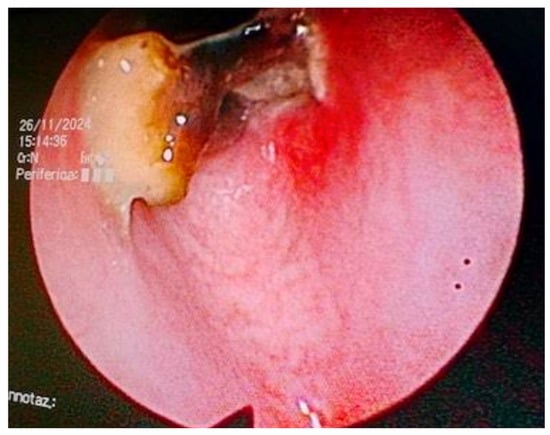
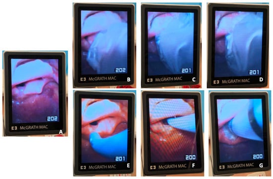
Objectives: To describe a rare case of delayed diagnosis of a subglottic foreign body (FB) in an adult, resulting in subglottic stenosis, and to evaluate the effectiveness of Suspension Microlaryngoscopy (SML) as a minimally invasive technique for its management. Case presentation: An adult patient presented with persistent respiratory symptoms and was ultimately diagnosed with a retained subglottic FB after a delayed clinical course. The FB was managed using endotracheal intubation and SML, which allowed precise visualization and extraction. Associated granulation tissue and mucosal overgrowth were treated with targeted resection and controlled cauterization. Discussion: The delayed presentation and nonspecific clinical manifestations posed significant diagnostic challenges, contributing to the persistence of the FB and the development of subglottic stenosis. SML provided excellent exposure of the subglottic region, enabling safe FB removal while minimizing trauma to surrounding tissues. Postoperative follow-up demonstrated complete symptom resolution and restoration of airway patency without complications. Conclusions: Subglottic FB impaction is an exceptionally rare condition in adults and may be overlooked because of its nonspecific presentation, leading to delayed diagnosis and airway complications. This case highlights the importance of maintaining a high index of suspicion for retained airway FBs in patients with persistent respiratory symptoms or unexplained subglottic stenosis. SML represents a safe and effective therapeutic option, offering precise visualization, atraumatic FB extraction, and successful management of associated granulation tissue, with favorable clinical outcomes.
Full article
(This article belongs to the Section Head and Neck Surgery)
►▼
Show Figures

Figure 1

Open AccessArticle
Benchmarking Large Language Model Responses Against Surgical Clinical Practice Guidelines for Chronic Rhinosinusitis: The Importance of User Prompts
by
Hetal Lad, Emily Kwon, Ayushi Chadha, Sean Z. Haimowitz, Brandon S. Gold, Rachel Kaye and Wayne D. Hsueh
J. Otorhinolaryngol. Hear. Balanc. Med. 2026, 7(2), 28; https://doi.org/10.3390/ohbm7020028 - 1 Aug 2026
Abstract
Background/Objectives: As patients increasingly rely on large language models (LLMs) for Chronic Rhinosinusitis (CRS) diagnosis, surgical candidacy, and perioperative care, evaluating the accuracy of LLM-generated information against established clinical practice guidelines for surgical management of CRS is essential. Methods: ChatGPT, Google
[...] Read more.
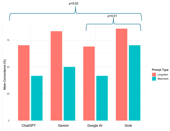
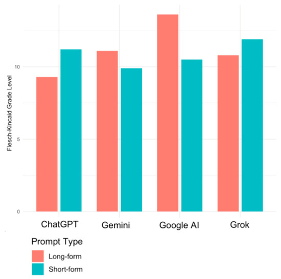
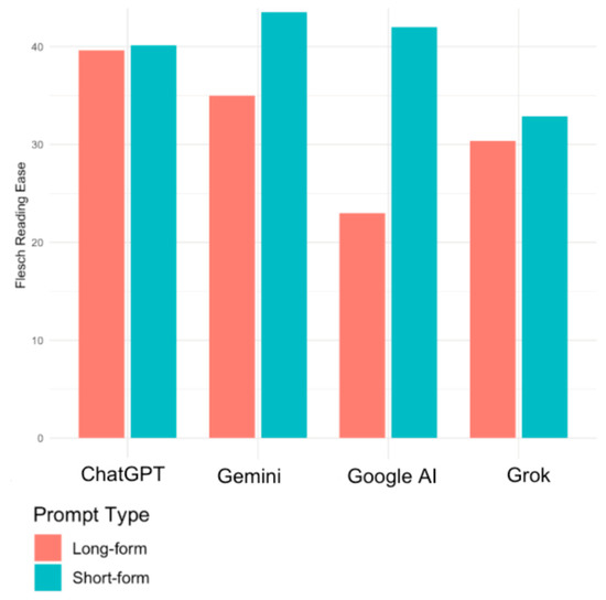
Background/Objectives: As patients increasingly rely on large language models (LLMs) for Chronic Rhinosinusitis (CRS) diagnosis, surgical candidacy, and perioperative care, evaluating the accuracy of LLM-generated information against established clinical practice guidelines for surgical management of CRS is essential. Methods: ChatGPT, Google AI, Google Gemini, and Grok were queried using a 21-question guideline-mapped prompt set (long) and a single patient-focused prompt (short). Two physician reviewers independently scored responses using a 3-point rubric across 21 fields. Primary outcomes were guideline-concordant scores; secondary outcomes included readability measured with the Flesch Reading Ease (FRE) and Flesch-Kincaid Grade Level (FKGL). Inter-rater reliability (IRR) was assessed using the intraclass correlation coefficient (ICC). Analyses were performed in SPSSv31. Results: Guideline concordance ranged from 55.36% to 77.98% (p > 0.05), highest for Grok (77.98%, 95% CI 63.67–92.28), followed by Google Gemini (66.67%, 95% CI 35.42–97.91), ChatGPT (55.95%, 95% CI 29.25–82.65), and Google AI (55.36%, 95% CI 29.97–80.75), with Grok significantly outperforming both ChatGPT and Google AI. Prompt structure significantly affected scores. Long-form prompting resulted in higher guideline concordance scores than short-form prompting (+26.19, p < 0.001). The CPG demonstrated a more readable structure, with a higher FRE (44.1), exceeding scores generated by Grok (31.7), ChatGPT (39.9), Gemini (39.2), and Google AI (32.5). In contrast, the CPG was a higher reading grade level (FKGL score of 11.7) than Grok (11.4), Gemini (10.5), and ChatGPT (10.3), but was lower in reading grade compared to Google AI, which produced the highest FKGL score (12.5). IRR was high (ICC = 0.961). Conclusions: LLMs demonstrated similar guideline concordance, suggesting patients can expect comparable accuracy across platforms. While LLMs generally improved FKGL scores compared to the AAO-HNS CPG, they demonstrated lower FRE scores, indicating mixed results on overall readability. However, longer prompt structure meaningfully influenced output quality, highlighting how a user’s ability to frame precise prompts is critical to obtaining accurate information.
Full article
(This article belongs to the Section Laryngology and Rhinology)
►▼
Show Figures

Figure 1
Open AccessArticle
Enhancing Pediatric Care Through a Multidisciplinary Hearing Disorder and Microtia (HDM) Clinic: A Comprehensive Analysis of Family Experiences and Outcomes
by
See Hui Chiu, Sonia Jaya Sankaran and Ching Yee Chan
J. Otorhinolaryngol. Hear. Balanc. Med. 2026, 7(2), 27; https://doi.org/10.3390/ohbm7020027 - 28 Jul 2026
Abstract
Background: The management of pediatric microtia and congenital hearing loss typically necessitates coordinated multidisciplinary input. Traditional fragmented care pathways across multiple specialties may contribute to delayed diagnosis and treatment intervention, inconsistent counselling, increased logistical burden, and suboptimal care integration. Objectives: We
[...] Read more.
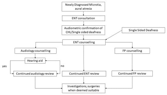
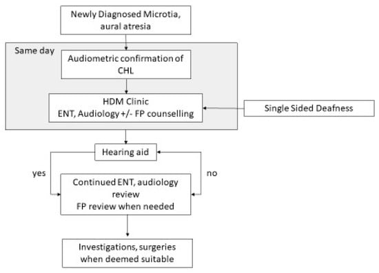
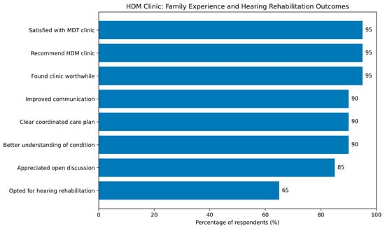
Background: The management of pediatric microtia and congenital hearing loss typically necessitates coordinated multidisciplinary input. Traditional fragmented care pathways across multiple specialties may contribute to delayed diagnosis and treatment intervention, inconsistent counselling, increased logistical burden, and suboptimal care integration. Objectives: We aimed to evaluate family-reported outcomes following implementation of a multidisciplinary Hearing Disorder and Microtia (HDM) clinic, focusing on family satisfaction, perceived care efficiency, understanding of disease and treatment, and early uptake of hearing rehabilitation. Methods: A prospective observational study was conducted on families attending the HDM clinic at a tertiary pediatric center between 2022 and 2024. A structured post-visit survey assessed satisfaction, perceived efficiency, understanding of disease and treatment options, and overall experience. Quantitative data were analyzed descriptively, while qualitative responses underwent thematic analysis. Results: Twenty families were included. Overall satisfaction was high, with 95% rating the clinic experience as very good to excellent and expressing willingness to recommend the service. Improved care coordination and clarification of counselling were reported by the majority of respondents, alongside reduced logistical burden. Notably, 65% of families initiated hearing rehabilitation at the first visit. Thematic analysis identified convenience, unified specialist communication, and clarity of management planning as key strengths of the multidisciplinary model. Conclusions: A structured multidisciplinary HDM clinic significantly improves caregiver experience and facilitates earlier hearing rehabilitation in children with microtia and congenital hearing loss. Integrated care delivery enhances communication, reduces fragmentation, and supports timely, family-centered decision-making. This model may serve as a framework for other complex pediatric otologic conditions requiring longitudinal multidisciplinary management.
Full article
(This article belongs to the Special Issue Etiology, Diagnosis, and Treatment of Congenital Hearing Loss)
►▼
Show Figures

Figure 1

Open AccessArticle
Combining Dexamethasone and D-Methionine Provides Greater Protection Against Cisplatin- and Radiation-Induced Cytotoxicity in the Organ-of-Corti-Derived OC-k3 Cell Line: An In Vitro Study
by
Genevieve Min Lee, Vanessa Yee Jueen Tan, Chwee Ming Lim, Jiun Fong Thong, Wong Kein Low, Kaman H. Dhillon, Siaw Wei Teng, Edward Zhiyong Zhang, Clara Ying Bei Low, Hanis Binte Abdul Kadir and Michelle Guet Khim Tan
J. Otorhinolaryngol. Hear. Balanc. Med. 2026, 7(2), 26; https://doi.org/10.3390/ohbm7020026 - 23 Jul 2026
Abstract
Background/Objectives: Nasopharyngeal cancer generally has a good survival rate with chemoradiotherapy, but a significant side effect is irreversible hearing loss, for which there is currently no preventive intervention. Given that dexamethasone (Dex) and D-methionine (dMet) have individually shown potential to prevent hearing
[...] Read more.
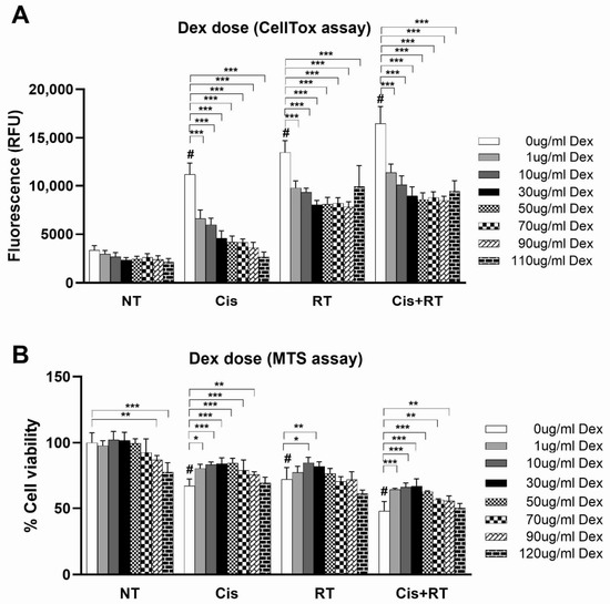
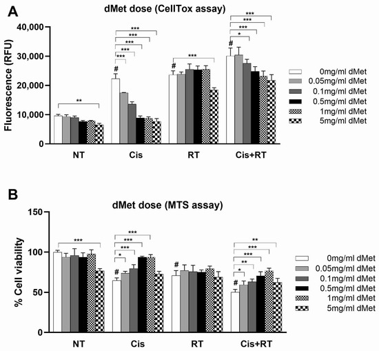
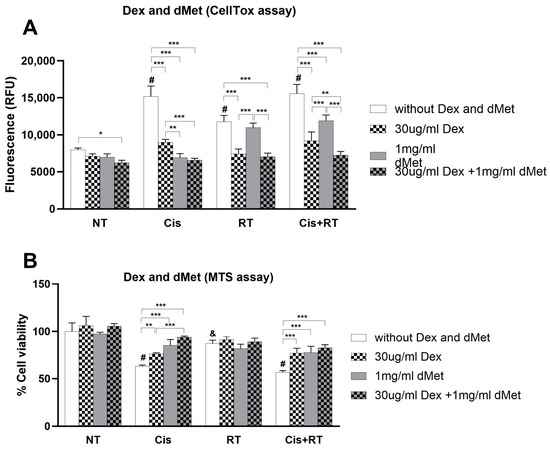
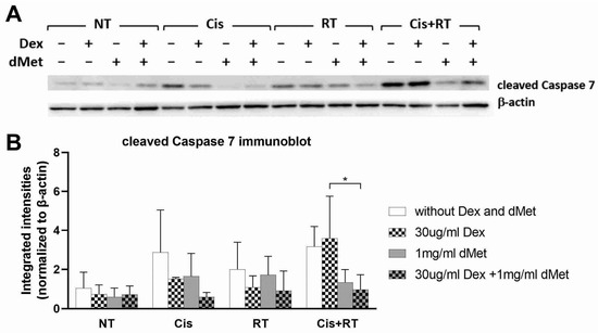
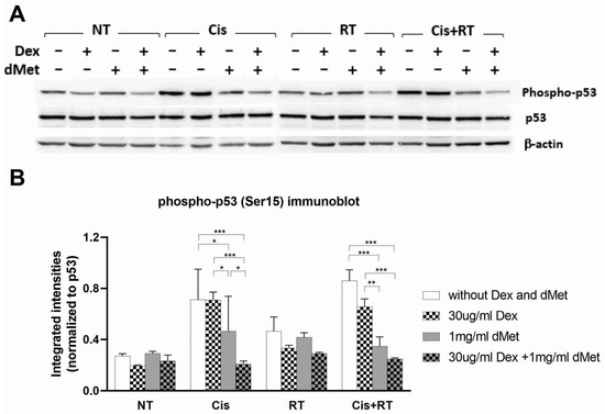
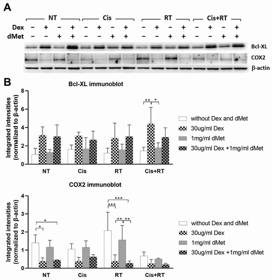
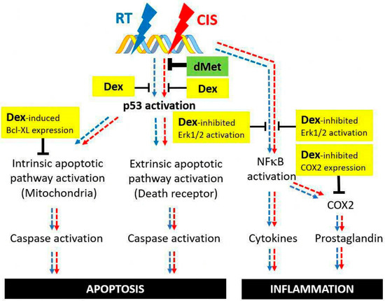
Background/Objectives: Nasopharyngeal cancer generally has a good survival rate with chemoradiotherapy, but a significant side effect is irreversible hearing loss, for which there is currently no preventive intervention. Given that dexamethasone (Dex) and D-methionine (dMet) have individually shown potential to prevent hearing loss in in vitro and animal studies, we hypothesized that combining Dex and dMet may offer greater protection against cytotoxicity induced by cisplatin and radiation treatments. Methods: Organ-of-Corti-derived OC-k3 cells exposed to cisplatin and/or radiation were used as an in vitro model of treatment-induced cytotoxicity to investigate the protective mechanisms of Dex and dMet. Results: Dex was associated with reduced apoptosis, accompanied by decreased p53 phosphorylation and increased Bcl-XL expression. Dex also reduced COX2 expression and showed trends toward attenuation of ERK1/2 and NFκB signaling. Conversely, dMet effectively blocked cisplatin-induced apoptosis but did not protect against radiation-induced cytotoxicity. Conclusions: The combination of Dex and dMet provided greater protection against cisplatin- and radiation-induced cytotoxicity in the OC-k3 cells than either agent alone. These findings support further investigation of combined Dex and dMet as a potential strategy to mitigate treatment-induced ototoxicity.
Full article
(This article belongs to the Section Otology and Neurotology)
►▼
Show Figures

Figure 1

Open AccessArticle
Factors Impacting Screening Documentation for Obstructive Sleep-Disordered Breathing at Well-Child Visits
by
Sarah R. Sutton, Caroline M. Fields, Christine B. San Giovanni, Paul J. Nietert and Phayvanh P. Pecha
J. Otorhinolaryngol. Hear. Balanc. Med. 2026, 7(2), 25; https://doi.org/10.3390/ohbm7020025 - 15 Jul 2026
Abstract
Background: The current literature suggests that screening for symptoms of pediatric obstructive sleep-disordered breathing (SDB) is low despite guidelines from the American Academy of Pediatrics to screen all children at well visits. Specific factors that affect screening rates for snoring have not been
[...] Read more.
Background: The current literature suggests that screening for symptoms of pediatric obstructive sleep-disordered breathing (SDB) is low despite guidelines from the American Academy of Pediatrics to screen all children at well visits. Specific factors that affect screening rates for snoring have not been well delineated. Materials and Methods: This study is a retrospective chart review of children ages 2–12 who presented for well-child visits at an academic hospital network. Multivariable logistic regression was used to identify factors associated with documentation for SDB screening. Results: Of 1001 charts reviewed, 809 children met inclusion criteria (median age 5 IQR [3–8] years, 50.3% female). Less than half (45.4%) of the children were screened for snoring, and 16.1% of those screened had symptoms of SDB. Adjusted multivariable regression showed that screening was significantly higher in older children (OR: 2.19 [1.46–3.29]; p < 0.001 for 5- to less than 8-year-old children; OR: 2.37 [1.57–3.57]; p < 0.001 for 8- to less than 12-year-old children). Children of the Other race group were also found to have significantly lower screening rates than Black and White children (OR 0.44 [0.23–0.84]; p = 0.014). The odds of being screened were 94% lower in visits in which a medical trainee was involved (p < 0.001). The odds of being screened at a well-child visit were 3.7 times higher in encounters in which a templated note was used (p < 0.001). Conclusions: Less than half of the children presenting for well visits were screened for snoring in this cohort. Differences in screening were attenuated by trainee involvement and the use of templated notes, which presents further avenues to explore meeting guideline recommendations for SDB screening.
Full article
(This article belongs to the Section Laryngology and Rhinology)
Open AccessReview
The Evolution of Laryngoscopy in Airway Management and Tracheal Intubation
by
Daniele Salvatore Paternò, Luigi La Via, Emilia Concetta Lo Giudice and Massimiliano Sorbello
J. Otorhinolaryngol. Hear. Balanc. Med. 2026, 7(2), 24; https://doi.org/10.3390/ohbm7020024 - 6 Jul 2026
Abstract
The work of anesthesiologists–intensivists, critical care specialists, and medical emergency teams is multifaceted and complex, with airway management representing a cornerstone and a common denominator of many procedures and interventions. Tracheal intubation has represented and still represents today the gold standard for airway
[...] Read more.
The work of anesthesiologists–intensivists, critical care specialists, and medical emergency teams is multifaceted and complex, with airway management representing a cornerstone and a common denominator of many procedures and interventions. Tracheal intubation has represented and still represents today the gold standard for airway control. This narrative review, conducted through a comprehensive search of PubMed/MEDLINE, EMBASE, and the Cochrane Library from database inception to December 2025 and supplemented by manual reference searches and retrieval of key historical sources, aims to trace the evolution of laryngoscopy for airway management and tracheal intubation—from its nineteenth-century origins to the current state of the art—while simultaneously addressing the clinical, educational, and safety implications of this evolution. Laryngoscopy evolution progressed in parallel with technological development and innovation, resulting in the expansion of clinical possibilities and in the improvement of patient safety. The evolution of laryngoscopy essentially took place between the late 1800s and the first half of the 1900s, culminating in the widespread adoption of the Macintosh laryngoscope. Almost 50 years later, a pivotal turning point occurred around the 2000s with the introduction of videolaryngoscopes. Along this path, the devices that succeeded one another introduced new problems, driving the search for new solutions. At present, tracheal intubation with videolaryngoscopy has achieved success and safety standards substantially superior to those of the early days of the technique. In this review we retrace the historical milestones of laryngoscopy, analyze the problems that emerged over time with various devices and the solutions adopted, examine the evolution of videolaryngoscopes and the impact of these devices on both technical and non-technical skills, discuss the ongoing debate surrounding their routine use (universal videolaryngoscopy) and the selection of optimal adjunct devices, and outline the emerging role of artificial intelligence in improving success rates and facilitating the learning curve. This journey, after 150 years of evolution, has reached a high level of maturity in terms of safety and efficacy.
Full article
(This article belongs to the Section Laryngology and Rhinology)
►▼
Show Figures

Figure 1

Open AccessReview
Positional Obstructive Sleep Apnea: A Model for Precision Sleep Medicine
by
Avneesh Prabakar, Shervin Eskandari, Tej Murudkar, Wenzhan Song, Xiaoyue Liu, Younghoon Kwon and William J. Healy
J. Otorhinolaryngol. Hear. Balanc. Med. 2026, 7(2), 23; https://doi.org/10.3390/ohbm7020023 - 28 Jun 2026
Abstract
Positional obstructive sleep apnea (POSA) is a subtype of obstructive sleep apnea in which the apnea–hypopnea index (AHI) is significantly greater in the supine position than in non-supine positions. However, POSA remains underrecognized and lacks a universally accepted clinical definition. POSA’s prevalence can
[...] Read more.
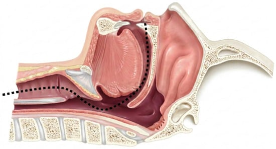
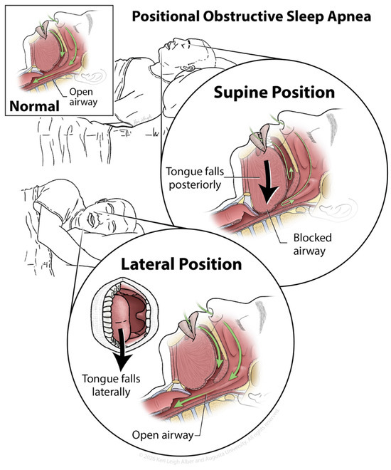
Positional obstructive sleep apnea (POSA) is a subtype of obstructive sleep apnea in which the apnea–hypopnea index (AHI) is significantly greater in the supine position than in non-supine positions. However, POSA remains underrecognized and lacks a universally accepted clinical definition. POSA’s prevalence can exceed 50% of OSA patients and occurs most frequently in patients with mild/moderate disease. In the supine position, gravity-dependent posterior displacement of the tongue and soft palate can increase upper airway collapsibility and drive the pathophysiology of POSA. These disturbances can carry meaningful cardiovascular consequences, including acute blood pressure surges that correlate with oxygen desaturation severity. As OSA is increasingly understood as a heterogeneous disorder shaped by variable anatomical and physiological traits, POSA offers a clinically actionable framework for precision sleep medicine. Current therapeutic strategies demonstrate that targeting the specific mechanisms of POSA can improve outcomes beyond what standardized CPAP treatment achieves alone. Racial and ethnic disparities in both POSA prevalence and treatment adherence further highlight the importance of individualized, culturally informed care. This review synthesizes the current understanding of the mechanisms, epidemiology, and therapeutic implications of POSA and evaluates its role as a model for individualized therapy in obstructive sleep apnea treatment.
Full article
(This article belongs to the Section Laryngology and Rhinology)
►▼
Show Figures

Figure 1
Open AccessReview
Horizontal Nystagmus as a Coupled-Integrator Network Phenotype: A Clinical–Conceptual Framework Linking Gaze Holding, Velocity Storage, and Nodulus–Uvula Supervision
by
Leonardo Manzari
J. Otorhinolaryngol. Hear. Balanc. Med. 2026, 7(1), 22; https://doi.org/10.3390/ohbm7010022 - 22 Jun 2026
Abstract
Horizontal nystagmus is still commonly interpreted at the bedside through a pragmatic peripheral-versus-central dichotomy. Although this heuristic is often clinically useful, it may be misleading because distributed brainstem–cerebellar disorders can generate peripheral-appearing phenotypes. This paper presents a narrative clinical–conceptual review proposing that a
[...] Read more.
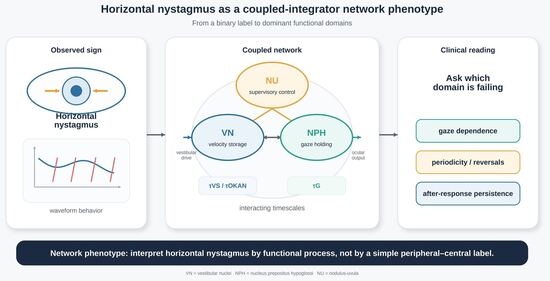
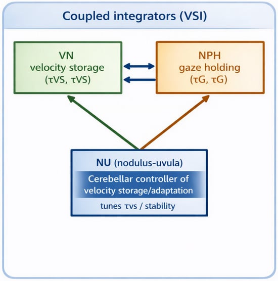
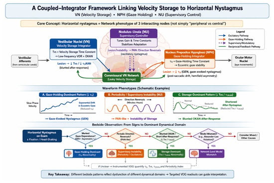
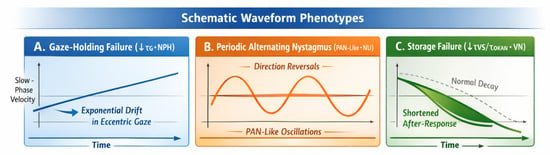
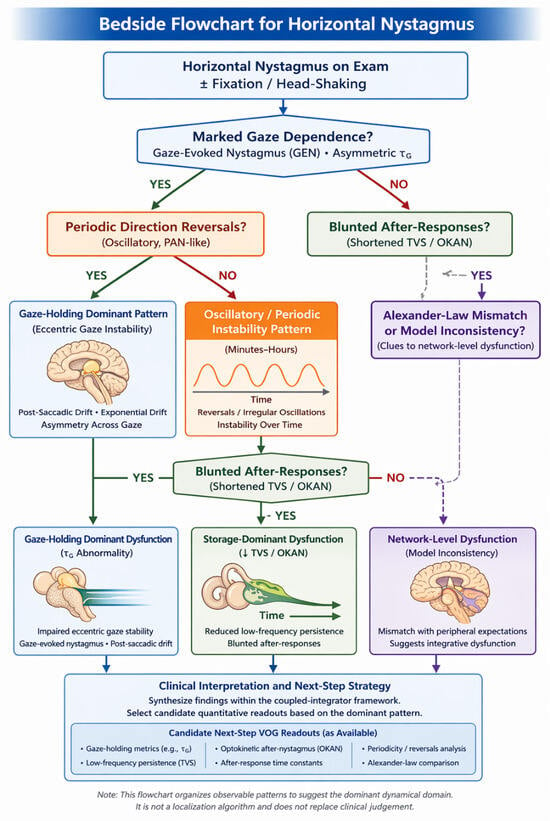
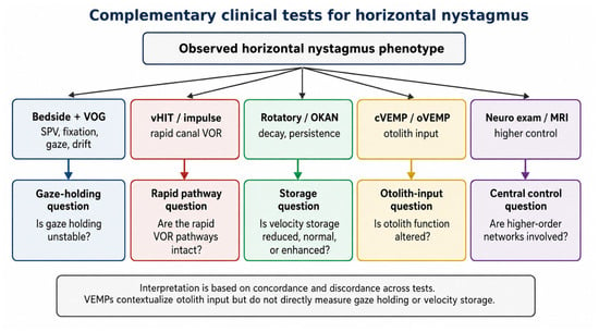
Horizontal nystagmus is still commonly interpreted at the bedside through a pragmatic peripheral-versus-central dichotomy. Although this heuristic is often clinically useful, it may be misleading because distributed brainstem–cerebellar disorders can generate peripheral-appearing phenotypes. This paper presents a narrative clinical–conceptual review proposing that a substantial subset of horizontal nystagmus patterns may be understood more coherently as expressions of dysfunction within a coupled vestibulo-ocular integrative network rather than as direct signatures of a single lesion site. Within this framework, two core dynamical domains are separated conceptually: a vestibular nuclei (VN)-centered velocity-storage process and an NPH-centered gaze-holding integrator. These processes are proposed to operate under cerebellar regulatory influence, with the nodulus–uvula (NU) acting as a plausible regulator of storage gain, temporal persistence, adaptation stability, and oscillatory behavior. Clinically, the velocity-storage domain is expressed through low-frequency vestibulo-ocular reflex behavior and optokinetic after-nystagmus-related dynamics, whereas the gaze-holding domain is expressed through eccentric gaze stability, gaze-evoked nystagmus, and post-saccadic drift. This framework carries a clinically relevant implication: horizontal nystagmus phenotypes may be interpreted more effectively by asking which functional process is predominantly abnormal—gaze holding, storage-related vestibular persistence, or cerebellar regulatory stability—rather than by relying solely on a binary peripheral–central label. On this basis, we outline a clinician-facing workflow linking gaze dependence, periodicity, direction reversals, head-shaking behavior, and Alexander-law mismatch to operational bedside criteria and candidate quantitative readouts. The proposed model is intended as a clinical–conceptual framework rather than a deterministic localization tool. Its main value lies in organizing discordant vestibular findings, strengthening the mechanistic interpretation of bedside and instrumented observations, and identifying testable directions for future validation studies in acute dizziness and ocular motor disorders.
Full article
(This article belongs to the Section Otology and Neurotology)
►▼
Show Figures

Graphical abstract
Open AccessCase Report
Primary Lingual Amyloidoma of the Tongue Base Causing Progressive Dysphagia: A Case Report
by
Carlos Carazo-Casas, Mar Domínguez-Fernández, Alejandra Véliz-Domínguez and Rafael Barberá-Durban
J. Otorhinolaryngol. Hear. Balanc. Med. 2026, 7(1), 21; https://doi.org/10.3390/ohbm7010021 - 4 Jun 2026
Abstract
Background and Clinical Significance: Localized amyloidoma rarely affects the tongue base, a site far less commonly involved than the larynx. When it does occur at this location, the clinical and radiological presentation can closely mimic oropharyngeal malignancy, making it a genuine diagnostic pitfall
[...] Read more.
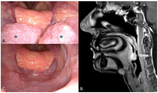
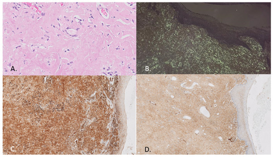
Background and Clinical Significance: Localized amyloidoma rarely affects the tongue base, a site far less commonly involved than the larynx. When it does occur at this location, the clinical and radiological presentation can closely mimic oropharyngeal malignancy, making it a genuine diagnostic pitfall for head and neck surgeons. Establishing the diagnosis demands histological proof, and ruling out systemic amyloidosis is an essential prerequisite. Case Presentation: An 80-year-old nonsmoking woman was evaluated for progressive dysphagia and globus sensation that had worsened over several years. Endoscopy revealed a firm, lobulated mass at the tongue base, centered on the glossotonsillar sulcus. Incisional biopsy under local anesthesia showed amyloid deposits. MRI demonstrated an avidly enhancing, well-circumscribed lesion without invasion of the deep neck spaces. A comprehensive systemic evaluation—including serum and urine protein electrophoresis with immunofixation, serum free light chains (κ/λ ratio 1.05), echocardiography, and bone marrow biopsy—yielded no evidence of systemic AL amyloidosis or plasma-cell dyscrasia. The mass was excised through transoral robotic surgery (TORS). Histopathology confirmed Congo red-positive, apple-green birefringent amyloid with lambda light-chain restriction; staining for AA amyloid and transthyretin was negative. The patient was diagnosed with primary localized lingual amyloidoma of the AL lambda type and remained symptom-free, with no endoscopic evidence of recurrence, at 12-month follow-up. Discussion: Amyloidoma deserves a place in the differential diagnosis of tongue base masses, particularly when the clinical picture does not fit squamous cell carcinoma. Congo red staining under polarized light, immunohistochemical light-chain typing, and a rigorous systemic workup are the cornerstones of diagnosis. Robotic-assisted transoral excision provides excellent access to the tongue base with low morbidity. Conclusions: This case underscores the need to consider amyloidoma when evaluating tongue base lesions, the central role of Congo red staining and light-chain typing, and the utility of TORS as an effective treatment option for selected symptomatic cases.
Full article
(This article belongs to the Section Laryngology and Rhinology)
►▼
Show Figures

Figure 1

Open AccessArticle
Hyperventilation-Induced Nystagmus in Patients with Vestibular Schwannoma: Pathophysiological and Clinical Considerations
by
Giuseppe Santopietro, Lucia Belen Musumano, Anna Lisa Giannuzzi and Elisabetta Rebecchi
J. Otorhinolaryngol. Hear. Balanc. Med. 2026, 7(1), 20; https://doi.org/10.3390/ohbm7010020 - 24 May 2026
Abstract
Background/Objectives: Hyperventilation-induced nystagmus (HVIN) has been described as a sensitive clinical sign in vestibular schwannoma (VS), potentially reflecting a reversible conduction block in partially demyelinated vestibular nerve fibers. However, its relationship with tumor size and instrumental vestibular deficits remains controversial. This study aimed
[...] Read more.
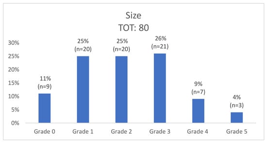
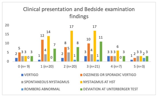
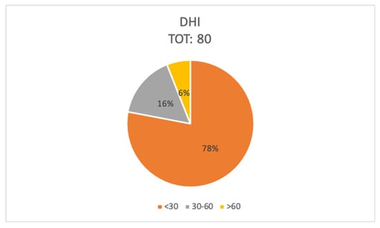
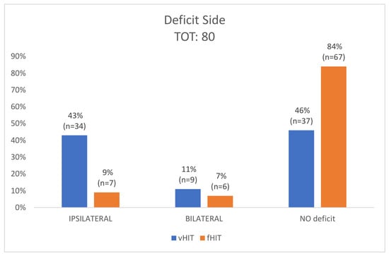
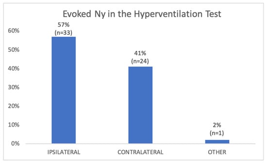
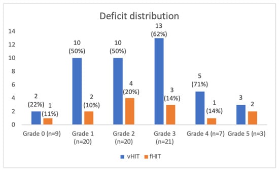
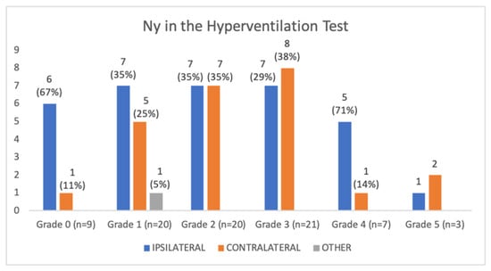
Background/Objectives: Hyperventilation-induced nystagmus (HVIN) has been described as a sensitive clinical sign in vestibular schwannoma (VS), potentially reflecting a reversible conduction block in partially demyelinated vestibular nerve fibers. However, its relationship with tumor size and instrumental vestibular deficits remains controversial. This study aimed to investigate the association between HVIN, tumor size, clinical presentation, and vestibular function assessed by the video Head Impulse Test (vHIT) and functional Head Impulse Test (fHIT). Methods: Eighty consecutive patients with unilateral VS were retrospectively evaluated. All underwent bedside vestibular examination, the vHIT, the fHIT, and the Hyperventilation Test (HVT). Tumors were classified according to the modified Koos–Sanna grading system. Associations between HVIN (presence and direction), demographic and clinical variables, vestibular deficits, and tumor size were analyzed using binary logistic regression, Fisher’s exact test, Welch’s independent samples t-test, and the Mann–Whitney U test. Results: HVIN was observed in 73% of patients. Among the patients with HVIN, ipsilesional horizontal nystagmus occurred in 57% of cases, 41% of the subjects showed contralesional nystagmus and one patient had downbeat nystagmus. vHIT abnormalities were identified in 54% of patients, while 30% of these also demonstrated fHIT deficits. No patient presented isolated fHIT abnormality. HVIN was detected even in patients with intracanalicular or small tumors and in some asymptomatic individuals. No statistically significant correlations were found between tumor size and HVIN presence, HVIN direction, or vestibular deficits in the vHIT/fHIT (all p > 0.05). Conclusions: HVIN is frequently observed in patients with VS, including those with small or asymptomatic lesions. However, neither its presence nor direction correlates with tumor size or objective vestibular deficit. The Hyperventilation Test should not be considered a prognostic tool but may serve as a simple and valuable adjunctive bedside examination in the early diagnostic suspicion of vestibular schwannoma.
Full article
(This article belongs to the Section Otology and Neurotology)
►▼
Show Figures

Figure 1
Open AccessCase Report
Spontaneous Intracranial Hypotension, Menière’s Disease and Secondary Benign Paroxysmal Positional Vertigo: Case Report
by
Rachael Arabian and Antonio Vintimilla
J. Otorhinolaryngol. Hear. Balanc. Med. 2026, 7(1), 19; https://doi.org/10.3390/ohbm7010019 - 23 May 2026
Abstract
Background/Objectives: Spontaneous intracranial hypotension (SIH) is a rare pathology that arises in the context of a known or suspected cerebral spinal fluid (CSF) leak. A key symptom of SIH is an orthostatic headache; however, additional neurological complications are common. This case study not
[...] Read more.
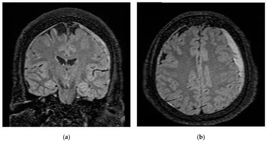
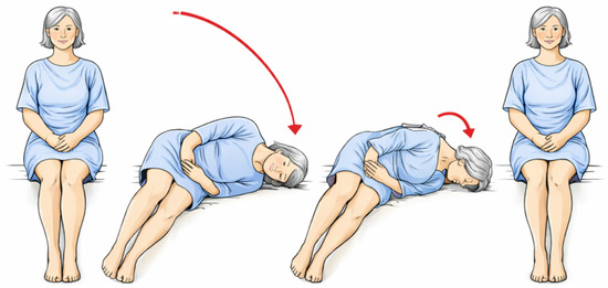
Background/Objectives: Spontaneous intracranial hypotension (SIH) is a rare pathology that arises in the context of a known or suspected cerebral spinal fluid (CSF) leak. A key symptom of SIH is an orthostatic headache; however, additional neurological complications are common. This case study not only highlights the co-existence of Menière’s disease and SIH but describes a subsequent complication of benign paroxysmal positional vertigo (BPPV) and management thereof. Case Description: The patient is a 61-year-old female who presented to the emergency department due to an intractable headache, right sided weakness and aphasia. CT/MRI revealed a subdural hematoma overlying the left cerebral hemisphere measuring up to 8 mm with 4 mm left to right midline shift. Fluoro-guided total spine myelogram, cisternogram, and lumbar epidural blood patch were performed for suspected SIH. As headache, right sided weakness and aphasia resolved, the patient began reporting onset of constant “spinning” dizziness, tinnitus and aural fullness mimicking symptoms of a Menière’s attack. The vestibular examination was consistent with compensated bilateral Menière’s disease (left > right) and right horizontal canalithiasis BPPV. The patient was treated with Gufoni and Lempert maneuvers with complete resolution of positional dizziness and associated nystagmus along with improved balance and gait. Discussion/Conclusions: This case study highlights the importance of multidisciplinary assessment in complex neurological cases and specifically recommends that patients with Menière’s disease accompanied by intractable headaches undergo extended neuroradiological examination of the brain to exclude underlying spontaneous intracranial hypotension syndrome.
Full article
(This article belongs to the Section Otology and Neurotology)
►▼
Show Figures

Figure 1
Open AccessArticle
Appropriate Listening Environment and Speaking Style for Individuals with Listening Difficulties Compared with Those with Normal Hearing and Hearing Loss
by
Chie Obuchi, Yuka Sasame and Yayoi Yamamoto
J. Otorhinolaryngol. Hear. Balanc. Med. 2026, 7(1), 18; https://doi.org/10.3390/ohbm7010018 - 21 May 2026
Abstract
Background/Objectives: Individuals with listening difficulties (LiD) report problems understanding speech despite normal hearing sensitivity. This study investigated how speech characteristics (clarity, speaking rate, and loudness) influence subjective speech understanding in adults with LiD under quiet and noisy conditions, compared with individuals with normal
[...] Read more.
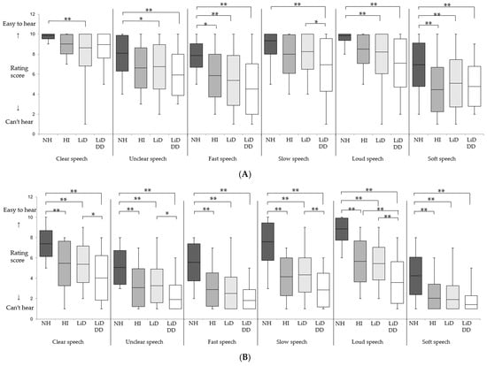
Background/Objectives: Individuals with listening difficulties (LiD) report problems understanding speech despite normal hearing sensitivity. This study investigated how speech characteristics (clarity, speaking rate, and loudness) influence subjective speech understanding in adults with LiD under quiet and noisy conditions, compared with individuals with normal hearing (NH) and those who are hard of hearing (HH). Methods: A questionnaire-based survey assessed perceived speech intelligibility across 12 listening conditions created by combining two environments (quiet and noisy) and six speaking styles. Participants included 77 adults with LiD (50 LiD without developmental disorders [DD] and 27 LiD and DD), 30 adults with NH, and 26 individuals with HH. Ratings were analyzed using analysis of variance and correlation analyses. Results: Both the LiD and HH groups demonstrated significantly reduced speech understanding in noisy environments when the speaking style was suboptimal, compared with the NH group. In contrast, the LiD group uniquely reported difficulties even under quiet conditions with clear, slow, and loud speech. Loud speech in quiet conditions was not consistently beneficial, particularly in the LiD and DD group, possibly reflecting auditory hypersensitivity. Conclusions: Listening difficulties in the LiD group are influenced by environmental and speaker-related factors and cannot be addressed solely by conventional strategies used for hearing loss. Although individualized communication approaches may be required, further investigation, including experimental studies, is necessary to validate this interpretation.
Full article
(This article belongs to the Section Otology and Neurotology)
►▼
Show Figures

Figure 1
Open AccessArticle
What Shapes Perceived Patient Understanding in Dysphagia and Voice Care? A Survey of Barriers and Facilitators
by
Chloe Doyon, Sophia Werden Abrams and Ashwini Namasivayam-MacDonald
J. Otorhinolaryngol. Hear. Balanc. Med. 2026, 7(1), 17; https://doi.org/10.3390/ohbm7010017 - 29 Apr 2026
Abstract
Background/Objectives: Patient understanding during healthcare appointments is essential for positive health outcomes and effective healthcare delivery. There is limited research exploring patient understanding for patients with swallowing difficulties (dysphagia) and voice disorders, where treatment engagement is often low. Dysphagia and voice disorders can
[...] Read more.
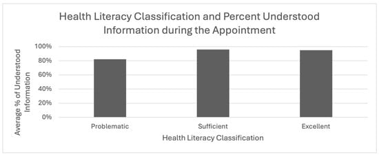
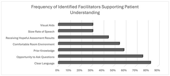
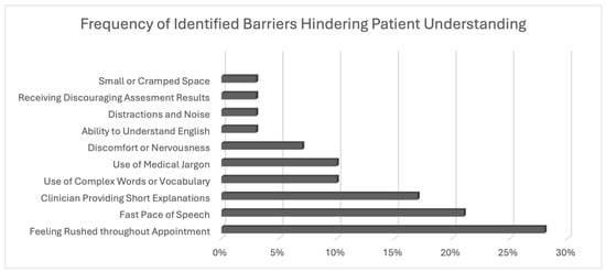
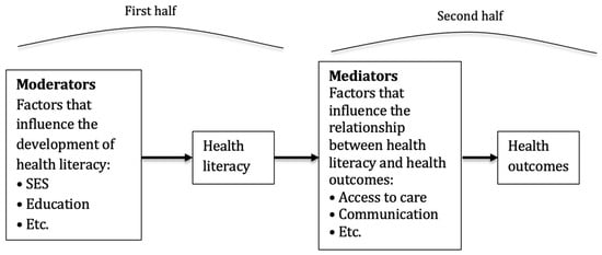
Background/Objectives: Patient understanding during healthcare appointments is essential for positive health outcomes and effective healthcare delivery. There is limited research exploring patient understanding for patients with swallowing difficulties (dysphagia) and voice disorders, where treatment engagement is often low. Dysphagia and voice disorders can affect communication, nutrition, and quality of life; therefore, it is essential to understand how to support this population effectively. This exploratory study aimed to identify barriers and facilitators to perceived patient understanding in clinical settings. Methods: A survey was administered to adults receiving assessment results for dysphagia or voice disorders from a speech–language pathologist or laryngologist at outpatient clinics in Ontario, Canada. The survey included demographics, health literacy levels (HLS19) and facilitators and barriers to comprehension. Descriptive statistics and content analysis were used to summarize responses. Results: Twenty-nine participants completed the survey (mean age of 64; 38% identifying as women). Facilitators to perceived understanding included the use of clear language (83%), being encouraged to ask questions (76%), and prior knowledge of their condition (59%). Reported barriers included rushing (28%), using a fast rate of speech (21%), and receiving short explanations (17%). Additionally, emotional and environmental factors such as the clinic atmosphere (55%) and the nature of assessment results (45%) influenced perceived patient comprehension. Conclusions: These findings suggest the need for consistent use of patient-centered communication strategies. Recommendations for clinicians include slowing speech, avoiding medical jargon, encouraging patient questions, incorporating visual aids, and creating a supportive environment. Implementing these strategies may improve understanding and health outcomes for patients with dysphagia and voice disorders.
Full article
(This article belongs to the Section Laryngology and Rhinology)
►▼
Show Figures

Figure 1
Open AccessCommentary
An Evidence-Based Framework for Simulation in Endoscopic Sinus Surgery: A Graded Approach to Training with 3D-Printed Models
by
Timothy Davies and Samuel Leong
J. Otorhinolaryngol. Hear. Balanc. Med. 2026, 7(1), 16; https://doi.org/10.3390/ohbm7010016 - 22 Apr 2026
Abstract
Background: Endoscopic sinus surgery (ESS) is a core operative technique in otolaryngology and is associated with a steep learning curve due to complex sinonasal anatomy, limited depth perception with two-dimensional endoscopy, and the requirement for precise bimanual coordination. Given the potential for serious
[...] Read more.
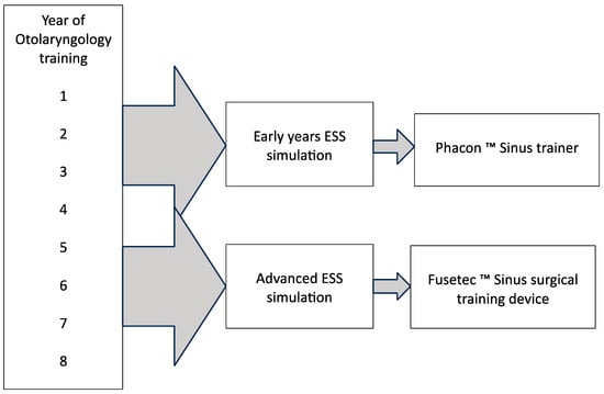
Background: Endoscopic sinus surgery (ESS) is a core operative technique in otolaryngology and is associated with a steep learning curve due to complex sinonasal anatomy, limited depth perception with two-dimensional endoscopy, and the requirement for precise bimanual coordination. Given the potential for serious complications, including cerebrospinal fluid leak and visual loss, simulation provides an important opportunity for trainees to develop technical skills in a controlled environment without risk to patients. Recent advances in three-dimensional (3D) printing have enabled the development of high-fidelity models for ESS training. Methods: We describe an evidence-based, graded approach to ESS simulation using two commercially available 3D printed sinus surgery models tailored to the trainee’s stage of training. Early-stage simulation focuses on development of anatomical orientation, endoscopic hand–eye coordination, tissue handling, and basic procedures such as middle meatal antrostomy and anterior ethmoidectomy. Advanced simulation targets more complex procedures, including frontal and sphenoid sinus surgery, transsphenoidal approaches, and management of intraoperative complications. Results: Validation studies demonstrate high face and content validity for both models. Early-stage simulators support acquisition of fundamental technical skills, while advanced models allow simulation of complex anatomy, pathology, and operative complications. Conclusions: A structured, stage-appropriate simulation strategy using high-fidelity 3D printed models may enhance technical skill acquisition and support safe and effective training in endoscopic sinus surgery.
Full article
(This article belongs to the Section Laryngology and Rhinology)
►▼
Show Figures

Figure 1
Open AccessArticle
Spatial Release from Masking with Simulated Electric–Acoustic and Cochlear Implant Speech
by
Nirmal Srinivasan, Bailey Borkowski, Morgan Barkhouse and Chhayakanta Patro
J. Otorhinolaryngol. Hear. Balanc. Med. 2026, 7(1), 15; https://doi.org/10.3390/ohbm7010015 - 16 Apr 2026
Abstract
Background/Objectives: Spatial release from masking (SRM) refers to the improvement in speech understanding that occurs when a target talker is spatially separated from competing speech. Although normal-hearing (NH) listeners benefit substantially from spatially separating the maskers from the target, cochlear implant (CI) users
[...] Read more.
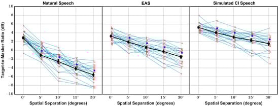
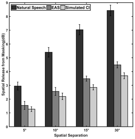
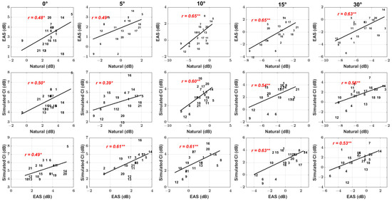
Background/Objectives: Spatial release from masking (SRM) refers to the improvement in speech understanding that occurs when a target talker is spatially separated from competing speech. Although normal-hearing (NH) listeners benefit substantially from spatially separating the maskers from the target, cochlear implant (CI) users experience markedly reduced advantages due to degraded spectral and binaural cue transmission. Electric–acoustic stimulation (EAS), which preserves low-frequency acoustic hearing in combination with electric stimulation, may partially restore these cues, but its benefits at small, conversationally relevant spatial separations remain poorly understood. Methods: This study measured speech identification thresholds using Coordinate Response Measure (CRM) sentences in NH listeners using natural, EAS, and simulated CI speech across five spatial configurations (0°, ±5°, ±10°, ±15°, ±30°). Speech identification thresholds were measured using a one-up/one-down adaptive procedure with Coordinate Response Measure (CRM) sentences. CI simulation used an eight-channel noise-band vocoder, whereas EAS simulation replaced the two lowest-frequency vocoder channels with low-pass speech (≤500 Hz). All stimuli were spatialized using head-related impulse responses generated from a validated virtual-acoustics model. Results: All stimulus types showed improved thresholds with increasing spatial separation; however, the magnitude of spatial release from masking (SRM) varied systematically. Natural speech produced the lowest thresholds and largest SRM, EAS speech yielded intermediate benefits, and simulated CI speech produced the smallest improvements. Notably, EAS and CI simulations were comparable at small separations, but EAS provided significantly greater SRM at ±15° and ±30°. Conclusions: These findings demonstrate that even partial low-frequency acoustic preservation enhances SRM at moderate spatial separations, highlighting the importance of EAS configurations for improving spatial hearing in CI-related listening environments.
Full article
(This article belongs to the Section Otology and Neurotology)
►▼
Show Figures

Figure 1
Open AccessArticle
Prestin as an Early Biomarker of Hearing Loss in Nasopharyngeal Cancer Patients Undergoing Induction Chemoradiation
by
Vanessa Y. J. Tan, Ng Jia Hui, Kiattisa Sommat, Thong Jiun Fong, Edward Z. Y. Zhang, Lai Oi Fah, Chia Xin Ni, Ang Mei Kim, Ong Ni Gin and Lim Chwee Ming
J. Otorhinolaryngol. Hear. Balanc. Med. 2026, 7(1), 14; https://doi.org/10.3390/ohbm7010014 - 6 Mar 2026
Abstract
Background/Objectives: Nasopharyngeal cancer (NPC) is prevalent in Southeast Asia, Southern China and North Africa. Up to 46% of NPC patients undergoing cisplatin chemoradiation treatment experience irreversible hearing loss. Prestin is a motor protein in the outer hair cells of the cochlea, and animal
[...] Read more.
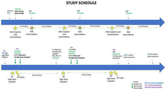
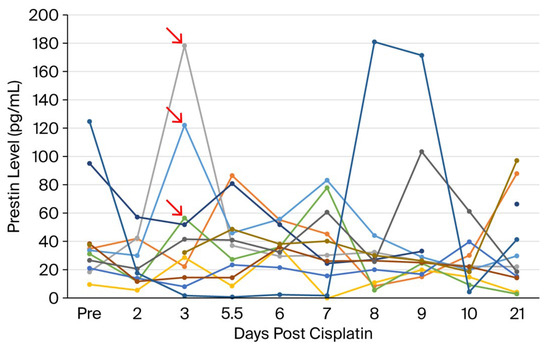
Background/Objectives: Nasopharyngeal cancer (NPC) is prevalent in Southeast Asia, Southern China and North Africa. Up to 46% of NPC patients undergoing cisplatin chemoradiation treatment experience irreversible hearing loss. Prestin is a motor protein in the outer hair cells of the cochlea, and animal studies have shown that blood prestin levels are elevated following cisplatin induced hearing loss. We investigated whether rising serum prestin levels can predict sensorineural hearing loss (SHNL) in NPC patients undergoing induction cisplatin chemotherapy (icCRT). Methods: Serum prestin levels were measured at ten time points during cisplatin chemotherapy. Pure tone audiogram and tinnitus handicap inventory (THI) were measured at baseline and at one and nine months after cisplatin administration. These outcomes were obtained to investigate whether rising prestin levels predict SNHL or worsening THI. Results: Of the 11 patients accrued, there was no association between prestin level and SNHL. An increase in THI was associated with higher prestin levels. There was significant hearing loss at 8 kHz at one (right ear, p = 0.012, left ear, p = 0.043) and nine months (right ear, p = 0.011) after treatment. After completing cisplatin, patients also had increased THI. Conclusions: Prestin was not identified as a biomarker of cisplatin-induced hearing loss in our cohort of NPC patients undergoing icCRT. NPC patients experience worsening of tinnitus with cumulative cisplatin, and hearing loss can persist at nine months post treatment. Future studies should focus on improved novel methods for measuring prestin or other cochlear proteins to better identify potential markers before permanent cisplatin induced hearing loss.
Full article
(This article belongs to the Section Otology and Neurotology)
►▼
Show Figures

Figure 1
Open AccessCase Report
Tracheal Rupture Secondary to an Iatrogenic Esophageal Perforation Presenting with Neck Insufflation on Valsalva: A Case Report
by
Lomesh Choudhary, Sophia Werden Abrams and Benjamin van der Woerd
J. Otorhinolaryngol. Hear. Balanc. Med. 2026, 7(1), 13; https://doi.org/10.3390/ohbm7010013 - 2 Mar 2026
Abstract
Background: Tracheal rupture is a rare, life-threatening condition that may follow blunt trauma, intubation, and, less commonly, esophageal instrumentation. The diagnosis may be challenging due to the subtle, delayed, and wide variety of symptoms. We present a unique case of delayed tracheal rupture
[...] Read more.
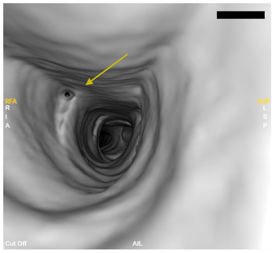
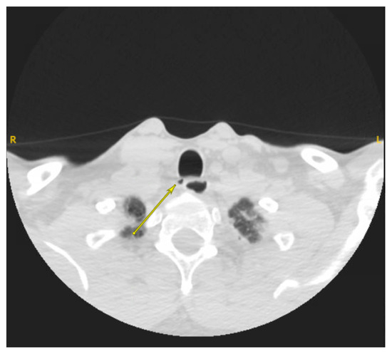
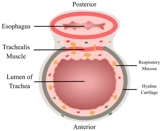
Background: Tracheal rupture is a rare, life-threatening condition that may follow blunt trauma, intubation, and, less commonly, esophageal instrumentation. The diagnosis may be challenging due to the subtle, delayed, and wide variety of symptoms. We present a unique case of delayed tracheal rupture secondary to iatrogenic esophageal perforation, presenting months later with a cervical neck insufflation on Valsalva, a novel clinical sign that has not been previously described in the literature. Case Presentation: A 44-year-old male initially presented with esophageal food impactation. Following endoscopic removal attempts, he developed signs consistent with esophageal perforation and was managed conservatively. In follow-up, he reported persistent neck discomfort, facial numbness, and the ability to insufflate the right side of his neck while doing a Valsalva. Flexible bronchoscopy and retrospective review of imaging revealed a proximal posterior tracheal defect, initially misinterpreted as a tracheal diverticulum. Surgical exploration confirmed a 6 cm dehiscence between the membranous trachea and the anterior wall of the esophagus. The defect was repaired using regional muscle flaps with tissue sealant reinforcement. Postoperatively, the patient had complete symptom resolution and no recurrence of neck insufflation. Conclusions: The case underscores the importance of maintaining a high index of suspicion for tracheal injury in patients with persistent or atypical symptoms following esophageal perforation or instrumentation. To our knowledge, we have described a novel clinical finding of neck insufflation upon Valsalva for diagnosing tracheal rupture.
Full article
(This article belongs to the Section Laryngology and Rhinology)
►▼
Show Figures

Figure 1
Highly Accessed Articles
Latest Books
E-Mail Alert
News
Topics