Case Report

Tracheal Rupture Secondary to an Iatrogenic Esophageal Perforation Presenting with Neck Insufflation on Valsalva: A Case Report


by
Lomesh Choudhary
1,
Sophia Werden Abrams
2 and
Benjamin van der Woerd
2,*
1
Michael G. DeGroote School of Medicine, McMaster University, 1280 Main Street West, Hamilton, ON L8S 4L8, Canada
2
Department of Surgery, Division of Otolaryngology-Head and Neck Surgery, McMaster University, 1200 Main Street West, Hamilton, ON L8S 4K1, Canada
*
Author to whom correspondence should be addressed.
J. Otorhinolaryngol. Hear. Balance Med. 2026, 7(1), 13; https://doi.org/10.3390/ohbm7010013
Submission received: 18 December 2025 / Revised: 14 February 2026 / Accepted: 26 February 2026 / Published: 2 March 2026
(This article belongs to the Section Laryngology and Rhinology)

Abstract

Background: Tracheal rupture is a rare, life-threatening condition that may follow blunt trauma, intubation, and, less commonly, esophageal instrumentation. The diagnosis may be challenging due to the subtle, delayed, and wide variety of symptoms. We present a unique case of delayed tracheal rupture secondary to iatrogenic esophageal perforation, presenting months later with a cervical neck insufflation on Valsalva, a novel clinical sign that has not been previously described in the literature. Case Presentation: A 44-year-old male initially presented with esophageal food impactation. Following endoscopic removal attempts, he developed signs consistent with esophageal perforation and was managed conservatively. In follow-up, he reported persistent neck discomfort, facial numbness, and the ability to insufflate the right side of his neck while doing a Valsalva. Flexible bronchoscopy and retrospective review of imaging revealed a proximal posterior tracheal defect, initially misinterpreted as a tracheal diverticulum. Surgical exploration confirmed a 6 cm dehiscence between the membranous trachea and the anterior wall of the esophagus. The defect was repaired using regional muscle flaps with tissue sealant reinforcement. Postoperatively, the patient had complete symptom resolution and no recurrence of neck insufflation. Conclusions: The case underscores the importance of maintaining a high index of suspicion for tracheal injury in patients with persistent or atypical symptoms following esophageal perforation or instrumentation. To our knowledge, we have described a novel clinical finding of neck insufflation upon Valsalva for diagnosing tracheal rupture.

1. Introduction

Tracheal rupture is an uncommon but potentially life-threatening condition that may arise from traumatic, iatrogenic, or spontaneous causes. Early diagnosis is important because delayed recognition can lead to life-threatening complications, such as airway compromise, respiratory distress, and mediastinitis, to list a few [1]. Although esophageal perforation is a recognized complication of endoscopic procedures, secondary tracheal injury is exceedingly rare and may present subtly [2,3].
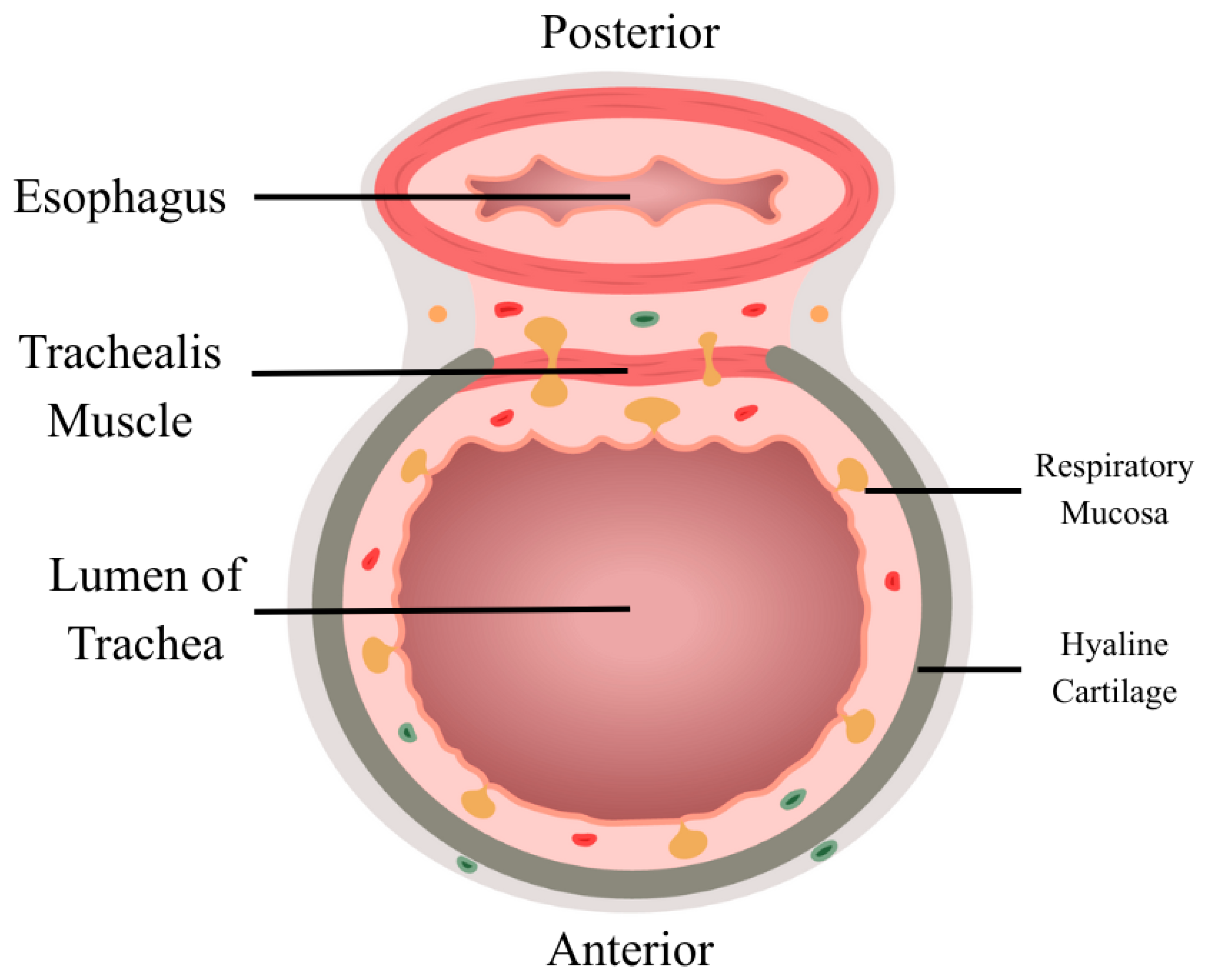
Tracheal rupture most commonly involves the posterior membranous trachea, where the airway lacks cartilaginous support and is more vulnerable [4]. Iatrogenic injury is most related to endotracheal intubation; however, injury due to esophageal instrumentation remains poorly described [5]. In cases of esophageal perforation, local inflammatory processes can weaken the shared tracheoesophageal plane, predisposing the posterior tracheal wall to delayed dehiscence [6]. Figure 1 displays the close relationship between the trachea and esophagus.
We present the case of a 44-year-old male who developed a delayed diagnosis of tracheal rupture following esophageal food impaction and iatrogenic perforation, initially managed conservatively but ultimately requiring surgical intervention after persistent symptoms such as neck insufflation. This case highlights the diagnostic challenges of identifying tracheal injuries in the context of esophageal perforation and highlights the importance of maintaining a high index of suspicion when evaluating persistent neck symptoms after esophageal instrumentation. This case report was prepared following the CARE guidelines [7].

2. Case Presentation

A 44-year-old male presented to the emergency department (ED) after having a piece of steak impacted in the esophagus and failed attempts to clear it by vomiting. On initial examination, he was non-toxic appearing, without cough, wheeze, or abnormal findings on the head and neck examination. His medical history includes benign esophageal strictures and three prior esophageal dilations, the last of which was performed eight years ago. He had no history of medication use, allergies, or systemic disease. His surgical history was notable for hemorrhoidectomy and prior esophagogastroduodenoscopies (EGDs). He had a 10-pack-year history of smoking but had quit a few years before. The patient did not report any alcohol or recreational drug use. There were no relevant family and psychosocial histories. The patient works as a carpenter and reports no occupational history of inhaled exposures, chemical irritants, or trauma that could explain his symptoms. He was discharged home with a course of oral steroids.
The patient returned to the ED the next day due to persistent symptoms associated with the impacted food. Gastroenterology was consulted and attempted endoscopic (EGD) removal of the suspected food bolus, which was unsuccessful. After the procedure, the patient experienced nausea and blood-tinged sputum, raising concern for an esophageal perforation. A computed tomography (CT) scan showed pneumomediastinum without pleural effusion, suggestive of a contained esophageal rupture injury. The patient was managed conservatively with nil per os (NPO) status, intravenous broad-spectrum antibiotics, and close monitoring. The thoracic surgery team performed an endoscopic evacuation of the food bolus and obtained multiple esophageal biopsies. The patient was admitted with broad-spectrum antibiotics and was later discharged after a few days since there was no overwhelming sepsis or uncontrolled perforation. Esophageal biopsy findings were concerning for eosinophilic esophagitis, which was later confirmed on pathology. There was also evidence of small intestinal metaplasia, focal erosions, and marked eosinophilic infiltration.
During the first follow-up appointment after discharge, the patient reported resolution of dysphagia symptoms but developed a persistent, unusual sensation in the left lateral neck and had intermittent left facial numbness. Approximately seven months later, he was referred to otolaryngology due to right-sided neck discomfort, needing to manually manipulate his thyroid cartilage for relief. There were also non-specific left-sided facial complaints associated. The patient described the release of air when he manipulates the trachea to the right-hand side. There was no overt respiratory compromise during this time, likely delaying suspicion for airway injury.
On physical examination, his nose, ears, oral cavity, and facial function (1/6 House-Brackmann, symmetric) exams were unremarkable. There were no vocal changes and no difficulty breathing. There were no lymphadenopathies noticed on physical exam. However, with a Valsalva maneuver, he was able to puff up the right side of his neck with air.
The ability to insufflate the neck with a Valsalva maneuver suggested the presence of communication between the airway and the surrounding cervical planes. This finding raised concern for air leak, dependent on pressure, from the trachea, given the patients past medical history of esophageal perforation and persistent neck symptoms.
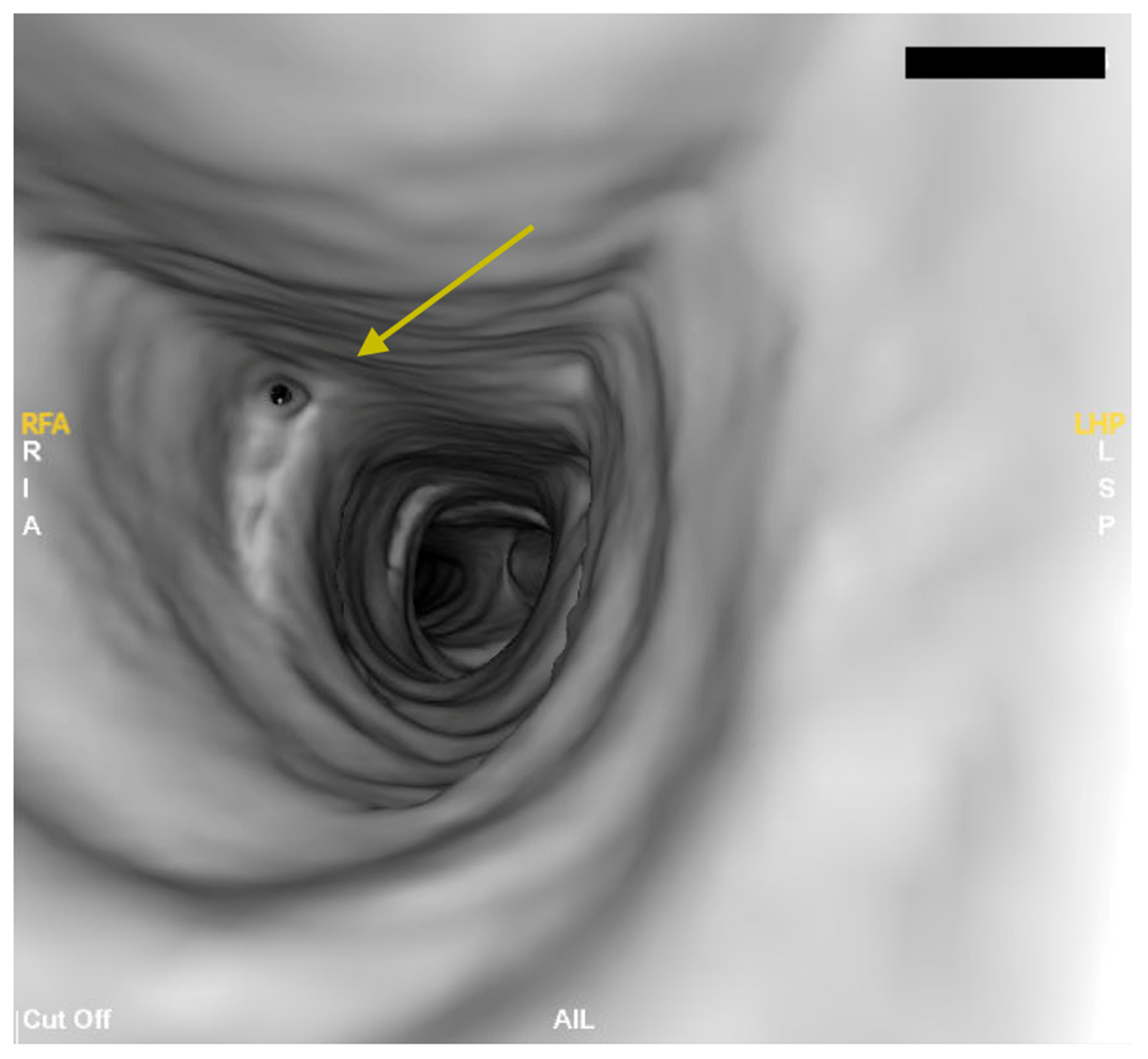
Given the patient’s persistent symptoms and unusual physical examination findings, a bronchoscopy was performed to rule out tracheal causes for the symptoms. A bedside bronchoscopy was performed, which showed the nasal cavity, oropharynx, hypopharynx, and supraglottic structures being within normal limits. Vocal fold mobility testing was normal. The trachea showed a normal caliber trachea with a small divot to the right posterior trachea and the proximal trachea, raising suspicion for a tracheal rupture (Figure 2).
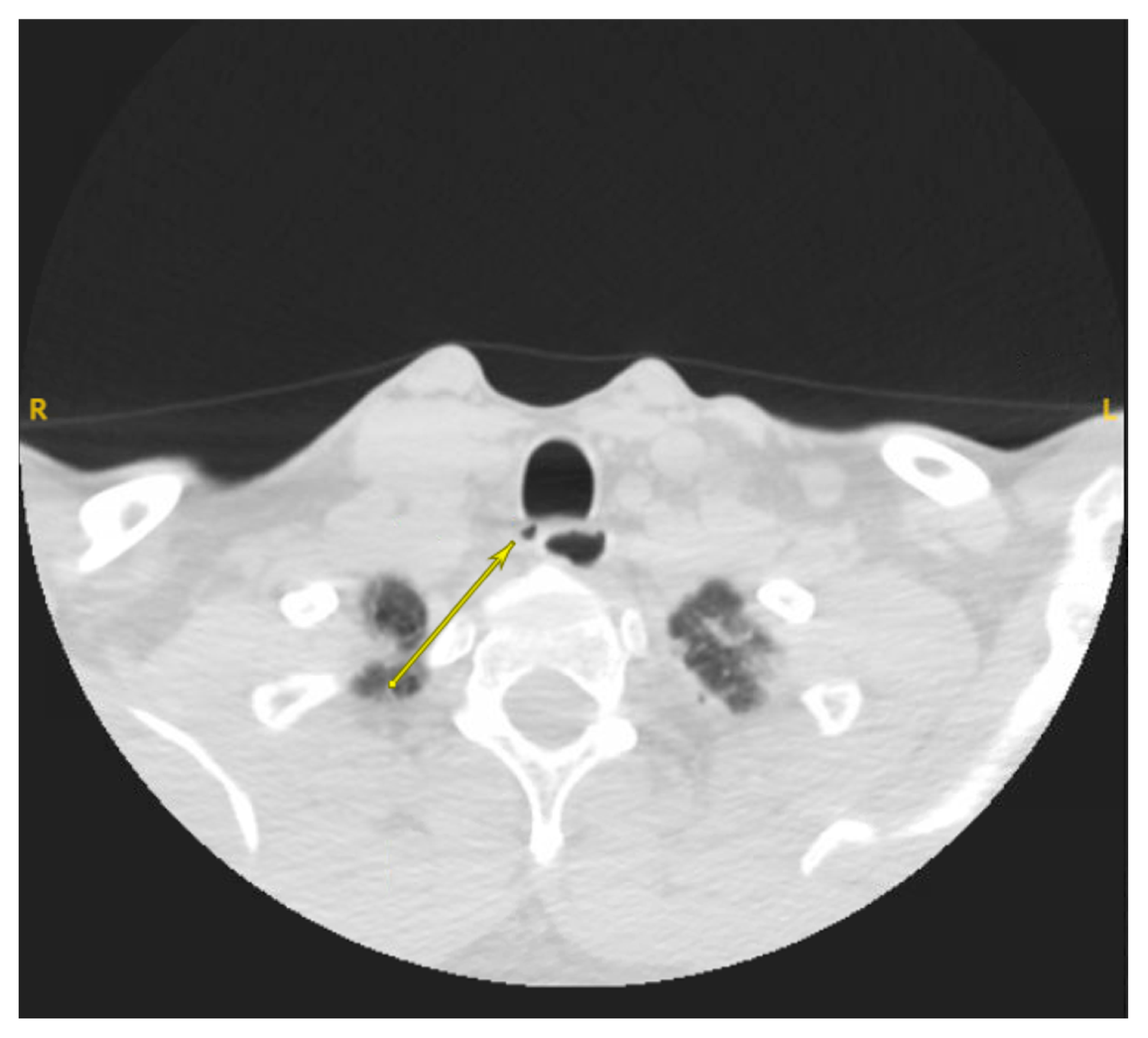
Upon review of prior CT scans from the time of the initial symptoms and onwards, a small tracheal rupture appeared in the proximal trachea (Figure 3). Previous radiologic reports repeatedly referred to the radiographic finding as a “tracheal diverticulum”; however, true tracheal diverticula are rare, and this finding should have raised suspicion for a more clinically relevant pathology, such as a tracheal rupture. Moreover, the radiolucent outpouching adjacent to the posterior tracheal wall showed characteristics that are inconsistent with a true tracheal diverticulum. True tracheal diverticulum is usually paratracheal and asymptomatic, which was not the case in this patient. However, there may have been limited clinical details given during the radiology requisition, leading to isolated radiologic interpretation. Given the persistence of symptoms and the imaging findings, the patient was scheduled for surgical repair involving right neck exploration and tracheal defect closure using sternocleidomastoid and strap muscle flaps.
During surgery, bronchoscopy revealed a right-sided tracheal defect at the junction between the membranous and cartilaginous trachea, associated with visible air bubbling. Despite the small size of the external tracheal perforation, exploration revealed a large, approximately 6 cm dehiscence between the membranous trachea and the anterior wall of the esophagus. A right neck exploration was performed. An incision was made below the cricoid cartilage and dissection proceeded through the platysma and strap muscles. The recurrent laryngeal nerve was identified and carefully retracted. Upon reaching the lateral trachea, a sizable air-filled pocket between the trachea and esophagus was encountered.
To repair the defect, a sternocleidomastoid muscle flap and a right strap muscle flap were harvested, rotated, and secured into place with 3-0 vicryl sutures. Tissue sealant was applied over the repair site, and a Jackson-Pratt (JP) drain was placed for postoperative drainage. Intraoperative flexible bronchoscopy confirmed successful repair without ongoing air leak under pressure. Reconstruction was successful, and the patient had an uncomplicated postoperative course.
At the three months postoperative appointment, the patient reported complete resolution of symptoms, with no recurrence of neck insufflation and no tracheal defects on follow-up bedside bronchoscopy. The patient was no longer able to expand his neck with a Valsalva maneuver. There was no recurrence of symptoms.

3. Discussion

Tracheal rupture is a rare and potentially life-threatening event commonly associated with traumatic intubation, blunt trauma, or external cervical injuries [1]. In this case, the patient’s tracheal injury was initially missed due to the management of an esophageal perforation after food bolus impaction and endoscopic intervention.
The diagnosis of tracheal injuries can be challenging, especially when there are few or no signs of respiratory distress [8]. In this patient, persistent neck discomfort and the ability to insufflate his neck during a Valsalva maneuver were atypical but important diagnostic clues. In fact, a literature search did not reveal any previously reported cases in which neck insufflation on Valsalva was described as a feature of tracheal rupture, suggesting this may be a novel clinical sign. Emphasizing this unique, novel presentation may help clinicians recognize subtle signs of tracheal rupture in otherwise healthy patients.
Moreover, the patient’s initial radiographic findings were called a “tracheal diverticulum.” However, it was important to consider the other clinical findings. Tracheal diverticula are usually asymptomatic. If symptomatic, they present with chronic cough, dyspnea, stridor, and other unique symptoms [9]. In this case, the imaging findings warranted greater scrutiny and suspicion, given the patient’s history of iatrogenic esophageal perforation and ongoing neck discomfort. Delayed recognition of tracheal injuries increases the risk of complications, including respiratory distress, mediastinitis, pneumothorax, scar formation, and airway stenosis [1].
This case also highlights the importance of remaining alert for tracheal complications after esophageal perforation, as a strong force that is sufficient to perforate the esophagus may also injure the posterior trachea. Close assessments of imaging as well as otolaryngology or thoracic surgery consultation should be considered when atypical symptoms continue to persist after the management of the esophageal tear.
Conservative management may carry the risk of delayed healing, persistent leaks, stenosis, and mediastinal infections [10]. Endoscopic stenting can offer relief; however, there remains a potential for stent-related complications, loss of patency, and need for repeat interventions. Moreover, studies have shown surgical intervention to have improved success rate and long-term results when compared to endoscopic stenting [11].
The decision to proceed with a surgical repair for this case was based on the presence of symptoms and failure of spontaneous resolution overtime. The surgical approach involved the use of both a sternocleidomastoid and strap muscle flap. These techniques are commonly used for the tension-free closure and reinforcement of tracheal injuries [12]. Regional muscle flaps provide well-vascularized tissue that reduce repair site tension and create an additional barrier between the trachea and esophagus [13]. This minimizes the risk of further rupturing or fistula formation between the trachea and esophagus. A sternocleidomastoid muscle flap was chosen due to its proximity to the cervical trachea, allowing for harvest of the flap without extensive dissection or a second incision. Moreover, it is perfused by multiple arteries which gives flexibility of size of the flap and increased chance of flap survival [14]. Similarly, infrahyoid muscles are thin, pliable, and well-vascularized tissues that are near the trachea [13,15]. Despite the delayed presentation, the successful outcome in this case highlights the need for operative management.
The strengths of this case include the successful delayed repair with full symptom resolution and no recurrence. Another strength includes the identification of atypical signs associated with tracheal rupture, such as the ability of the patient to insufflate air into the neck with a Valsalva. Limitations of this case include the potential to identify persistent symptoms sooner to provide an earlier diagnosis.

4. Conclusions

This case highlights the necessity to maintain a high level of suspicion for tracheal injury following esophageal instrumentation. This is needed in patients who develop persistent neck symptoms or unusual physical findings, such as cervical air insufflation. This case also highlights the limitations of isolated radiology interpretation without clinical correlation. Early bronchoscopy can facilitate a timely diagnosis and prompt quick surgical repair. Lastly, reconstruction using regional muscle flaps can achieve excellent outcomes even in delayed presentations.

Author Contributions

L.C., S.W.A. and B.v.d.W. wrote, reviewed, and edited the manuscript. B.v.d.W. proposed the idea for this case report. The intervention and management of this patient was provided by B.v.d.W. All authors have read and agreed to the published version of the manuscript.

Funding

This research received no external funding.

Institutional Review Board Statement

The study was conducted in accordance with the Declaration of Helsinki. Our institution does not require ethics approval for reporting individual cases or case series.

Informed Consent Statement

Informed consent was obtained from the study subject involved in the study. Written informed consent has been obtained from the patient to publish this paper.

Data Availability Statement

The data presented in this study are available on request from the corresponding author due to protection of patient privacy.

Conflicts of Interest

The authors declare no conflicts of interest.

References

  1. Ak, A.K.; Anjum, F. Tracheobronchial Tear. In StatPearls; StatPearls Publishing: Treasure Island, FL, USA, 2025. Available online: http://www.ncbi.nlm.nih.gov/books/NBK560900/ (accessed on 18 December 2025).
  2. Rollins, R.; Tocino, I. Early radiographic signs of tracheal rupture. Am. J. Roentgenol. 1987, 148, 695–698. [Google Scholar] [CrossRef] [PubMed]
  3. Atkins, J.H.; Mandel, J.E.; Metz, D.C. Sudden Tracheal Collapse during EGD and Subsequent Anesthetic Management with Dexmedetomidine-Ketamine in a Patient with Achalasia and Tracheomalacia. Case Rep. Anesthesiol. 2011, 2011, 281679. [Google Scholar] [CrossRef] [PubMed]
  4. Marty-Ané, C.-H.; Picard, E.; Jonquet, O.; Mary, H. Membranous tracheal rupture after endotracheal intubation. Ann. Thorac. Surg. 1995, 60, 1367–1371. [Google Scholar] [CrossRef] [PubMed]
  5. Hofmann, H.; Rettig, G.; Radke, J.; Neef, H.; Silber, R. Iatrogenic ruptures of the tracheobronchial tree. Eur. J. Cardiothorac. Surg. 2002, 21, 649–652. [Google Scholar] [CrossRef] [PubMed]
  6. Li, G.; Wang, Z.; Liu, J.; Fu, S.; Xue, L.; Liao, Z.; Yang, R.; Huang, S.; Xu, R.; Chen, P.; et al. Comprehensive Insights into Tracheoesophageal Fistula Pathophysiology, Diagnosis, Treatment, and Future Directions. Adv. Sci. 2025, 12, e02825. [Google Scholar] [CrossRef] [PubMed]
  7. Riley, D.S.; Barber, M.S.; Kienle, G.S.; Aronson, J.K.; Von Schoen-Angerer, T.; Tugwell, P.; Kiene, H.; Helfand, M.; Altman, D.G.; Sox, H.; et al. CARE guidelines for case reports: Explanation and elaboration document. J. Clin. Epidemiol. 2017, 89, 218–235. [Google Scholar] [CrossRef] [PubMed]
  8. Ye, D.; Shen, Z.; Zhang, Y.; Qiu, S.; Kang, C. Clinical features and management of closed injury of the cervical trachea due to blunt trauma. Scand. J. Trauma Resusc. Emerg. Med. 2013, 21, 60. [Google Scholar] [CrossRef] [PubMed]
  9. Tanrivermis Sayit, A.; Elmali, M.; Saglam, D.; Celenk, C. The diseases of airway-tracheal diverticulum: A review of the literature. J. Thorac. Dis. 2016, 8, E1163–E1167. [Google Scholar] [CrossRef] [PubMed]
  10. Carretta, A.; Melloni, G.; Bandiera, A.; Negri, G.; Voci, C.; Zannini, P. Conservative and surgical treatment of acute posttraumatic tracheobronchial injuries. World J. Surg. 2011, 35, 2568–2574. [Google Scholar] [CrossRef] [PubMed]
  11. Mandour, M.; Remacle, M.; Van de Heyning, P.; Elwany, S.; Tantawy, A.; Gaafar, A. Chronic subglottic and tracheal stenosis: Endoscopic management vs. surgical reconstruction. Eur. Arch. Otorhinolaryngol. 2003, 260, 374–380. [Google Scholar] [CrossRef] [PubMed]
  12. Welter, S.; Essaleh, W. Management of tracheobronchial injuries. J. Thorac. Dis. 2020, 12, 6143–6151. [Google Scholar] [CrossRef] [PubMed]
  13. Rammos, C.K.; Anderson, R.C.; Taege, S.M.; King, V.A.; Yoo, A. Treatment of Synchronous Traumatic Perforations of the Trachea and the Esophagus With Two Inferiorly Based Strap Muscle Interposition Flaps: An Innovative Approach. J. Craniofac. Surg. 2019, 30, e330–e332. [Google Scholar] [CrossRef] [PubMed]
  14. Jones, L.F.; Farrar, E.M.; Roberts, D.J.H.; Moor, J.W. Revisiting the sternocleidomastoid flap as a reconstructive option in head and neck surgery. J. Laryngol. Otol. 2019, 133, 742–746. [Google Scholar] [CrossRef] [PubMed]
  15. Pisanu, A.; Reccia, I.; Nieddu, R.; Uccheddu, A. Sternohyoid muscle flap interposition in the treatment of an acquired tracheoesophageal fistula. Head Neck 2009, 31, 962–967. [Google Scholar] [CrossRef]
Figure 1. Cross-sectional schematic demonstrating the close anatomical relationship between the posterior tracheal membrane and the anterior esophageal wall. Horizontal Section of Trachea Icon by DBCLS (https://togotv.dbcls.jp/en/pics.html, accessed on 1 February 2026) is licensed under CC-BY 4.0 Unported https://creativecommons.org/licenses/by/4.0/ (accessed on 1 February 2026). Labels were added by the authors to highlight key anatomic structures.
Ohbm 07 00013 g001
Figure 2. Bronchoscope image demonstrating a right posterior tracheal defect at the junction of the membranous and cartilaginous trachea. The defect, as shown by the yellow arrow, appears as a divot in the tracheal wall, consistent with a tracheal rupture.
Ohbm 07 00013 g002
Figure 3. Axial computed tomography (CT) scan of the neck demonstrating a right posterior proximal tracheal, as shown by the arrow. This finding was initially misinterpreted as a tracheal diverticulum; however, in the clinical context and on retrospective review of imaging, the finding is consistent with a tracheal rupture.
Ohbm 07 00013 g003
Disclaimer/Publisher’s Note: The statements, opinions and data contained in all publications are solely those of the individual author(s) and contributor(s) and not of MDPI and/or the editor(s). MDPI and/or the editor(s) disclaim responsibility for any injury to people or property resulting from any ideas, methods, instructions or products referred to in the content.