1. Introduction
The term amyloidosis refers to a family of diseases united by a common mechanism: extracellular accumulation of misfolded proteins arranged in beta-pleated sheet fibrils [
1,
2]. Over time, these insoluble deposits disrupt tissue architecture and compromise organ function. Because treatment hinges on identifying the offending precursor protein—whether immunoglobulin light chain (AL), serum amyloid A (AA), transthyretin (ATTR), or one of more than a dozen rarer subtypes—accurate typing is indispensable. Laser-capture mass spectrometry is now regarded as the reference standard for this purpose [
2,
3].
Distinct from its systemic counterpart, localized amyloidoma confines amyloid deposition to a single anatomic site [
4]. The AL subtype predominates in these focal lesions: a small, indolent clone of plasma cells at the site of involvement secretes amyloidogenic light chains that polymerize locally [
5,
6]. Importantly, the long-term outlook is favorable. In a landmark Mayo Clinic cohort of 413 patients followed over more than four decades, no case progressed to systemic disease [
4]. More recent cohorts have reinforced this observation: in the Cleveland series, no patient progressed to systemic disease, and overall survival was excellent [
7].
Within the head and neck, localized amyloidoma has a well-documented predilection for the larynx [
8,
9]. The tongue, by contrast, is seldom a primary site; when amyloid does involve the tongue, it is nearly always in the setting of systemic AL disease presenting as diffuse macroglossia [
10,
11]. Truly localized amyloidoma of the tongue is uncommon, and base-of-tongue involvement is rarer still—fewer than 30 cases have appeared in the literature [
10,
12,
13]. The clinical challenge is that such lesions can look indistinguishable from squamous cell carcinoma, plasmacytoma, or lymphoma on both endoscopy and cross-sectional imaging [
8].
Here, an elderly woman with long-standing dysphagia caused by a primary localized amyloidoma of the tongue base is presented. The diagnostic pathway, including the hematological workup required to exclude systemic disease, is detailed, and the role of transoral robotic surgery (TORS) as a minimally invasive treatment option for this rare entity is discussed.
2. Detailed Case Description
2.1. Patient Information and Presenting Symptoms
The patient was an 80-year-old woman, a lifelong nonsmoker, who had been experiencing slowly worsening difficulty swallowing—particularly with mixed-texture foods—for several years. She also described a persistent sensation of a lump in her throat. She reported no pain on swallowing, unintentional weight loss, ear pain, fever, night sweats, or any other systemic symptoms. Her past medical history included hypertension treated with enalapril, bronchial asthma managed with a combination inhaler (fluticasone furoate/vilanterol), chronic rhinosinusitis with nasal polyps (previously operated on endoscopically), and allergic rhinitis. There was no personal history of malignancy, tobacco or alcohol use, or autoimmune disease.
2.2. Clinical Findings and Initial Workup
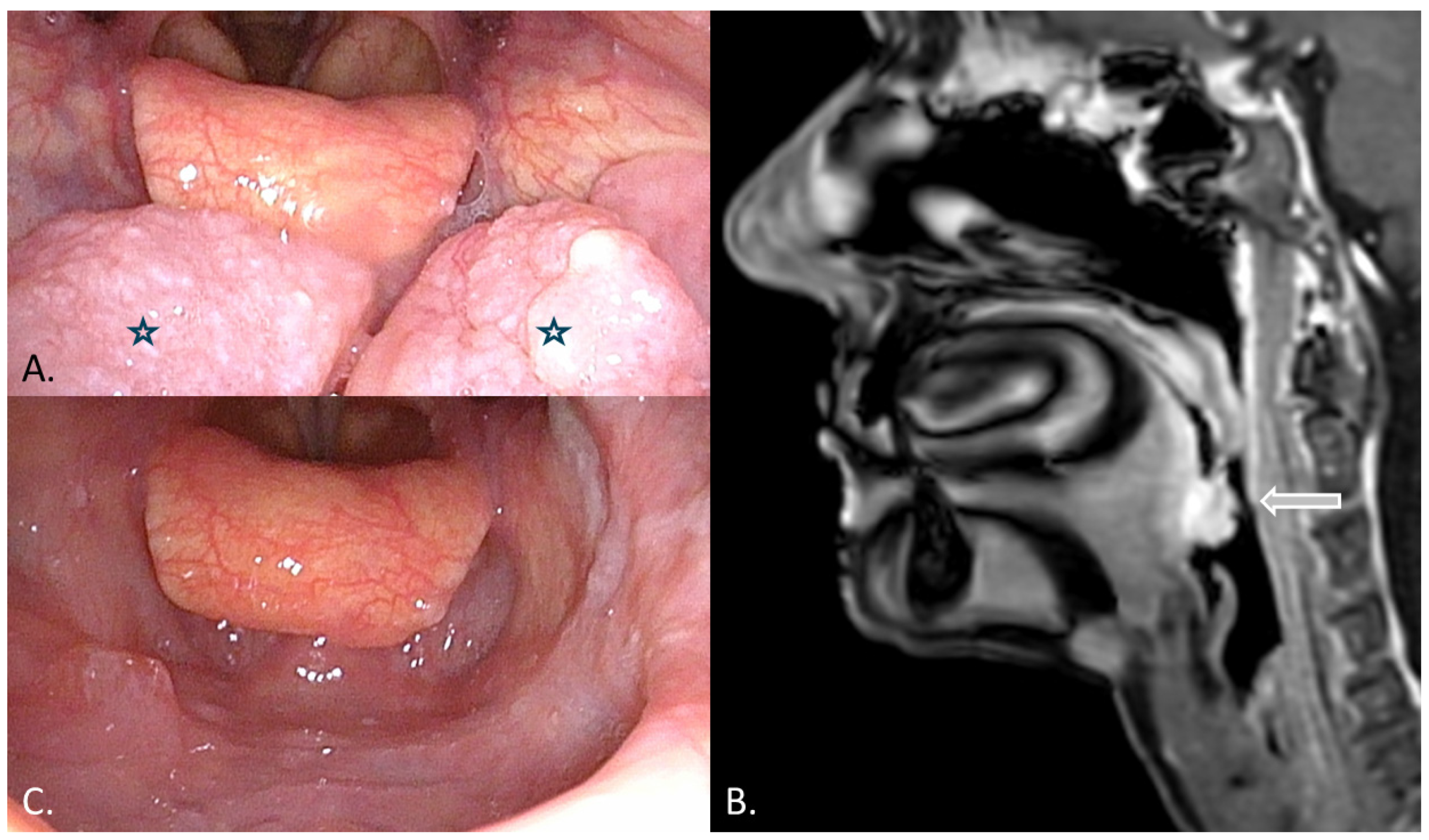
Flexible nasopharyngolaryngoscopy at the initial visit revealed a firm, lobulated, exophytic mass occupying the tongue base bilaterally, though more prominent on the left side, centered at the glossotonsillar sulcus (
Figure 1A). Under narrow-band imaging (NBI), the lesion displayed a vascular pattern considered suspicious. Superficial ulceration was noted over part of its surface. The vallecula, the V-shaped row of circumvallate papillae, and the laryngeal inlet were all spared, and both vocal cords moved normally. The hypopharynx and piriform sinuses appeared unremarkable. No lymph nodes were palpable in the neck.
A punch biopsy was taken under local anesthesia in the outpatient clinic. Histopathology showed squamous mucosa overlying dense subepithelial deposits of amorphous, eosinophilic material consistent with amyloid. Interstitial plasma cells expressing immunoglobulin lambda light chain were scattered throughout the deposits. The pathologist recommended ruling out an underlying hematologic disorder.
2.3. Diagnostic Imaging
On contrast-enhanced MRI, the lesion involved both hemibases of the tongue, enhancing avidly with increased vascularity on dynamic perfusion sequences. The initial radiology report flagged it as suspicious for a primary oropharyngeal malignancy. Notably, the mass was well circumscribed, with no extension into the parapharyngeal or carotid spaces and no pathologic cervical lymph nodes (
Figure 1B). A staging CT of the chest and abdomen was unremarkable, showing no distant deposits or organomegaly.
2.4. Systemic Evaluation
The patient was referred to the hematology service for a full workup to exclude systemic AL amyloidosis and an underlying plasma-cell neoplasm. Blood counts and coagulation studies were normal. Cardiac biomarkers were reassuring: troponin was undetectable and brain natriuretic peptide (BNP) measured 34 pg/mL. Neither serum nor urine protein electrophoresis with immunofixation detected a monoclonal paraprotein, and the serum free light-chain kappa-to-lambda ratio was 1.05—well within the normal range. Urinalysis showed no proteinuria. Transthoracic echocardiography demonstrated a left ventricle of normal size with moderate hypertrophy but preserved systolic function, normal wall motion, and a borderline interventricular septum thickness. Bone marrow biopsy was free of plasma-cell infiltration and amyloid deposits. Taken together, these results excluded systemic amyloidosis, monoclonal gammopathy, and myeloma, confirming that the tongue base deposits represented a primary localized amyloidoma.
2.5. Surgical Treatment
Because the patient had progressive swallowing difficulty and a definitive tissue diagnosis was needed on the full specimen, excision was planned using the da Vinci Xi robotic system via a transoral approach. Under general anesthesia with nasotracheal intubation, a Davies retractor with a large aspiration blade was positioned to expose the oropharynx. A 30-degree endoscope provided magnified, angled visualization of the tongue base. The right robotic arm carried a spatula tip cautery instrument; the left, a Maryland bipolar forceps. The entire tongue base mass was resected en bloc, together with the left palatine tonsil, which appeared grossly involved at the glossotonsillar sulcus. Hemostasis was straightforward, achieved entirely with the Maryland bipolar forceps of the da Vinci system, and no sutures were needed across the surgical bed. Setup took 22 min; the console time was 61 min. No tracheostomy or nasogastric tube was required. Estimated blood loss was minimal, and the postoperative course allowed early resumption of oral intake without airway compromise or bleeding. Two specimens, tongue base and left tonsil, were sent separately for pathological examination.
2.6. Histopathological Findings
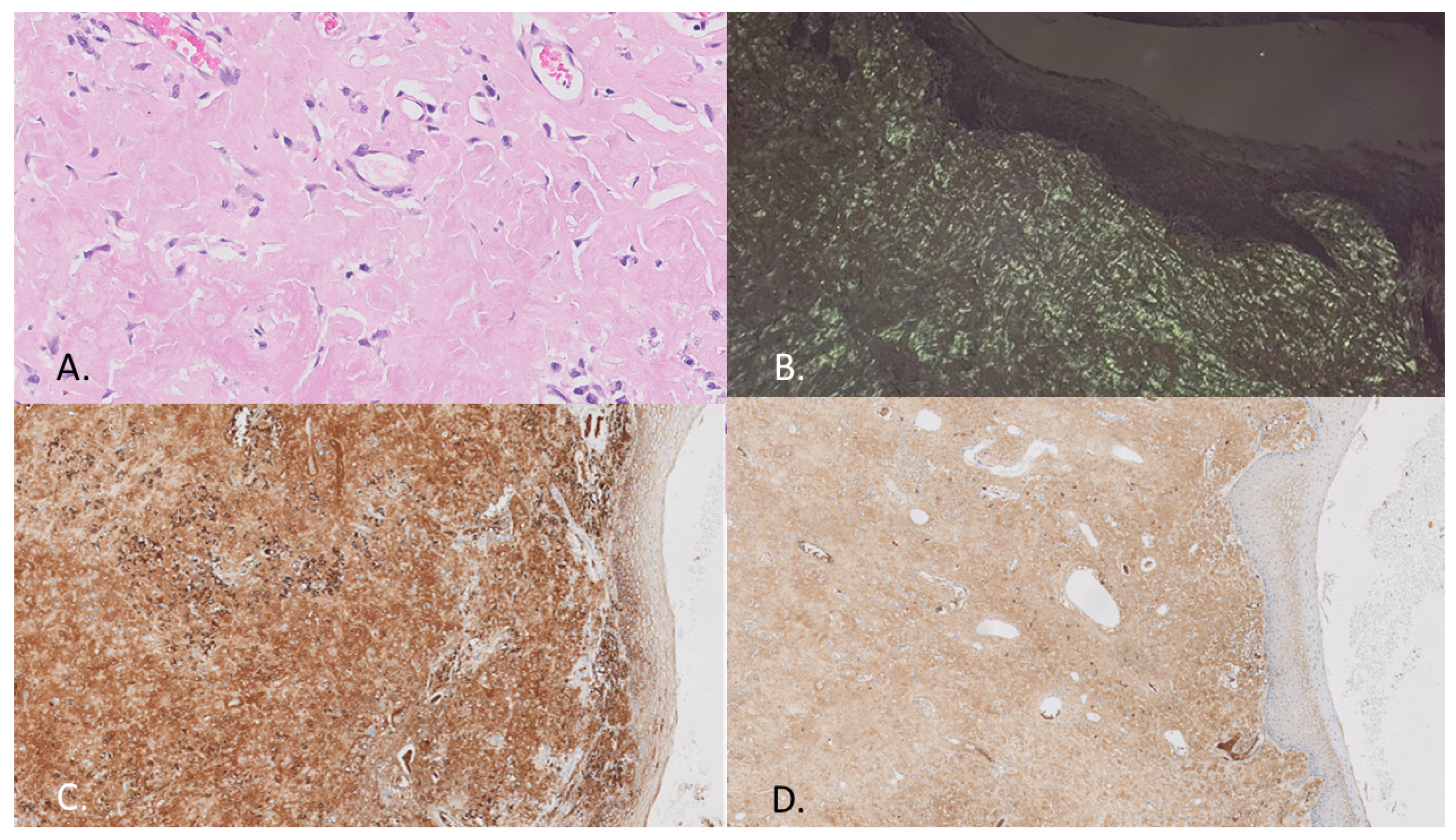
Both specimens showed extensive subepithelial expansion by amorphous, eosinophilic material (
Figure 2A). Congo red staining was unequivocally positive, producing the hallmark apple-green birefringence under polarized-light microscopy (
Figure 2B). Immunohistochemistry identified scattered plasma cells within the deposits expressing lambda light chain, with markedly weaker kappa staining (
Figure 2C,D), while stains for AA amyloid (serum amyloid A) and prealbumin (transthyretin) were negative. These findings confirmed the AL lambda subtype. Critically, neither specimen harbored sheets of atypical plasma cells, malignant epithelial proliferation, or dysplasia of any kind.
2.7. Follow-Up and Outcomes
Recovery was uncomplicated. Mild lingual edema and ecchymosis appeared in the first few postoperative days and resolved on their own without specific treatment. There was no significant pain, bleeding, wound infection, or airway compromise. The patient returned to a normal diet promptly. At 12-month follow-up, flexible endoscopy showed a clean, well-healed surgical bed with no evidence of residual or recurrent disease (
Figure 1C). She remained asymptomatic, tolerated a normal diet, and reported no new local or systemic symptoms. Hematology review at the same time point reaffirmed the absence of systemic involvement. A long-term surveillance plan was established: periodic endoscopic checks, with repeat systemic investigations to be triggered only if new symptoms develop.
3. Discussion
Among head and neck sites, the larynx accounts for the vast majority of localized amyloidomas [
8,
9]. Penner and Müller, in a clinicopathologic study spanning 15 cases, found involvement outside the larynx (including the tongue) to be relatively uncommon [
9]. Singh et al. reported a single-center experience of seven patients with head and neck amyloidosis; three had lingual deposits, all managed surgically without recurrence [
14]. The tongue base, as opposed to the dorsum, is an especially uncommon location. In a review by Adamo et al. that pooled 21 published cases of localized tongue amyloidosis, nearly three quarters involved the dorsum and presented as solitary nodules; excision was performed in nine, with recurrence in two [
10]. The present case adds to the small body of literature on tongue base amyloidoma, a location that poses particular diagnostic confusion because of its overlap with oropharyngeal carcinoma [
13].
At the cellular level, localized AL amyloidoma appears to arise from a minute, non-aggressive plasma-cell clone confined to the affected tissue. These cells overproduce immunoglobulin light chains—most often lambda—that undergo misfolding and are converted into insoluble fibrils, likely by tissue macrophages [
2,
6,
13]. Multinucleated giant cells are frequently observed in the deposits, lending support to this mechanism [
13,
15]. A key conceptual distinction from systemic AL amyloidosis is that, in the localized form, the amyloidogenic process does not spill over into the circulation [
4,
5]. Martínez et al. have gone further, characterizing localized AL as a self-limited plasmacytic lymphoproliferative disorder and arguing that the rare reports of systemic progression more likely reflect cases that were incompletely staged at the outset rather than genuine biological transformation [
5].
Before labeling an amyloidoma as localized, systemic AL amyloidosis must be rigorously excluded, because the management of the two entities could not be more different. The standard workup calls for serum and urine protein electrophoresis with immunofixation, serum free light chains (with attention to the kappa-to-lambda ratio), a urine sample to check for proteinuria, echocardiography for occult cardiac infiltration, and bone marrow biopsy [
1,
3,
16]. Every one of these studies was unremarkable in the present patient. The kappa-to-lambda ratio of 1.05 showed no clonal skewing, and normal troponin and BNP levels made clinically significant cardiac involvement unlikely.
From a radiological standpoint, amyloidomas do not have a signature appearance. They can enhance brightly on MRI and appear as soft-tissue-density masses on CT; when ulceration or prominent vascularity is present, the picture may be frankly worrisome for malignancy [
8]. This is precisely what happened in the present case: the MRI was initially read as suspicious for a primary oropharyngeal tumor. The differential diagnosis for any tongue base mass is broad, including squamous cell carcinoma, extramedullary plasmacytoma, lymphoma, granular cell tumor, and minor salivary gland neoplasm [
8]. Certain clinical features may tilt the balance toward amyloidoma: a protracted symptom history without constitutional decline, no palpable cervical adenopathy, and a well-demarcated mass. Ultimately, however, the diagnosis rests on tissue: Congo red-positive deposits with apple-green birefringence under polarized light, combined with light-chain typing, settle the matter [
1,
3].
In this patient, squamous cell carcinoma was the main initial diagnostic concern because of the ulcerated surface, suspicious vascular pattern on narrow-band imaging, and avid enhancement on MRI [
8]. Extramedullary plasmacytoma was also considered because of the presence of lambda-restricted plasma cells, but the absence of sheets of atypical plasma cells, the negative bone marrow biopsy, and the absence of monoclonal protein in serum and urine argued against this diagnosis [
1,
3,
16]. Lymphoma and minor salivary gland tumors were also part of the differential diagnosis of a tongue-base mass, although histology showed Congo red-positive amyloid deposition without malignant lymphoid or epithelial proliferation [
8].
Determining the amyloid subtype is not merely academic. In this patient, immunohistochemistry demonstrated lambda light-chain restriction while stains for AA amyloid and transthyretin came back negative, establishing the AL lambda subtype. When immunohistochemistry is equivocal, mass spectrometry-based proteomic analysis is the definitive arbiter and is now considered the gold standard for amyloid typing [
1,
3].
Treatment decisions in localized lingual amyloidoma hinge on symptoms. Small, asymptomatic deposits can reasonably be watched, whereas a tongue base mass causing dysphagia or airway concern generally warrants surgical removal [
4,
10,
13]. TORS offers clear advantages in this anatomic region: three-dimensional magnified visualization, tremor-filtered instrumentation, and the ability to work comfortably in deep, curved spaces that are awkward to reach with conventional instruments [
17]. In the present case, the entire excision was completed robotically in just over an hour, without the need for sutures, and the patient resumed eating within days. These observations are in line with a recent systematic review that endorsed TORS as a feasible and safe approach for benign tongue base pathology, though the authors noted that the evidence base remains limited to case reports and small series [
17].
There is no role for adjuvant chemotherapy or radiation after excision of a localized amyloidoma; systemic treatment is reserved for bona fide systemic AL amyloidosis [
1,
3,
16]. As for the risk of recurrence, Basset et al. studied 293 patients with localized AL and reported 5- and 10-year local progression-free survival rates of 62% and 44%, respectively. Notably, patients in whom a clonal plasma-cell infiltrate could be identified at the deposition site were at higher risk of local relapse [
6]. In a separate, more recent cohort of 146 patients followed for up to 14 years, Dima et al. found that none progressed to systemic disease, and 10-year overall survival stood at 83% [
7]. On the basis of these data, a strategy of periodic endoscopic surveillance, with systemic reassessment reserved for new or recurrent symptoms, seems both prudent and sufficient [
4,
7].
Several aspects of this report merit comment. The case is documented in unusual detail, from the NBI endoscopic findings through the minute-by-minute operative record to the specific hematologic results. Every component of the recommended systemic workup was performed, lending confidence to the localized diagnosis. Additionally, the use of TORS for tongue base amyloidoma has seldom been described. Although the follow-up period is now longer than in the initial submission, this remains a single-case report, and longer surveillance is still required to draw firm conclusions regarding long-term local control. Mass spectrometry-based amyloid typing was not performed, which is another limitation; however, Congo red positivity with apple-green birefringence, lambda light-chain predominance, negative AA and transthyretin staining, and a negative systemic workup strongly supported the diagnosis of localized AL lambda amyloidoma.




 ,
,