Feature Papers in Cell Biology and Pathology

A topical collection in Biomedicines (ISSN 2227-9059). This collection belongs to the section "Cell Biology and Pathology".

Viewed by 45208

Editor


E-Mail Website
Collection Editor
Forschungszentrum für Bio-Makromoleküle (FZ BIOmac), University of Bayreuth, Bayreuth, Germany
Interests: NMR-spectroscopy; protein structures; viral proteins; bacterial proteins; transcription; translation; allergens; nuclear magnetic resonance; crystallography; electron microscopy; fluorescence spectroscopy
Special Issues, Collections and Topics in MDPI journals

Topical Collection Information

Dear Colleagues,

The special edition “Feature Papers in Cell Biology and Pathology” will comprise important contributions by scholars in the field of cell biology and the Editorial Board members of the section Cell Biology and Pathology in Biomedicines. Their broad expertise will result in a comprehensive array of the latest findings in this field, and thus we encourage submissions of high-quality research papers or review articles.

Contributions that explore the origin of diseases at the molecular level, such as structural, biochemical, and genetic studies of receptors, oncogenes, tumor suppressor and regulatory proteins, signal pathways, and RNA/DNA, as well as articles that propose new routes for curing and preventing diseases, are of particular interest. Cutting-edge diagnostic tools ranging from PCR to magnetic resonance as applied to the detection of cellular or organismic pathological dysfunctions on all levels are also appropriate topics for this special edition. We look forward to your submissions on the above-listed research areas of cell biology and pathology.

Prof. Dr. Paul Rösch
Collection Editor

Manuscript Submission Information

Manuscripts should be submitted online at www.mdpi.com by registering and logging in to this website. Once you are registered, click here to go to the submission form. Manuscripts can be submitted until the deadline. All submissions that pass pre-check are peer-reviewed. Accepted papers will be published continuously in the journal (as soon as accepted) and will be listed together on the collection website. Research articles, review articles as well as short communications are invited. For planned papers, a title and short abstract (about 100 words) can be sent to the Editorial Office for announcement on this website.

Submitted manuscripts should not have been published previously, nor be under consideration for publication elsewhere (except conference proceedings papers). All manuscripts are thoroughly refereed through a single-blind peer-review process. A guide for authors and other relevant information for submission of manuscripts is available on the Instructions for Authors page. Biomedicines is an international peer-reviewed open access monthly journal published by MDPI.

Please visit the Instructions for Authors page before submitting a manuscript. The Article Processing Charge (APC) for publication in this open access journal is 2600 CHF (Swiss Francs). Submitted papers should be well formatted and use good English. Authors may use MDPI's English editing service prior to publication or during author revisions.

Published Papers (14 papers)

2024

Jump to: 2023, 2022

14 pages, 3475 KiB  
Article
Gallic Acid Induces HeLa Cell Lines Apoptosis via the P53/Bax Signaling Pathway
by Umut Sarı, Fuat Zaman, İlhan Özdemir, Şamil Öztürk and Mehmet Cudi Tuncer
Biomedicines 2024, 12(11), 2632; https://doi.org/10.3390/biomedicines12112632 - 18 Nov 2024
Cited by 2 | Viewed by 1450
Abstract
Background: Cervical cancer is a type of cancer that originates from the endometrium and is more common in developed countries and its incidence is increasing day by day in developing countries. The most commonly prescribed chemotherapeutic drugs limit their use due to serious [...] Read more.
Background: Cervical cancer is a type of cancer that originates from the endometrium and is more common in developed countries and its incidence is increasing day by day in developing countries. The most commonly prescribed chemotherapeutic drugs limit their use due to serious side effects and the development of drug resistance. For this reason, interest in new active ingredients obtained from natural products is increasing. This study aimed to reveal the apoptotic and antiproliferative effects of gallic acid and doxorubicin combination therapy against the HeLa cell line. Methods: We investigated the anti-cancer effects of doxorubicin and gallic acid in the human HeLa cervical cell line by using the MTT test, Nucblue staining for the identification of apoptotic cells due to nuclear condensation using fluorescent substance, and apoptotic markers P53 and Bax for the RT-PCR test. Results: The highest cytotoxic effect obtained in the study, the highest increase in apoptotic induction, and a significant difference in P53/Bax levels were seen in the gallic acid/doxorubicin combination. Additionally, it was determined that gallic acid exhibited an effective cytotoxic effect on HeLa and HaCat cells within 48 and 72 h of application. Conclusions: The obtained findings show that the gallic acid/doxorubicin combination applied to HeLa cells may be an alternative treatment against both the cytotoxic effect size and the side effects of the chemotherapy agent. Full article
Show Figures

Figure 1

15 pages, 592 KiB  
Review
Characterizing Fibroblast Heterogeneity in Diabetic Wounds Through Single-Cell RNA-Sequencing
by Helen H. Wang, Maria Korah, Serena L. Jing, Charlotte E. Berry, Michelle F. Griffin, Michael T. Longaker and Michael Januszyk
Biomedicines 2024, 12(11), 2538; https://doi.org/10.3390/biomedicines12112538 - 7 Nov 2024
Cited by 1 | Viewed by 1825
Abstract
Diabetes mellitus is an increasingly prevalent chronic metabolic disorder characterized by physiologic hyperglycemia that, when left uncontrolled, can lead to significant complications in multiple organs. Diabetic wounds are common in the general population, yet the underlying mechanism of impaired healing in such wounds [...] Read more.
Diabetes mellitus is an increasingly prevalent chronic metabolic disorder characterized by physiologic hyperglycemia that, when left uncontrolled, can lead to significant complications in multiple organs. Diabetic wounds are common in the general population, yet the underlying mechanism of impaired healing in such wounds remains unclear. Single-cell RNA-sequencing (scRNAseq) has recently emerged as a tool to study the gene expression of heterogeneous cell populations in skin wounds. Herein, we review the history of scRNAseq and its application to the study of diabetic wound healing, focusing on how innovations in single-cell sequencing have transformed strategies for fibroblast analysis. We summarize recent research on the role of fibroblasts in diabetic wound healing and describe the functional and cellular heterogeneity of skin fibroblasts. Moreover, we highlight future opportunities in diabetic wound fibroblast research, with a focus on characterizing distinct fibroblast subpopulations and their lineages. Leveraging single-cell technologies to explore fibroblast heterogeneity and the complex biology of diabetic wounds may reveal new therapeutic targets for improving wound healing and ultimately alleviate the clinical burden of chronic wounds. Full article
Show Figures

Graphical abstract

19 pages, 24345 KiB  
Review
Remarks on Selected Morphological Aspects of Cancer Neuroscience: A Microscopic Photo Review
by Ewa Iżycka-Świeszewska, Jacek Gulczyński, Aleksandra Sejda, Joanna Kitlińska, Susana Galli, Wojciech Rogowski and Dawid Sigorski
Biomedicines 2024, 12(10), 2335; https://doi.org/10.3390/biomedicines12102335 - 14 Oct 2024
Viewed by 1452
Abstract
Background: This short review and pictorial essay presents a morphological insight into cancer neuroscience, which is a complex and dynamic area of the pathobiology of tumors. Methods: We discuss the different methods and issues connected with structural research on tumor innervation, interactions between [...] Read more.
Background: This short review and pictorial essay presents a morphological insight into cancer neuroscience, which is a complex and dynamic area of the pathobiology of tumors. Methods: We discuss the different methods and issues connected with structural research on tumor innervation, interactions between neoplastic cells and the nervous system, and dysregulated neural influence on cancer phenotypes. Results: Perineural invasion (PNI), the most-visible cancer–nerve relation, is briefly presented, focusing on its pathophysiology and structural diversity as well as its clinical significance. The morphological approach to cancer neurobiology further includes the analysis of neural density/axonogenesis, neural network topographic distribution, and composition of fiber types and size. Next, the diverse range of neurotransmitters and neuropeptides and the neuroendocrine differentiation of cancer cells are reviewed. Another morphological area of cancer neuroscience is spatial or quantitative neural-related marker expression analysis through different detection, description, and visualization methods, also on experimental animal or cellular models. Conclusions: Morphological studies with systematic methodologies provide a necessary insight into the structure and function of the multifaceted tumor neural microenvironment and in context of possible new therapeutic neural-based oncological solutions. Full article
Show Figures

Figure 1

18 pages, 2732 KiB  
Article
Increased Myocardial MAO-A, Atrogin-1, and IL-1β Expression in Transgenic Mice with Pancreatic Carcinoma—Benefit of MAO-A Inhibition for Cardiac Cachexia
by Kira Stelter, Annalena Alabssi, Gabriel Alejandro Bonaterra, Hans Schwarzbach, Volker Fendrich, Emily P. Slater, Ralf Kinscherf and Wulf Hildebrandt
Biomedicines 2024, 12(9), 2009; https://doi.org/10.3390/biomedicines12092009 - 3 Sep 2024
Viewed by 1326
Abstract
Cancer cachexia (CC) continues to challenge clinicians by massively impairing patients’ prognosis, mobility, and quality of life through skeletal muscle wasting. CC also includes cardiac cachexia as characterized by atrophy, compromised metabolism, innervation and function of the myocardium through factors awaiting clarification for [...] Read more.
Cancer cachexia (CC) continues to challenge clinicians by massively impairing patients’ prognosis, mobility, and quality of life through skeletal muscle wasting. CC also includes cardiac cachexia as characterized by atrophy, compromised metabolism, innervation and function of the myocardium through factors awaiting clarification for therapeutic targeting. Because monoamine oxidase-A (MAO-A) is a myocardial source of H2O2 and implicated in myofibrillar protein catabolism and heart failure, we presently studied myocardial MAO-A expression, inflammatory cells, and capillarization together with transcripts of pro-inflammatory, -angiogenic, -apoptotic, and -proteolytic signals (by qRT-PCR) in a 3x-transgenic (LSL-KrasG12D/+; LSL-TrP53R172H/+; Pdx1-Cre) mouse model of orthotopic pancreatic ductal adenoarcinoma (PDAC) compared to wild-type (WT) mice. Moreover, we evaluated the effect of MAO-A inhibition by application of harmine hydrochloride (HH, 8 weeks, i.p., no sham control) on PDAC-related myocardial alterations. Myocardial MAO-A protein content was significantly increased (1.69-fold) in PDAC compared to WT mice. PDAC was associated with an increased percentage of atrogin-1+ (p < 0.001), IL-1β+ (p < 0.01), COX2+ (p < 0.001), and CD68+ (p > 0.05) cells and enhanced transcripts of pro-inflammatory IL-1β (2.47-fold), COX2 (1.53-fold), TNF (1.87-fold), and SOCS3 (1.64-fold). Moreover, PDAC was associated with a reduction in capillary density (−17%, p < 0.05) and transcripts of KDR (0.46-fold) but not of VEGFA, Notch1, or Notch3. Importantly, HH treatment largely reversed the PDAC-related increases in atrogin-1+, IL-1β+, and TNF+ cell fraction as well as in COX2, IL-1β, TNF, and SOCS3 transcripts, whereas capillary density and KDR transcripts failed to improve. In mice with PDAC, increased myocardial pro-atrophic/-inflammatory signals are attributable to increased expression of MAO-A, because they are significantly improved with MAO-A inhibition as a potential novel therapeutic option. The PDAC-related loss in myocardial capillary density may be due to other mechanisms awaiting evaluation with consideration of cardiomyocyte size, cardiac function and physical activity. Full article
Show Figures

Figure 1

15 pages, 1515 KiB  
Review
Understanding Tendon Fibroblast Biology and Heterogeneity
by Sarah E. DiIorio, Bill Young, Jennifer B. Parker, Michelle F. Griffin and Michael T. Longaker
Biomedicines 2024, 12(4), 859; https://doi.org/10.3390/biomedicines12040859 - 12 Apr 2024
Cited by 8 | Viewed by 5117
Abstract
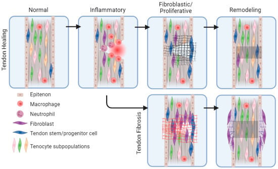
Tendon regeneration has emerged as an area of interest due to the challenging healing process of avascular tendon tissue. During tendon healing after injury, the formation of a fibrous scar can limit tendon strength and lead to subsequent complications. The specific biological mechanisms [...] Read more.
Tendon regeneration has emerged as an area of interest due to the challenging healing process of avascular tendon tissue. During tendon healing after injury, the formation of a fibrous scar can limit tendon strength and lead to subsequent complications. The specific biological mechanisms that cause fibrosis across different cellular subtypes within the tendon and across different tendons in the body continue to remain unknown. Herein, we review the current understanding of tendon healing, fibrosis mechanisms, and future directions for treatments. We summarize recent research on the role of fibroblasts throughout tendon healing and describe the functional and cellular heterogeneity of fibroblasts and tendons. The review notes gaps in tendon fibrosis research, with a focus on characterizing distinct fibroblast subpopulations in the tendon. We highlight new techniques in the field that can be used to enhance our understanding of complex tendon pathologies such as fibrosis. Finally, we explore bioengineering tools for tendon regeneration and discuss future areas for innovation. Exploring the heterogeneity of tendon fibroblasts on the cellular level can inform therapeutic strategies for addressing tendon fibrosis and ultimately reduce its clinical burden. Full article
Show Figures

Figure 1

16 pages, 2899 KiB  
Article
PD-L1 Expression in Neoplastic and Immune Cells of Thymic Epithelial Tumors: Correlations with Disease Characteristics and HDAC Expression
by Ioanna E. Stergiou, Kostas Palamaris, Georgia Levidou, Maria Tzimou, Stavros P. Papadakos, Georgios Mandrakis, Christos Masaoutis, Dimitra Rontogianni and Stamatios Theocharis
Biomedicines 2024, 12(4), 772; https://doi.org/10.3390/biomedicines12040772 - 31 Mar 2024
Cited by 2 | Viewed by 1889
Abstract
Background: Programmed death-ligand 1 (PD-L1) expression in neoplastic and immune cells of the tumor microenvironment determines the efficacy of antitumor immunity, while it can be regulated at the epigenetic level by various factors, including HDACs. In this study, we aim to evaluate the [...] Read more.
Background: Programmed death-ligand 1 (PD-L1) expression in neoplastic and immune cells of the tumor microenvironment determines the efficacy of antitumor immunity, while it can be regulated at the epigenetic level by various factors, including HDACs. In this study, we aim to evaluate the expression patterns of PD-L1 in thymic epithelial tumors (TETs), while we attempt the first correlation analysis between PD-L1 and histone deacetylases (HDACs) expression. Methods: Immunohistochemistry was used to evaluate the expression of PD-L1 in tumor and immune cells of 91 TETs with SP263 and SP142 antibody clones, as well as the expressions of HDCA1, -2, -3, -4, -5, and -6. Results: The PD-L1 tumor proportion score (TPS) was higher, while the immune cell score (IC-score) was lower in the more aggressive TET subtypes and in more advanced Masaoka–Koga stages. A positive correlation between PD-L1 and HDAC-3, -4, and -5 cytoplasmic expression was identified. Conclusions: Higher PD-L1 expression in neoplastic cells and lower PD-L1 expression in immune cells of TETs characterizes more aggressive and advanced neoplasms. Correlations between PD-L1 and HDAC expression unravel the impact of epigenetic regulation on the expression of immune checkpoint molecules in TETs, with possible future applications in combined therapeutic targeting. Full article
Show Figures

Figure 1

19 pages, 4655 KiB  
Article
The FLNC Ala1186Val Variant Linked to Cytoplasmic Body Myopathy and Cardiomyopathy Causes Protein Instability
by Marion Onnée, Audrey Bénézit, Sultan Bastu, Aleksandra Nadaj-Pakleza, Béatrice Lannes, Flavie Ader, Corinne Thèze, Pascal Cintas, Claude Cances, Robert-Yves Carlier, Corinne Metay, Mireille Cossée and Edoardo Malfatti
Biomedicines 2024, 12(2), 322; https://doi.org/10.3390/biomedicines12020322 - 30 Jan 2024
Cited by 3 | Viewed by 1980
Abstract
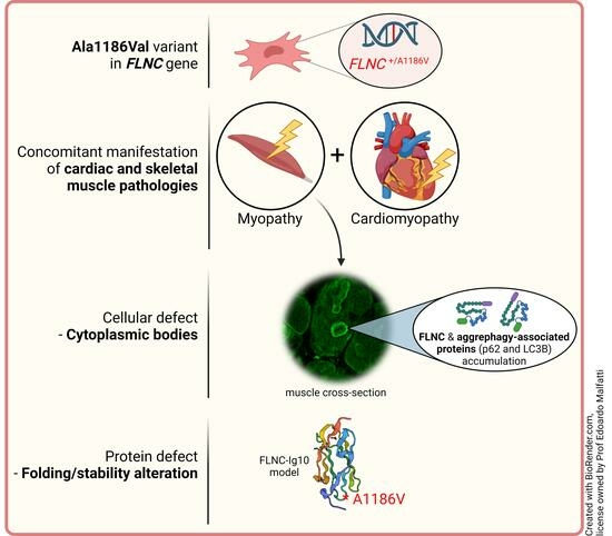
Filamin C-related disorders include myopathies and cardiomyopathies linked to variants in the FLNC gene. Filamin C belongs to a family of actin-binding proteins involved in sarcomere stability. This study investigates the pathogenic impact of the FLNC c.3557C > T (p.Ala1186Val) pathogenic variant associated [...] Read more.
Filamin C-related disorders include myopathies and cardiomyopathies linked to variants in the FLNC gene. Filamin C belongs to a family of actin-binding proteins involved in sarcomere stability. This study investigates the pathogenic impact of the FLNC c.3557C > T (p.Ala1186Val) pathogenic variant associated with an early-onset cytoplasmic body myopathy and cardiomyopathy in three unrelated patients. We performed clinical imaging and myopathologic and genetic characterization of three patients with an early-onset myopathy and cardiomyopathy. Bioinformatics analysis, variant interpretation, and protein structure analysis were performed to validate and assess the effects of the filamin C variant. All patients presented with a homogeneous clinical phenotype marked by a severe contractural myopathy, leading to loss of gait. There was prominent respiratory involvement and restrictive or hypertrophic cardiomyopathies. The Ala1186Val variant is located in the interstrand loop involved in intradomain stabilization and/or interdomain interactions with neighbor Ig-like domains. 3D modeling highlights local structural changes involving nearby residues and probably impacts the protein stability, causing protein aggregation in the form of cytoplasmic bodies. Myopathologic studies have disclosed the prominent aggregation and upregulation of the aggrephagy-associated proteins LC3B and p62. As a whole, the Ala1186Val variant in the FLNC gene provokes a severe myopathy with contractures, respiratory involvement, and cardiomyopathy due to protein aggregation in patients’ muscles. Full article
Show Figures

Graphical abstract

2023

Jump to: 2024, 2022

12 pages, 806 KiB  
Review
Chemical Transdifferentiation of Somatic Cells: Unleashing the Power of Small Molecules
by Yu Zhang, Xuefeng Li, Jianyu Xing, Jinsong Zhou and Hai Li
Biomedicines 2023, 11(11), 2913; https://doi.org/10.3390/biomedicines11112913 - 27 Oct 2023
Cited by 7 | Viewed by 2875
Abstract
Chemical transdifferentiation is a technique that utilizes small molecules to directly convert one cell type into another without passing through an intermediate stem cell state. This technique offers several advantages over other methods of cell reprogramming, such as simplicity, standardization, versatility, no ethical [...] Read more.
Chemical transdifferentiation is a technique that utilizes small molecules to directly convert one cell type into another without passing through an intermediate stem cell state. This technique offers several advantages over other methods of cell reprogramming, such as simplicity, standardization, versatility, no ethical and safety concern and patient-specific therapies. Chemical transdifferentiation has been successfully applied to various cell types across different tissues and organs, and its potential applications are rapidly expanding as scientists continue to explore new combinations of small molecules and refine the mechanisms driving cell fate conversion. These applications have opened up new possibilities for regenerative medicine, disease modeling, drug discovery and tissue engineering. However, there are still challenges and limitations that need to be overcome before chemical transdifferentiation can be translated into clinical practice. These include low efficiency and reproducibility, incomplete understanding of the molecular mechanisms, long-term stability and functionality of the transdifferentiated cells, cell-type specificity and scalability. In this review, we compared the commonly used methods for cell transdifferentiation in recent years and discussed the current progress and future perspective of the chemical transdifferentiation of somatic cells and its potential impact on biomedicine. We believe that with ongoing research and technological advancements, the future holds tremendous promise for harnessing the power of small molecules to shape the cellular landscape and revolutionize the field of biomedicine. Full article
Show Figures

Figure 1

16 pages, 2185 KiB  
Systematic Review
Prospect of Mesenchymal Stem-Cell-Conditioned Medium in the Treatment of Acute Pancreatitis: A Systematic Review
by Ke Pang, Fanyi Kong and Dong Wu
Biomedicines 2023, 11(9), 2343; https://doi.org/10.3390/biomedicines11092343 - 23 Aug 2023
Cited by 4 | Viewed by 2222
Abstract
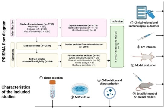
Mesenchymal stem cells (MSCs) have demonstrated potential in both clinical and pre-clinical research for mitigating tissue damage and inflammation associated with acute pancreatitis (AP) via paracrine mechanisms. Hence, there has been a recent surge of interest among researchers in utilizing MSC cultured medium [...] Read more.
Mesenchymal stem cells (MSCs) have demonstrated potential in both clinical and pre-clinical research for mitigating tissue damage and inflammation associated with acute pancreatitis (AP) via paracrine mechanisms. Hence, there has been a recent surge of interest among researchers in utilizing MSC cultured medium (CM) and its components for the treatment of AP, which is recognized as the primary cause of hospitalization for gastrointestinal disorders globally. A systematic review was conducted by searching the MEDLINE, EMBASE, and Web of Science databases. Studies that involve the administration of MSC-CM, extracellular vesicles/microvesicles (EVs/MVs), or exosomes to AP animal models are included. A total of six research studies, including eight experiments, were identified as relevant. The findings of this study provide evidence in favor of a beneficial impact of MSC-CM on both clinical and immunological outcomes. Nevertheless, prior to clinical trials, large animal models should be used and prolonged observation periods conducted in pre-clinical research. Challenges arise due to the lack of standardization and consensus on isolation processes, quantifications, and purity testing, making it difficult to compare reports and conduct meta-analyses in MSC-CM-based therapies. Full article
Show Figures

Graphical abstract

13 pages, 2529 KiB  
Article
Naturally Occurring N-Terminal Fragments of Bovine Milk Osteopontin Are Transported across Models of the Intestinal Barrier
by Brian Christensen, Nanna R. Nielsen, Marie R. Sørensen, Lotte N. Jacobsen, Marie S. Ostenfeld and Esben S. Sørensen
Biomedicines 2023, 11(3), 893; https://doi.org/10.3390/biomedicines11030893 - 14 Mar 2023
Cited by 8 | Viewed by 2291
Abstract
Osteopontin (OPN) is a bioactive integrin-binding protein found in high concentrations in milk, where it is present both as a full-length protein and as several N-terminally derived fragments. OPN resists gastric digestion, and via interaction with receptors in the gut or by crossing [...] Read more.
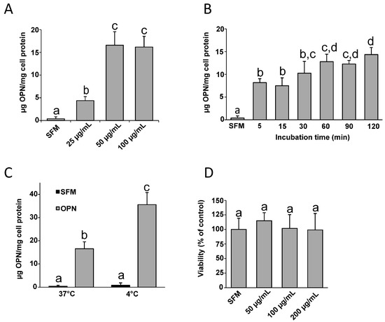
Osteopontin (OPN) is a bioactive integrin-binding protein found in high concentrations in milk, where it is present both as a full-length protein and as several N-terminally derived fragments. OPN resists gastric digestion, and via interaction with receptors in the gut or by crossing the intestinal barrier into circulation, ingested milk OPN may influence physiological processes. The aim of this study was to investigate OPN interaction with intestinal cells and its transport across models of the intestinal barrier. Immunodetection of OPN incubated with Caco-2 cells at 4 °C and 37 °C showed that OPN binds to the intestinal cells, but it is not internalised. Transepithelial transport was studied using mono- and co-cultures of Caco-2 cells and mucus-producing HT29-MTX cells in transwell membranes. OPN was shown to cross the barrier models in a time-, temperature-, and energy-dependent process inhibited by wortmannin, indicating that the transport takes place via the transcytosis pathway. Analyses of the naturally occurring milk mixture of full-length and N-terminal fragments showed that the N-terminal fragments of OPN bound intestinal cells most effectively and that the fragments were transported across the intestinal membrane models. This suggests that proteolytic processing of OPN increases its biological activity after ingestion. Full article
Show Figures

Figure 1

13 pages, 1646 KiB  
Article
Smooth Muscle Cells of Dystrophic (mdx) Mice Are More Susceptible to Hypoxia; The Protective Effect of Reducing Ca2+ Influx
by Arkady Uryash, Alfredo Mijares, Eric Estève, Jose A. Adams and Jose R. Lopez
Biomedicines 2023, 11(2), 623; https://doi.org/10.3390/biomedicines11020623 - 19 Feb 2023
Cited by 1 | Viewed by 1927
Abstract
Duchenne muscular dystrophy (DMD) is an inherited muscular disorder caused by mutations in the dystrophin gene. DMD patients have hypoxemic events due to sleep-disordered breathing. We reported an anomalous regulation of resting intracellular Ca2+ ([Ca2+]i) in vascular smooth [...] Read more.
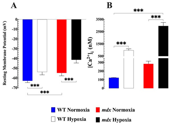
Duchenne muscular dystrophy (DMD) is an inherited muscular disorder caused by mutations in the dystrophin gene. DMD patients have hypoxemic events due to sleep-disordered breathing. We reported an anomalous regulation of resting intracellular Ca2+ ([Ca2+]i) in vascular smooth muscle cells (VSMCs) from a mouse (mdx) model of DMD. We investigated the effect of hypoxia on [Ca2+]i in isolated and quiescent VSMCs from C57BL/10SnJ (WT) and C57BL/10ScSn-Dmd (mdx) male mice. [Ca2+]i was measured using Ca2+-selective microelectrodes under normoxic conditions (95% air, 5% CO2) and after hypoxia (glucose-free solution aerated with 95% N2-5% CO2 for 30 min). [Ca2+]i in mdx VSMCs was significantly elevated compared to WT under normoxia. Hypoxia-induced [Ca2+]i overload, which was significantly greater in mdx than in WT VSMCs. A low Ca2+ solution caused a reduction in [Ca2+]i and prevented [Ca2+]i overload secondary to hypoxia. Nifedipine (10 µM), a Ca2+ channel blocker, did not modify resting [Ca2+]i in VSMCs but partially prevented the hypoxia-induced elevation of [Ca2+]i in both genotypes. SAR7334 (1 µM), an antagonist of TRPC3 and TRPC6, reduced the basal and [Ca2+]i overload caused by hypoxia. Cell viability, assessed by tetrazolium salt (3-(4,5-dimethylthiazol-2-yl)-2,5-diphenyltetrazolium bromide, was significantly reduced in mdx compared to WT VSMCs. Pretreatment with SAR7341 increases cell viability in normoxic mdx (p < 0.001) and during hypoxia in WT and mdx VSMCs. These results provide evidence that the lack of dystrophin makes VSMCs more susceptible to hypoxia-induced [Ca2+]i overload, which appears to be mediated by increased Ca2+ entry through L-type Ca2+ and TRPC channels. Full article
Show Figures

Figure 1

2022

Jump to: 2024, 2023

22 pages, 2630 KiB  
Review
The Role of Hsp27 in Chemotherapy Resistance
by Marios Lampros, Nikolaos Vlachos, Spyridon Voulgaris and George A. Alexiou
Biomedicines 2022, 10(4), 897; https://doi.org/10.3390/biomedicines10040897 - 14 Apr 2022
Cited by 33 | Viewed by 4925
Abstract
Heat shock protein (Hsp)-27 is a small-sized, ATP-independent, chaperone molecule that is overexpressed under conditions of cellular stress such as oxidative stress and heat shock, and protects proteins from unfolding, thus facilitating proteostasis and cellular survival. Despite its protective role in normal cell [...] Read more.
Heat shock protein (Hsp)-27 is a small-sized, ATP-independent, chaperone molecule that is overexpressed under conditions of cellular stress such as oxidative stress and heat shock, and protects proteins from unfolding, thus facilitating proteostasis and cellular survival. Despite its protective role in normal cell physiology, Hsp27 overexpression in various cancer cell lines is implicated in tumor initiation, progression, and metastasis through various mechanisms, including modulation of the SWH pathway, inhibition of apoptosis, promotion of EMT, adaptation of CSCs in the tumor microenvironment and induction of angiogenesis. Investigation of the role of Hsp27 in the resistance of various cancer cell types against doxorubicin, herceptin/trastuzumab, gemcitabine, 5-FU, temozolomide, and paclitaxel suggested that Hsp27 overexpression promotes cancer cell survival against the above-mentioned chemotherapeutic agents. Conversely, Hsp27 inhibition increased the efficacy of those chemotherapy drugs, both in vitro and in vivo. Although numerous signaling pathways and molecular mechanisms were implicated in that chemotherapy resistance, Hsp27 most commonly contributed to the upregulation of Akt/mTOR signaling cascade and inactivation of p53, thus inhibiting the chemotherapy-mediated induction of apoptosis. Blockage of Hsp27 could enhance the cytotoxic effect of well-established chemotherapeutic drugs, especially in difficult-to-treat cancer types, ultimately improving patients’ outcomes. Full article
Show Figures

Figure 1

23 pages, 19124 KiB  
Review
Ion Channels and Transporters as Therapeutic Agents: From Biomolecules to Supramolecular Medicinal Chemistry
by Giacomo Picci, Silvia Marchesan and Claudia Caltagirone
Biomedicines 2022, 10(4), 885; https://doi.org/10.3390/biomedicines10040885 - 12 Apr 2022
Cited by 32 | Viewed by 10082
Abstract
Ion channels and transporters typically consist of biomolecules that play key roles in a large variety of physiological and pathological processes. Traditional therapies include many ion-channel blockers, and some activators, although the exact biochemical pathways and mechanisms that regulate ion homeostasis are yet [...] Read more.
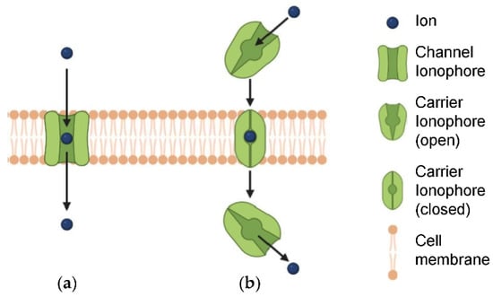
Ion channels and transporters typically consist of biomolecules that play key roles in a large variety of physiological and pathological processes. Traditional therapies include many ion-channel blockers, and some activators, although the exact biochemical pathways and mechanisms that regulate ion homeostasis are yet to be fully elucidated. An emerging area of research with great innovative potential in biomedicine pertains the design and development of synthetic ion channels and transporters, which may provide unexplored therapeutic opportunities. However, most studies in this challenging and multidisciplinary area are still at a fundamental level. In this review, we discuss the progress that has been made over the last five years on ion channels and transporters, touching upon biomolecules and synthetic supramolecules that are relevant to biological use. We conclude with the identification of therapeutic opportunities for future exploration. Full article
Show Figures

Figure 1

15 pages, 1303 KiB  
Review
Checkpoint Inhibitors and Induction of Celiac Disease-like Condition
by Aaron Lerner and Carina Benzvi
Biomedicines 2022, 10(3), 609; https://doi.org/10.3390/biomedicines10030609 - 4 Mar 2022
Cited by 4 | Viewed by 3849
Abstract
Immune checkpoint inhibitors herald a new era in oncological therapy-resistant cancer, thus bringing hope for better outcomes and quality of life for patients. However, as with other medications, they are not without serious side effects over time. Despite this, their advantages outweigh their [...] Read more.
Immune checkpoint inhibitors herald a new era in oncological therapy-resistant cancer, thus bringing hope for better outcomes and quality of life for patients. However, as with other medications, they are not without serious side effects over time. Despite this, their advantages outweigh their disadvantages. Understanding the adverse effects will help therapists locate, apprehend, treat, and perhaps diminish them. The major ones are termed immune-related adverse events (irAEs), representing their auto-immunogenic capacity. This narrative review concentrates on the immune checkpoint inhibitors induced celiac disease (CD), highlighting the importance of the costimulatory inhibitors in CD evolvement and suggesting several mechanisms for CD induction. Unraveling those cross-talks and pathways might reveal some new therapeutic strategies. Full article
Show Figures

Figure 1

Back to TopTop