applsci-logo

Journal Browser

Journal Browser

Advances in Medical Imaging: Techniques and Applications

A special issue of Applied Sciences (ISSN 2076-3417). This special issue belongs to the section "Biomedical Engineering".

Deadline for manuscript submissions: 20 November 2025 | Viewed by 2601

Special Issue Editors


E-Mail Website
Guest Editor
Medical Imaging and Radiotherapy Department, University of Algarve, Faro, Portugal
Interests: diagnostic radiology; computed tomography; magnetic resonance; diagnostic imaging artificial intelligence in medical imaging; digital health innovation; patient safety and quality in medical imaging; healthcare health literacy
Special Issues, Collections and Topics in MDPI journals

E-Mail Website
Guest Editor
Department of Computer Science and Engineering, University of North Texas, Denton, TX 76203, USA
Interests: medical image and video analysis

Special Issue Information

Dear Colleagues,

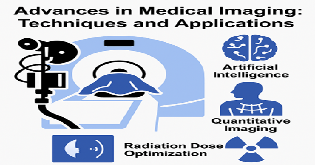
Medical imaging is fundamental to modern healthcare, supporting accurate diagnosis, therapeutic planning, and patient follow-up across a broad spectrum of diseases. Recent advances in imaging techniques, computational methods, and hardware development have significantly expanded the capabilities and applications of diagnostic imaging. This Special Issue aims to present cutting-edge research and innovative developments across various modalities, including computed tomography (CT), magnetic resonance imaging (MRI), ultrasound, radiography, interventional radiology and hybrid imaging techniques. Particular attention will be given to novel acquisition methods, image reconstruction algorithms, quantitative imaging, and image-guided interventions. Furthermore, the integration of artificial intelligence, deep learning, and big data analytics into medical imaging workflows is transforming clinical practice and decision-making processes. Contributions that address patient safety, radiation dose optimization, and quality assurance in imaging are also welcome. The goal of this Special Issue is to bring together researchers, clinicians, and industry experts to share knowledge, foster collaboration, and highlight future directions in medical imaging science and technology.

Prof. Dr. Rui Pedro Pereira De Almeida
Dr. JungHwan Oh
Guest Editors

Manuscript Submission Information

Manuscripts should be submitted online at www.mdpi.com by registering and logging in to this website. Once you are registered, click here to go to the submission form. Manuscripts can be submitted until the deadline. All submissions that pass pre-check are peer-reviewed. Accepted papers will be published continuously in the journal (as soon as accepted) and will be listed together on the special issue website. Research articles, review articles as well as short communications are invited. For planned papers, a title and short abstract (about 100 words) can be sent to the Editorial Office for announcement on this website.

Submitted manuscripts should not have been published previously, nor be under consideration for publication elsewhere (except conference proceedings papers). All manuscripts are thoroughly refereed through a single-blind peer-review process. A guide for authors and other relevant information for submission of manuscripts is available on the Instructions for Authors page. Applied Sciences is an international peer-reviewed open access semimonthly journal published by MDPI.

Please visit the Instructions for Authors page before submitting a manuscript. The Article Processing Charge (APC) for publication in this open access journal is 2400 CHF (Swiss Francs). Submitted papers should be well formatted and use good English. Authors may use MDPI's English editing service prior to publication or during author revisions.

Keywords

  • medical imaging
  • diagnostic radiology
  • computed tomography
  • magnetic resonance imaging
  • image reconstruction
  • artificial intelligence
  • quantitative imaging
  • image-guided interventions
  • radiation dose optimization
  • image quality

Benefits of Publishing in a Special Issue

  • Ease of navigation: Grouping papers by topic helps scholars navigate broad scope journals more efficiently.
  • Greater discoverability: Special Issues support the reach and impact of scientific research. Articles in Special Issues are more discoverable and cited more frequently.
  • Expansion of research network: Special Issues facilitate connections among authors, fostering scientific collaborations.
  • External promotion: Articles in Special Issues are often promoted through the journal's social media, increasing their visibility.
  • Reprint: MDPI Books provides the opportunity to republish successful Special Issues in book format, both online and in print.

Further information on MDPI's Special Issue policies can be found here.

Published Papers (3 papers)

Order results
Result details
Select all
Export citation of selected articles as:

Research

Jump to: Review

15 pages, 20890 KB  
Article
Development of an XAI-Enhanced Deep-Learning Algorithm for Automated Decision-Making on Shoulder-Joint X-Ray Retaking
by Konatsu Sekiura, Takaaki Yoshimura and Hiroyuki Sugimori
Appl. Sci. 2025, 15(19), 10534; https://doi.org/10.3390/app151910534 - 29 Sep 2025
Viewed by 171
Abstract
Purpose: To develop and validate a two-stage system for automated quality assessment of shoulder true-AP radiographs by combining joint localization with quality classification. Materials and Methods: From the MURA “SHOULDER” subset, 2956 anteroposterior images were identified; 59 images with negative–positive inversion, excessive metallic [...] Read more.
Purpose: To develop and validate a two-stage system for automated quality assessment of shoulder true-AP radiographs by combining joint localization with quality classification. Materials and Methods: From the MURA “SHOULDER” subset, 2956 anteroposterior images were identified; 59 images with negative–positive inversion, excessive metallic implants, extreme exposure, or presumed fluoroscopy were excluded, yielding a class-balanced set of 2800 images (1400 OK/1400 NG). A YOLOX-based detector localized the glenohumeral joint, and classifiers operated on both whole images and detector-centered crops. To enhance interpretability, we integrated Grad-CAM into both whole-image and local classifiers and assessed attention patterns against radiographic criteria. Results: The detector achieved AP@0.5 = 1.00 and a mean Dice similarity coefficient of 0.967. The classifier attained AUC = 0.977 (F1 = 0.943) on a held-out test set. Heat map analyses indicated anatomically focused attention consistent with expert-defined regions, and coverage metrics favored local over whole-image models. Conclusions: The two-stage, XAI-integrated approach provides accurate and interpretable assessment of shoulder true-AP image quality, aligning model attention with radiographic criteria. Full article
(This article belongs to the Special Issue Advances in Medical Imaging: Techniques and Applications)
Show Figures

Figure 1

12 pages, 3159 KB  
Article
Optimizing Knee MRI: Diagnostic Performance of a 3D PDW SPAIR-Based Short Protocol
by Marco Pinnizzotto, Maria Ragusi, Cesare Maino, Pietro Allegranza, Cammillo Talei Franzesi, Stefania Pellegatta, Davide Gandola, Marco Turati, Rocco Corso and Davide Ippolito
Appl. Sci. 2025, 15(16), 8870; https://doi.org/10.3390/app15168870 - 12 Aug 2025
Viewed by 896
Abstract
Objectives: This study aimed to evaluate the usefulness of a short magnetic resonance (MR) protocol for knee evaluation, using 3D PDW SPAIR sequences compared with traditional 2D ones. Methods: A prospective analysis included 76 patients with knee pain. MR was performed using a [...] Read more.
Objectives: This study aimed to evaluate the usefulness of a short magnetic resonance (MR) protocol for knee evaluation, using 3D PDW SPAIR sequences compared with traditional 2D ones. Methods: A prospective analysis included 76 patients with knee pain. MR was performed using a 1.5 T scanner. The standard protocol consisted of multiplanar 2D proton density-weighted (PDW) SPectral Attenuated Inversion Recovery (SPAIR) and additional T1-weighted (T1W) and T2-weighted (T2W) sequences, with a total acquisition time of 17 min. The simulated short protocol included a 3D PDW SPAIR sequence with isotropic voxels and a slice thickness of 0.6 mm, coronal T1W, and gradient echo (GRE) axial sequences, with a total acquisition time of 9 min. Two radiologists independently reviewed images and collected pathological processes. Results: The 3D PDW SPAIR sequence demonstrated a significantly higher subjective image quality compared to standard 2D sequences [κ = 0.712 (p < 0.001) vs. κ = 0.144 (p = 0.63); p < 0.001]. Artifacts were not significantly different between the two protocols (p = 0.201). Qualitative assessments showed superior ratings for 3D images (excellent quality: 72.4% vs. 26.3% for 3D and 2D, respectively; p < 0.001). Diagnostic performance was comparable between the two protocols for ACL injuries, medial and lateral collateral ligament injuries, and chondropathies. Three-dimensional sequences were more effective in detecting medial meniscal injuries (p < 0.001), particularly radial and complex tears, likely due to higher spatial resolution and multiplanar reconstruction capability. Conclusions: The 3D PDW SPAIR sequence offers advantages in knee MRI study, including improved image quality, reduced acquisition time, and superior detection of meniscal injuries. Full article
(This article belongs to the Special Issue Advances in Medical Imaging: Techniques and Applications)
Show Figures

Figure 1

Review

Jump to: Research

21 pages, 1335 KB  
Review
Machine Learning in Stroke Lesion Segmentation and Recovery Forecasting: A Review
by Simi Meledathu Sasidharan, Sibusiso Mdletshe and Alan Wang
Appl. Sci. 2025, 15(18), 10082; https://doi.org/10.3390/app151810082 - 15 Sep 2025
Viewed by 984
Abstract
Introduction: Stroke remains a major cause of disability worldwide, and precise identification of stroke lesions is essential for prognosis and rehabilitation planning. Machine learning has emerged as a powerful tool for automating stroke lesion segmentation and outcome prediction; however, these tasks are often [...] Read more.
Introduction: Stroke remains a major cause of disability worldwide, and precise identification of stroke lesions is essential for prognosis and rehabilitation planning. Machine learning has emerged as a powerful tool for automating stroke lesion segmentation and outcome prediction; however, these tasks are often studied in isolation. The two strategies are inherently interdependent since segmentation provides lesion-based features that directly inform prediction models. Methods: This narrative review synthesises studies published between 2010 and 2024 on the application of machine learning in stroke lesion segmentation and recovery forecasting. A total of 23 relevant studies were reviewed, including 10 focused on lesion segmentation and 13 on recovery prediction. Results: Convolutional Neural Networks (CNNs), including architectures such as U-Net, have improved segmentation accuracy on the Anatomical Tracings of Lesions After Stroke (ATLAS) V2 dataset; however, dataset bias and inconsistent evaluation metrics limit comparability. Integrating imaging-derived lesion characteristics with clinical features improves predictive accuracy at a higher level. Furthermore, semi-supervised and self-supervised methods enhanced performance where annotated datasets are scarce. Discussion: The review highlights the interdependence between segmentation and outcome prediction. Reliable segmentation provides biologically meaningful features that underpin recovery forecasting, while prediction tasks validate the clinical relevance of segmentation outputs. This bidirectional relationship underlines the need for unified pipelines integrating lesion segmentation with outcome prediction. Future research can improve generalisability and foster clinically robust models by advancing semi-supervised and self-supervised learning, bridging the gap between automated image analysis and patient-centred prognosis. Conclusion: Accurate lesion segmentation and outcome prediction should be viewed not as separate goals but as mutually reinforcing components of a single pipeline. Progress in segmentation strengthens recovery forecasting, while predictive modelling emphasises the clinical importance of segmentation outputs. This interdependence provides a pathway for developing more effective, generalisable, and relevant AI-driven stroke care tools. Full article
(This article belongs to the Special Issue Advances in Medical Imaging: Techniques and Applications)
Show Figures

Figure 1

Back to TopTop