Article

Optimizing Knee MRI: Diagnostic Performance of a 3D PDW SPAIR-Based Short Protocol

1
Department of Diagnostic Radiology, Fondazione IRCCS San Gerardo dei Tintori, Via Pergolesi 33, 20900 Monza, MB, Italy
2
School of Medicine, University of Milano-Bicocca, Via Cadore 48, 20900 Monza, MB, Italy
3
Orthopedic Department, Fondazione IRCCS San Gerardo dei Tintori, 20900 Monza, MB, Italy
*
Author to whom correspondence should be addressed.
Appl. Sci. 2025, 15(16), 8870; https://doi.org/10.3390/app15168870
Submission received: 1 July 2025 / Revised: 30 July 2025 / Accepted: 4 August 2025 / Published: 12 August 2025
(This article belongs to the Special Issue Advances in Medical Imaging: Techniques and Applications)

Abstract

Objectives: This study aimed to evaluate the usefulness of a short magnetic resonance (MR) protocol for knee evaluation, using 3D PDW SPAIR sequences compared with traditional 2D ones. Methods: A prospective analysis included 76 patients with knee pain. MR was performed using a 1.5 T scanner. The standard protocol consisted of multiplanar 2D proton density-weighted (PDW) SPectral Attenuated Inversion Recovery (SPAIR) and additional T1-weighted (T1W) and T2-weighted (T2W) sequences, with a total acquisition time of 17 min. The simulated short protocol included a 3D PDW SPAIR sequence with isotropic voxels and a slice thickness of 0.6 mm, coronal T1W, and gradient echo (GRE) axial sequences, with a total acquisition time of 9 min. Two radiologists independently reviewed images and collected pathological processes. Results: The 3D PDW SPAIR sequence demonstrated a significantly higher subjective image quality compared to standard 2D sequences [κ = 0.712 (p < 0.001) vs. κ = 0.144 (p = 0.63); p < 0.001]. Artifacts were not significantly different between the two protocols (p = 0.201). Qualitative assessments showed superior ratings for 3D images (excellent quality: 72.4% vs. 26.3% for 3D and 2D, respectively; p < 0.001). Diagnostic performance was comparable between the two protocols for ACL injuries, medial and lateral collateral ligament injuries, and chondropathies. Three-dimensional sequences were more effective in detecting medial meniscal injuries (p < 0.001), particularly radial and complex tears, likely due to higher spatial resolution and multiplanar reconstruction capability. Conclusions: The 3D PDW SPAIR sequence offers advantages in knee MRI study, including improved image quality, reduced acquisition time, and superior detection of meniscal injuries.

1. Introduction

The knee joint is a critical weight-bearing structure that ensures mobility, stability during physical activities, and balance while standing [1]. Its function depends on the integrity of multiple soft-tissue components that maintain proper alignment of the bones. Traumatic knee injuries occur when forces exceed the joint’s physiological tolerance, affecting either osseous or soft-tissue structures. Osseous injuries may result from direct bone impact, compressive forces due to bone collision, or traction forces during avulsion injuries [2,3,4].
Magnetic Resonance Imaging (MRI) is the reference standard for evaluating musculoskeletal injuries, offering high-resolution soft-tissue contrast. It is highly effective in detecting acute injuries and provides comprehensive visualization of the articular components [4,5,6,7]. However MRI has limitations, including relatively long scan times, limited availability, and high costs [8]. Therefore, optimizing MRI protocols is essential to ensure accurate and timely diagnosis.
To address these challenges, abbreviated MRI protocols have been developed, enabling faster and yet reliable imaging. These shorter protocols can increase MRI accessibility by allowing more examinations within the same timeframe, reducing patient discomfort, and improving throughput. Such protocols are especially beneficial in pediatric populations, who often struggle to tolerate prolonged MRI exams due to the duration, noise, and confined environment [9,10,11,12]. Moderate sedation or general anesthesia is sometimes required in children [13], but these approaches carry risks, increase costs, and can cause stress for patients and families [14,15,16]. Consequently, the development of standardized, abbreviated protocols tailored to specific clinical indications is underway, significantly reducing scan times [17,18].
The standard knee MRI protocol typically involves multiplanar imaging (axial, coronal, and sagittal planes) using a combination of fluid-sensitive sequences such as T2-weighted fat-saturated (T2W FS) or proton density-weighted fat-saturated (PDW FS) sequences, alongside T1-weighted non-fat-saturated (T1W) images with slice thickness ≤ 3 mm [19]. Most protocols include at least one high-resolution PDW FS sequence, valued for its superior spatial resolution and sensitivity over T2W FS in detecting meniscal pathology and detailing joint anatomy. Fat suppression is achieved using either Short Tau Inversion Recovery (STIR) or Spectral Attenuated Inversion Recovery (SPAIR) sequences. While STIR effectively suppresses fat and highlights fluid-sensitive changes such as subchondral bone edema, it suffers from a lower signal-to-noise ratio (SNR), which can be partially mitigated by shortening the echo time at the expense of T2 weighting [20]. SPAIR sequences are less sensitive to magnetic field inhomogeneities and typically offer higher SNR compared to STIR [20,21]. T1W images, usually acquired without fat saturation, are essential for evaluating bone marrow abnormalities or detecting fractures. Additionally, dedicated cartilage-sensitive sequences, such as T2 gradient echo (GRE), are often included [19,22,23,24].
Most knee MRI exams utilize bi-dimensional (2D) sequences. Although 2D sequences provide high in-plane spatial resolution, their relatively thick slices (>2 mm) and interslice gaps can cause partial volume artifacts, potentially leading to underdiagnosis of subtle lesions. In contrast, three-dimensional (3D) sequences offer isotropic voxels with slice thicknesses under 1 mm and no interslice gaps, enhancing spatial resolution and potentially improving diagnostic accuracy. This makes 3D sequences a promising alternative to traditional 2D imaging [25].
Based on these considerations, this study aims to evaluate whether a simulated abbreviated MRI protocol using a single 3D PDW SPAIR sequence can effectively replace the standard protocol that relies on multiple 2D multiplanar PDW SPAIR sequences.

2. Materials and Methods

2.1. Patients

This prospective study enrolled outpatients who underwent knee MRI between January and November 2023. All adult and pediatric patients presenting with knee pain or suspected injury were included. Exclusion criteria were (1) contraindications to MRI, such as cardiac pacemakers or non-MRI-compatible metallic implants, and (2) incomplete MRI protocols.
Written informed consent was obtained from all participants in accordance with hospital guidelines. Due to the anonymized nature of the study, approval from the Institutional Ethics Committee was waived.
Given the study design and outpatient recruitment pathway, arthroscopic evaluation data were not available.

2.2. Imaging Technique

All scans were performed on a 1.5 T scanner (Ingenia, Philips Medical Systems, Eindhoven, The Netherlands) using a 16-channel knee coil to ensure full coverage of the knee joint. Patients were positioned supine, feet-first, with the knee flexed approximately 10°.
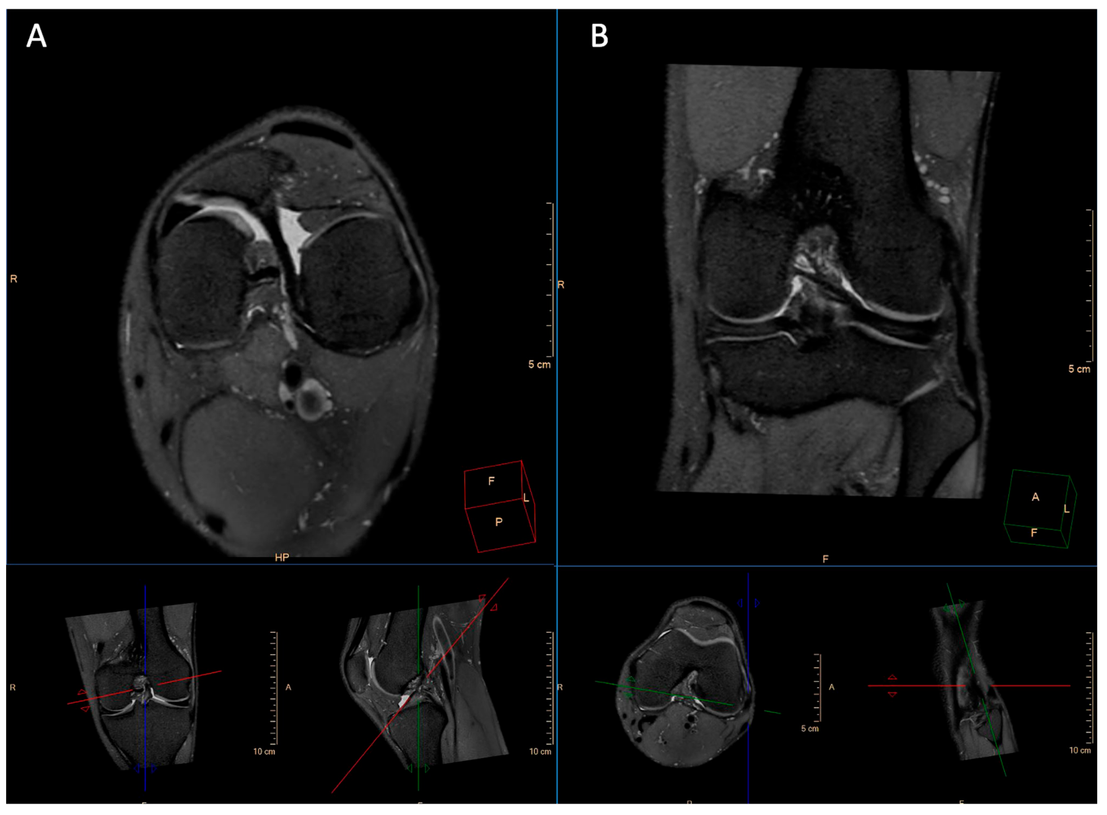
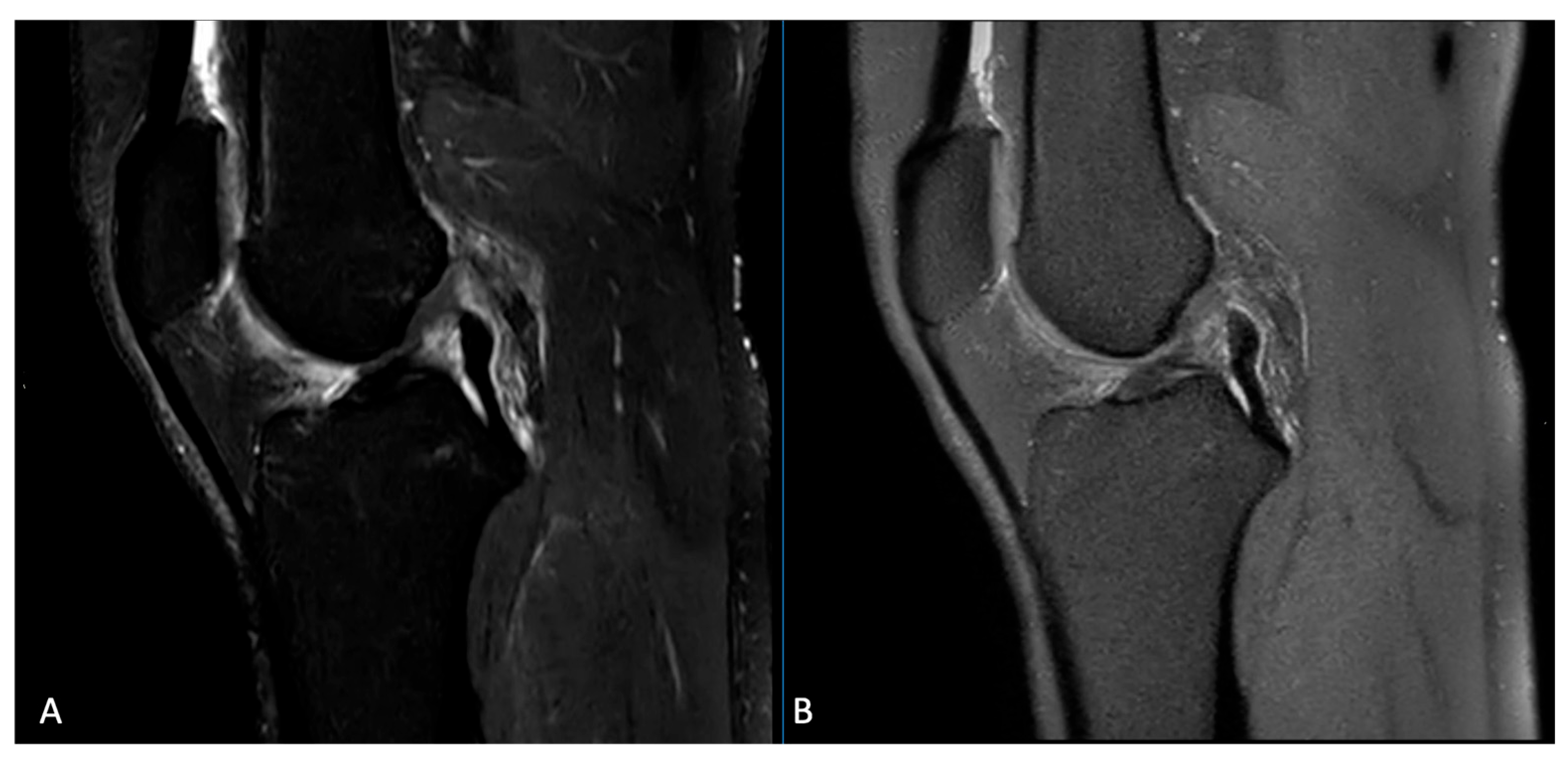
The imaging protocol included standard sequences: axial and coronal PD-weighted SPAIR, sagittal T2-weighted SPAIR, coronal T1-weighted turbo spin echo (TSE), and axial T2-weighted fast field echo (FFE). Additionally, a 3D PD-weighted SPAIR sequence was acquired in all patients (see Figure 1 and Figure 2).
Table 1 summarizes the standard and short protocols.

2.3. Image Analysis

All images were independently reviewed by two radiologists with 2 and 10 years of experience in musculoskeletal imaging. Prior to the reading sessions, both radiologists jointly evaluated 10 randomly selected, anonymized cases provided by a third reader to calibrate their subjective assessments using a five-point Likert scale. Subsequent readings were conducted independently, blindly, in random order, and at separate time points to minimize recall bias. The more experienced radiologist assessed standard anatomical structures across all sequences included in both protocols, and also documented all pathological findings and the presence of artifacts. A summary of the collected data is provided in Table 2.
Readers were able to generate multiplanar reconstructions (MPRs) of the 3D PDW SPAIR sequence in the axial, sagittal, and coronal planes, as well as along various oblique planes tailored to the anatomical structures under evaluation.

2.4. Statistical Analysis

Continuous variables were reported as mean ± standard deviation (SD) and compared using the Mann–Whitney U test due to non-normal distribution. Categorical variables were summarized as frequencies and percentages, or as median and interquartile range (IQR) when appropriate, and compared using the Chi-square test or the Friedman test, depending on the data structure and number of related groups. To assess the reliability of agreement between the two radiologists, categorical variables were analyzed with weighted Fleiss’ or Cohen’s kappa (κ) statistics, as appropriate and as follows: <0 poor, 0.01–0.20 slight, 0.21–0.40 fair, 0.41–0.60 moderate, 0.61–0.80 substantial, and 0.81–1.00 almost perfect [26].

3. Results

A total of 76 patients were enrolled, 44 males (57.9%) and 32 females (42.1%), with a mean age of 47 ± 18 years. The total acquisition time for the simulated short protocol was significantly shorter than the standard protocol, lasting 9 min compared to 17 min—a 47% reduction.

3.1. Image Quality

Table 3 and Table 4 reported subjective image quality and artifacts reported by the two readers in the standard and short protocol.
The evaluation of the agreement between the two readers regarding the diagnostic quality between the standard and 3D PDW sequences showed that the image quality of the 3D ones was qualitatively superior for both readers compared to the standard ones [κ = 0.712 (95% CIs = 0.699–0.7301; p < 0.001) vs. κ = 0.244 (95% CIs = 0.199–0.339; p = 0.63)]. Further analyses were performed on data collected by the most experienced reader.

3.2. Artifacts

The frequency of artifacts did not differ significantly between the two sequences, with 17 cases (22.4%) for the 3D sequence and 18 cases (23.7%) for the 2D sequence (χ2 = 1.63; p = 0.201). Likewise, sequences with artifacts impacting diagnostic quality showed no significant difference, occurring in four cases (5.2%) for 3D and two cases (2.6%) for 2D (χ2 = 0.716; p = 0.560).
However, qualitative assessment using the Likert scale demonstrated that images acquired with the 3D technique were rated significantly higher than those obtained with the 2D sequences. Specifically, 72.4% of 3D images were rated as excellent compared to 26.3% of 2D images, while 18.4% of 3D images were rated good versus 52.6% for 2D. Diagnostic-quality ratings accounted for 6.6% in 3D and 19.7% in 2D, and poor-quality images were 2.6% for 3D and 1.3% for 2D. The distribution of ratings between the two sequences was statistically significant (χ2 = 32.9; p < 0.001).

3.3. Pathological Entities

For partial or complete anterior cruciate ligament (ACL) injuries, no statistically significant difference was observed between the two groups, with both 3D and 2D sequences detecting 13 cases each (17.1%; p = 1.0). No partial or complete posterior cruciate ligament (PCL) injuries were identified in the study cohort.
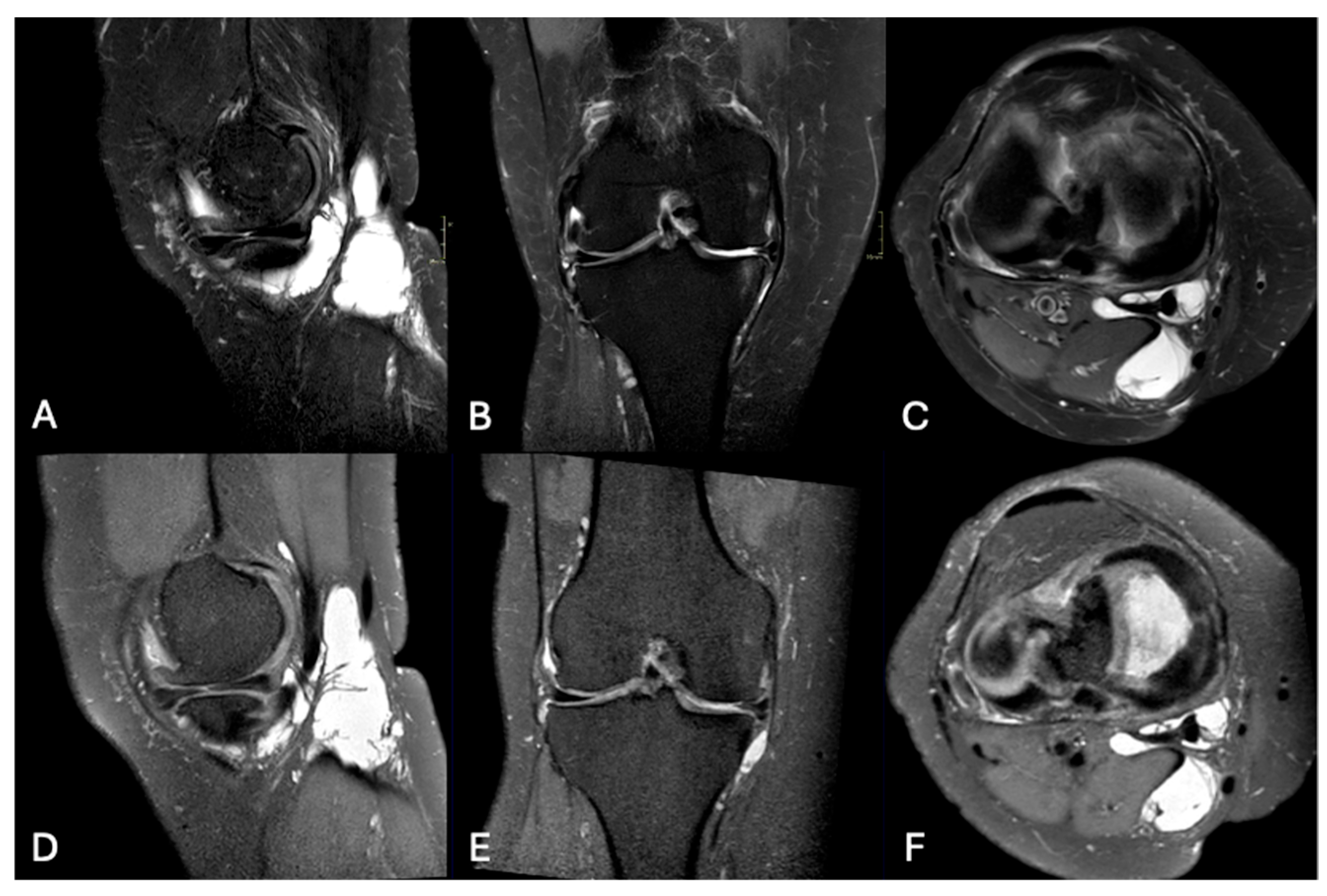
In contrast, the detection of medial meniscus injuries differed significantly between groups, with the 3D sequence identifying 37 cases (48.7%) compared to 27 cases (35.5%) with the 2D sequence (χ2 = 37.9; p < 0.001) (see Figure 3).
Similar findings were observed for lateral meniscus injuries, with the 3D sequence detecting 17 cases (22.4%) and the 2D sequence 12 cases (15.8%), showing a statistically significant difference (χ2 = 39.4; p = 0.006). Regarding the type of medial meniscus injury—classified as vertical, horizontal, radial, or complex (Figure 4)—no significant difference was found between subgroups (κ = 0.735). Likewise, lateral meniscus injury subtypes showed excellent agreement between sequences (κ = 1).
For partial or complete medial collateral ligament (MCL) injuries, no significant difference was observed between sequences, with both detecting eight cases (10.5%; p = 1). The MCL injuries were similarly classified by grade (I, II, or III) across both protocols (κ = 1), and no significant differences were found between subgroups (p > 0.05).
Similarly, partial or complete lateral collateral ligament (LCL) injuries showed no significant difference between groups, with three cases (3.9%) detected by 3D and two cases (2.6%) by 2D (p = 0.990). The two LCL injuries were both classified as grade II (κ = 1).
The presence of femoropatellar chondropathy was evaluated using both sequences, with no significant difference between the groups [3D: 40 cases (52.6%) vs. 2D: 44 cases (57.9%); p = 0.620]. Furthermore, the grading of femoropatellar chondropathy (grades I–IV) did not differ significantly between protocols (p = 0.941).
Similarly, tibiofemoral chondropathy was identified in 37 cases (48.7%) on 3D imaging and 35 cases (46.1%) on 2D imaging, without significant difference (p = 0.530). No significant differences were found between subgroups for tibiofemoral chondropathy grades I–IV (p = 0.836).
Bone marrow edema was detected at similar rates by both sequences, with positivity in 51.3% of cases on 3D and 50% on 2D (n = 39 vs. n = 38). Fracture detection was also comparable, with three cases identified by each sequence (3.9% each).
A summary of these findings is provided in Table 5.

4. Discussion

The inter-reader agreement demonstrated that 3D PDW SPAIR sequences provide superior image quality compared to 2D sequences. This improvement is primarily due to the thin, continuous slices of the 3D technique, which reduce partial volume effects and enable high-resolution multiplanar reconstructions. Such reconstructions allow comprehensive joint evaluation in all spatial planes from a single acquisition, eliminating the need for multiple 2D scans. While some authors recommend dedicated oblique sequences for better visualization of specific anatomical structures to improve their depiction [27,28], 3D sequences offer a versatile alternative.
Our study found no significant differences between 2D and 3D sequences in diagnosing ACL injuries, collateral ligament injuries, femoropatellar and femorotibial chondropathy, bone marrow edema, or fractures. However, 3D sequences detected significantly more medial meniscus lesions, particularly radial and complex tears, likely due to thinner slice thickness, the absence of inter-slice gaps, and the ability to reorient imaging planes according to anatomy [29,30]. These features enhance anatomical detail and facilitate detection of subtle lesions [24,25,30,31,32], consistent with previous reports.
Currently, 3D sequences are primarily used for evaluating cartilage pathology, while their application to other joint structures remains limited. Meta-analyses and studies by Shakoor et al. [33], Chagas-Neto et al. [34], and Kijowski et al. [35] have shown comparable diagnostic performance between 3D and routine 2D sequences for ligament and meniscal assessments. Our findings align with this literature, confirming comparable ligament and cartilage injury detection and superior meniscal lesion identification with 3D imaging, as also reported by del Grande et al. [36].
A notable advantage of 3D sequences is the reduction in overall examination time, as a single 3D scan replaces multiple 2D acquisitions in orthogonal planes, decreasing scan duration by nearly 50% [37]. This shortens the MRI protocol significantly, as seen in our institution, where 3D protocols save approximately 8–10 min per knee exam. This time efficiency can translate into 4–5 additional daily exams per scanner, improving patient access and reducing waiting times, especially in high-demand or resource-limited settings [38,39,40,41,42,43,44].
Operationally, shorter scan times enhance scanner utilization and lower per-exam costs by reducing energy consumption and staff time. They also improve patient comfort by minimizing time in the scanner and motion artifacts, an important factor for pediatric patients and trauma cases where rapid imaging is critical.
Two-dimensional sequences are limited by anisotropic voxels with thicker slices relative to in-plane resolution, leading to partial volume artifacts and limited multiplanar reconstruction capabilities [27]. In contrast, 3D isotropic imaging allows retrospective reformatting in any plane, reducing dependence on technologist plane planning and repeat acquisitions [37].
An additional approach to further shorten protocols could involve omitting the T2W FFE sequence for cartilage evaluation, as PDW sequences reliably assess cartilage, synovial fluid interfaces, and subchondral bone [45,46,47].
Limitations of 3D sequences include their availability and the necessity for high image quality, since relying on a single sequence means that inadequate scans require fallback to standard 2D protocols. Our study’s main limitation is the lack of arthroscopic correlation, the gold standard, to validate whether the increased detection of meniscal lesions by 3D imaging reflects true pathology or increased sensitivity.
Fritz et al. [48] have validated the clinical accuracy of 3D sequences against arthroscopy, confirming high diagnostic performance for meniscal, skeletal, and cartilage lesions, including in pediatric populations. Future research should focus on arthroscopic validation of 3D PDW SPAIR sequences to confirm their role as a non-invasive alternative for patients unsuitable for arthroscopy.

5. Conclusions

The proposed simulated short protocol, comprising the 3D PDW sequence along with coronal T1W and axial T2W sequences, offers a reliable alternative to the traditional protocol by maintaining high image quality and lesion detection while significantly reducing acquisition time.

Author Contributions

Conceptualization: D.I.; Methodology: D.I.; Software: M.P.; Validation: D.I. and R.C.; Formal Analysis: C.M.; Investigation: M.P., M.R., D.G., P.A., and S.P.; Resources: D.I. and M.P.; Data Curation: M.P., M.R., D.G., P.A., S.P., and M.T.; Writing—Original Draft: M.P., M.R., and C.M.; Writing—Review and Editing: M.P., M.R., C.M., and D.I.; Visualization: D.I., R.C., and M.T.; Supervision: R.C., C.T.F., and M.T.; Project Administration: D.I.; Funding Acquisition: not applicable. All authors have read and agreed to the published version of the manuscript.

Funding

This research received no external funding.

Institutional Review Board Statement

Ethical review and approval were waived for this study due to its retrospective and anonymous nature.

Informed Consent Statement

Informed consent was obtained from all subjects involved in the study.

Data Availability Statement

Data are available upon request.

Conflicts of Interest

The authors declare no conflicts of interest.

Abbreviations

FFE: fast field echo; GRE: gradient echo; MPRs: multiplanar reconstructions; STIR: Short Tau Inversion Recovery; SPAIR: SPectral Attenuated Inversion Recovery.

References

  1. Bull, A. Musculoskeletal biomechanics. J. Bone Joint Surg. Br. 2004, 86, 311. [Google Scholar] [CrossRef]
  2. Shelburne, K.B.; Torry, M.R.; Pandy, M.G. Contributions of muscles, ligaments, and the ground-reaction force to tibiofemoral joint loading during normal gait. J. Orthop. Res. 2006, 24, 1983–1990. [Google Scholar] [CrossRef] [PubMed]
  3. Shelburne, K.B.; Torry, M.R.; Pandy, M.G. Muscle, Ligament, and Joint-Contact Forces at the Knee during Walking. Med. Sci. Sports Exerc. 2005, 37, 1948–1956. [Google Scholar] [CrossRef]
  4. Sanders, T.G.; Medynski, M.A.; Feller, J.F.; Lawhorn, K.W. Bone Contusion Patterns of the Knee at MR Imaging: Footprint of the Mechanism of Injury. RadioGraphics 2000, 20 (Suppl. S1), S135–S151. [Google Scholar] [CrossRef]
  5. Bencardino, J.T.; Rosenberg, Z.S.; Brown, R.R.; Hassankhani, A.; Lustrin, E.S.; Beltran, J. Traumatic Musculotendinous Injuries of the Knee: Diagnosis with MR Imaging. RadioGraphics 2000, 20 (Suppl. S1), S103–S120. [Google Scholar] [CrossRef] [PubMed]
  6. Mink, J.H.; Deutsch, A.L. Occult cartilage and bone injuries of the knee: Detection, classification, and assessment with MR imaging. Radiology 1989, 170, 823–829. [Google Scholar] [CrossRef]
  7. Oei, E.H.G.; Ginai, A.Z.; Hunink, M.G.M. MRI for Traumatic Knee Injury: A Review. Semin. Ultrasound CT MRI 2007, 28, 141–157. [Google Scholar] [CrossRef]
  8. Carbonell, G.; Taouli, B. Abbreviated MR Protocols for Chronic Liver Disease and Liver Cancer. Magn. Reson. Imaging Clin. N. Am. 2021, 29, 321–327. [Google Scholar] [CrossRef]
  9. Canellas, R.; Rosenkrantz, A.B.; Taouli, B.; Sala, E.; Saini, S.; Pedrosa, I.; Sahani, D.V. Abbreviated MRI Protocols for the Abdomen. RadioGraphics 2019, 39, 744–758. [Google Scholar] [CrossRef]
  10. Pozzi-Mucelli, R.M.; Rinta-Kiikka, I.; Wünsche, K.; Laukkarinen, J.; Labori, K.J.; Ånonsen, K.; Kartalis, N. Pancreatic MRI for the surveillance of cystic neoplasms: Comparison of a short with a comprehensive imaging protocol. Eur. Radiol. 2017, 27, 41–50. [Google Scholar] [CrossRef]
  11. Dong, S.; Zhu, M.; Bulas, D. Techniques for minimizing sedation in pediatric MRI. J. Magn. Reson. Imaging 2019, 50, 1047–1054. [Google Scholar] [CrossRef]
  12. Jaimes, C.; Gee, M.S. Strategies to minimize sedation in pediatric body magnetic resonance imaging. Pediatr. Radiol. 2016, 46, 916–927. [Google Scholar] [CrossRef] [PubMed]
  13. Kannikeswaran, N.; Sethuraman, U.; Sivaswamy, L.; Chen, X.; Mahajan, P.V. Children with and Without Developmental Disabilities. Pediatr. Emerg. Care. 2012, 28, 1036–1040. [Google Scholar] [CrossRef]
  14. Janos, S.; Schooler, G.R.; Ngo, J.S.; Davis, J.T. Free-breathing unsedated MRI in children: Justification and techniques. J. Magn. Reson. Imaging 2019, 50, 365–376. [Google Scholar] [CrossRef]
  15. Bjur, K.A.; Payne, E.T.; Nemergut, M.E.; Hu, D.; Flick, R.P. Anesthetic-Related Neurotoxicity and Neuroimaging in Children: A Call for Conversation. J. Child. Neurol. 2017, 32, 594–602. [Google Scholar] [CrossRef] [PubMed]
  16. Barton, K.; Nickerson, J.P.; Higgins, T.; Williams, R.K. Pediatric anesthesia and neurotoxicity: What the radiologist needs to know. Pediatr. Radiol. 2018, 48, 31–36. [Google Scholar] [CrossRef] [PubMed]
  17. Chavhan, G.B.; Babyn, P.S.; Vasanawala, S.S. Abdominal MR Imaging in Children: Motion Compensation, Sequence Optimization, and Protocol Organization. RadioGraphics 2013, 33, 703–719. [Google Scholar] [CrossRef]
  18. Ahmad, R.; Hu, H.H.; Krishnamurthy, R.; Krishnamurthy, R. Reducing sedation for pediatric body MRI using accelerated and abbreviated imaging protocols. Pediatr. Radiol. 2018, 48, 37–49. [Google Scholar] [CrossRef]
  19. Parkar, A.P.; Adriaensen, M.E.A.P.M. ESR essentials: MRI of the knee—Practice recommendations by ESSR. Eur. Radiol. 2024, 34, 6590–6599. [Google Scholar] [CrossRef]
  20. Del Grande, F.; Santini, F.; Herzka, D.A.; Aro, M.R.; Dean, C.W.; Gold, G.E.; Carrino, J.A. Fat-Suppression Techniques for 3-T MR Imaging of the Musculoskeletal System. RadioGraphics 2014, 34, 217–233. [Google Scholar] [CrossRef]
  21. Dalto, V.F.; Assad, R.L.; Lorenzato, M.M.; Crema, M.D.; Louzada-Junior, P.; Nogueira-Barbosa, M.H. Comparison between STIR and T2-weighted SPAIR sequences in the evaluation of inflammatory sacroiliitis: Diagnostic performance and signal-to-noise ratio. Radiol. Bras. 2020, 53, 223–228. [Google Scholar] [CrossRef]
  22. Chien, A.; Weaver, J.S.; Kinne, E.; Omar, I. Magnetic resonance imaging of the knee. Pol. J. Radiol. 2020, 85, 509–531. [Google Scholar] [CrossRef]
  23. American College of Radiology. ACR–SPR–SSR Practice Parameter for the Performance and Interpretation of Magnetic Resonance (MRI) of the Knee. Available online: https://www.acr.org/-/media/ACR/Files/Practice-Parameters/MR-Knee.pdf (accessed on 15 June 2025).
  24. Crema, M.D.; Roemer, F.W.; Marra, M.D.; Burstein, D.; Gold, G.E.; Eckstein, F.; Guermazi, A. Articular Cartilage in the Knee: Current MR Imaging Techniques and Applications in Clinical Practice and Research. RadioGraphics 2011, 31, 37–61. [Google Scholar] [CrossRef]
  25. Kijowski, R.; Davis, K.W.; Woods, M.A.; Lindstrom, M.J.; De Smet, A.A.; Gold, G.E.; Busse, R.F. Knee Joint: Comprehensive Assessment with 3D Isotropic Resolution Fast Spin-Echo MR Imaging—Diagnostic Performance Compared with That of Conventional MR Imaging at 3.0 T. Radiology 2009, 252, 486–495. [Google Scholar] [CrossRef] [PubMed]
  26. McHugh, M.L. Interrater reliability: The kappa statistic. Biochem. Med. 2012, 22, 276–282. [Google Scholar] [CrossRef]
  27. Gold, G.E.; Busse, R.F.; Beehler, C.; Han, E.; Brau, A.C.; Beatty, P.J.; Beaulieu, C.F. Isotropic MRI of the Knee with 3D Fast Spin-Echo Extended Echo-Train Acquisition (XETA): Initial Experience. Am. J. Roentgenol. 2007, 188, 1287–1293. [Google Scholar] [CrossRef] [PubMed]
  28. Naraghi, A.; White, L.M. Three-Dimensional MRI of the Musculoskeletal System. Am. J. Roentgenol. 2012, 199, W283–W293. [Google Scholar] [CrossRef]
  29. Smith, J.P.; Barrett, G.R. Medial and Lateral Meniscal Tear Patterns in Anterior Cruciate Ligament-Deficient Knees. Am. J. Sports Med. 2001, 29, 415–419. [Google Scholar] [CrossRef]
  30. Endler, C.H.J.; Faron, A.; Isaak, A.; Katemann, C.; Mesropyan, N.; Kupczyk, P.A.; Luetkens, J.A. Fast 3D Isotropic Proton Density-Weighted Fat-Saturated MRI of the Knee at 1.5 T with Compressed Sensing: Comparison with Conventional Multiplanar 2D Sequences. In RöFo—Fortschritte auf dem Gebiet der Röntgenstrahlen und der Bildgebenden Verfahren; Georg Thieme Verlag KG: Stuttgart, Germany, 2021. [Google Scholar] [CrossRef]
  31. Kijowski, R. Three-Dimensional Magnetic Resonance Imaging of Joints. Top. Magn. Reson. Imaging 2010, 21, 297–313. [Google Scholar] [CrossRef]
  32. Nguyen, J.C.; De Smet, A.A.; Graf, B.K.; Rosas, H.G. MR Imaging–based Diagnosis and Classification of Meniscal Tears. RadioGraphics 2014, 34, 981–999. [Google Scholar] [CrossRef]
  33. Shakoor, D.; Guermazi, A.; Kijowski, R.; Fritz, J.; Roemer, F.W.; Jalali-Farahani, S.; Demehri, S. Cruciate ligament injuries of the knee: A meta-analysis of the diagnostic performance of 3D MRI. J. Magn. Reson. Imaging 2019, 50, 1545–1560. [Google Scholar] [CrossRef]
  34. Chagas-Neto, F.A.; Nogueira-Barbosa, M.H.; Lorenzato, M.M.; Salim, R.; Kfuri-Junior, M.; Crema, M.D. Diagnostic performance of 3D TSE MRI versus 2D TSE MRI of the knee at 1.5 T, with prompt arthroscopic correlation, in the detection of meniscal and cruciate ligament tears. Radiol. Bras. 2016, 49, 69–74. [Google Scholar] [CrossRef] [PubMed]
  35. Kijowski, R.; Blankenbaker, D.G.; Klaers, J.L.; Shinki, K.; De Smet, A.A.; Block, W.F. Vastly Undersampled Isotropic Projection Steady-State Free Precession Imaging of the Knee: Diagnostic Performance Compared with Conventional MR. Radiology 2009, 251, 185–194. [Google Scholar] [CrossRef] [PubMed]
  36. Del Grande, F.; Delcogliano, M.; Guglielmi, R.; Raithel, E.; Stern, S.E.; Papp, D.F.; Fritz, J. Fully Automated 10-Minute 3D CAIPIRINHA SPACE TSE MRI of the Knee in Adults. Investig. Radiol. 2018, 53, 689–697. [Google Scholar] [CrossRef] [PubMed]
  37. Altahawi, F.; Pierce, J.; Aslan, M.; Li, X.; Winalski, C.S.; Subhas, N. 3D MRI of the Knee. Semin. Musculoskelet. Radiol. 2021, 25, 455–467. [Google Scholar] [CrossRef]
  38. Subhas, N. Abbreviated Musculoskeletal MRI Protocols: Point—Improved Patient Access and Reduced Health Care Costs. Am. J. Roentgenol. 2021, 216, 33–34. [Google Scholar] [CrossRef]
  39. Ippolito, D.; Giandola, T.; Maino, C.; Gandola, D.; Ragusi, M.; Brambilla, P.; Sironi, S. Diagnostic Value of Whole-Body MRI Short Protocols in Bone Lesion Detection in Multiple Myeloma Patients. Diagnostics 2021, 11, 1053. [Google Scholar] [CrossRef]
  40. Puhr-Westerheide, D.; Froelich, M.F.; Solyanik, O.; Gresser, E.; Reidler, P.; Fabritius, M.P.; Kazmierczak, P.M. Cost-effectiveness of short-protocol emergency brain MRI after negative non-contrast CT for minor stroke detection. Eur. Radiol. 2022, 32, 1117–1126. [Google Scholar] [CrossRef]
  41. Peters, S.; Gärtner, F.; Austein, F.; Wodarg, F.; Jansen, O.; Hensler, J. Evaluation of an Ultra-Short MRI Protocol for Cerebral Staging Examinations in Melanoma Patients. In RöFo—Fortschritte auf dem Gebiet der Röntgenstrahlen und der Bildgebenden Verfahren; Georg Thieme Verlag KG: Stuttgart, Germany, 2022; Volume 194, pp. 409–415. [Google Scholar] [CrossRef]
  42. Kuhl, C.K. Abbreviated Breast MRI: State of the Art. Radiology 2024, 310, e221822. [Google Scholar] [CrossRef]
  43. van de Braak, C.; Willemssen, F.E.J.A.; de Man, R.A.; van der Lugt, A.; Uyl-de Groot, C.A.; Bos, D.; Dwarkasing, R.S. Non-contrast short MRI surveillance for HCC screening: The study protocol of the SMS-HCC prospective multicenter study. Eur. Radiol. Exp. 2024, 8, 29. [Google Scholar] [CrossRef]
  44. Ghorra, C.; Pommier, R.; Piveteau, A.; Rubbia-Brandt, L.; Vilgrain, V.; Terraz, S.; Ronot, M. The diagnostic performance of a simulated “short” gadoxetic acid-enhanced MRI protocol is similar to that of a conventional protocol for the detection of colorectal liver metastases. Eur. Radiol. 2021, 31, 2451–2460. [Google Scholar] [CrossRef]
  45. Potter, H.G.; Linklater, I.J.M.; Allen, A.A.; Hannafin, J.A.; Haas, S.B. Magnetic Resonance Imaging of Articular Cartilage in the Knee. An Evaluation with Use of Fast-Spin-Echo Imaging. J. Bone Joint Surg. 1998, 80, 1276–1284. [Google Scholar] [CrossRef]
  46. Mohr, A. The value of water-excitation 3D FLASH and fat-saturated PDw TSE MR imaging for detecting and grading articular cartilage lesions of the knee. Skelet. Radiol. 2003, 32, 396–402. [Google Scholar] [CrossRef]
  47. Schaefer, F.K.W.; Kurz, B.; Schaefer, P.J.; Fuerst, M.; Hedderich, J.; Graessner, J.; Heller, H. Accuracy and precision in the detection of articular cartilage lesions using magnetic resonance imaging at 1.5 tesla in an in vitro study with orthopedic and histopathologic correlation. Acta Radiol. 2007, 48, 1131–1137. [Google Scholar] [CrossRef]
  48. Fritz, J.; Ahlawat, S.; Fritz, B.; Thawait, G.K.; Stern, S.E.; Raithel, E.; Lee, R.J. 10-Min 3D Turbo Spin Echo MRI of the Knee in Children: Arthroscopy-Validated Accuracy for the Diagnosis of Internal Derangement. J. Magn. Reson. Imaging 2019, 49, e139–e151. [Google Scholar] [CrossRef]
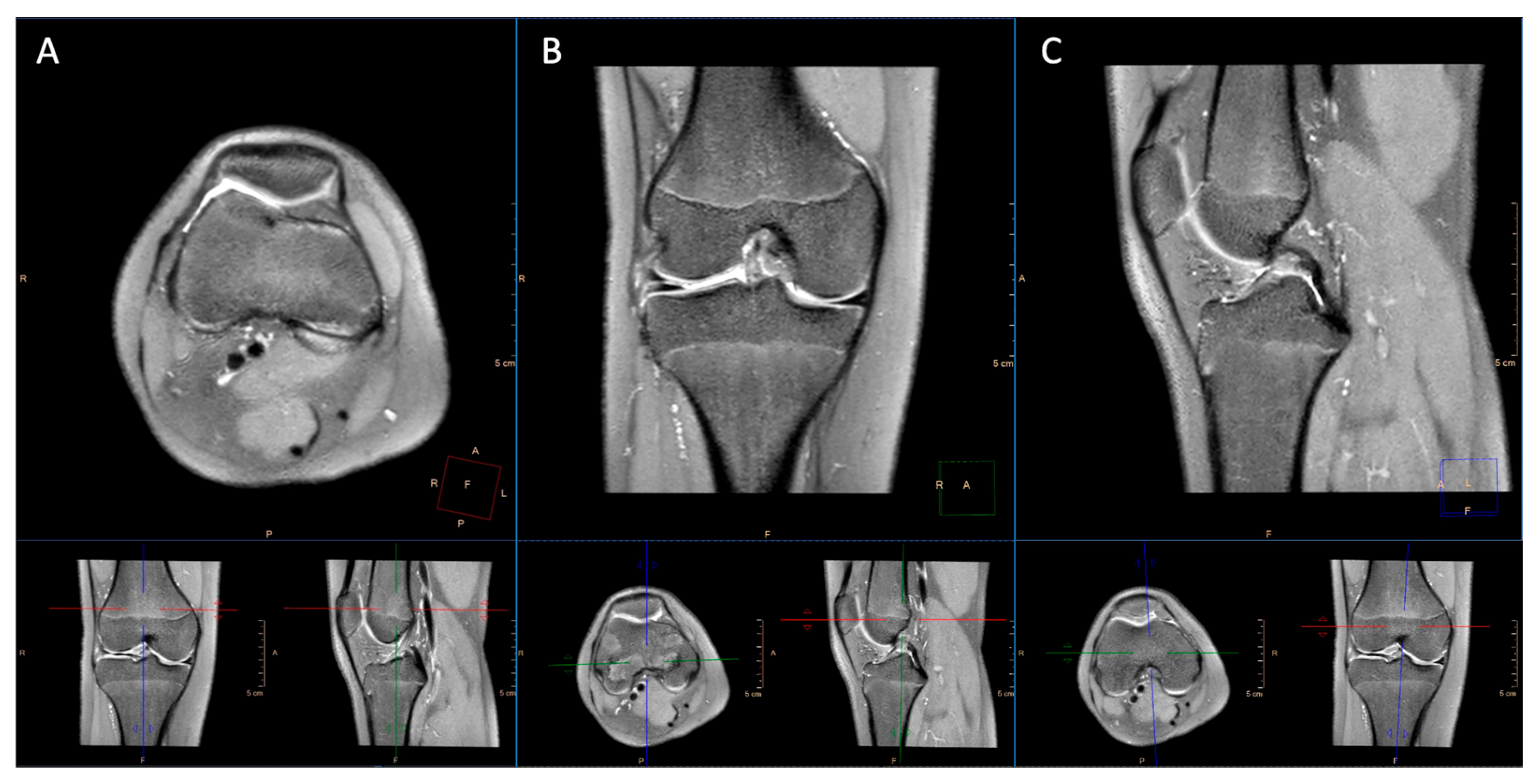
Figure 1. Three-dimensional PDW SPAIR sequence with MPRs on axial (A), coronal (B), and sagittal (C) planes.
Applsci 15 08870 g001
Figure 2. Three-dimensional PDW SPAIR sequence with retrospective reconstruction along the long axis of the anterior cruciate ligament (A) and the lateral collateral ligament (B).
Applsci 15 08870 g002
Figure 3. Anterior cruciate ligament injury assessed on the sagittal 2D T2W SPAIR scan (A) and on the 3D PDW SPAIR scan reconstructed on the sagittal plane (B).
Applsci 15 08870 g003
Figure 4. Radial lesion of the medial meniscus. Standard protocol: 2D T2W SPAIR sagittal sequence (A), 2D PDW SPAIR coronal sequence (B), and 2D PDW SPAIR axial sequence (C). Short protocol: 3D PDW SPAIR sequence with retrospective MPRs in sagittal (D), coronal (E), and axial planes aligned with the long axis of the medial meniscus for an enhanced overview of the lesion (F).
Applsci 15 08870 g004
Table 1. MRI scanning parameters at 1.5 T of standard and short protocols. FOV: field of view; TE: echo time; TR: repetition time; FA: flip angle. Thickness is expressed in mm while time in minutes.
SequenceThicknessFOVMatrixTE (ms)TR (ms)FA (°)Time
Standard protocol
T2W SPAIR sag3160*160*92308*238603585903.57
PDW SPAIR cor379*160*160268*255303000903.42
T1W TSE cor386*160*160380*25717500903.17
PDW SPAIR ax3160*79*160268*256303000903.18
T2W FFE3160*92*160324*25918.42Shortest303.15
Short protocol
3D PD SPAIR0.6*0.6*0.6157*140*165232*262311100905.54
T1W TSE cor386*160*160380*25717500903.17
T2W FFE3160*92*160324*25918.42Shortest303.15
Table 2. Pathological conditions collected in both MR protocols.
Pathological AlterationDescription
Anterior cruciate ligament injuryTotal or partial
Posterior cruciate ligament injury Total or partial
Medial meniscal tearType of tear
Lateral meniscal tearType of tear
Lateral collateral ligament injuryGrade I, Grade II, Grade III
Medial collateral ligament injuryGrade I, Grade II, Grade III
Patellofemoral chondromalaciaGrade I, Grade II, Grade III, Grade IV
Tibiofemoral chondromalaciaGrade I, Grade II, Grade III, Grade IV
Bone edema
Fracture
Table 3. Results of image quality assessment using the Likert scale by readers.
Likert Scale ValueReader 1Reader 2
2D Sequences3D Sequences2D Sequences3D Sequences
1 (n, %)0000
2 (n, %)1 (1.3)2 (2.6)02 (2.6)
3 (n, %)15 (19.7)5 (6.6)27 (35.5)10 (13.1)
4 (n, %)40 (52.6)14 (18.4)42 (55.3)26 (34.2)
5 (n, %)20 (26.4)55 (72.4)7 (9.2)38 (50.1)
Table 4. Numbers of artifacts and knee injuries reported by the most experienced reader in both sequences. A p-value < 0.05 was considered statistically significant.
Pathological Alteration2D Sequences3D Sequencesp-Value
Artifacts (n, %)17 (22.4)18 (23.7)0.801
Anterior cruciate ligament injury (n, %)13 (17.1)13 (17.1)1.0
Posterior cruciate ligament injury (n, %)0 0-
Medial meniscal tear (n, %)27 (35.5)37 (48.7)< 0.001
Lateral meniscal tear (n, %)12 (15.8)17 (22.4)< 0.001
Lateral collateral ligament injury (n, %)2 (2.6)3 (3.9)0.990
Medial collateral ligament injury (n, %)8 (10.5)8 (10.5)0.890
Femoropatellar chondropathy (n, %)44 (57.9)40 (52.6)0.620
Tibiofemoral chondropathy (n, %)35 (46.1)37 (48.7)0.500
Bone edema (n, %)38 (50)39 (51.3)0.977
Fracture (n, %)3 (3.9)3 (3.9)1.0
Table 5. Pathological findings evaluated by the most experienced reader in 2D and 3D sequences. p-values reported were computed by the two groups. Statistical information regarding subgroups analysis is reported in the text.
Pathological Alteration2D Sequences3D Sequencesp-Value
Anterior cruciate ligament injuryComplete (n, %)8 (10.5)8 (10.5)1.0
Partial (n, %)5 (6.6)5 (6.5)
Medial meniscus injuryVertical (n, %)2 (2.6)1 (1.3)<0.001
Horizontal (n, %)16 (21)21 (27.6)
Radial (n, %)1 (1.3)4 (5.2)
Complex (n, %)8 (10.4)11 (14.3)
Lateral meniscus injuryVertical (n, %)3 (3.9)4 (5.2)0.006
Horizontal (n, %)4 (5.2)5 (6.5)
Radial (n, %)1 (1.3)4 (5.2)
Complex (n, %)4 (5.2)4 (5.2)
Lateral collatarl ligament injuryGrade 1 (n, %)2 (2.6)3 (3.9)0.990
Grade 2 (n, %)00
Grade 3 (n, %)00
Medial collatarl ligament injuryGrade 1 (n, %)4 (5.2)4 (5.2)1.0
Grade 2 (n, %)3 (3.9)3 (3.9)
Grade 3 (n, %)1 (1.3)1 (1.3)
Femoropatellar chondropathyGrade 1 (n, %)6 (7.8)3 (3.9)0.620
Grade 2 (n, %)13 (16.9)12 (15.6)
Grade 3 (n, %)12 (15.6)13 (16.9)
Grade 4 (n, %)13 (16.9)12 (15.6)
Tibiofemoral chondromalaciaGrade 1 (n, %)2 (2.6)2 (2.6)0.530
Grade 2 (n, %)6 (7.8)7 (9.1)
Grade 3 (n, %)4 (5.2)5 (6.5)
Grade 4 (n, %)23 (29.9)23 (29.9)
Disclaimer/Publisher’s Note: The statements, opinions and data contained in all publications are solely those of the individual author(s) and contributor(s) and not of MDPI and/or the editor(s). MDPI and/or the editor(s) disclaim responsibility for any injury to people or property resulting from any ideas, methods, instructions or products referred to in the content.