
Performance Evaluation of Selenite (SeO32−) Reduction by Enterococcus spp.



Abstract
1. Introduction
2. Results
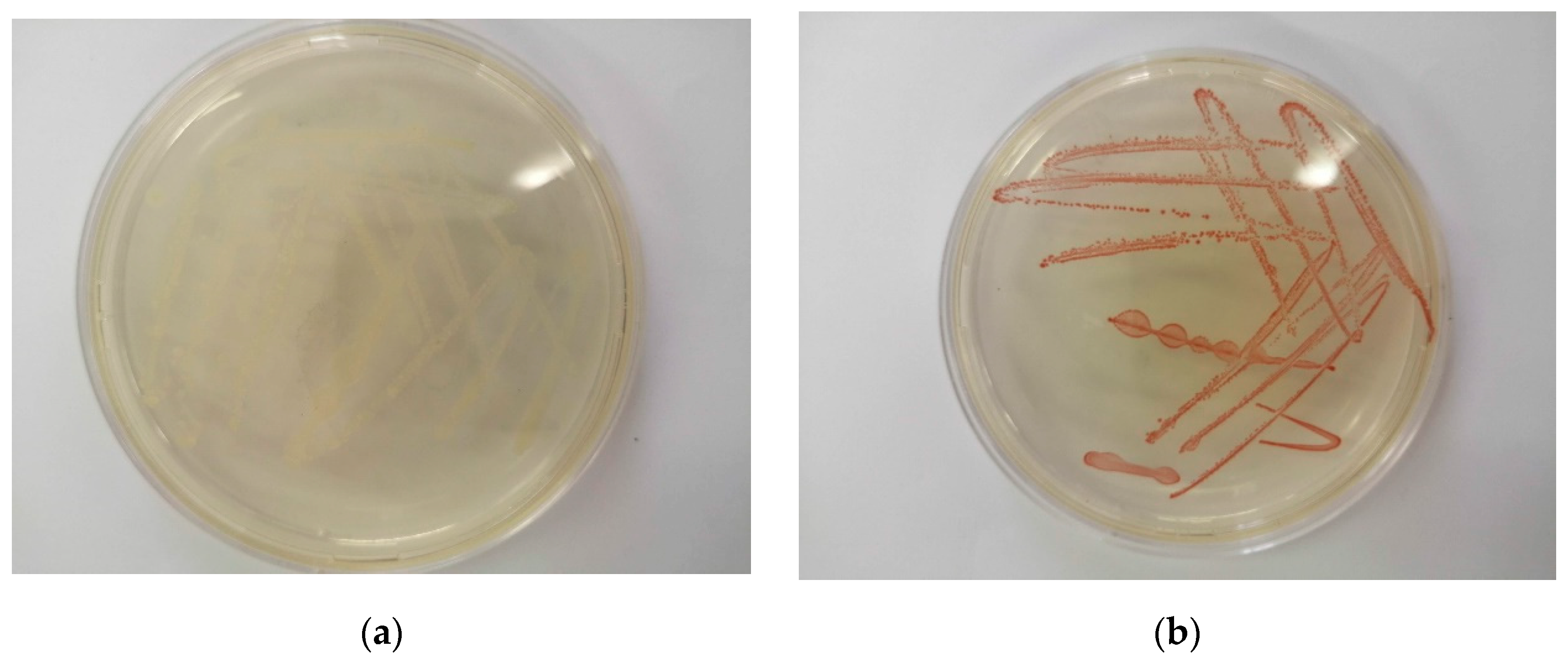
2.1. Microbial Characterisation
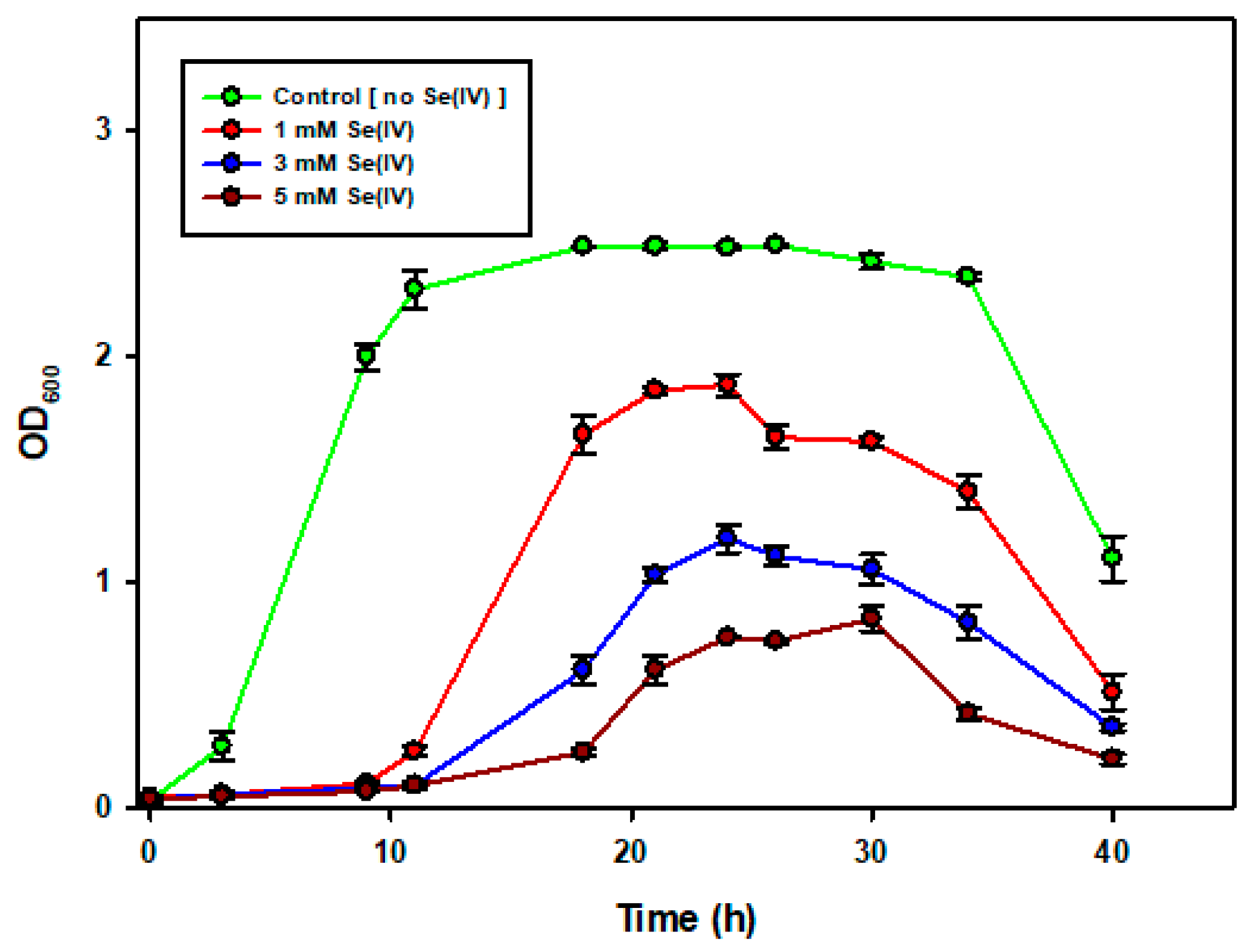
2.2. The Growth of Enterococcus spp. in the Presence of Increasing SeO32− Concentrations
2.3. Minimum Inhibitory Concentration (MIC) Assay for SeO32− Tolerance
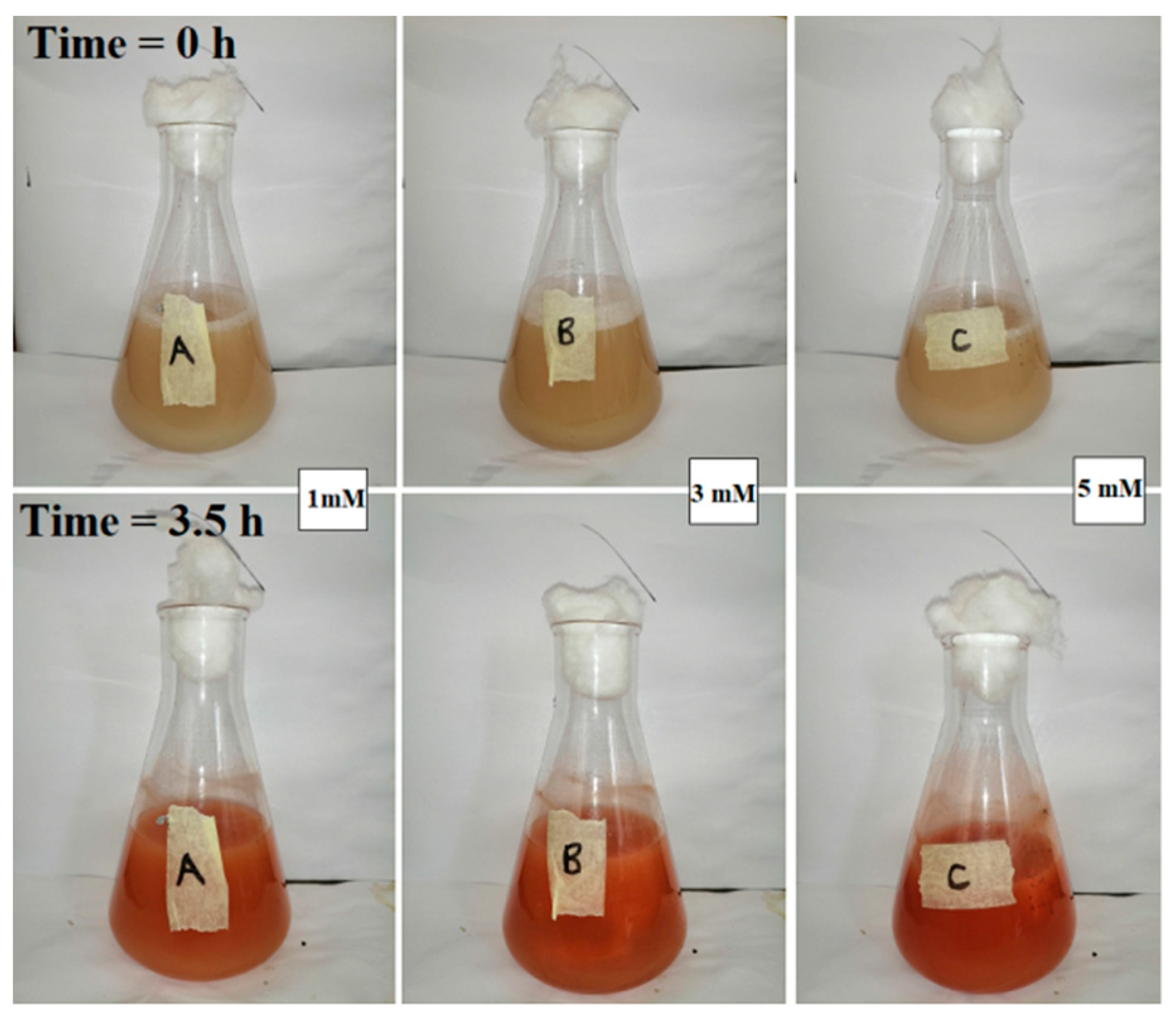
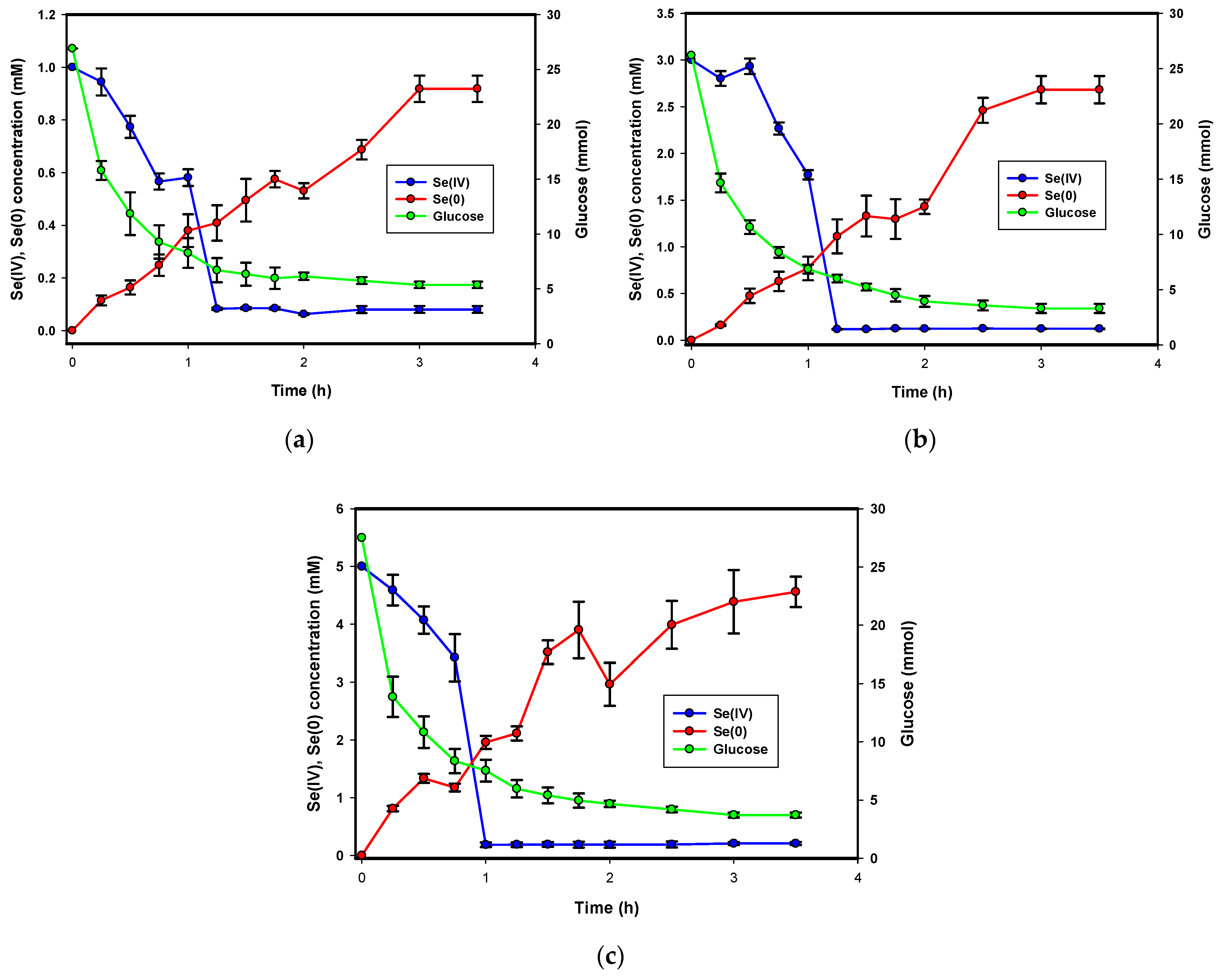
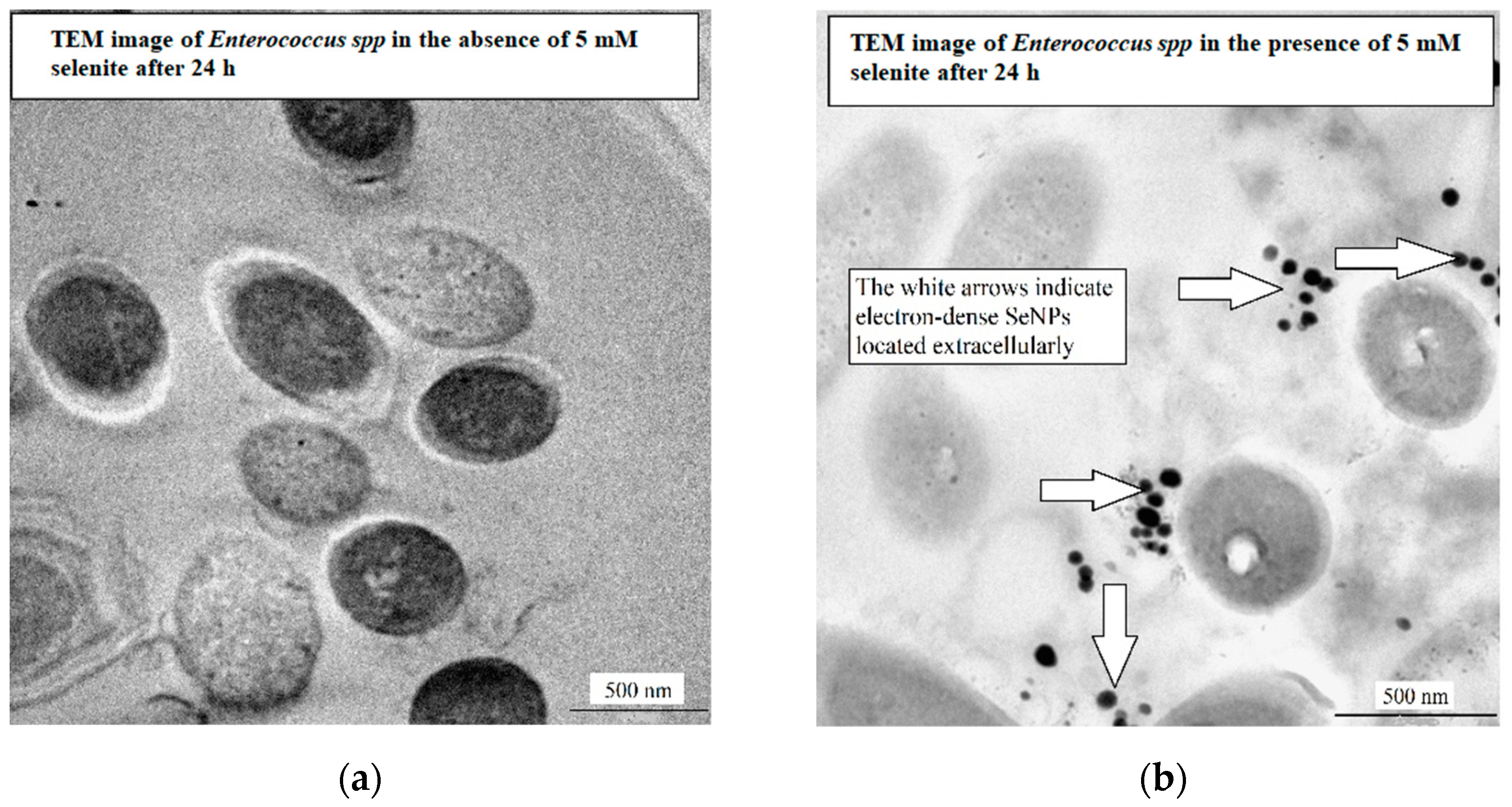
2.4. SeO32− Depletion and Se0 Formation
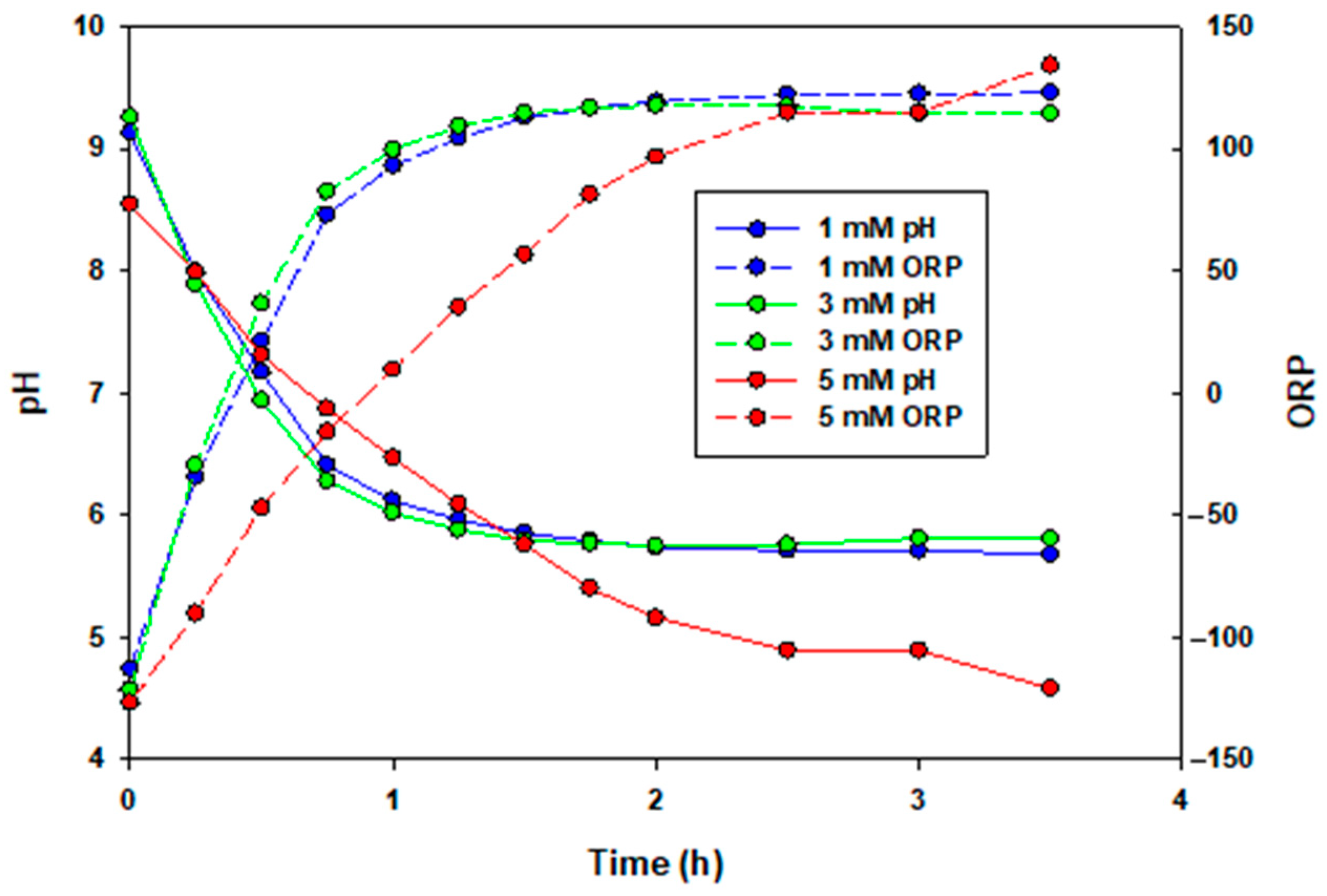
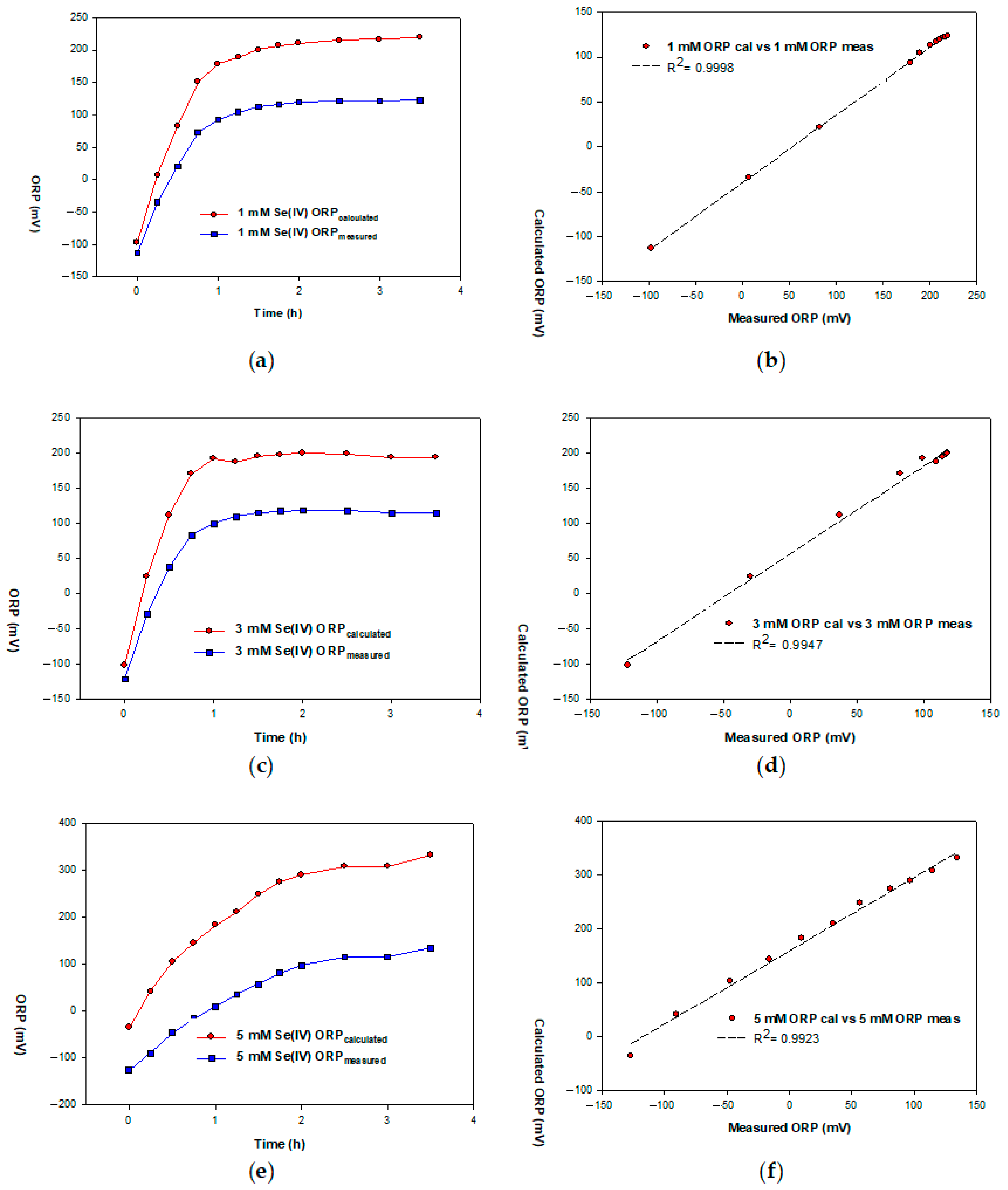
2.5. Redox Potential Measurements as A Monitoring Technique for SeO32− Reduction
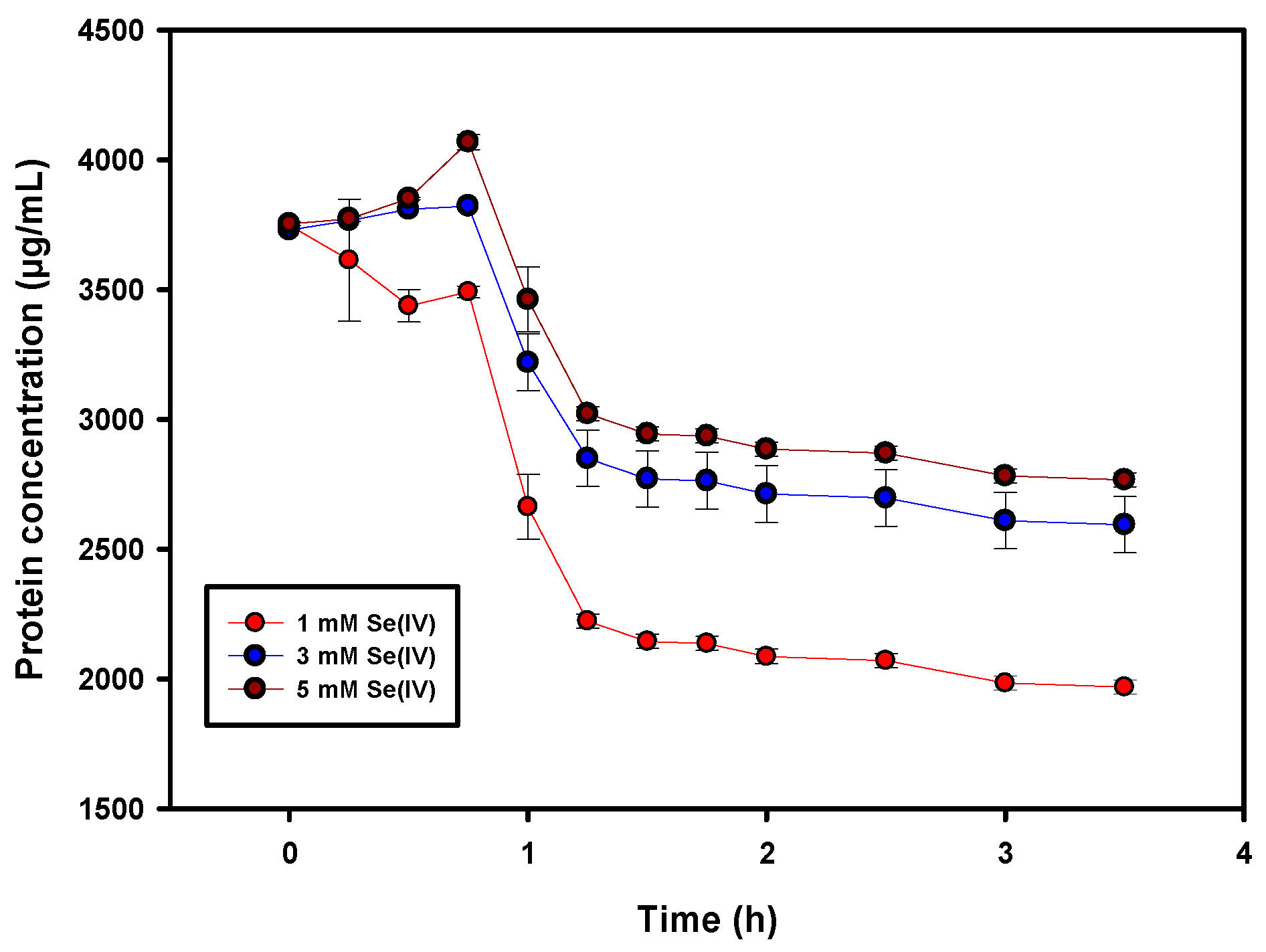
2.6. Protein Assay
3. Materials and Methods
3.1. Growth Media
3.2. Mineral Salt Medium (MSM)
3.3. SeO32− Stock Solution
3.4. Culture Isolation, Identification, Cultivation and Storage
3.5. SeO32− and Se0 Measurement
3.6. pH and ORP Measurement
3.7. Glucose Measurement
3.8. Protein Assay
3.9. TEM Analysis
4. Conclusions
Supplementary Materials
Author Contributions
Funding
Data Availability Statement
Acknowledgments
Conflicts of Interest
References
- Nancharaiah, Y.V.; Lens, P.N.L. Ecology and Biotechnology of Selenium-Respiring Bacteria. Microbiol. Mol. Biol. Rev. 2015, 79, 61–80. [Google Scholar] [CrossRef] [Scilit]
- Dungan, R.S.; Frankenberger, W.T. Reduction of Selenite to Elemental Selenium by Enterobacter cloacae SLD1a-1. J. Environ. Qual. 1998, 27, 1301–1306. [Google Scholar] [CrossRef] [Scilit]
- Stewart, A.R.; Grosell, M.; Buchwalter, D.B.; Fisher, N.S.; Luoma, S.N.; Mathews, T.; Orr, P.L.; Wang, W.-X. Bioaccumulation and Trophic Transfer of Selenium. In Ecological Assessment of Selenium in the Aquatic Environment; SETAC Press: Pensacola, FL, USA, 2010; pp. 93–139. [Google Scholar]
- Kuroda, M.; Notaguchi, E.; Sato, A.; Yoshioka, M.; Hasegawa, A.; Kagami, T.; Narita, T.; Yamashita, M.; Sei, K.; Soda, S.; et al. Characterization of Pseudomonas stutzeri NT-I capable of removing soluble selenium from the aqueous phase under aerobic conditions. J. Biosci. Bioeng. 2011, 112, 259–264. [Google Scholar] [CrossRef] [Scilit] [PubMed]
- Frankenberger, W.T.; Engberg, R.A. Environmental Chemistry of Selenium. Environ. Chem. Selenium 1998, 736. [Google Scholar] [CrossRef] [Scilit]
- Ečimović, S.; Velki, M.; Vuković, R.; Čamagajevac, I.Š.; Petek, A.; Bošnjaković, R.; Grgić, M.; Engelmann, P.; Bodó, K.; Filipović-Marijić, V.; et al. Acute toxicity of selenate and selenite and their impacts on oxidative status, efflux pump activity, cellular and genetic parameters in earthworm Eisenia andrei. Chemosphere 2018, 212, 307–318. [Google Scholar] [CrossRef] [Scilit] [PubMed]
- Garousi, F. The toxicity of different selenium forms and compounds—Review. Acta Agrar. Debreceniensis 2015, 64, 33–38. [Google Scholar] [CrossRef] [Scilit]
- Hunter, W.J.; Manter, D.K. Reduction of Selenite to Elemental Red Selenium by Pseudomonas sp. Strain CA5. Curr. Microbiol. 2009, 58, 493–498. [Google Scholar] [CrossRef] [Scilit]
- Tendenedzai, J.T.; Brink, H.G. The Effect of Nitrogen on the Reduction of Selenite to Elemental Selenium by Pseudomonas stutzeri NT-I. Chemical Engineering Transanctions. CET 2019, 74, 529–534. [Google Scholar] [CrossRef] [Scilit]
- Shoeibi, S.; Mashreghi, M. Biosynthesis of selenium nanoparticles using Enterococcus faecalis and evaluation of their antibacterial activities. J. Trace Elements Med. Biol. 2017, 39, 135–139. [Google Scholar] [CrossRef] [Scilit]
- Oremland, R.S.; Herbel, M.J.; Blum, J.S.; Langley, S.; Beveridge, T.J.; Ajayan, P.M.; Sutto, T.; Ellis, A.; Curran, S. Structural and Spectral Features of Selenium Nanospheres Produced by Se-Respiring Bacteria. Appl. Environ. Microbiol. 2004, 70, 52–60. [Google Scholar] [CrossRef] [Scilit]
- Cremonini, E.; Zonaro, E.; Donini, M.; Lampis, S.; Boaretti, M.; Dusi, S.; Melotti, P.; Lleo, M.M.; Vallini, G. Biogenic selenium nanoparticles: Characterization, antimicrobial activity and effects on human dendritic cells and fibroblasts. Microb. Biotechnol. 2016, 9, 758–771. [Google Scholar] [CrossRef] [Scilit] [PubMed]
- Kessi, J.; Ramuz, M.; Wehrli, E.; Spycher, M.; Bachofen, R. Reduction of selenite and detoxification of elemental selenium by the phototrophic bacterium Rhodospirillum rubrum. Appl. Environ. Microbiol. 1999, 65, 4734–4740. [Google Scholar] [CrossRef] [Scilit]
- Painter, E.P. The Chemistry and Toxicity of Selenium Compounds, with Special Reference to the Selenium Problem. Chem. Rev. 1941, 28, 179–213. [Google Scholar] [CrossRef] [Scilit]
- Cortese, M.; Paszczynski, A.; Lewis, T.A.; Sebat, J.; Borek, V.; Crawford, R.L. Metal chelating properties of pyridine-2,6-bis(thiocarboxylic acid) produced by Pseudomonas spp. and the biological activities of the formed complexes. BioMetals 2002, 15, 103–120. [Google Scholar] [CrossRef] [Scilit]
- Zawadzka, A.M.; Crawford, R.L.; Paszczynski, A.J. Pyridine-2,6-Bis(Thiocarboxylic Acid) Produced by Pseudomonas stutzeri KC Reduces and Precipitates Selenium and Tellurium Oxyanions. Appl. Environ. Microbiol. 2006, 72, 3119–3129. [Google Scholar] [CrossRef] [Scilit]
- Harrison, J.; Ceri, H.; Turner, R.J. Multimetal resistance and tolerance in microbial biofilms. Nat. Rev. Genet. 2007, 5, 928–938. [Google Scholar] [CrossRef] [Scilit]
- Li, D.-B.; Cheng, Y.-Y.; Wu, C.; Li, W.-W.; Li, N.; Yang, Z.-C.; Tong, Z.-H.; Yu, H.-Q. Selenite reduction by Shewanella oneidensis MR-1 is mediated by fumarate reductase in periplasm. Sci. Rep. 2014, 4, 3735. [Google Scholar] [CrossRef] [Scilit]
- Ji, Y.; Wang, Y.-T. Selenium Reduction by Batch Cultures of Escherichia coli Strain EWB32213. J. Environ. Eng. 2017, 143, 04017009. [Google Scholar] [CrossRef] [Scilit]
- Rutkowski, T.; Conroy, K.; Gusek, J. Past, present and future for treating selenium-impacted water. In Tailings and Mine Waste ’08; Informa UK Limited: London, UK, 2008; pp. 281–290. [Google Scholar]
- Hanchi, H.; Mottawea, W.; Sebei, K.; Hammami, R. The Genus Enterococcus: Between Probiotic Potential and Safety Concerns—An Update. Front. Microbiol. 2018, 9, 1791. [Google Scholar] [CrossRef] [Scilit] [PubMed]
- Fisher, K.; Phillips, C. The ecology, epidemiology and virulence of Enterococcus. Microbiology 2009, 155, 1749–1757. [Google Scholar] [CrossRef] [Scilit] [PubMed]
- Ramos, S.; Silva, V.; Dapkevicius, M.; Igrejas, G.; Poeta, P. Enterococci, from Harmless Bacteria to a Pathogen. Microorganisms 2020, 8, 1118. [Google Scholar] [CrossRef] [Scilit] [PubMed]
- Meade, E.; Slattery, M.A.; Garvey, M. Bacteriocins, Potent Antimicrobial Peptides and the Fight against Multi Drug Resistant Species: Resistance Is Futile? Antibiotics 2020, 9, 32. [Google Scholar] [CrossRef] [Scilit] [PubMed]
- Vandera, E.; Lianou, A.; Kakouri, A.; Feng, J.; Koukkou, A.-I.; Samelis, J. Enhanced Control of Listeria monocytogenes by Enterococcus faecium KE82, a Multiple Enterocin–Producing Strain, in Different Milk Environments. J. Food Prot. 2016, 80, 74–85. [Google Scholar] [CrossRef] [Scilit] [PubMed]
- Henning, C.; Gautam, D.; Muriana, P. Identification of Multiple Bacteriocins in Enterococcus spp. Using an Enterococcus-Specific Bacteriocin PCR Array. Microorganisms 2015, 3, 1–16. [Google Scholar] [CrossRef] [Scilit]
- Goncalves, O.; Legrand, J. (Eds.) 2—Spoilage of Egg Products. In Alteration of Ovoproducts; Elsevier: Philadelphia, PA, USA, 2018; pp. 51–156. [Google Scholar]
- Pal, A.; Paul, A.K. Microbial extracellular polymeric substances: Central elements in heavy metal bioremediation. Indian J. Microbiol. 2008, 48, 49–64. [Google Scholar] [CrossRef] [Scilit] [PubMed]
- Jan, A.T. Outer Membrane Vesicles (OMVs) of Gram-negative Bacteria: A Perspective Update. Front. Microbiol. 2017, 8, 1053. [Google Scholar] [CrossRef] [Scilit]
- Zhang, X.; Fan, W.-Y.; Yao, M.-C.; Yang, C.-W.; Sheng, G.-P. Redox state of microbial extracellular polymeric substances regulates reduction of selenite to elemental selenium accompanying with enhancing microbial detoxification in aquatic environments. Water Res. 2020, 172, 115538. [Google Scholar] [CrossRef] [Scilit]
- Hörstmann, C.; Brink, H.G.; Chirwa, E.M. Pb(II) Bio-Removal, Viability, and Population Distribution of an Industrial Microbial Consortium: The Effect of Pb(II) and Nutrient Concentrations. Sustainability 2020, 12, 2511. [Google Scholar] [CrossRef] [Scilit]
- Lampis, S.; Zonaro, E.; Bertolini, C.; Cecconi, D.; Monti, F.; Micaroni, M.; Turner, R.J.; Butler, C.S.; Vallini, G. Selenite biotransformation and detoxification by Stenotrophomonas maltophilia SeITE02: Novel clues on the route to bacterial biogenesis of selenium nanoparticles. J. Hazard. Mater. 2017, 324, 3–14. [Google Scholar] [CrossRef] [Scilit]
- Fresneda, M.A.R.; Martín, J.D.; Bolívar, J.G.; Cantos, M.V.F.; Bosch-Estévez, G.; Moreno, M.F.M.; Merroun, M.L. Green synthesis and biotransformation of amorphous Se nanospheres to trigonal 1D Se nanostructures: Impact on Se mobility within the concept of radioactive waste disposal. Environ. Sci. Nano 2018, 5, 2103–2116. [Google Scholar] [CrossRef] [Scilit]
- Dwivedi, S.; Al-Khedhairy, A.; Ahamed, M.; Musarrat, J. Biomimetic Synthesis of Selenium Nanospheres by Bacterial Strain JS-11 and Its Role as a Biosensor for Nanotoxicity Assessment: A Novel Se-Bioassay. PLoS ONE 2013, 8, e57404. [Google Scholar] [CrossRef] [Scilit]
- Zheng, S.; Su, J.; Wang, L.; Yao, R.; Wang, D.; Deng, Y.; Wang, R.; Wang, G.; Rensing, C. Selenite reduction by the obligate aerobic bacterium Comamonas testosteroni S44 isolated from a metal-contaminated soil. BMC Microbiol. 2014, 14, 204. [Google Scholar] [CrossRef] [Scilit]
- Kagami, T.; Narita, T.; Kuroda, M.; Notaguchi, E.; Yamashita, M.; Sei, K.; Soda, S.; Ike, M. Effective selenium volatilization under aerobic conditions and recovery from the aqueous phase by Pseudomonas stutzeri NT-I. Water Res. 2013, 47, 1361–1368. [Google Scholar] [CrossRef] [Scilit] [PubMed]
- Siefert, K.S.; Lampert, K.E. ORP for Chemical Dosage Control in Metal Precipitation; Nalco Chemical Company: Naperville, IL, USA, 2002. [Google Scholar]
- Aigberua, A.; Tarawou, J.; Abasi, C. Effect of Oxidation-Reduction Fluctuations on Metal Mobility of Speciated Metals and Arsenic in Bottom Sediments of Middleton River, Bayelsa State, Nigeria. J. Appl. Sci. Environ. Manag. 2018, 22, 1511. [Google Scholar] [CrossRef] [Scilit]
- Doi, R. Determination of the selenium (VI)/(IV) standard redox potential by cyclic voltammetry. J. Nucl. Sci. Technol. 2013, 51, 56–63. [Google Scholar] [CrossRef] [Scilit]
- Schafer, F.Q.; Buettner, G. Redox environment of the cell as viewed through the redox state of the glutathione disulfide/glutathione couple. Free. Radic. Biol. Med. 2001, 30, 1191–1212. [Google Scholar] [CrossRef] [Scilit]
- Ioka, S.; Muraoka, H.; Matsuyama, K.; Tomita, K. In situ redox potential measurements as a monitoring technique for hot spring water quality. Sustain. Water Resour. Manag. 2016, 2, 353–358. [Google Scholar] [CrossRef] [Scilit]
- Nernst Equation. 2020. Available online: https://byjus.com/jee/nernst-equation/#:~:text=Limitations%20of%20Nernst%20Equation,equal%20to%20the%20ion%20activity (accessed on 14 September 2020).
- Dunne, N.; Viggiani, M.T.; Pardo, S.; Robinson, C.; Parkes, J.L. Accuracy Evaluation of CONTOUR®PLUS Compared with Four Blood Glucose Monitoring Systems. Diabetes Ther. 2015, 6, 377–388. [Google Scholar] [CrossRef] [Scilit] [PubMed]
- Bradford, M.M. A rapid and sensitive method for the quantitation of microgram quantities of protein utilizing the principle of protein-Dye binding. Anal. Biochem. 1976, 72, 248–254. [Google Scholar] [CrossRef]
- He, F. Bradford Protein Assay. Bio-Protocol 2011, 1, 45. [Google Scholar] [CrossRef] [Scilit]
- Becker, J.M.; Caldwell, G.A.; Zachgo, E.A. Exercise 13—Protein Assays. In Biotechnology, 2nd ed.; Becker, J.M., Caldwell, G.A., Zachgo, E.A., Eds.; Academic Press: San Diego, CA, USA, 1996; pp. 119–124. [Google Scholar]
- Altschul, S.F.; Madden, T.L.; Schäffer, A.A.; Zhang, J.; Zhang, Z.; Miller, W.; Lipman, D.J. Gapped BLAST and PSI-BLAST: A new generation of protein database search programs. Nucleic Acids Res. 1997, 25, 3389–3402. [Google Scholar] [CrossRef] [Scilit] [PubMed]





| Bacteria | SeO32− MIC (mM) | Reference |
|---|---|---|
| Enterococcus spp. | >100 | This study |
| Pseudomonas aeruginosa JS-11 | ≤100 | [34] |
| Comamonas testosteroni S44 | ≤100 | [35] |
| Stenotrophomonas bentonitica BII-R7 | >100 | [33] |
| Name of Primer | Target | Sequence (5′ to 3′) |
|---|---|---|
| 16S-27F | 16S rDNA sequence | AGAGTTTGATCMTGGCTCAG |
| 16S-1492R | 16S rDNA sequence | CGGTTACCTTGTTACGACTT |
Publisher’s Note: MDPI stays neutral with regard to jurisdictional claims in published maps and institutional affiliations. |
© 2021 by the authors. Licensee MDPI, Basel, Switzerland. This article is an open access article distributed under the terms and conditions of the Creative Commons Attribution (CC BY) license (https://creativecommons.org/licenses/by/4.0/).
Share and Cite
Tendenedzai, J.T.; Chirwa, E.M.N.; Brink, H.G. Performance Evaluation of Selenite (SeO32−) Reduction by Enterococcus spp. Catalysts 2021, 11, 1024. https://doi.org/10.3390/catal11091024
Tendenedzai JT, Chirwa EMN, Brink HG. Performance Evaluation of Selenite (SeO32−) Reduction by Enterococcus spp. Catalysts. 2021; 11(9):1024. https://doi.org/10.3390/catal11091024
Chicago/Turabian StyleTendenedzai, Job T., Evans M. N. Chirwa, and Hendrik G. Brink. 2021. "Performance Evaluation of Selenite (SeO32−) Reduction by Enterococcus spp." Catalysts 11, no. 9: 1024. https://doi.org/10.3390/catal11091024
APA StyleTendenedzai, J. T., Chirwa, E. M. N., & Brink, H. G. (2021). Performance Evaluation of Selenite (SeO32−) Reduction by Enterococcus spp. Catalysts, 11(9), 1024. https://doi.org/10.3390/catal11091024