Review

Farnesoid X Receptor (FXR) Agonists and Protein Kinase Regulation in NAFLD and NASH: Mechanisms and Therapeutic Potential

1
Department of Biological Sciences, Asian University for Women, Chittagong 4000, Bangladesh
2
Department of Biology, Geology, and Environmental Science, University of Tennessee at Chattanooga, 615 McCallie Ave, Chattanooga, TN 37403, USA
3
Department of Biotechnology and Genetic Engineering, Noakhali Science and Technology University, Noakhali 3814, Bangladesh
*
Author to whom correspondence should be addressed.
These authors contributed equally to this work.
Kinases Phosphatases 2025, 3(3), 16; https://doi.org/10.3390/kinasesphosphatases3030016
Submission received: 28 March 2025 / Revised: 21 June 2025 / Accepted: 26 June 2025 / Published: 11 July 2025

Abstract

Non-alcoholic fatty liver disease (NAFLD) is a common metabolic condition characterized by hepatic lipid deposits, insulin resistance, and inflammation which may progress to non-alcoholic steatohepatitis (NASH) and fibrosis. Protein kinases play an important role in NAFLD development by regulating metabolic and inflammatory pathways. Mitogen-activated protein kinases (MAPKs), protein kinase C (PKC), AMP-activated protein kinase (AMPK), phosphoinositide 3-kinase (PI3K)/AKT, and mechanistic target of rapamycin (mTOR) are all involved in NAFLD and NASH progression. Emerging evidence indicates that Farnesoid X Receptor (FXR) agonists have therapeutic potential by modulating bile acid metabolism, lipid balance, and inflammatory responses. This review examines the mechanistic interplay between FXR agonists and important protein kinases in NAFLD and NASH. FXR agonists activate AMPK, which promotes fatty acid oxidation and reduces hepatic steatosis. They also regulate MAPK signaling, which reduces c-Jun NH2-terminal kinase (JNK)- and p38 MAPK-mediated inflammation. Furthermore, FXR agonists activate the PI3K/AKT pathway, enhancing insulin sensitivity and modulating mTOR signaling to reduce hepatic fibrosis. Clinical studies in NAFLD/NASH indicate that FXR agonists confer metabolic and anti-inflammatory benefits, although optimizing efficacy and minimizing adverse effects remain challenging. Future studies should focus on combination therapies targeting FXR alongside specific kinases to improve therapeutic outcomes. This review highlights the potential of FXR agonists to modulate protein kinase signaling, opening new avenues for targeted NAFLD/NASH therapy.

1. Introduction

Non-alcoholic fatty liver disease (NAFLD) is characterized by an accumulation of excessive fat in the liver without significant alcohol consumption [1]. It currently has a global prevalence of about 25% and is predicted to become the leading cause of cirrhosis requiring liver transplantation in the next decade [2]. NAFLD is defined by macrovesicular steatosis, or abnormal fat retention, in >5% of hepatocytes. Approximately 20% of individuals with NAFLD have non-alcoholic steatohepatitis (NASH) [3]. The evolution from the simple steatosis that characterizes NAFLD results from complex interactions involving hepatic cell populations as well as pathological signals originating from other organs, such as adipose tissue and the gut [4]. The disease progresses due to a triggered inflammatory process, oxidative stress, and reduced insulin sensitivity in different pathways. Several pathological stimuli can enhance the inflammatory response and fibrogenesis via the activation of macrophages that guide monocytes and leukocytes from the circulation. These concurrent events eventually culminate in the activation of stellate cells, subsequently leading to the excessive production and deposition of the extracellular matrix (ECM). Protein kinases, such as MAPK, PKC, PI3K/AKT, ErbB, and mTOR, control most of the pathological pathways and act on numerous downstream critical points in NAFLD/NASH and modulate both hepatic inflammation and glucoliponeogenesis. It has previously been shown that the suppression or activation of certain protein kinase pathways can alleviate liver damage and in turn prevent disease progression to NASH. Protein kinases trigger these different intracellular signals by covalently attaching phosphate to the side chain of either tyrosine, serine, or threonine of the substrate proteins, which can control many biological processes, such as apoptosis, autophagy, cellular differentiation, inflammation, and glucoliponeogenesis. The involvement of different protein kinase isoforms and their exact pathways for stimulating NAFLD and NASH, inflammation, and fibrosis are still unclear [5].
Current management strategies primarily rely on lifestyle modifications, like dietary modifications and increased physical exercise, which have been shown to reduce hepatic fat and improve metabolic health [2]. However, these interventions often fail, and their effectiveness varies drastically among patients. Pharmacological strategies, like insulin sensitizers (i.e., pioglitazone) and lipid-lowering agents (i.e., statins), have been explored, but both have shown limited ability in reversing liver pathology or preventing NAFLD progression [3]. Therefore, there is a critical need for targeted therapies that can directly address the underlying mechanisms of NAFLD and NASH.
One potential approach is the activation of the Farnesoid X Receptor (FXR) nuclear receptor, which is one of the central players in bile acid metabolism, lipid regulation, and hepatic inflammation [4]. FXR agonists have shown promising results in inhibiting hepatic steatosis, fibrosis, and inflammation by regulating bile acid signaling and lipid homeostasis [4,5]. Clinical studies suggest that FXR agonists can significantly improve liver histology and biochemical markers associated with disease progression, making them a promising therapeutic strategy for NAFLD and NASH treatment [5]. Emerging studies show that FXR activity can alter specific protein kinases, connecting signal transduction pathways to metabolic regulation and potential treatment in metabolic disorders [4,5]. However, the strategies by which FXR agonists control various kinases across different metabolic pathways and their therapeutic uses in controlling NAFLD remain limitedly explored. This review aims to clarify the mechanistic interplay between FXR agonists and key protein kinases involved in the progression of NAFLD and NASH. It also highlights the potential of FXR agonists to modulate protein kinase signaling, thereby opening new avenues for targeted NAFLD/NASH therapy.

2. Pathophysiology of NAFLD/NASH

NAFLD is progressively recognized as the hepatic expression of metabolic dysfunction, closely related to obesity, dyslipidemia, and insulin resistance. This disorder encompasses steatosis to NASH, which may advance to fibrosis, cirrhosis, and liver cancer. Environmental factors, an impaired immune system, and genetic predispositions contribute to NAFLD and NASH pathogenesis, highlighting the complexity of these conditions as metabolic diseases rather than isolated hepatic conditions.

2.1. Insulin Resistance and Metabolic Dysfunction

Insulin resistance is a central driver of NAFLD and NASH progression, linking metabolic dysfunction with hepatic lipid accumulation. In states of impaired insulin sensitivity, adipose or fat tissue fails to adequately inhibit lipolysis, leading to an increased influx of free fatty acids (FFAs) into the liver. This excess lipid burden promotes hepatic steatosis and impairs insulin signaling pathways, exacerbating hepatic glucose production and worsening systemic metabolic dysfunction [6]. Additionally, emerging evidence suggests that hepatic platelet-derived growth factor-AA (PDGF-AA) signaling plays a critical role in mediating insulin resistance in obesity-associated type 2 diabetes, further implicating liver dysfunction in metabolic syndrome [7]. The interaction between insulin resistance and hepatic lipid deposition establishes a vicious cycle that accelerates NAFLD/NASH progression toward more severe disease states.

2.2. Lipotoxicity and Hepatic Inflammation

Beyond simple lipid accumulation, lipotoxicity plays a significant role in NAFLD and NASH pathogenesis by inducing hepatocellular injury and inflammation. The toxic effects of excessive FFAs, particularly saturated fatty acids, contribute to mitochondrial dysfunction, endoplasmic reticulum stress, and oxidative stress within hepatocytes [8]. One key factor implicated in hepatic lipotoxicity is the CD36 receptor, a lipid transporter that facilitates fatty acid uptake in the liver. Dysregulated CD36 expression has been shown to exacerbate lipid accumulation, triggering inflammatory responses and fibrotic pathways [9]. Moreover, overnutrition-induced metabolic stress promotes hepatic inflammation through the activation of Kupffer cells and the release of pro-inflammatory cytokines, additionally driving disease progression. Protein kinases have also been shown to play a role in lipotoxicity and hepatic inflammation.

2.3. Oxidative Stress in NAFLD/NASH

Oxidative stress plays a central and causative role in the pathogenesis and progression of NAFLD and NASH. This condition is characterized by an imbalance where the generation of reactive oxygen species (ROS) surpasses the liver’s antioxidant capacity to detoxify them [10]. Key sources of excessive ROS production in NAFLD include mitochondria, particularly from electron leakage due to enhanced fatty acid oxidation not matched by the electron transport chain’s capacity, and non-ETC sources like long- and very-long-chain acyl-CoA dehydrogenases (LCAD and VLCAD) [11]. Other contributors include the endoplasmic reticulum (ER) and peroxisomes, as well as enzymes like NADPH oxidase (NOX), xanthine oxidase, and cytochrome P450 2E1 in the cytosol and plasma membranes. Furthermore, alterations in gut microbiota can lead to increased lipopolysaccharide (LPS) levels, inducing ROS release in the liver [12]. The consequences of this elevated ROS generation are multifaceted: it leads to cellular dysfunction, induces damage to normal lipid metabolism, impairs hepatic metabolism, and is a significant factor in insulin resistance. Oxidative stress also causes damage to cellular macromolecules such as DNA, lipids (leading to lipid peroxidation products like MDA and 4-HNE), and proteins, which in turn triggers hepatic stress pathways, profibrogenic processes, and chronic inflammatory responses, thereby driving the progression from simple steatosis to more severe NASH [13].

3. Key Protein Kinases in NAFLD and NASH Progression

NAFLD and NASH progression is affected by several signaling pathways, where protein kinases play an important role in regulating lipid metabolism, fibrosis, inflammation, and so on. Figure 1 presents key protein kinases involved in the progression of NAFLD and NASH.

3.1. Mitogen-Activated Protein Kinases (MAPKs)

Mitogen-activated protein kinases (MAPKs) have been the center of study in determining the role that protein kinases play in the liver’s metabolism. The family of MAPKs primarily encompasses the stress-reactive MAPKs, p38 MAPK and c-Jun NH2-terminal kinase (JNK), and the growth factor-responsive extracellular signal-regulated kinases 1 and 2 (ERK1/2) (Figure 1). The pathways of these kinases include the MAPK kinase kinases (MAPKKs) that are activated by stimuli such as cellular stress, growth factors, and cytokines [14]. They phosphorylate and stimulate particular MAPK kinases, and Ras phosphorylates Raf to the membrane where it enters an active state. The protein kinases then translocate to the cell nucleus where they can control several transcription factors involved in hepatic fat accumulation [15]. The key role that they play in NAFLD/NASH progression is in the promotion of lipid accumulation. The stress-activated protein kinase JNK has a pathway that is triggered specifically by inflammatory cytokines, FFAs, growth factors, and cellular stress. JNK isomers can also be phosphorylated by MAPK kinases and MAPKKs. These JNKs play important roles in apoptotic pathways and may promote metabolic syndrome including NAFLD and NASH. Excessive lipid deposition in the liver has been shown to enhance JNK and activator protein 1 signaling, and studies have shown that among rats fed a high-fat diet, there was an increase in liver JNK signaling activity [16,17]. This increase also pointed to insulin resistance development, increased body weight, and liver injury, which shows the importance of JNK activity in metabolic regulation.
p38 MAPK is categorized into four isoforms: α, β, γ and δ. Prior investigations have shown that the p38α/β MAPK isoforms appear predominantly in hepatocytes. Different mechanisms for how p38 MAPKs improve hepatic steatosis have been hypothesized. p38 MAPK activation seems to decrease de novo lipogenesis (DNL) by downregulating the expression of lipid synthesis genes, encompassing fatty acid synthetase (FAS), farnesyl pyrophosphate synthase, hydroxy-3-methylglutaryl coenzyme A reductase, sterol regulatory element-binding protein 1 (SREBP1), and its coactivator PGC1β. p38α MAPK also enhances the expression of genes related to fatty acid oxidation, including carnitine palmitoyltransferase 1A, peroxisome proliferator-activated receptor α (PPARα), and peroxisomal acyl-coenzyme oxidase 1, which together significantly suppress hepatic lipid accumulation. Conversely, p38α MAPK could increase hepatic bile acid synthesis by increasing the gene expression of cholesterol 7-α-hydroxylase and PGC1α, therefore inhibiting the progression of hepatic steatosis. Recent evidence indicates that the inhibition of p38α/β isomers decreases ER stress and maintains glucose homeostasis. Furthermore, p38 MAPK promotes hepatic gluconeogenesis by increasing the transcription of its controlling enzymes, including glucose-6-phosphatase (G6Pase) and phosphoenolpyruvate carboxykinase (PEPCK), and PGC1α inflammation [4].

3.2. Protein Kinase C (PKC)

Protein kinase C (PKC) is an intracellular signaling serine–threonine kinase that plays a role in regulating various cellular processes. PKC is divided into three categories: conventional (cPKC; α, β, and γ), novel (nPKC; δ, ε, and θ), and atypical (aPKC; ζ and ι/λ). cPKC and nPKC are activated by cellular stress and lipids such as diacylglycerol (DAG), while aPKC does not rely on lipid second messengers or calcium for activation [18]. Elucidating the function of PKC signaling in the advancement of NAFLD/NASH is important, as PKC can interfere with several components of the insulin signaling pathway [19]. PKC receptor activation reduces insulin signaling by increasing inflammation, disrupting the AKT pathway, and promoting JNK signaling, which enhances the phosphorylation of IRS1 [20]. Additionally, PKC isoforms have been linked to hyperglycemia by boosting de novo DAG synthesis in response to elevated levels of glycerol-3-phosphate, its precursor, in a hyperglycemic environment [5]. In the human liver, nPKCε is the predominant isoform expressed. However, evidence suggests that the activation of nPKCε contributes to the development of NAFLD-associated insulin resistance in humans, by either negatively regulating or directly phosphorylating IRS, leading to receptor degradation. Taken together, a potential interaction between PKCε and S6K has been found in promoting hepatic insulin resistance [21].

3.3. AMP-Activated Protein Kinase (AMPK)

The involvement of AMPK in the progression of NAFLD and NASH has become increasingly understood (Figure 1). High levels of phosphorylated AMPK are associated with a lower severity of NAFLD. AMPK can help mitigate NAFLD and NASH by regulating signaling pathways related to lipid metabolism, inflammation, oxidative stress, autophagy, and insulin resistance. Following activation, AMPK reduces hepatic lipogenesis and lipid deposition by suppressing acetyl-CoA carboxylase 1 (ACC1), FAS, and SREBP1, concurrently promoting the upregulation of PPARα. Activated AMPK also potentiates autophagy by phosphorylating Beclin1 at the Thr388 site and stimulating the phosphorylation of mammalian target of rapamycin (mTOR) and unc-51-like kinase 1 (ULK1), which facilitates lipid catabolism [22]. Research has shown that insulin resistance is a key factor driving liver fat accumulation, and AMPK plays a role in regulating this process. AMPK activation reduces insulin resistance by enhancing insulin sensitivity, increasing glucose uptake, and boosting fatty acid oxidation. Furthermore, AMPK regulates oxidative stress and inflammation, with its activation helping reduce reactive oxygen species (ROS) and inflammatory markers in hepatic tissue. As a result, maintaining AMPK activation has emerged as a promising strategy for controlling NAFLD and NASH [23].

3.4. Phosphoinositide 3-Kinase (PI3K)/AKT Pathway

PI3K is a member of the lipid kinase family that phosphorylates phosphatidylinositol; it is an essential regulator of cellular metabolism, proliferation, and survival, and a crucial component of the PI3K/AKT signaling pathway. Comprehensive data indicate that the PI3K-AKT pathway is a core signaling pathway implicated in several biological processes and plays a critical role in metabolic disorders such as type 2 diabetes mellitus, NAFLD, and NASH [24]. Protein kinase B (AKT), a serine/threonine protein kinase, comprises a family of three members: AKT1, AKT2, and AKT3, also known as PKBα, PKBβ, and PKBγ (Figure 1). The AKT signaling pathway regulates processes like cell proliferation, metabolism, and angiogenesis in response to extracellular signals, such as growth factors, cytokines, hormones, and nutrients [25]. AKT signaling is activated by PI3K, and complete activation requires the phosphorylation of Thr308 by phosphoinositide-dependent protein kinase-1 and Ser473 by mTOR complex 2 (mTORC2).
AKT activity plays a role in various physiological and pathological cellular pathways, including lipid metabolism, sparking interest in its role in regulating hepatic lipid balance. In human hepatic tissue, AKT signaling enhances the production of proteins and transcription factors involved in DNL, including ATP-citrate lyase, ACCα, stearoyl-CoA desaturase, FAS, and SREBP1 [26,27]. Contemporary research on hepatocytes has shown that AKT is activated by brain and muscle Arnt-like protein-1, a basic helix–loop–helix transcription factor, which exerts a significant metabolic influence. For example, mice lacking this protein develop insulin resistance and have impaired lipid homeostasis. Some studies have reported that AKT signaling has a protective effect against hepatic fat accumulation. For instance, rats with NAFLD exhibited reduced AKT phosphorylation levels, and another study found significant downregulation of AKT in patients with NAFLD. Research has shown that knocking out AKT1 in mammary epithelial cells disrupts cellular metabolism, encompassing lipid synthesis. Moreover, activating AKT1 increases the intracellular lipid level by stimulating SREBP1c and mTOR complex 1 (mTORC1) signaling, which inhibits the tuberous sclerosis complex (TSC), activated by Rheb, a Ras-related small G protein [28]. Conversely, a mouse model with hepatic TSC1 knockout demonstrated protection against both age- and diet-induced hepatic steatosis due to reduced hepatic AKT signaling.
AKT can also stimulate lipogenesis through mTORC1-independent pathways, bypassing mTORC1-driven feedback mechanisms that contribute to insulin resistance [29]. Additionally, knocking out AKT2 in hepatocytes significantly diminishes hepatic AKT activity, confirming that AKT2 is the predominant AKT isoform in the liver. Beyond lipid metabolism, the AKT pathway is involved in other essential metabolic processes [30]. In a diabetic mouse model, hyperglycemia suppressed AKT phosphorylation, downregulated SREBP1, FAS, and ACC expression, and reduced intracellular triglyceride and cholesterol levels. Moreover, the AKT pathway is closely associated with mitochondrial function in diabetic rat models [5].

3.5. Mechanistic Target of Rapamycin (mTOR)

Another important protein in protein kinase signaling is mTOR, an atypical serine/threonine kinase that senses diverse substrates including glucose, lipids, growth factors, amino acids, and cytokines (Figure 1). The mTOR signaling pathway regulates processes like metabolism, the cell cycle, cellular proliferation, and gene transcription. mTOR exists in two distinct complexes [31]. The first, mTORC1, is inhibited by rapamycin and is known as the master growth regulator, mediating cellular processes such as cell growth, protein translation, and autophagy [31]. The second, mTORC2, primarily governs cellular survival by activating AKT and regulating cytoskeleton remodeling through the activation of PKCs [32]. Additionally, mTOR signaling contributes to the pathogenesis of various diseases, including cancer, cardiovascular disorders, and NAFLD.
Investigations have demonstrated that mTORC1 induces hepatic DNL by activating SREBP in an S6K1-independent manner [33]. This occurs by inhibiting CREB-regulated transcription coactivator 2 (a key regulator of gluconeogenesis) and negatively regulating Lipin-1, which promotes the de novo synthesis of phospholipids and triglycerides. However, while mTORC1 signaling is necessary for activating SREBP-dependent hepatic DNL, it is not sufficient on its own. Additionally, mTORC1 has been implicated in adipogenesis, stimulating adipocyte formation in response to insulin and high-fat diet feeding in mouse models [34]. Moreover, deleting TSC2 protein, a negative regulator of mTORC1, in mouse embryo fibroblasts enhanced adipocyte differentiation by modulating 4E-BP1 and regulating PPARγ translation. Moreover, elevated mTORC1 levels have been associated with hepatic insulin resistance, mediated by a negative feedback loop that impacts the upstream regulators of AKT signaling. However, liver-specific genetic models are required to more precisely define the role of mTORC1 in regulating insulin signaling and lipogenesis within hepatocytes.
On the other hand, mTORC2 knockout in adipocytes has been shown to cause insulin resistance, attributed to reduced AKT activity and a decrease in lipid synthesis due to lower expression of the ChREBP gene. mTORC2 also contributes to DNL by elevating acetyl-CoA production from acetate through acetyl-CoA synthetase-2 [5].

3.6. Other Kinases Involved in Hepatic Lipid Metabolism

The ErbB receptor family comprises four transmembrane tyrosine kinase receptors: epidermal growth factor receptor (EGFR, also known as ErbB1/HER1), ErbB2/HER2/neu, ErbB3/HER3, and ErbB4/HER4. Research on ErbB receptors has highlighted their significant role in initiating various intracellular signaling cascades that govern cellular proliferation, differentiation, apoptosis, and lipid homeostasis. This family, especially EGFR/ErbB1, is recognized as a crucial factor in the progression of NAFLD, as it plays an essential role in lipid equilibrium and the stimulation of NAFLD/NASH development [35]. The kinase activity of EGFR results in the activation of transcription factors such as SREBP1, ChREBP, and PPARγ, all of which play a central role in lipid synthesis. Moreover, it promotes acetyl-CoA carboxylase [36] activity via PI3K/AKT, a critical enzyme in DNL [37].
Glycogen synthase kinase 3 beta (GSK3β), a serine/threonine kinase, plays a pivotal role in the pathogenesis of NAFLD by negatively modulating β-catenin through phosphorylation, resulting in its degradation. After AKT activation via the PI3K pathway, it inhibits GSK3β, allowing β-catenin to stabilize and translocate to the nucleus. Stable β-catenin enhances the expression of genes that suppress lipid deposition, inflammation, and fibrogenesis, thereby contributing to NAFLD inhibition. Conversely, active GSK3β disrupts this defensive mechanism and accelerates disease advancement [38]. Thereby, the interplay between GSK3β inhibition and β-catenin stabilization is critical in modulating NAFLD/NASH.
Transcription factors such as NFκB, Nrf2, and STAT3 also play pivotal roles in the progression of NAFLD by regulating genes involved in inflammation, oxidative stress, and fibrosis in response to upstream kinase signaling. NFκB contributes to NAFLD progression by transcriptionally upregulating pro-inflammatory cytokines in response to JNK and IKK signaling [39]. In contrast, Nrf2, activated by AMPK and SIRT1, drives the expression of antioxidant genes such as HO-1, mitigating oxidative stress and lipid peroxidation. STAT3, stimulated via JAK and MAPK pathways, promotes fibrogenesis and insulin resistance through the regulation of profibrotic gene expression [39]. The coordinated interplay between these transcription factors and their upstream kinases orchestrates the key molecular events underlying metabolic inflammation and liver injury in NAFLD.

4. FXR Agonists as Therapeutic Agents for NAFLD and NASH

FXR agonists have emerged as promising therapeutic agents for NAFLD and NASH. FXR is a nuclear receptor that plays a crucial role in regulating bile acid metabolism, lipid and glucose homeostasis, and inflammation, all of which are key factors in the pathogenesis of NAFLD and NASH.

4.1. Mechanism of Action of FXR Agonists

FXR activation exerts beneficial effects on NAFLD and NASH through the mechanisms outlined below.

4.1.1. Activation of FXR

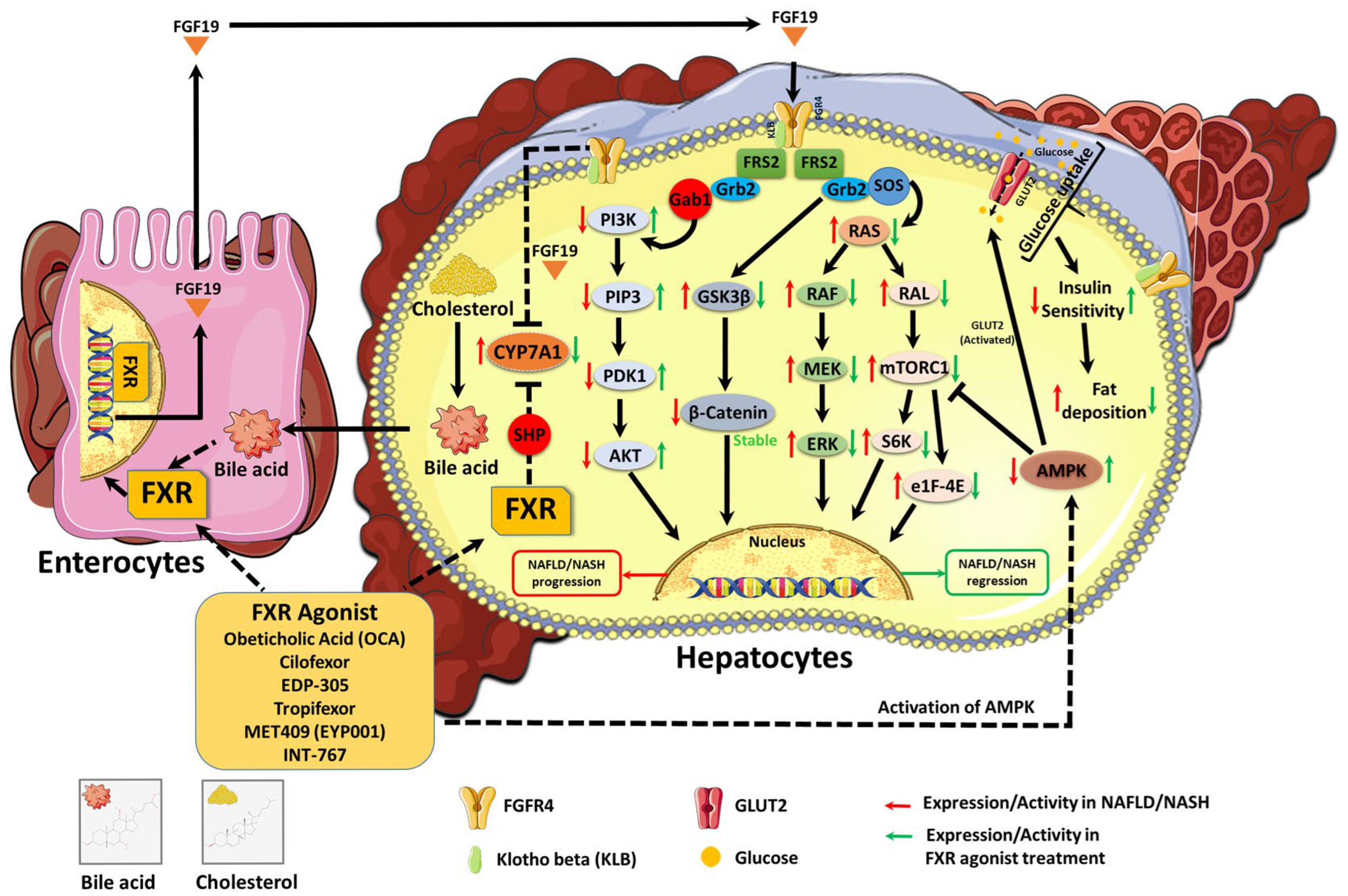
FXRs are nuclear receptors primarily expressed in the liver and intestine, among other tissues like adipose tissue. FXRs are associated with the maintenance of bile acid homeostasis, lipid and glucose metabolism, and immune responses. FXR agonists are synthetic ligands that activate FXRs, influencing the expression of target genes involved in key biological processes related to NAFLD and NASH [40]. Hepatic FXR activation results in a reduction in bile acid synthesis and an increase in bile acid excretion, both of which reduce the accumulation of toxic bile acids in the liver. Bile acids serve as endogenous ligands for FXR as part of a negative feedback loop [41] (Figure 2). When bile acids are present at high concentrations in hepatocytes, FXR activation is triggered by negative feedback and stimulates the transcriptional upregulation of SHP [42].

4.1.2. Regulation of Bile Acid Metabolism and Lipid Homeostasis

Bile acids serve as the primary endogenous activators of FXR, functioning within a negative feedback loop. This process is primarily driven by the interplay between FXR and SHP. When bile acid concentrations rise within hepatocytes, FXR is activated, leading to increased SHP expression (Figure 2) [43]. SHP then binds to and suppresses genes responsible for the synthesis of bile acid from cholesterol, such as cholesterol 7alpha-hydroxylase (CYP7A1) and sterol 12-alpha-hydroxylase (CYP8B1). Additionally, FXR activation regulates the expression of bile acid transporters in both distal ileal enterocytes and hepatocytes, highlighting its critical role in the enterohepatic circulation of bile acids [42].
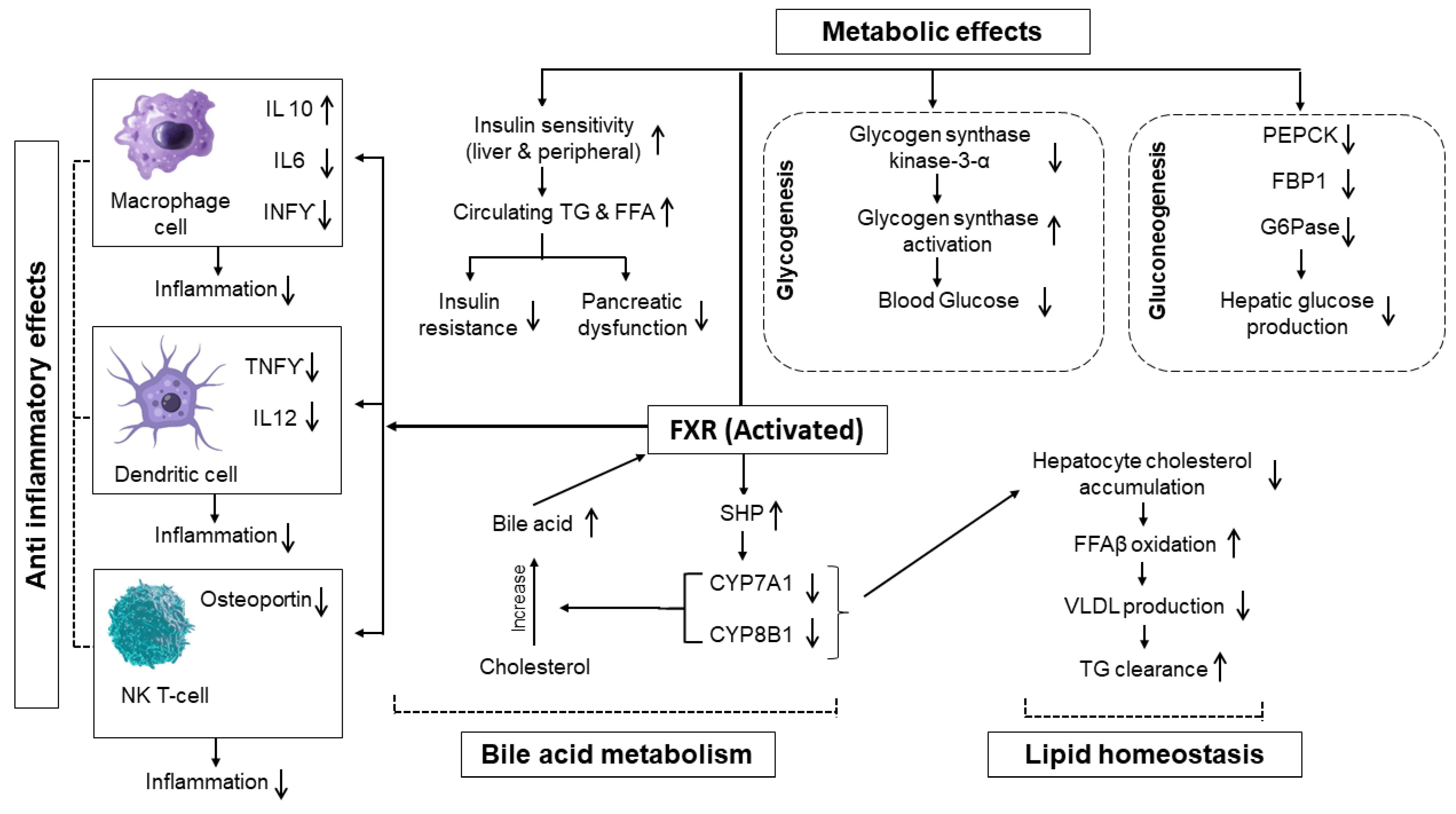
The activation of FXR reduces hepatocyte cholesterol accumulation, elevates free fatty acid beta oxidation, suppresses hepatic lipogenesis, curtails VLDL production, and enhances triglyceride clearance (Figure 2). FXR also plays a key role in lipid metabolism by reducing cholesterol accumulation in hepatocytes through the inhibition of CYP7A1 and CYP8B1, mediated by SHP and fibroblast growth factor 19 (FGF19) activation. Beyond their role in metabolism, FXR agonists also have immunomodulatory effects on innate immune cells. Dendritic cells and macrophages show co-expression of GPBAR1 and FXR receptors, while evidence suggests that FXR expression is restricted to natural killer T cells [42].

4.1.3. Anti-Inflammatory and Metabolic Effects

In macrophages, bile acid activation of FXRs promotes a transition toward the anti-inflammatory M2 phenotype, characterized by increased interleukin-10 (IL-10) expression and decreased levels of the pro-inflammatory cytokines interleukin-6 (IL-6) and interferon-gamma (IFN-γ) (Figure 2) [44]. Bile acids in dendritic cells suppress tumor necrosis factor α (TNFα) and IL12 production, both of which are critical for Th1 cell activation and inflammatory responses. Additionally, bile acids reduce osteopontin expression in natural killer T cells, a key factor in the pathogenesis of autoimmune diseases and inflammation processes. Given these effects, FXR agonists hold potential as therapeutic interventions for inflammatory bowel disease and other autoimmune disorders [42]. Comprehensive activation of FXR elicits a complex response, modulating the expression of numerous genes involved in metabolic and anti-inflammatory regulation, as evidenced by recent genome-wide studies of FXR binding sites [44]. Moreover, FXR plays a crucial role in glucose metabolism by controlling several metabolic pathways in the liver and intestine.
FXR activation improves insulin sensitivity in both hepatic and peripheral tissues—specifically within skeletal muscle and adipose tissue—by reducing circulating triglycerides and free fatty acids, which are key contributors to pancreatic dysfunction, insulin resistance, and lipotoxicity [45]. By attenuating lipotoxicity, FXR activation promotes glycogen synthesis and inhibits gluconeogenesis. Specifically, FXR upregulates glycogen synthase kinase 3-alpha gene transcription while repressing key gluconeogenic enzymes, including phosphoenolpyruvate carboxykinase, fructose-1,6-bisphosphatase 1, and G6Pase, through an SHP-dependent mechanism [42].

4.1.4. Interplay of FXR, PPARs, and LXRs in NAFLD/NASH Pathogenesis

Peroxisome proliferator-activated receptor α (PPARα) is predominantly expressed in tissues with high fatty acid catabolism, such as the liver, where it plays a key role in coordinating pathways involved in fatty acid metabolism and inflammation. In contrast, PPARβ/δ is more ubiquitously distributed and is notably expressed in hepatocytes, hepatic stellate cells (HSCs), and Kupffer cells, indicating its involvement in regulating hepatic inflammation and fibrosis. One of its key actions includes the activation of stearoyl-CoA desaturase 1 (SCD1), which catalyzes the conversion of saturated fatty acids into monounsaturated fatty acids (MUFAs), thereby reducing lipotoxicity. Meanwhile, elevated PPARγ mRNA expression in the liver is a hallmark of steatotic livers in both humans and animal models [46,47,48]. In hepatocytes, PPARγ drives hepatic lipid accumulation, contributing to steatosis and the progression of non-alcoholic fatty liver disease (NAFLD). Additionally, liver X receptors (LXRs), activated by oxysterols (cholesterol derivatives), help maintain cholesterol homeostasis by suppressing cholesterol synthesis and uptake, while promoting its conversion into bile acids and enhancing excretion ultimately improving the lipoprotein profile [49].
PPARs, FXR, and LXRs are intricately interconnected “energy vectors” that collectively regulate nutrient and energy homeostasis within the gut–liver–adipose axis, and their dysfunction contributes significantly to the development and progression of NAFLD and NASH [49]. Specific cross-regulatory mechanisms highlight this relationship: PPARα, a key regulator of fatty acid metabolism, can indirectly coordinate the expression of sterol regulatory element-binding protein 1c (SREBP1c), a master regulator of lipogenesis, through cross-regulation with the LXR signaling pathway. Furthermore, hepatic FXR activation, through the induction of the small heterodimer partner (SHP), can reduce lipogenesis and increase fatty acid oxidation, partly via PPARα. However, this interaction is primarily observed in humans, as the murine PPARα promoter lacks a functional FXR-responsive element [50,51]. In hepatic stellate cells (HSCs), the administration of the FXR agonist obeticholic acid (OCA) has been shown to induce PPARγ expression, which in turn contributes to a reduction in collagen gene induction, demonstrating the direct influence of FXR on PPARγ’s antifibrotic actions. LXRs, as cholesterol sensors, directly induce SREBP1c expression, thereby promoting the synthesis of triglycerides and fatty acids, and also regulate carbohydrate regulatory element-binding protein (ChREBP), which works synergistically with LXR and SREBP1c to induce lipogenic genes [52]. The interplay extends to systemic effects, where intestinal FXR activation induces the enterokine FGF15/19 (mouse/human), which travels to the liver to downregulate bile acid synthesis and plays a broader role in preventing steatosis, inflammation, fibrosis, and metabolic syndrome. These receptors often converge on common metabolic and inflammatory pathways, acting in concert to maintain liver health, and their dysregulation represents a critical aspect of NASH pathogenesis.

4.2. Clinical Trials and Evidence Supporting Role of FXR Agonist in NAFLD/NASH

Given FXR’s central role in the regulation of metabolic pathways disrupted in NAFLD and NASH, as well as its protective effects against hepatic inflammation and fibrogenesis, rigorous clinical evaluation of FXR agonists is essential [4]. Clinical trials are required to establish the therapeutic efficacy, safety profile, and long-term outcomes of FXR activation in human subjects. These investigations will clarify whether pharmacological activation of FXR can effectively control obesity, reduce hepatic steatosis, attenuate inflammatory responses, reduce insulin resistance, and prevent or reverse fibrotic progression (Figure 3). The successful clinical translation of FXR-targeted therapies may offer a novel and effective approach for managing the growing global burden of NAFLD and NASH. FXR agonists have garnered attention in the treatment of NAFLD/NASH, as FXR plays a key role in regulating liver metabolism, inflammation, and bile acid homeostasis, as mentioned previously. FXR agonists clinically tested in NASH include bile acid-derived compounds (OCA), steroidal non-BA-derived FXR agonists (EDP305), nonsteroidal FXR agonists (PX-104TERN-10, cilofexor, tropifexor, vonafexor MET409), and partial FXR agonists (nidufexor). Some of these promising agonists have been discussed here. Below is an overview of the clinical trials and evidence supporting the role of FXR agonists in the management of NAFLD/NASH.

4.2.1. Summary of Key Clinical Trials and Their Outcomes

Obeticholic Acid (OCA)
One of the landmark studies evaluating FXR agonists in NASH was the FLINT trial, which studied the impact of OCA in patients with non-cirrhotic NASH (Table 1). This multicenter, randomized, placebo-controlled trial demonstrated that patients treated with OCA experienced significant improvements in liver histology, along with reduced fibrosis and decreased hepatocellular ballooning. However, pruritus (itching) was a notable adverse effect associated with OCA treatment [53]. This preliminary trial also showed that OCA was well tolerated in patients with NAFLD and led to improvements in insulin sensitivity, as well as markers of hepatic inflammation and fibrosis. Histopathological evaluation showed that OCA improved NAFLD in 45% of patients when compared to 23% in the placebo group over a period of 72 weeks. This was the first experiment that demonstrated that FXR activity may be a critical target in enhancing the histological activity of NAFLD [42].
Cilofexor
Another FXR agonist, cilofexor, was evaluated in a phase II, randomized, controlled trial involving patients with non-cirrhotic NASH (Table 1). The investigation found that cilofexor administration resulted in reduced hepatic lipid content and improved markers of liver injury, suggesting its potential as a therapeutic agent for NASH. Cilofexor for a duration of 24 weeks exhibited favorable tolerability and provided significant reductions in hepatic steatosis, hepatic biochemical parameters, and serum bile acids in patients with NASH; however, the study also documented mild-to-moderate pruritus and gastrointestinal side effects in a subset of patients [55]. Overall, cilofexor was shown to reduce steatosis and downstage hepatic fibrosis without worsening steatohepatitis when used in combination with firsocostat (an acetyl-CoA carboxylase inhibitor) [42].
EDP305
EDP305 is an FXR agonist that selectively activates FXR; it is at present under investigation as a putative therapy for patients with NASH and liver fibrosis (Table 1) [56]. In vitro studies showed that EDP305 modulates bile acid and lipid metabolism, and decreases the transcription of inflammation-inducing and fibrogenic genes. A phase II study assessed the efficacy of EDP305, a novel FXR agonist, in subjects with NASH. The trial noted a dose-dependent reduction in liver enzyme levels and markers of inflammation. Although EDP305 showed promising results in improving liver health, some patients experienced pruritus, which is a common side effect observed with FXR activation [56]. The primary outcome focused on change in alanine aminotransferase (ALT) between baseline and Week 12 of treatment, and the key secondary endpoint was mean change in hepatic fat accumulation during the same time frame. Across the study cohort, EDP305 reduced ALT levels and hepatic fat content, thereby providing justification for an extended clinical trial assessing histological endpoints in patients with NAFLD.
Tropifexor
Tropifexor is a strong, potent, non-bile-acid FXR agonist that has demonstrated encouraging effects in patients with NASH/NAFLD (Table 1). In a phase II, randomized, placebo-controlled trial, 12 weeks of tropifexor treatment showed a significant reduction in ALT levels, liver fat content, and markers of hepatic inflammation [57]. In line with other FXR agonists, pruritus was developed in a small percentage of patients despite the compound’s decent tolerance. Notably, tropifexor also demonstrated dose-dependent improvement in biomarkers of fibrosis, making it a prospective candidate for further implementation in the treatment of NAFLD and NASH [60].
MET409 (EYP001)
MET409 (EYP001) is another nonsteroidal FXR agonist under trail for NASH therapy (Table 1). FXR agonism by MET409 suppresses NF-κB signaling to reduce inflammation of the liver and inhibit lipogenesis, collectively counteracting hepatic steatosis and NAFLD/NASH progression [61]. In clinical trials, MET409 showed strong FXR activation with efficient pharmacokinetics and limited systemic distribution. Outcomes from a phase I/II trail indicated significant reductions in hepatic fat and improvements in liver enzyme levels after 12 weeks of treatment [59]. MET409 showed high tolerability, with a reduced incidence and severity of pruritus compared with other FXR agonists, indicating that its distinct pharmacological profile might give an improved therapeutic index for NAFLD and NASH management.
Vonafexor
Vonafexor is a selective FXR agonist that has undergone a phase IIa, double-blind study (Table 1) [58]. It works by improving bile acid homeostasis and inducing potent liver fat reduction, which are key factors in managing conditions like NAFLD/NASH. In clinical trials, vonafexor has shown promising results in enhancing metabolic functions and weight loss [58]. However, some mild side effects have been reported, including pruritus and fatigue. These side effects are generally manageable and do not overshadow the potential therapeutic benefits of vonafexor.
INT767
INT767 is a dual agonist of FXR and TGR5, a G-protein-coupled bile acid receptor, which can target numerous pathways involved in NAFLD/NASH pathophysiology. TGR5 is a bile acid receptor abundantly expressed throughout the liver, intestinal tract, monocytes, and macrophages [62,63]. It has been shown to inhibit LPS-induced NF-κB activation and to activate the AKT–mTORC1 (alpha serine/threonine protein kinase–mammalian target of rapamycin) pathway [64]. Preclinical studies have demonstrated that INT767 reduces liver inflammation and steatosis more successfully than selective FXR agonists. In early-phase clinical trials, INT767 again showed reductions in hepatic fat and improved metabolic parameters. While clinical data are still emerging, the dual mechanism of action suggests therapeutic potential for reducing hepatic fat, inflammation, and fibrosis in patients with NAFLD/NASH.

4.2.2. Comparison with Other Therapeutic Agents

Compared to other therapeutic strategies for NASH or NAFLD, FXR agonists have shown potential advantages in targeting multiple pathways involved in disease progression. FXR, whether activated by bile acids or other compounds, plays a vital role in controlling lipid metabolism and maintaining intestinal homeostasis, thus playing a major role in the pathogenesis of NAFLD/NASH. Therefore, pharmaceutical modulation of FXR is pivotal for the effective treatment of NAFLD and NASH. While current treatment options such as lifestyle modifications and vitamin E supplementation primarily focus on metabolic regulation and oxidative stress, FXR agonists directly regulate bile acid metabolism, inflammation, and fibrosis [53,56]. Furthermore, unlike GLP1 receptor agonists, which mainly exert metabolic benefits, FXR agonists address both metabolic dysfunction and fibrotic pathways, making them a compelling candidate for NASH treatment [55]. However, the challenge of pruritus as a side effect remains a key limitation that requires further research. For example, the FDA rejected the marketing approval request of OCA, suggesting that managing common adverse reactions such as pruritus, increased LDL cholesterol, and reduced HDL cholesterol remains a major challenge for the advancement of FXR agonists. Nevertheless, this does not imply that FXR agonists have been excluded from the treatment of NAFLD; rather, their side effects related to disrupted lipid metabolism can be successfully managed through combination therapy with statins [65]. Overall, FXR agonists are still a promising class of therapeutics for NAFLD and NASH, with ongoing clinical trials further elucidating their long-term efficacy and safety profiles.

5. FXR Agonists and Protein Kinase Regulation in NAFLD and NASH

The efficacy of FXR agonists is largely attributed to their ability to modulate various protein kinase pathways involved in lipid metabolism, inflammation, insulin sensitivity, and hepatic fibrosis. The dysregulation of these pathways plays a pivotal role in the pathogenesis and progression of NAFLD and NASH, making them critical targets for therapeutic intervention. The activation of FXR leads to FGF19 gene transcription in humans, which produces a hormone released into the portal circulation (Figure 4). Then, FGF19 moves to the liver, where it binds to and activates a membrane tyrosine kinase receptor, fibroblast growth factor receptor 4 (FGFR4). This activation triggers a signaling cascade that suppresses bile acid synthesis by inhibiting the transcription of the CYP7A1 gene, which encodes the CYP7A1 enzyme. CYP7A1 is responsible for the rate-limiting step in bile acid production. Similarly, FGF19 analog administration in humans has been shown to reduce bile acid synthesis in healthy individuals.

5.1. Effect of FXR Agonists on AMPK Activation and Lipid Metabolism

FXR agonists have been found to affect several metabolic processes, including lipid metabolism, by regulating the activity of AMPK (Figure 4). AMPK is an important energy sensor that modulates lipid and glucose metabolism. In healthy hepatocytes, AMPK activation suppresses lipid synthesis by inhibiting acetyl-CoA carboxylase [36] and SREBP1c, while simultaneously promoting fatty acid oxidation via PPARα activation [66]. However, in NAFLD, AMPK activity is often impaired due to chronic energy surplus and insulin resistance, leading to excessive lipid accumulation, mitochondrial dysfunction, and endoplasmic reticulum stress [67]. This dysregulation exacerbates hepatic steatosis, a hallmark of NAFLD.
FXR agonists have been shown to activate AMPK, thereby restoring metabolic homeostasis in NAFLD and NASH. Studies indicate that FXR activation upregulates genes involved in fatty acid oxidation, like carnitine palmitoyl transferase 1 (CPT1), while downregulating those related to lipogenesis, such as FAS and SREBP1c [68]. Additionally, FXR agonists reduce hepatic lipid accumulation by decreasing triglyceride synthesis and enhancing mitochondrial β-oxidation [69]. These mechanisms collectively contribute to a reduction in hepatic steatosis, highlighting the therapeutic potential of FXR agonists in lipid metabolism disorders.

5.2. FXR Agonists’ Role in Modulating MAPK Signaling and Inflammation

FXR agonists also play a central role in modulating numerous biological processes, like the MAPK signaling pathway. For cellular growth, survival, and inflammation, MAPK signaling is a crucial pathway. FXR agonists have been shown to interact with MAPK signaling pathways as discussed below.
The MAPK family, including JNK and p38 MAPK, plays a key role in inflammatory responses in NAFLD and NASH. The JNK pathway is particularly involved in mediating hepatocyte apoptosis and insulin resistance by phosphorylating insulin receptor substrate 1 (IRS1), resulting in insulin resistance [70]. Moreover, the p38 MAPK pathway contributes to the activation of pro-inflammatory cytokines, such as interleukin 6 (IL6) and TNFα, which exacerbate hepatic inflammation and fibrosis [71]. Persistent activation of these pathways promotes disease progression from simple steatosis to NASH. FXR agonists exhibit potent anti-inflammatory properties by modulating MAPK signaling pathways. Studies have demonstrated that FXR activation suppresses JNK phosphorylation, thereby improving insulin sensitivity and reducing hepatocyte apoptosis [72]. Furthermore, FXR agonists inhibit the p38 MAPK pathway, leading to decreased expression of inflammatory mediators, such as TNFα and IL1β [73]. These anti-inflammatory effects contribute to the hepatoprotective benefits of FXR agonists, making them valuable candidates for NAFLD and NASH therapy.

5.3. FXR Agonists’ Influence on PI3K/AKT and Insulin Sensitivity

In addition, FXR agonists have been shown to utilize the PI3K/AKT signaling pathway to control NAFLD and insulin sensitivity (Figure 4). The PI3K/AKT pathway is another major signaling cascade associated with insulin-controlled glucose uptake and metabolism. Under normal physiological conditions, PI3K gets activated by insulin binding to its receptor, which in turn stimulates AKT phosphorylation. This activation promotes glycogen synthesis, inhibits gluconeogenesis, and enhances lipid metabolism [74]. However, in NAFLD, chronic inflammation and lipid overload impair PI3K/AKT signaling, leading to hepatic insulin resistance. This results in unchecked glucose production, exacerbating hyperglycemia and metabolic dysfunction [75].
As discussed before, FXR agonists can enhance insulin sensitivity by restoring PI3K/AKT signaling. The activation of FXR reduces hepatic gluconeogenesis by suppressing G6Pase and phosphoenolpyruvate carboxykinase (PEPCK), two key enzymes in glucose production [76]. Additionally, FXR agonists promote AKT phosphorylation, thereby improving insulin receptor signaling and glucose uptake [77]. These effects facilitate the overall metabolic benefits of FXR agonists in NAFLD, positioning them as promising agents for addressing insulin resistance in the disease.

5.4. FXR Agonist and mTOR Pathway in NAFLD/NASH Progression

Research has shown that the FXR agonist also exhibits an important connection with the mTOR pathway since both play critical roles in liver metabolism and NAFLD/NASH progression (Figure 4). The mTOR pathway plays a crucial role in cell growth, metabolism, and autophagy. mTOR dysregulation signaling is associated with the progression of NAFLD and NASH, particularly in hepatic steatosis and fibrosis. The overactivation of mTORC1 promotes lipogenesis by upregulating SREBP1c and FAS, leading to excessive lipid accumulation in hepatocytes [78]. Additionally, mTORC1 activation has been associated with hepatic stellate cell (HSC) activation and collagen deposition, which contribute to fibrosis in advanced stages of NAFLD [79].
FXR agonists have demonstrated the ability to modulate mTOR signaling, thereby mitigating hepatic lipid accumulation and fibrosis. By inhibiting mTORC1 activity, FXR activation reduces lipogenesis and enhances autophagy, promoting lipid clearance in hepatocytes [80]. Furthermore, FXR agonists suppress the activation of HSCs, thereby attenuating fibrotic processes in NAFLD. These findings emphasize the potential of FXR agonists in targeting mTOR-mediated mechanisms to prevent disease progression. Therefore, FXR activation has been linked to the modulation of the mTOR pathway, which is crucial for autophagy and lipid metabolism [81]. Additionally, FXR influences the activity of AMPK, a vital regulator of energy homeostasis, thereby affecting lipid and glucose metabolism [82]. Moreover, studies have demonstrated that FXR activation can impact the JNK pathway, which is linked with inflammation and insulin resistance in NAFLD [83]. The modulation of specific protein kinases, such as AMPK, mTOR, and JNK, through FXR activation presents promising strategies for drug development. Targeting these kinases could ameliorate metabolic dysregulation and inflammation associated with NAFLD and NASH.

6. Potential of Combining FXR Agonist with Kinase-Targeting Drugs

It is known that FXR activity can be modulated by the phosphorylation of protein kinases, meaning that protein kinase drugs could potentially influence the effectiveness of FXR agonists by altering FXR’s phosphorylation status and subsequent transcriptional activity [84]. In addition to FXR agonists, new synthetic small molecules directly or indirectly targeting protein kinases are also emerging as promising agents. Some of these molecules can activate AMPK, and these are already on the market to treat risk factors of NAFLD/NASH such as diabetes mellitus and hypercholesterolemia [85]. For example, the novel glucagon-like peptide-1 receptor agonist AWR demonstrated the ability to improve hepatic steatosis, significantly reducing FFA levels in high-energy diet-fed mice by modulating the PI3K/AKT pathway. This regulation also inhibited hepatic inflammation and improved glucose metabolism in hepatocytes [86]. Another glucagon-like peptide-1 activator (ALT801) has started to be clinically evaluated to treat fatty liver disease and shows an impact on AMPK signaling [87]. Atorvastatin has proven effective in preventing lipid accumulation in hepatocytes via the activation of the protein kinase pathway through AMPK. This protein kinase pathway activation serves as a protective measure against lipid deposition in hepatocytes, which promotes an improvement in liver health [88].
A study demonstrated that FRX transcriptional activity is directly regulated by PKC, showing that PKCα-mediated phosphorylation of FXR regulates its agonist activation [89]. The study also revealed FXR to be a phosphoprotein and highlighted the involvement of the calcium-dependent PKC pathway in regulating FXR activity. Another work showed that PKC phosphorylates SRC3, enhancing estrogen receptor-dependent gene transcription [90]. FXR phosphorylation by PKCα and PKCβI was confirmed, but other PKC isoforms’ roles remain unclear, requiring further research on the specific kinases involved in FXR regulation and their impact on coactivators, complicating therapeutic development. Some agents targeting protein kinases related to inflammation pathways, like NF-κB and ERK, showed potential, though many targeted ERK less extensively [36,91]. Additionally, a phase 2 agent selectively inhibiting JNK protein emphasizes the importance of protein kinase-targeting drugs [92]. The complex network of protein kinase isomers can lead to varying outcomes, as demonstrated by diacerein and triptorelin, which both activate JNK and inhibit ERK-MAPK, highlighting the challenge of balancing beneficial and unfavorable effects in protein kinase-targeting treatments. This complexity underscores the difficulties in developing effective therapies that target multiple intracellular and intercellular pathways.
FXR agonists have strong therapeutic potential in NAFLD and NASH by modulating bile acid, lipid, and glucose metabolism, and their combination with protein kinase-targeting agents may improve clinical outcomes by addressing complementary metabolic and inflammatory pathways. For example, in a randomized, placebo-controlled, double-blind CONTROL phase 2 trial (NCT02633956), co-administration of atorvastatin effectively managed the LDL elevation usually induced by the FXR agonist obeticholic acid (OCA) (Table 2) [93]. In a similar effort, a 12-week phase 2 trial (NCT04702490) worked on the combination of MET409 (a potent non-bile-acid sustained FXR agonist) and empagliflozin (a sodium–glucose cotransporter-2 (SGLT2) inhibitor) in patients with type 2 diabetes and NASH. This trial reported significant liver fat reduction with minimal pruritus (0–6% across groups), which is a favorable safety profile for patients (Table 2) [94]. Empagliflozin’s therapeutic effects are partially attributed to AMPK activation, which improves energy metabolism and suppresses inflammatory signaling (Table 2) [95]. In parallel, a phase 2, randomized, double-blind trial is currently assessing the combined efficacy of licogliflozin (a dual SGLT1/2 inhibitor) and tropifexor, with primary endpoints targeting NASH resolution without worsening fibrosis or ≥1-stage fibrosis improvement without worsening NASH at Week 48 (Table 2) [96]. Notably, licogliflozin also modulates multiple protein kinases implicated in the pathogenesis of NASH and NAFLD [97], offering a mechanistic synergy with FXR agonists. Likewise, in a phase 2b trial, the combination of cilofexor and firsocostat demonstrated good tolerability and meaningful improvements in NASH activity, along with potential antifibrotic effects, in patients with advanced fibrosis (F3–F4) over 48 weeks (Table 2) [98]. Together, these findings support the rationale for combining FXR agonists with complementary agents to achieve broader and more effective disease modulation.
Several challenges hinder the optimal use of FXR agonists in treating NAFLD and NASH, including the difficulty in maximizing therapeutic efficacy while minimizing adverse effects such as pruritus, elevated LDL cholesterol, and reduced HDL cholesterol concerns that contributed to the FDA’s rejection of obeticholic acid’s marketing approval. Furthermore, the precise roles of different protein kinase isoforms and their signaling pathways in driving NAFLD, NASH, inflammation, and fibrosis remain unclear. Moreover, future challenges in developing FXR agonists and protein kinase-targeted therapies include the difficulty of selectively targeting specific kinases involved in FXR regulation, the potential for contradictory effects when targeting multiple pathways, and the variability in patient responses to combination therapies. There has also been limited exploration of how FXR agonists regulate various kinases across metabolic pathways. Future directions should focus on identifying more selective and effective protein kinase inhibitors, optimizing combination therapies to enhance both efficacy and safety, and conducting long-term clinical trials to evaluate the broader impacts of these treatments on liver health and metabolic disorders. Therefore, future directions in personalized medicine for NAFLD/NASH include leveraging genetic testing to identify high-risk individuals, utilizing advanced imaging techniques for disease staging, incorporating gut microbiome analysis to tailor treatment strategies, developing targeted therapies based on specific molecular profiles, and utilizing machine learning to integrate diverse patient data for better prediction and treatment decision-making.

7. Conclusions

In summary, this review clarifies that protein kinases such as MAPK, PI3K/AKT, ErbB, PKC, and mTOR are critical in controlling cellular processes involved in NAFLD/NASH pathogenesis, including inflammation, hepatic metabolism, and lipid deposition. The regulation of these pathways, especially through FXR activation, offers a potential therapeutic strategy to prevent disease progression to NASH. FXR agonists play an important role in regulating protein kinase pathways, enhancing insulin sensitivity, suppressing hepatic lipogenesis, and reducing inflammation. These agents also promote β-oxidation, reduce de novo lipogenesis, and reduce oxidative stress, thereby controlling hepatic steatosis and fibrosis. This comprehensive modulation underscores the significant promise of integrating FXR-targeted therapies with protein kinase regulation for treating NAFLD and NASH. However, further long-term studies are essential to fully ascertain the efficacy and safety profiles of FXR agonists in clinical settings, emphasizing that a deeper understanding of the combined effects of FXR activation and protein kinase regulation will be crucial for the future management of NAFLD/NASH and its associated complications.

Author Contributions

Conceptualization, A.S. and J.F.; methodology, A.S., E.W. and L.O.; validation, A.S., E.W. and L.O.; formal analysis, E.W. and L.O.; investigation, A.S., M.M., E.W., L.O. and K.M.; resources, E.W. and L.O.; data curation, K.M.; writing—original draft preparation, E.W., L.O., A.S. and J.F.; writing—review and editing, A.S., M.M., S.D.G. and J.F.; visualization, A.S.; supervision, A.S., and J.F.; project administration, A.S., S.D.G. and J.F. All authors have read and agreed to the published version of the manuscript.

Funding

This research is supported by the start-up fund of Jannatul Ferdoush at the University of Tennessee at Chattanooga.

Conflicts of Interest

The authors declare no conflict of interest.

References

  1. Mahlapuu, M.; Caputo, M.; Xia, Y.; Cansby, E. GCKIII kinases in lipotoxicity: Roles in NAFLD and beyond. Hepatol. Commun. 2022, 6, 2613–2622. [Google Scholar] [CrossRef] [PubMed]
  2. Maurice, J.; Manousou, P. Non-alcoholic fatty liver disease. Clin. Med. 2018, 18, 245–250. [Google Scholar] [CrossRef]
  3. Kawanaka, M.; Nishino, K.; Morimoto, Y.; Ishii, K.; Tanikawa, T.; Urata, N.; Suehiro, M.; Sasai, T.; Haruma, K.; Kawamoto, H. Progression from nonalcoholic fatty liver to nonalcoholic steatohepatitis cirrhosis confirmed by liver histology after 14 years. Intern. Med. 2021, 60, 1397–1401. [Google Scholar] [CrossRef] [PubMed]
  4. Pierantonelli, I.; Svegliati-Baroni, G. Nonalcoholic fatty liver disease: Basic pathogenetic mechanisms in the progression from NAFLD to NASH. Transplantation 2019, 103, e1–e13. [Google Scholar] [CrossRef] [PubMed]
  5. Alshehade, S.; Alshawsh, M.A.; Murugaiyah, V.; Asif, M.; Alshehade, O.; Almoustafa, H.; Al Zarzour, R.H. The role of protein kinases as key drivers of metabolic dysfunction-associated fatty liver disease progression: New insights and future directions. Life Sci. 2022, 305, 120732. [Google Scholar] [CrossRef]
  6. Stefan, N.; Häring, H.U.; Cusi, K. Non-alcoholic fatty liver disease: Causes, diagnosis, cardiometabolic consequences, and treatment strategies. Lancet Diabetes Endocrinol. 2019, 7, 313–324. [Google Scholar] [CrossRef]
  7. Kwiecinski, M.; Noetel, A.; Elfimova, N.; Trebicka, J.; Schievenbusch, S.; Strack, I.; Molnar, L.; von Brandenstein, M.; Töx, U.; Nischt, R. Hepatocyte growth factor (HGF) inhibits collagen I and IV synthesis in hepatic stellate cells by miRNA-29 induction. PLoS ONE 2011, 6, e24568. [Google Scholar] [CrossRef]
  8. Marra, F.; Svegliati-Baroni, G. Lipotoxicity and the gut-liver axis in NASH pathogenesis. J. Hepatol. 2018, 68, 280–295. [Google Scholar] [CrossRef]
  9. Feng, Y.; Sun, W.; Sun, F.; Yin, G.; Liang, P.; Chen, S.; Liu, X.; Jiang, T.; Zhang, F. Biological mechanisms and related natural inhibitors of CD36 in nonalcoholic fatty liver. Drug Des. Dev. Ther. 2022, 16, 3829–3845. [Google Scholar] [CrossRef]
  10. Hong, H.; Jin, Z.; Qian, T.; Xu, X.; Zhu, X.; Fei, Q.; Yang, J.; Sui, C.; Xu, M. Falcarindiol enhances cisplatin chemosensitivity of hepatocellular carcinoma via down-regulating the STAT3-modulated PTTG1 pathway. Front. Pharmacol. 2021, 12, 656697. [Google Scholar] [CrossRef]
  11. Chen, Z.; Tian, R.; She, Z.; Cai, J.; Li, H. Role of oxidative stress in the pathogenesis of nonalcoholic fatty liver disease. Free Radic. Biol. Med. 2020, 152, 116–141. [Google Scholar] [CrossRef] [PubMed]
  12. Ashraf, N.U.; Sheikh, T.A. Endoplasmic reticulum stress and oxidative stress in the pathogenesis of non-alcoholic fatty liver disease. Free Radic. Res. 2015, 49, 1405–1418. [Google Scholar] [CrossRef] [PubMed]
  13. Kitase, A.; Hino, K.; Furutani, T.; Okuda, M.; Gondo, T.; Hidaka, I.; Hara, Y.; Yamaguchi, Y. In situ detection of oxidized n-3 polyunsaturated fatty acids in chronic hepatitis C: Correlation with hepatic steatosis. J. Gastroenterol. 2005, 40, 617–624. [Google Scholar] [CrossRef] [PubMed]
  14. Kyriakis, J.M.; Avruch, J. Mammalian MAPK signal transduction pathways activated by stress and inflammation: A 10-year update. Physiol. Rev. 2012, 92, 689–737. [Google Scholar] [CrossRef]
  15. Chang, L.; Karin, M. Mammalian MAP kinase signalling cascades. Nature 2001, 410, 37–40. [Google Scholar] [CrossRef]
  16. Hirosumi, J.; Tuncman, G.; Chang, L.; Görgün, C.Z.; Uysal, K.T.; Maeda, K.; Karin, M.; Hotamisligil, G.S. A central role for JNK in obesity and insulin resistance. Nature 2002, 420, 333–336. [Google Scholar] [CrossRef]
  17. Yuan, M.; Konstantopoulos, N.; Lee, J.; Hansen, L.; Li, Z.-W.; Karin, M.; Shoelson, S.E. Reversal of obesity-and diet-induced insulin resistance with salicylates or targeted disruption of Ikkβ. Science 2001, 293, 1673–1677. [Google Scholar] [CrossRef]
  18. Newton, A.C. Protein kinase C: Perfectly balanced. Crit. Rev. Biochem. Mol. Biol. 2018, 53, 208–230. [Google Scholar] [CrossRef]
  19. Samuel, V.T.; Shulman, G.I. Mechanisms for insulin resistance: Common threads and missing links. Cell 2012, 148, 852–871. [Google Scholar] [CrossRef]
  20. Petersen, M.C.; Madiraju, A.K.; Gassaway, B.M.; Marcel, M.; Nasiri, A.R.; Butrico, G.; Marcucci, M.J.; Zhang, D.; Abulizi, A.; Zhang, X.-M. Insulin receptor Thr 1160 phosphorylation mediates lipid-induced hepatic insulin resistance. J. Clin. Investig. 2016, 126, 4361–4371. [Google Scholar] [CrossRef]
  21. Liu, R.; Qian, M.P.; Cui, Y.Y. Protein kinases: The key contributors in pathogenesis and treatment of nonalcoholic fatty liver disease-derived hepatocellular carcinoma. Metabolism 2023, 147, 155665. [Google Scholar] [CrossRef] [PubMed]
  22. Zhang, D.; Wang, W.; Sun, X.; Xu, D.; Wang, C.; Zhang, Q.; Wang, H.; Luo, W.; Chen, Y.; Chen, H.; et al. AMPK regulates autophagy by phosphorylating BECN1 at threonine 388. Autophagy 2016, 12, 1447–1459. [Google Scholar] [CrossRef] [PubMed]
  23. Cai, Y.; Fang, L.; Chen, F.; Zhong, P.; Zheng, X.; Xing, H.; Fan, R.; Yuan, L.; Peng, W.; Li, X. Targeting AMPK related signaling pathways: A feasible approach for natural herbal medicines to intervene non-alcoholic fatty liver disease. J. Pharm. Anal. 2024, 15, 101052. [Google Scholar] [CrossRef] [PubMed]
  24. Shaywitz, A.J.; Courtney, K.D.; Patnaik, A.; Cantley, L.C. PI3K enters beta-testing. Cell Metab. 2008, 8, 179–181. [Google Scholar] [CrossRef]
  25. Shamsan, E.; Almezgagi, M.; Gamah, M.; Khan, N.; Qasem, A.; Chuanchuan, L.; Haining, F. The role of PI3k/AKT signaling pathway in attenuating liver fibrosis: A comprehensive review. Front. Med. 2024, 11, 1389329. [Google Scholar] [CrossRef]
  26. Czech, M.P. Insulin action and resistance in obesity and type 2 diabetes. Nat. Med. 2017, 23, 804–814. [Google Scholar] [CrossRef]
  27. Foretz, M.; Guigas, B.; Bertrand, L.; Pollak, M.; Viollet, B. Metformin: From mechanisms of action to therapies. Cell Metab. 2014, 20, 953–966. [Google Scholar] [CrossRef]
  28. Wan, M.; Leavens, K.F.; Saleh, D.; Easton, R.M.; Guertin, D.A.; Peterson, T.R.; Kaestner, K.H.; Sabatini, D.M.; Birnbaum, M.J. Postprandial hepatic lipid metabolism requires signaling through Akt2 independent of the transcription factors FoxA2, FoxO1, and SREBP1c. Cell Metab. 2011, 14, 516–527. [Google Scholar] [CrossRef]
  29. Yecies, J.L.; Zhang, H.H.; Menon, S.; Liu, S.; Yecies, D.; Lipovsky, A.I.; Gorgun, C.; Kwiatkowski, D.J.; Hotamisligil, G.S.; Lee, C.H.; et al. Akt stimulates hepatic SREBP1c and lipogenesis through parallel mTORC1-dependent and independent pathways. Cell Metab. 2011, 14, 21–32. [Google Scholar] [CrossRef]
  30. Götting, I.; Jendrossek, V.; Matschke, J. A New Twist in Protein Kinase B/AKT Signaling: Role of Altered Cancer Cell Metabolism in AKT-Mediated Therapy Resistance. Int. J. Mol. Sci. 2020, 21, 8563. [Google Scholar] [CrossRef]
  31. Saxton, R.A.; Sabatini, D.M. mTOR Signaling in Growth, Metabolism, and Disease. Cell 2017, 168, 960–976. [Google Scholar] [CrossRef] [PubMed]
  32. Oh, W.J.; Jacinto, E. mTOR complex 2 signaling and functions. Cell Cycle 2011, 10, 2305–2316. [Google Scholar] [CrossRef] [PubMed]
  33. Uehara, K.; Sostre-Colón, J.; Gavin, M.; Santoleri, D.; Leonard, K.-A.; Jacobs, R.L.; Titchenell, P.M. Activation of liver mTORC1 protects against NASH via dual regulation of VLDL-TAG secretion and de novo lipogenesis. Cell. Mol. Gastroenterol. Hepatol. 2022, 13, 1625–1647. [Google Scholar] [CrossRef]
  34. Ye, Y.; Liu, H.; Zhang, F.; Hu, F. mTOR signaling in Brown and Beige adipocytes: Implications for thermogenesis and obesity. Nutr. Metab. 2019, 16, 74. [Google Scholar] [CrossRef] [PubMed]
  35. Murphrey, M.B.; Quaim, L.; Rahimi, N.; Varacallo, M. Biochemistry, epidermal growth factor receptor. In StatPearls [Internet]; StatPearls Publishing: Treasure Island, FL, USA, 2023. [Google Scholar]
  36. Galve-Roperh, I.; Chiurchiù, V.; Díaz-Alonso, J.; Bari, M.; Guzmán, M.; Maccarrone, M. Cannabinoid receptor signaling in progenitor/stem cell proliferation and differentiation. Prog. Lipid Res. 2013, 52, 633–650. [Google Scholar] [CrossRef]
  37. Zhai, T.; Cai, L.; Jia, X.; Xia, M.; Bian, H.; Gao, X.; Pan, C.; Li, X.; Xia, P. IGFBP2 functions as an endogenous protector against hepatic steatosis via suppression of the EGFR-STAT3 pathway. Mol. Metab. 2024, 89, 102026. [Google Scholar] [CrossRef]
  38. Khurana, C.; Bedi, O. Proposed hypothesis of GSK-3 β inhibition for stimulating Wnt/β-catenin signaling pathway which triggers liver regeneration process. Naunyn-Schmiedeberg’s Arch. Pharmacol. 2022, 395, 377–380. [Google Scholar] [CrossRef]
  39. Farage, A.E.; Abdo, W.; Osman, A.; Abdel-Kareem, M.A.; Hakami, Z.H.; Alsulimani, A.; Bin-Ammar, A.; Alanazi, A.S.; Alsuwayt, B.; Alanazi, M.M.; et al. Betulin prevents high-fat diet-induced non-alcoholic fatty liver disease by mitigating oxidative stress and upregulating Nrf2 and SIRT1 in rats. Life Sci. 2023, 322, 121688. [Google Scholar] [CrossRef]
  40. Brendel, C.; Schoonjans, K.; Botrugno, O.A.; Treuter, E.; Auwerx, J. The small heterodimer partner interacts with the liver X receptor α and represses its transcriptional activity. Mol. Endocrinol. 2002, 16, 2065–2076. [Google Scholar] [CrossRef]
  41. Garg, S.; Kagal, U. Gene Expression of Cyp7a1 in Liver Tissue of Type 2 Diabetic Rats Treated with Fenugreek and/or Metformin. Int. J. Appl. Basic Med. Res. 2025, 15, 11–17. [Google Scholar] [CrossRef]
  42. Almeqdadi, M.; Gordon, F.D. Farnesoid X receptor agonists: A promising therapeutic strategy for gastrointestinal diseases. Gastro Hep Adv. 2024, 3, 344–352. [Google Scholar] [CrossRef] [PubMed]
  43. Chiang, J.Y. Negative feedback regulation of bile acid metabolism: Impact on liver metabolism and diseases. Hepatology 2015, 62, 1315–1317. [Google Scholar] [CrossRef]
  44. Jaroonwitchawan, T.; Arimochi, H.; Sasaki, Y.; Ishifune, C.; Kondo, H.; Otsuka, K.; Tsukumo, S.-I.; Yasutomo, K. Stimulation of the farnesoid X receptor promotes M2 macrophage polarization. Front. Immunol. 2023, 14, 1065790. [Google Scholar] [CrossRef]
  45. Modica, S.; Gadaleta, R.M.; Moschetta, A. Deciphering the nuclear bile acid receptor FXR paradigm. Nucl. Recept. Signal. 2010, 8, e005. [Google Scholar] [CrossRef]
  46. Braissant, O.; Foufelle, F.; Scotto, C.; Dauca, M.; Wahli, W. Differential expression of peroxisome proliferator-activated receptors (PPARs): Tissue distribution of PPAR-alpha, -beta, and -gamma in the adult rat. Endocrinology 1996, 137, 354–366. [Google Scholar] [CrossRef]
  47. Mandard, S.; Muller, M.; Kersten, S. Peroxisome proliferator-activated receptor alpha target genes. Cell. Mol. Life Sci. 2004, 61, 393–416. [Google Scholar] [CrossRef] [PubMed]
  48. Girroir, E.E.; Hollingshead, H.E.; He, P.; Zhu, B.; Perdew, G.H.; Peters, J.M. Quantitative expression patterns of peroxisome proliferator-activated receptor-beta/delta (PPARβ/δ) protein in mice. Biochem. Biophys. Res. Commun. 2008, 371, 456–461. [Google Scholar] [CrossRef]
  49. Cariello, M.; Piccinin, E.; Moschetta, A. Transcriptional regulation of metabolic pathways via lipid-sensing nuclear receptors PPARs, FXR, and LXR in NASH. Cell. Mol. Gastroenterol. Hepatol. 2021, 11, 1519–1539. [Google Scholar] [CrossRef] [PubMed]
  50. Liu, Y.; Han, X.; Bian, Z.; Peng, Y.; You, Z.; Wang, Q.; Chen, X.; Qiu, D.; Ma, X. Activation of liver X receptors attenuates endotoxin-induced liver injury in mice with nonalcoholic fatty liver disease. Dig. Dis. Sci. 2012, 57, 390–398. [Google Scholar] [CrossRef]
  51. Endo-Umeda, K.; Makishima, M. Liver X receptors regulate cholesterol metabolism and immunity in hepatic non-parenchymal cells. Int. J. Mol. Sci. 2019, 20, 5045. [Google Scholar] [CrossRef]
  52. Horton, J.D.; Goldstein, J.L.; Brown, M.S. SREBPs: Activators of the complete program of cholesterol and fatty acid synthesis in the liver. J. Clin. Investig. 2002, 109, 1125–1131. [Google Scholar] [CrossRef] [PubMed]
  53. Neuschwander-Tetri, B.A.; Loomba, R.; Sanyal, A.J.; Lavine, J.E.; Van Natta, M.L.; Abdelmalek, M.F.; Chalasani, N.; Dasarathy, S.; Diehl, A.M.; Hameed, B. Farnesoid X nuclear receptor ligand obeticholic acid for non-cirrhotic, non-alcoholic steatohepatitis (FLINT): A multicentre, randomised, placebo-controlled trial. Lancet 2015, 385, 956–965. [Google Scholar] [CrossRef] [PubMed]
  54. Pate, J.; Gutierrez, J.A.; Frenette, C.T.; Goel, A.; Kumar, S.; Manch, R.A.; Mena, E.A.; Pockros, P.J.; Satapathy, S.K.; Yimam, K.K.; et al. Practical strategies for pruritus management in the obeticholic acid-treated patient with PBC: Proceedings from the 2018 expert panel. BMJ Open Gastroenterol. 2019, 6, e000256. [Google Scholar] [CrossRef]
  55. Patel, K.; Harrison, S.A.; Elkhashab, M.; Trotter, J.F.; Herring, R.; Rojter, S.E.; Kayali, Z.; Wong, V.W.S.; Greenbloom, S.; Jayakumar, S. Cilofexor, a nonsteroidal FXR agonist, in patients with noncirrhotic NASH: A phase 2 randomized controlled trial. Hepatology 2020, 72, 58–71. [Google Scholar] [CrossRef] [PubMed]
  56. Ratziu, V.; Rinella, M.E.; Neuschwander-Tetri, B.A.; Lawitz, E.; Denham, D.; Kayali, Z.; Sheikh, A.; Kowdley, K.V.; Desta, T.; Elkhashab, M. EDP-305 in patients with NASH: A phase II double-blind placebo-controlled dose-ranging study. J. Hepatol. 2022, 76, 506–517. [Google Scholar] [CrossRef]
  57. Sanyal, A.J.; Lopez, P.; Lawitz, E.J.; Lucas, K.J.; Loeffler, J.; Kim, W.; Goh, G.B.; Huang, J.-F.; Serra, C.; Andreone, P. Tropifexor for nonalcoholic steatohepatitis: An adaptive, randomized, placebo-controlled phase 2a/b trial. Nat. Med. 2023, 29, 392–400. [Google Scholar] [CrossRef]
  58. Ratziu, V.; Harrison, S.A.; Loustaud-Ratti, V.; Bureau, C.; Lawitz, E.; Abdelmalek, M.; Alkhouri, N.; Francque, S.; Girma, H.; Darteil, R. Hepatic and renal improvements with FXR agonist vonafexor in individuals with suspected fibrotic NASH. J. Hepatol. 2023, 78, 479–492. [Google Scholar] [CrossRef]
  59. Harrison, S.A.; Bashir, M.R.; Lee, K.-J.; Shim-Lopez, J.; Lee, J.; Wagner, B.; Smith, N.D.; Chen, H.C.; Lawitz, E.J. A structurally optimized FXR agonist, MET409, reduced liver fat content over 12 weeks in patients with non-alcoholic steatohepatitis. J. Hepatol. 2021, 75, 25–33. [Google Scholar] [CrossRef]
  60. Tully, D.C.; Rucker, P.V.; Chianelli, D.; Williams, J.; Vidal, A.; Alper, P.B.; Mutnick, D.; Bursulaya, B.; Schmeits, J.; Wu, X.; et al. Discovery of Tropifexor (LJN452), a Highly Potent Non-bile Acid FXR Agonist for the Treatment of Cholestatic Liver Diseases and Nonalcoholic Steatohepatitis (NASH). J. Med. Chem. 2017, 60, 9960–9973. [Google Scholar] [CrossRef]
  61. Gai, Z.; Visentin, M.; Gui, T.; Zhao, L.; Thasler, W.E.; Häusler, S.; Hartling, I.; Cremonesi, A.; Hiller, C.; Kullak-Ublick, G.A. Effects of farnesoid X receptor activation on arachidonic acid metabolism, NF-kB signaling, and hepatic inflammation. Mol. Pharmacol. 2018, 94, 802–811. [Google Scholar] [CrossRef]
  62. Fiorucci, S.; Biagioli, M.; Zampella, A.; Distrutti, E. Bile acids activated receptors regulate innate immunity. Front. Immunol. 2018, 9, 1853. [Google Scholar] [CrossRef] [PubMed]
  63. Pols, T.W.; Noriega, L.G.; Nomura, M.; Auwerx, J.; Schoonjans, K. The bile acid membrane receptor TGR5 as an emerging target in metabolism and inflammation. J. Hepatol. 2011, 54, 1263–1272. [Google Scholar] [CrossRef]
  64. Wang, Y.D.; Chen, W.D.; Yu, D.; Forman, B.M.; Huang, W. The G-Protein-coupled bile acid receptor, Gpbar1 (TGR5), negatively regulates hepatic inflammatory response through antagonizing nuclear factor kappa light-chain enhancer of activated B cells (NF-κB) in mice. Hepatology 2011, 54, 1421–1432. [Google Scholar] [CrossRef]
  65. Tang, Y.; Fan, Y.; Wang, Y.; Wang, D.; Huang, Q.; Chen, T.; Cao, X.; Wen, C.; Shen, X.; Li, J. A Current Understanding of FXR in NAFLD: The multifaceted regulatory role of FXR and novel lead discovery for drug development. Biomed. Pharmacother. 2024, 175, 116658. [Google Scholar] [CrossRef] [PubMed]
  66. Clifford, B.L.; Sedgeman, L.R.; Williams, K.J.; Morand, P.; Cheng, A.; Jarrett, K.E.; Chan, A.P.; Brearley-Sholto, M.C.; Wahlström, A.; Ashby, J.W. FXR activation protects against NAFLD via bile-acid-dependent reductions in lipid absorption. Cell Metab. 2021, 33, 1671–1684.e1674. [Google Scholar] [CrossRef] [PubMed]
  67. Xu, J.-Y.; Li, Z.-P.; Zhang, L.; Ji, G. Recent insights into farnesoid X receptor in non-alcoholic fatty liver disease. World J. Gastroenterol. WJG 2014, 20, 13493. [Google Scholar] [CrossRef]
  68. Lin, C.; Yu, B.; Chen, L.; Zhang, Z.; Ye, W.; Zhong, H.; Bai, W.; Yang, Y.; Nie, B. Obeticholic acid induces hepatoxicity via FXR in the NAFLD mice. Front. Pharmacol. 2022, 13, 880508. [Google Scholar] [CrossRef]
  69. Xu, X.; Poulsen, K.L.; Wu, L.; Liu, S.; Miyata, T.; Song, Q.; Wei, Q.; Zhao, C.; Lin, C.; Yang, J. Targeted therapeutics and novel signaling pathways in non-alcohol-associated fatty liver/steatohepatitis (NAFL/NASH). Signal Transduct. Target. Ther. 2022, 7, 287. [Google Scholar] [CrossRef]
  70. Sun, H.; Kemper, J.K. MicroRNA regulation of AMPK in nonalcoholic fatty liver disease. Exp. Mol. Med. 2023, 55, 1974–1981. [Google Scholar] [CrossRef]
  71. Liu, Y.; Song, A.; Yang, X.; Zhen, Y.; Chen, W.; Yang, L.; Wang, C.; Ma, H. Farnesoid X receptor agonist decreases lipid accumulation by promoting hepatic fatty acid oxidation in db/db mice. Int. J. Mol. Med. 2018, 42, 1723–1731. [Google Scholar] [CrossRef]
  72. Chiang, J.Y.; Ferrell, J.M. Bile acid receptors FXR and TGR5 signaling in fatty liver diseases and therapy. Am. J. Physiol.-Gastrointest. Liver Physiol. 2020, 318, G554–G573. [Google Scholar] [CrossRef] [PubMed]
  73. Vitulo, M.; Gnodi, E.; Rosini, G.; Meneveri, R.; Giovannoni, R.; Barisani, D. Current therapeutical approaches targeting lipid metabolism in NAFLD. Int. J. Mol. Sci. 2023, 24, 12748. [Google Scholar] [CrossRef] [PubMed]
  74. Lee, Y.A.; Friedman, S.L. Inflammatory and fibrotic mechanisms in NAFLD—Implications for new treatment strategies. J. Intern. Med. 2022, 291, 11–31. [Google Scholar] [CrossRef]
  75. Rausch, M.; Samodelov, S.L.; Visentin, M.; Kullak-Ublick, G.A. The farnesoid X receptor as a master regulator of hepatotoxicity. Int. J. Mol. Sci. 2022, 23, 13967. [Google Scholar] [CrossRef] [PubMed]
  76. Yang, S.; Duan, Z.; Zhang, S.; Fan, C.; Zhu, C.; Fu, R.; Ma, X.; Fan, D. Ginsenoside Rh4 improves hepatic lipid metabolism and inflammation in a model of NAFLD by targeting the gut liver axis and modulating the FXR signaling pathway. Foods 2023, 12, 2492. [Google Scholar] [CrossRef]
  77. Dai, X.; Hou, H.; Zhang, W.; Liu, T.; Li, Y.; Wang, S.; Wang, B.; Cao, H. Microbial Metabolites: Critical Regulators in NAFLD. Front. Microbiol. 2020, 11, 567654. [Google Scholar] [CrossRef]
  78. Amanatidou, A.I.; Dedoussis, G.V. Construction and analysis of protein-protein interaction network of non-alcoholic fatty liver disease. Comput. Biol. Med. 2021, 131, 104243. [Google Scholar] [CrossRef]
  79. Wang, R.; Wang, X.; Zhuang, L. Gene expression profiling reveals key genes and pathways related to the development of non-alcoholic fatty liver disease. Ann. Hepatol. 2016, 15, 190–199. [Google Scholar] [PubMed]
  80. Ali, A.H.; Carey, E.J.; Lindor, K.D. Recent advances in the development of farnesoid X receptor agonists. Ann. Transl. Med. 2015, 3, 5. [Google Scholar]
  81. Wang, H.; Liu, Y.; Wang, D.; Xu, Y.; Dong, R.; Yang, Y.; Lv, Q.; Chen, X.; Zhang, Z. The Upstream Pathway of mTOR-Mediated Autophagy in Liver Diseases. Cells 2019, 8, 1597. [Google Scholar] [CrossRef]
  82. Jiao, Y.; Lu, Y.; Li, X.Y. Farnesoid X receptor: A master regulator of hepatic triglyceride and glucose homeostasis. Acta Pharmacol. Sin. 2015, 36, 44–50. [Google Scholar] [CrossRef] [PubMed]
  83. Yang, H.; Park, C.-Y. Implications for Farnesoid X Receptor Signaling on Bile Acid Metabolism as a Potential Therapeutic Strategy for Nonalcoholic Fatty Liver Disease. Korean J. Obes. 2016, 25, 167–175. [Google Scholar] [CrossRef]
  84. Mahmoudi, A.; Butler, A.E.; De Vincentis, A.; Jamialahmadi, T.; Sahebkar, A. Microarray-based Detection of Critical Overexpressed Genes in the Progression of Hepatic Fibrosis in Non-alcoholic Fatty Liver Disease: A Protein-protein Interaction Network Analysis. Curr. Med. Chem. 2024, 31, 3631–3652. [Google Scholar] [CrossRef] [PubMed]
  85. Zhang, C.; Yang, M. Current options and future directions for NAFLD and NASH treatment. Int. J. Mol. Sci. 2021, 22, 7571. [Google Scholar] [CrossRef]
  86. Jin, L.; Sun, Y.; Li, Y.; Zhang, H.; Yu, W.; Li, Y.; Xin, Y.; Alsareii, S.A.; Wang, Q.; Zhang, D. A synthetic peptide AWRK6 ameliorates metabolic associated fatty liver disease: Involvement of lipid and glucose homeostasis. Peptides 2021, 143, 170597. [Google Scholar] [CrossRef] [PubMed]
  87. Light, P.E.; Manning Fox, J.E.; Riedel, M.J.; Wheeler, M.B. Glucagon-like peptide-1 inhibits pancreatic ATP-sensitive potassium channels via a protein kinase A-and ADP-dependent mechanism. Mol. Endocrinol. 2002, 16, 2135–2144. [Google Scholar] [CrossRef]
  88. Zhang, B.; Zhang, C.; Zhang, X.; Li, N.; Dong, Z.; Sun, G.; Sun, X. Atorvastatin promotes AMPK signaling to protect against high fat diet-induced non-alcoholic fatty liver in golden hamsters. Exp. Ther. Med. 2020, 19, 2133–2142. [Google Scholar] [CrossRef]
  89. Gineste, R.; Sirvent, A.; Paumelle, R.; Helleboid, S.; Aquilina, A.; Darteil, R.; Hum, D.W.; Fruchart, J.-C.; Staels, B. Phosphorylation of farnesoid X receptor by protein kinase C promotes its transcriptional activity. Mol. Endocrinol. 2008, 22, 2433–2447. [Google Scholar] [CrossRef]
  90. Yi, P.; Feng, Q.; Amazit, L.; Lonard, D.M.; Tsai, S.Y.; Tsai, M.-J.; O’Malley, B.W. Atypical protein kinase C regulates dual pathways for degradation of the oncogenic coactivator SRC-3/AIB1. Mol. Cell 2008, 29, 465–476. [Google Scholar] [CrossRef]
  91. Tang, H.-Y.; Lin, H.-Y.; Zhang, S.; Davis, F.B.; Davis, P.J. Thyroid hormone causes mitogen-activated protein kinase-dependent phosphorylation of the nuclear estrogen receptor. Endocrinology 2004, 145, 3265–3272. [Google Scholar] [CrossRef]
  92. Greenberg, S.; Horan, G.; Bennett, B.; Blease, K.; Ye, Y.; Azaryan, A.; Ramirez-Valle, F.; Ceres, R.; Schafer, P. Late breaking abstract-evaluation of the JNK inhibitor, CC-90001, in a phase 1b pulmonary fibrosis trial. Eur. Respir. J. 2017, 50 (Suppl. S61), OA474. [Google Scholar]
  93. Pockros, P.J.; Fuchs, M.; Freilich, B.; Schiff, E.; Kohli, A.; Lawitz, E.J.; Hellstern, P.A.; Owens-Grillo, J.; Van Biene, C.; Shringarpure, R. CONTROL: A randomized phase 2 study of obeticholic acid and atorvastatin on lipoproteins in nonalcoholic steatohepatitis patients. Liver Int. 2019, 39, 2082–2093. [Google Scholar] [CrossRef] [PubMed]
  94. Zhao, S.; Zhang, L.; Zhao, J.; Kota, V.G.; Venkat, K.M.; Tasnim, F.; Yu, H. Characteristics of contemporary drug clinical trials regarding the treatment of non-alcoholic steatohepatitis. Diabetes Metab. Syndr. Clin. Res. Rev. 2024, 18, 102921. [Google Scholar] [CrossRef]
  95. Fu, J.; Xu, H.; Wu, F.; Tu, Q.; Dong, X.; Xie, H.; Cao, Z. Empagliflozin inhibits macrophage inflammation through AMPK signaling pathway and plays an anti-atherosclerosis role. Int. J. Cardiol. 2022, 367, 56–62. [Google Scholar] [CrossRef]
  96. Zhang, C.Y.; Liu, S.; Yang, M. Treatment of liver fibrosis: Past, current, and future. World J. Hepatol. 2023, 15, 755. [Google Scholar] [CrossRef] [PubMed]
  97. He, Y.L.; Haynes, W.; Meyers, C.D.; Amer, A.; Zhang, Y.; Mahling, P.; Mendonza, A.E.; Ma, S.; Chutkow, W.; Bachman, E. The effects of licogliflozin, a dual SGLT1/2 inhibitor, on body weight in obese patients with or without diabetes. Diabetes Obes. Metab. 2019, 21, 1311–1321. [Google Scholar] [CrossRef]
  98. Alkhouri, N.; Herring, R.; Kabler, H.; Kayali, Z.; Hassanein, T.; Kohli, A.; Huss, R.S.; Zhu, Y.; Billin, A.N.; Damgaard, L.H. Safety and efficacy of combination therapy with semaglutide, cilofexor and firsocostat in patients with non-alcoholic steatohepatitis: A randomised, open-label phase II trial. J. Hepatol. 2022, 77, 607–618. [Google Scholar] [CrossRef]
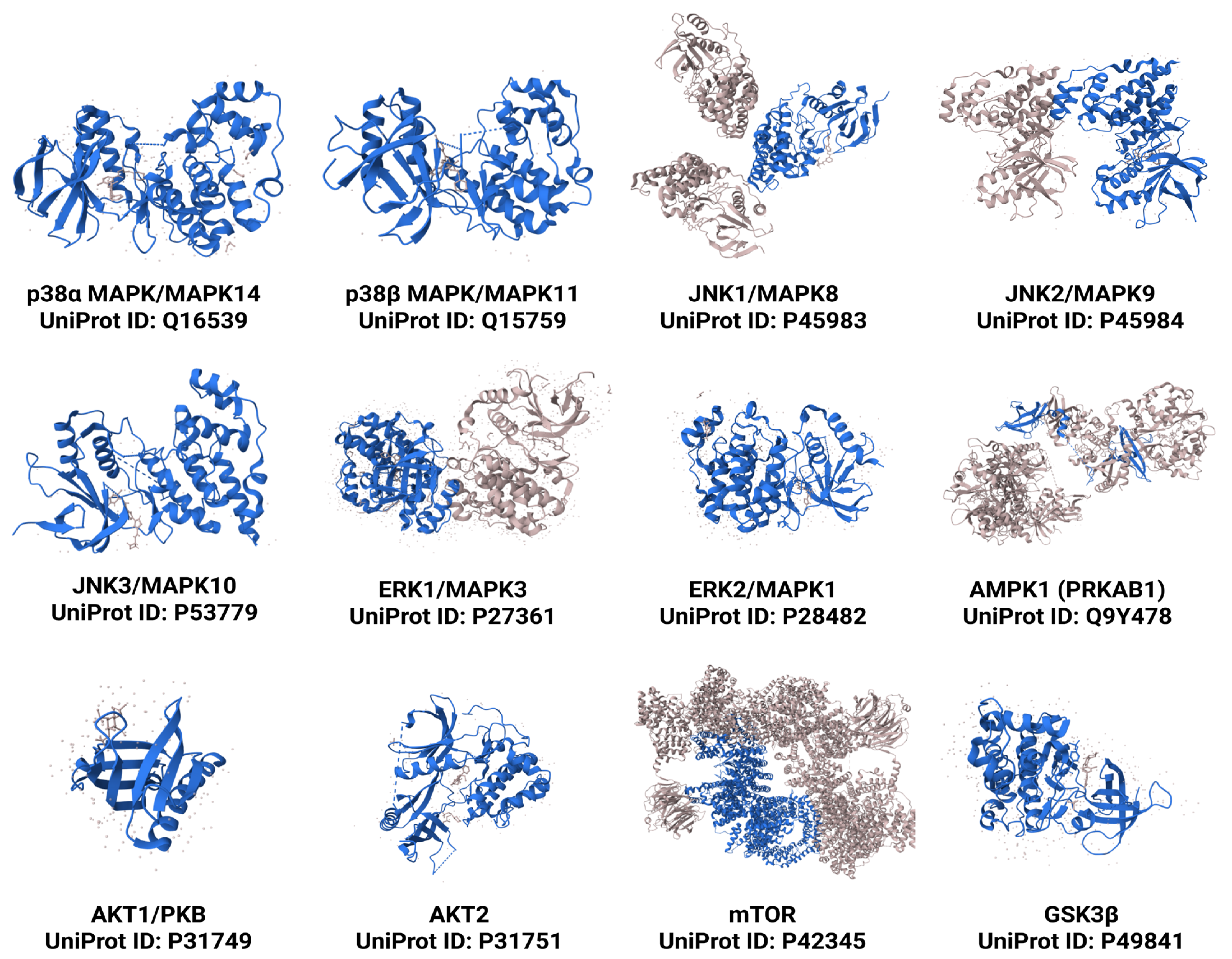
Figure 1. 3D structures of key protein kinases involved in NAFLD and NASH progression, retrieved from PDBe-KB (Protein Data Bank in Europe—Knowledge Base). Observed regions are shown in darker shades, while unobserved regions appear semi-transparent.
Kinasesphosphatases 03 00016 g001
Figure 2. Impact of FXR activation on glucose, lipid, and immune homeostasis. Figure 2 illustrates the diverse effects of FXR activation on metabolism and inflammation. FXR activation modulates glucose homeostasis and improves insulin sensitivity by regulating key enzymes, and it enhances lipid profiles by inhibiting cholesterol accumulation and promoting fatty acid oxidation. It also impacts bile acid metabolism by SHP modulation of CYP7A1/CYP8B1. Moreover, FXR activation exerts anti-inflammatory effects by regulating cytokine production in macrophages, dendritic cells, and natural killer cells (NK cells), thereby reducing systemic inflammation. Upward arrow indicates overexpression, while downward arrow indicates lower expression.
Kinasesphosphatases 03 00016 g002
Figure 3. Impact of FXR dysregulation and therapeutic activation on NAFLD/NASH pathogenesis.This schematic illustrates the contrasting effects of FXR dysregulation (red arrows) and therapeutic FXR activation (green dashed arrows) on liver health and the progression of NAFLD/NASH. FXR dysregulation promotes key pathogenic features, including increased hepatic fat accumulation, oxidative stress, inflammation, insulin resistance, fibrosis progression, and disrupted gut–liver communication. In contrast, therapeutic activation of FXR exerts protective effects by reducing steatosis, oxidative stress, and inflammation, improving insulin sensitivity and glucose balance, attenuating fibrogenesis, and restoring gut–liver axis homeostasis. Together, these opposing pathways highlight FXR’s central role in liver metabolic regulation and its potential as a therapeutic target in NAFLD/NASH. Solid lines indicate the effects of FXR dysregulation, while dotted lines represent the impact of therapeutic FXR activation.
Kinasesphosphatases 03 00016 g003
Figure 4. FGF19 signaling and FXR agonist effects in NAFLD/NASH pathogenesis and potential therapeutic targets. Figure 4 outlines the complex relationship between FGF19 signaling and FXR activation in the context of NAFLD and NASH. FGF19, secreted from the intestines (Enterocytes) upon FXR activation by bile acids, binds to its receptor FGFR4 on hepatocytes, where Klotho beta (KLB) is a co-receptor. This stimulates an array of signaling pathways including RAS/RAF/MEK/ERK, PI3K/AKT, and other pathways, ultimately influencing fat deposition, insulin sensitivity, and glucose uptake. FXR agonists, like obeticholic Acid (OCA), EDP-305, tropifexor, cilofexor, and MET409 (EYP001), target FXR in both hepatocytes and enterocytes. FXR activation later influences bile acid synthesis (CYP7A1) and impacts NAFLD/NASH pathogenesis and NAFLD/NASH regression through several pathways, including AMPK activation. Green arrows represent the direction of change following FXR agonist treatment, while red arrows indicate overexpression or activity in the NAFLD/NASH condition.
Kinasesphosphatases 03 00016 g004
Table 1. Summary of key clinical trials on FXR agonists in NAFLD/NASH.
FXR Agonist and StructureTrial Name/Phase/ApprovalMechanism of ActionAdverse EffectsRefs.
Kinasesphosphatases 03 00016 i001
Obeticholic Acid (OCA)
FLINT Trial (Multicenter, Randomized,
Placebo-Controlled)

Accelerated approval (2016) for primary biliary cholangitis, full approval pending
  • Activation of hepatic FXR
  • Increased SHP expression
  • Decreased CYP7A1/CYP8B1 and bile acid synthesis
  • Reduced hepatic lipogenesis
  • Increased β-oxidation
  • Anti-inflammatory effects
  • Insulin-sensitizing effects
Pruritus, ↑ LDL, ↓ HDL cholesterol[53,54]
Kinasesphosphatases 03 00016 i002
Cilofexor
Phase II, Randomized Controlled Trial
  • Nonsteroidal FXR agonist
  • Modulates bile acid metabolism and lipid/glucose pathways
  • Reduces de novo lipogenesis (DNL) and increases lipid clearance
  • Downregulates inflammatory genes
Mild-to-moderate pruritus, GI disturbances[55]
Kinasesphosphatases 03 00016 i003EDP305Phase II, Dose-Ranging Study
  • Highly selective FXR agonist
  • Modulates FXR in liver and intestine
  • Regulates bile acid and lipid metabolism
  • Exhibits anti-inflammatory and antifibrotic effects
Pruritus (dose-dependent)[56]
Kinasesphosphatases 03 00016 i004
Tropifexor
FLIGHT-FXR, Phase II
  • Potent nonsteroidal FXR agonist
  • Targets bile acid metabolism, inflammation, and fibrosis
Pruritus, GI upset, transient ↑ LDL[57]
Kinasesphosphatases 03 00016 i005
Vonafexor
Phase IIa, Proof-of-Concept Study
  • Selective FXR agonist
  • Improves bile acid homeostasis and metabolic control
Mild pruritus, fatigue[58]
Kinasesphosphatases 03 00016 i006
MET409
Phase I/II, Dose-Escalation Study
  • Highly potent non-bile-acid FXR agonist
  • Targets bile acid synthesis and metabolic pathways
Mild pruritus, headache[59]
↑ (up arrow) means increase or upregulation; ↓ (down arrow) means decrease or downregulation.
Table 2. Impact of second drugs on protein kinases in combination with FXR agonists in clinical trials.
NameFXR Agonist2nd DrugImpact of 2nd Drug on Protein KinasesReferences
CONTROL
Phase 2 (NCT02633956)
Obeticholic Acid (OCA)AtorvastatinHMG-CoA reductase inhibitor that activates AKT pathway[93]
ELIVATE
Phase 2
(NCT04065841)
TropifexorLicoglifozinIndirectly targets multiple kinases[96]
ATLAS
Phase 2
(NCT03449446)
CilofexorFirsocostat,
Selonsertib
Selonsertib inhibiting apoptosis signal-regulating kinase 1 (ASK1)[98]
Phase 2 (NCT04702490)MET409EmpagliflozinActivates AMPK[94]
Disclaimer/Publisher’s Note: The statements, opinions and data contained in all publications are solely those of the individual author(s) and contributor(s) and not of MDPI and/or the editor(s). MDPI and/or the editor(s) disclaim responsibility for any injury to people or property resulting from any ideas, methods, instructions or products referred to in the content.