Article

Porphyra sensu lato Species as Source for Biological UV Photoprotectors and Antioxidants to Develop Cosmeceutical Products


by
Débora Tomazi Pereira
1,*,
Julia Vega
1,
José Bonomi Barufi
2,
Nathalie Korbee
1 and
Félix L. Figueroa
1
1
Department of de Ecology and Geology, University Institute of Blue Biotechnology and Development (IBYDA), University of Malaga, Campus of Teatinos s/n, 29071 Málaga, Spain
2
Phycology Laboratory, Federal University of Santa Catarina, Florianópolis 88040-535, Brazil
*
Author to whom correspondence should be addressed.
Phycology 2026, 6(2), 59; https://doi.org/10.3390/phycology6020059
Submission received: 1 April 2026 / Revised: 15 May 2026 / Accepted: 26 May 2026 / Published: 28 May 2026
(This article belongs to the Special Issue Development of Algal Biotechnology, Second Edition)

Abstract

Porphyra sensu lato is a promising source of natural photoprotective and antioxidant compounds for cosmeceutical applications. This study evaluated the geographical and temporal variability of bioactive compounds in Porphyra and Pyropia spp. from Chile and Spain and assessed their applicability in topical photoprotective formulations. Antioxidant capacity, soluble polyphenols, phycobiliproteins, and mycosporine-like amino acids (MAAs) were quantified in different species, collection sites, and sampling periods. In parallel, selected Pyropia elongata extracts were incorporated into water-in-oil creams, alone or combined with vegetable oils and physical UV filters, and in vitro SPF/UVAPF values were determined using the PMMA plate transmittance method. Marked variability was observed among species, sites, and collection dates. In P. elongata, total MAAs ranged approximately from 3 to 6 mg.g−1 DW, while porphyra-334 was the dominant MAA, accounting for about 70–100% of total MAAs across samples. A positive correlation was found between soluble polyphenol concentration and ABTS antioxidant activity. In the formulations, Pyropia extracts increased absorbance mainly in the UVB–UVA range and improved SPF and UVA-related protection, particularly when combined with karanja and calophyll oils and physical filters. These results support Porphyra sensu lato biomass as a flexible natural reservoir for developing sustainable bio-based photoprotective products.

1. Introduction

The red macroalgae Porphyra sensu lato, widely known as Nori, is considered as one of the most economically important and extensively cultivated algae worldwide, particularly in Asian countries [1,2,3,4]. The term Porphyra sensu lato is used here to refer to the broader group of Bangiales species historically assigned to Porphyra, including taxa currently classified within related genera such as Pyropia and Neopyropia. Molecular studies based on the genes encoding the large subunit of RuBisCO and the small ribosomal subunit concluded that all Porphyra species reported in Brazil actually belong to the genus Pyropia [5]. Both genera belong to the order Bangiales and share very similar morphological characteristics, differing mainly in genetic traits. In addition to Brazil, Pyropia has also been reported in Japan, Australia, Mexico, and the Mediterranean Sea [5]. Its relevance is due not only to its nutritional value, being a rich source of proteins, iron, and vitamins [6], but also to its abundance in bioactive compounds (BACs) with significant health benefits. These compounds include nitrogenous substances such as phycobiliproteins, proteins, and mycosporine-like amino acids (MAAs), as well as non-nitrogenous components like polysaccharides and polyphenols [7,8]. Among these compounds, MAAs, phycobiliproteins, and polyphenols are particularly relevant to this study because they contribute to UV absorption, antioxidant capacity, and biological photoprotection [9,10,11,12]. Among them, MAAs have attracted special attention due to their ability to absorb UV radiation and their potential application as eco-friendly sunscreens, in addition to their antioxidant and anti-inflammatory properties [13,14,15,16,17]. Considering all these advantages, Porphyra has emerged as a promising resource for both the food and pharmaceutical industries, stimulating growing scientific interest.
In its natural habitat, Porphyra sensu lato thrives in the supralittoral zone, where it is exposed to extreme and fluctuating conditions such as high light intensity, osmotic stress, desiccation, and UV radiation [18,19]. These environmental challenges shape its biochemical composition and highlight its natural adaptability [2]. One contributing factor to this resilience is its high surface-area-to-volume ratio, which facilitates efficient nutrient absorption and supports rapid growth and BACs production [8,20]. These characteristics not only enable Porphyra to perform well in dynamic marine ecosystems but also make it a compelling candidate for biotechnological applications such as bioremediation and integrated multi-trophic aquaculture (IMTA) systems [21].
The species’ adaptability is closely linked to its biochemical plasticity, particularly in the production of BACs in response to abiotic stressors. Laboratory experiments have shown that Porphyra’s biochemical profile can be modulated by environmental cues. For instance, reintroduction of nitrate after a nutrient-deprivation period in cultivation systems led to a twofold increase in both MAAs and soluble proteins, accompanied by enhanced antioxidant activity [8]. Likewise, exposure to blue light significantly increased the accumulation of MAAs, phycobiliproteins, and total proteins in Porphyra leucosticta [22], while UVA radiation boosted MAA content by 29% in Porphyra columbina [23]. Based on these responses, MAAs are expected to covary mainly with UV radiation, light intensity, and nitrogen availability, reflecting their role as UV-screening and nitrogen-containing compounds. Phenolic compounds are expected to be more closely associated with oxidative stress-related drivers, such as high irradiance, desiccation/emersion stress, salinity fluctuations, and other conditions that enhance reactive oxygen species formation. In contrast, phycobiliproteins are expected to covary primarily with nitrogen availability and light quality or intensity, as they are nitrogen-rich photosynthetic antenna pigments involved in photoacclimation. Among these BACs, some MAAs may be constitutively produced, whereas others are facultatively upregulated in response to UV radiation or nutrient availability, demonstrating their functional plasticity and ecological significance [24].
These observations suggest that specific geographic regions, particularly those characterized by intense UV exposure and/or natural nutrient upwelling, may favor the occurrence of Porphyra strains enriched in valuable BACs, offering a promising strategy for their biotechnological exploitation in the pharmaceutical and food sectors. Therefore, identifying suitable collection sites and harvesting periods represents a relevant bioprospecting strategy for selecting biomass with higher photoprotective and antioxidant potential. Although extraction conditions can further influence BAC recovery [25] and bioavailability, extraction optimization was not the focus of the present study.
Thus, it is important to know the natural variability of BACs, such as phenolics, pigments, and MAAs, is essential to determine how species identity, collection site, and harvesting period influence the photoprotective and antioxidant potential of Porphyra sensu lato biomass. This variability may directly affect the selection of extracts for cosmeceutical formulations and their performance in terms of UV absorption, antioxidant capacity, and in vitro SPF/UVAPF. MAAs and other antioxidant compounds derived from Porphyra biomass are increasingly explored for use in cosmeceutical products. Notable examples include Helioguard™365, developed by Mibelle Biochemistry Group, and Helionori®, produced by Gelyma Group, both using P. umbilicalis extracts of which leverage the UV-protective and anti-aging properties. These examples illustrate the commercial interest in Porphyra-derived ingredients for cosmetic formulations. However, their broader application still depends on overcoming key challenges related to compound stability, formulation compatibility, reproducible photoprotective performance, and regulatory safety and efficacy testing.
In this context, the present study aims to deepen our understanding of the seasonal and geographic variability in the photoprotective and antioxidant compounds profile of Porphyra spp. from Chile and Spain. In this sense, we hypothesized that species identity, collection site, and harvesting period would significantly influence total MAAs, MAA composition, phycobiliproteins, phenolic compounds, and antioxidant capacity. We also hypothesized that the incorporation of Pyropia extracts into water-in-oil creams would increase UV absorbance and improve in vitro photoprotective performance, particularly SPF, UVAPF, and UVA-related biological effective protection factors. By linking biochemical diversity to environmental conditions and optimizing extraction protocols, this research provides a foundation for the development of innovative, bio-based photoprotective products with potential applications in skin care and anti-aging treatments.

2. Material and Methods

2.1. Samples

The gametophytes of Porphyra sensu lato (Porphyra columbina, Porphyra dioica, Porphyra linearis, Pyropia elongata, and Neopyropia leucosticta—species identification was performed by the BIOMOL Scientific Service at the University of Las Palmas de Gran Canaria using DNA barcoding. Genomic DNA was extracted using the CTAB method, and the raw sequences were edited with Sequencing Analysis Software v. 6.0 for Sanger sequencing and aligned using Geneious v. 7.1. The resulting sequences were compared against international molecular identification databases, including NCBI GenBank/BLAST and BOLD Systems, were collected at different sites and times (Table 1 and Table 2) and were transported to the laboratory in plastic bags filled with seawater and kept in a thermal box. The sampling areas are located in similar latitudes as the Northern and Southern Hemispheres (Table 1). For each collection site and sampling date, the biological sample consisted of pooled biomass (approximately 100 g fresh biomass) obtained from multiple healthy thalli collected from the same population. Immediately upon arrival at the laboratory, the thalli were rinsed with diluted seawater; healthy portions were freeze-dried and then ground using a coffee grinder to obtain a powder (≤1 mm). Dried ground algal tissues were used for various analyses, including evaluation of bioactive compounds (phycobiliproteins, MAAs and phenolic compounds) and also for antioxidant capacity assays. Moreover, algal dried material was utilized to prepare and evaluate photoprotection cosmeceutical formulations. These procedures are detailed further below. With the obtained samples, two different comparisons were performed: (i) comparison among Porphyra spp. collected in different regions (Table 1); and (ii) temporal variability of Pyropia elongata collected in Torre del Pino Beach, Malaga (Southern Iberian Peninsula) (Table 2). Pyropia elongata was selected because it occurs locally in Málaga, providing more accessible biomass for the study.
The global radiation (350–1500 nm) of each collection site was obtained from the Spanish Agency of Meteorology (AEMET), the Galician Meteorological Service (MeteoGalicia) and from the Chilean Meteorological Service (MeteoChile). In the case of Spain, the data came from Malaga (36°43′05″ N, 4°28′53″ W), Almería (36°50′47″ N, 2°12′52″ W), Cádiz station (36°29′59″ N, 6°15′28″ W) and A Coruña (43°23′10″ N, 8°24′38″ W). In the case of Chile, data were obtained from Concepción (36°46′ S, 73°03′ W) and Concon (32°55′ S, 71°31′ W). The monthly mean of daily values was calculated for the sampling month and the previous one.
The air temperature near the sampling area was obtained from the same stations. Only in the case of Andalucia, values were obtained from the stations of Junta de Andalucia in Institute for Agricultural and Fisheries Research and Training (IFAPA) in Málaga, Vélez-Málaga, Tarifa and Almería.

2.2. Phycobiliproteins

For phycobiliproteins determination, 20 mg of dry weight (DW) were homogenized in 1 mL of phosphate buffer (pH 6.5, 0.1 M). After overnight incubation at 4 °C, samples were centrifuged (2721.6× g; 15 min) (Heraeus Labofuge 400R, Thermo Scientific, Waltham, MA, USA), and the supernatant was collected and measured using a spectrophotometer (UV-2600, Shimadzu, Duisburg, Germany) at the following wavelengths: 455, 564, 592, 618, 645 and 730 nm. Quantification (mg·g−1 DW) was made using the chromatic formulas described [26] for phycoerythrin (PE) and phycocyanin (PC). After, the ratio between both phycobiliproteins was calculated (PE:PC ratio).
P E = [ ( A b s 564 A b s 592 ) ( A b s 455 A b s 592 ) × 0.20 ] × 0.12
P C = [ ( A b s 618 A b s 645 ) ( A b s 592 A b s 645 ) × 0.51 ] × 0.15

2.3. Phenolic Compounds

The Folin–Ciocalteu reducing capacity of the aqueous buffer-soluble fraction was determined according to [27], with slight modifications. Briefly, 20 mg of dry weight (DW) was extracted with 1 mL of phosphate buffer (0.1 M, pH 6.5) and left overnight at 4 °C. After centrifugation, 100 μL of the extract was mixed with 700 μL of distilled water and 50 μL of Folin–Ciocalteu reagent (Sigma-Aldrich, St. Louis, MO, USA). The mixture was vortexed, followed by the addition of 150 μL of 20% Na2CO3, and vortexed again. Samples were then incubated for 2 h at 4 °C, and absorbance was measured at 760 nm using the spectrophotometer described above. Gallic acid (Sigma-Aldrich, St. Louis, MO, USA) was used as the standard.

2.4. Mycosporine-like Amino Acids (MAAs)

MAAs were determined using uHPLC according to [23], with some modifications based on [28]. Briefly, 20 mg of dry weight (DW) was extracted in 1 mL of 20% methanol in water and incubated for 2 h at 45 °C in a water bath. Samples were then centrifuged, and 700 μL of the supernatant was dried under vacuum (Speed-Vac SPD210, Thermo Scientific, Waltham, MA, USA). The dried extracts were re-suspended in 700 μL of ultrapure H2O, filtered through a 0.2 μm membrane filter, and injected into the uHPLC system (1260 Agilent InfinityLab Series, Santa Clara, CA, USA). MAAs were separated using a Luna C8 column (5 µm, 250 mm length, 4.6 mm diameter; Phenomenex, Aschaffenburg, Germany) maintained at 20 °C, with an injection volume of 10 µL, under isocratic conditions at a flow rate of 0.5 mL min−1, with a mobile phase consisting of 1.5% methanol and 0.15% acetic acid in ultrapure H2O. Detection was performed using a photodiode array detector at 330 nm. MAAs isolated by HPCCC [29] were used as standards. Quantification was carried out using the molar extinction coefficients (ε) of the different MAAs [30]. Results were expressed as mg g−1 DW.

2.5. Antioxidant Capacity (ABTS Assay)

The ABTS assay was performed according to [31], with slight modifications. ABTS radical cations (ABTS•+) were generated by reacting 7 mM ABTS (2,2′-azinobis(3-ethylbenzothiazoline-6-sulfonic acid); Sigma-Aldrich, St. Louis, MO, USA) with 2.45 mM K2S2O8 in phosphate buffer (0.1 M, pH 7.0). The reaction mixture was kept at room temperature for 12–16 h in the dark to allow complete radical formation. The ABTS•+ solution was then diluted with phosphate buffer (1:30) until an absorbance of 0.75 ± 0.05 at 727 nm was reached. The same extracts used for phycobiliprotein and phenolic compound quantification were also used for this assay. For the reaction, 50 μL of extract was mixed with 950 μL of diluted ABTS•+ solution. The mixture was incubated for 8 min at room temperature in the dark, and absorbance was measured at 727 nm. The antioxidant compound concentration was calculated using a standard curve of Trolox (6-hydroxy-2,5,7,8-tetramethylchroman-2-carboxylic acid), (Sigma-Aldrich) (20 to 100 µg mL−1—R2 = 0.99; y = 15.8928×) and the results were expressed as µmol of Trolox equivalent antioxidant capacity (TEAC) per g of DW.

2.6. Development of Cosmeceutical Formulations

A water-in-oil (W/O) base cream formulation was elaborated according to the requirements of natural cosmetics with the aim of evaluating the potential photoprotective properties of Pyropia elongata extract. The cosmetic formula used in the study is shown in Supplementary Table S1. Pyropia elongata was selected because it occurs locally in Málaga, providing more accessible biomass for the study. Three different approaches were followed in the design of the creams: (i) combination of different oils in the lipid phase, such as karanja (Aroma-Zone, Spain) and calophyll (Camassia, Spain), to enhance UV-absorbing capacity and bioactivities; (ii) addition of Pyropia elongata extracts to the formulation in different percentages (5, 10 and 15%) to assess dose-dependent photoprotective performance; and (iii) addition of physical filters (5% of titanium dioxide and 2.5% of zinc oxide) to the formulation and its combination with the best Pyropia elonagata extract (10%).
For the Pyropia extract used in the formulation, 20 g of dry weight was homogenized in 1 L of distilled water. The biomass was disrupted with the help of a blender, incubated for 2 h at 45 °C in a water bath, concentrated in a rotoevaporador and freeze-dried to obtain a powder that was included in the creams.

2.7. Photoprotection Assay of the Creams

Different photoprotection factors (PPFs) were determined: sun protection factor (SPF), UVA protection factor (UVAPF) and biological effective protection factors (BEPFs). For the determination of all photoprotection factors, creams were spread in PMMA (polymethyl methacrylate) plates as suggested in ISO 24443 (2012) [32] and ref. [33]. PMMA plates (Schönberg, Hamburg, Germany) presented a roughness (Ra) of 6 µm and a surface of 50 mm × 50 mm. Creams were spread at a concentration of 1.2 mg cream·cm−2 (32.5 mg over the total surface of the plate) using a gloved fingertip (previously saturated with the cream) and for no more than 1 min. After 15 min of incubation at room temperature and darkness, transmittance through the plates was measured using a spectrophotometer (UV-2600, Shimadzu, Duisburg, Germany) with an integrated sphere (ISR-2600Plus, Shimadzu, Duisburg, Germany). The transmittance (T) values were converted to absorbance (A) values as A = −log(T). Three plates were spread for each cream and two measurements per plate were carried out.
The in vitro SPF was calculated using the erythematic action spectrum [34] as described in [35] with modifications. The in vitro UVAPF was calculated using the persistent pigment darkening (PPD) action spectrum [35], as described in ISO 24443 (2012), with some modifications. The BEPFs were calculated as described by [36] against different action spectra: photocarcinogenesis [37], immunosuppression [38], 1980), elastosis [39], singlet oxygen formation [40] and photoaging [41]. The following formula was used in all cases, changing the action spectrum depending on the PPF calculated:
PPFs   =   λ = 290 λ   = 400 Act . Sp ( λ )   ×   I ( λ )   ×   d ( λ ) λ = 290 λ = 400 Act . Sp ( λ )   ×   I ( λ )   ×   10 Abs ( λ )   ×   d ( λ )
where:
  • Act.Sp (λ) = action spectra (normalized from 0 to 1);
  • I (λ) = spectral irradiance of a sunny midday in summer in Malaga (W·m−2);
  • d (λ) = wavelength step (1 nm);
  • Abs (λ) = absorbance values (normalized from 0 to 1).
The critical lambda (λc) was also calculated as the wavelength (from 290 to 400 nm) at which the creams absorbed 90% of the radiation [30].
As a control a commercial sunscreen was used. The sunscreen ALGA MARIS® SPF50 (Laboratoires de Biarritz, Biarritz, France), was selected, as it is a cream with physical filters and algae extract, similar to the one that we have developed.

2.8. Statistical Analysis

Data normality and homogeneity of variances were assessed using the Shapiro–Wilk and Bartlett tests, respectively. Data were considered suitable for parametric analysis when both tests were non-significant (p ≥ 0.05). For each collection site and sampling date, three independent pooled biomass samples were prepared from multiple healthy thalli collected from the same population and treated as biological replicates. Subsequently, a one-way ANOVA was applied, considering the species combined with place and time of collection as the factor of influence, totaling eight combinations to evaluate Porphyra spp. In the case of the temporal assessment of P. elongata, time of sampling was the independent variable, containing seven levels. When significance was observed, ANOVAs were followed by Tukey’s post hoc test (p ≤ 0.05) using the Statistica software package (Release 7.0). After, dependent variables were submitted to Pearson correlation analyses.

3. Results

3.1. Comparison of Different Porphyra sensu lato Species

Through a screening of various Porphyra sensu lato species collected across different regions of Chile and Spain, antioxidant activity (ABTS assay) ranged from 3.60 to 16.82 µmol TEAC·g−1 DW. Neopyropia leucosticta collected in Lagos (May 2022) showed the highest activity, whereas the lowest activity was observed in N. leucosticta from La Araña (May 2024) (Table 3).
Total soluble polyphenols peaked in P. dioica from La Coruña, followed by P. columbina from Concepción, P. linearis from Tarifa, and N. leucosticta from Guadalmar and Lagos (May 2022). In contrast, the lowest polyphenol contents were recorded for N. leucosticta from La Araña and Almería (Table 3). Additionally, a positive Pearson correlation between polyphenol concentration and antioxidant activity (ABTS) was observed (r = 0.5590, p = 0.02, n = 24).
Regarding phycobiliproteins, phycoerythrin (PE) ranged from 1.23 to 8.26 mg·g−1 DW, with the highest value in P. linearis from Tarifa, and the lowest PE values were found in N. leucosticta from Almería and from La Araña. Considering the phycocyanin (PC), it ranged from 0.21 to 3.86 mg·g−1 DW, reaching its maximum in N. leucosticta from Guadalmar and remaining high in P. columbina from Concon and P. linearis from Tarifa. The average PE:PC ratio was highest in N. leucosticta from Almeria and lowest in N. leucosticta from Lagos (May 2022) (Table 3).
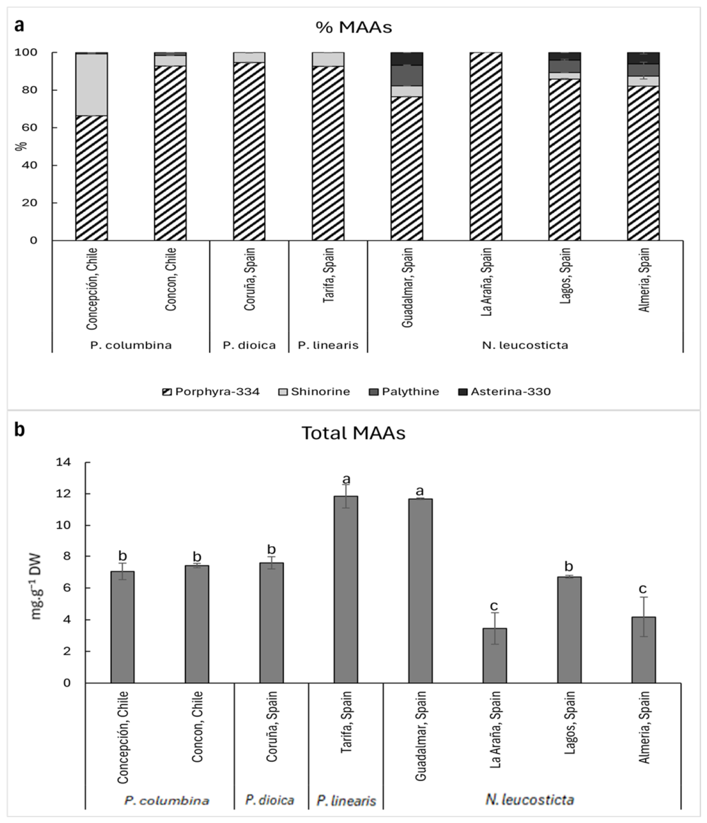
Across the screened Porphyra sensu lato samples, MAA profiles were consistently dominated by porphyra-334, which accounted for the majority of total MAAs in all cases (approximately 70–100%). Shinorine was generally the second most abundant compound, showing a marked contribution in P. columbina from Concepcion (March 2022). In contrast, palythine and asterina-330 were usually minor components, but their relative contribution increased in some N. leucosticta samples (notably Lagos and Guadalmar, May 2022, and Almeria, February 2025) (Figure 1a). The concentrations of individual MAAs, expressed as mg g−1 DW, are presented in Supplementary Table S2.
The total MAA content (mg·g−1 DW) differed significantly among samples, where the highest total MAAs were observed in P. linearis from Tarifa ( March 2022) and N. leucosticta from Guadalmar (May 2022), whereas the lowest total MAAs were recorded for N. leucosticta from La Araña (May 2024) and Almería (February 2025) (Figure 1b).
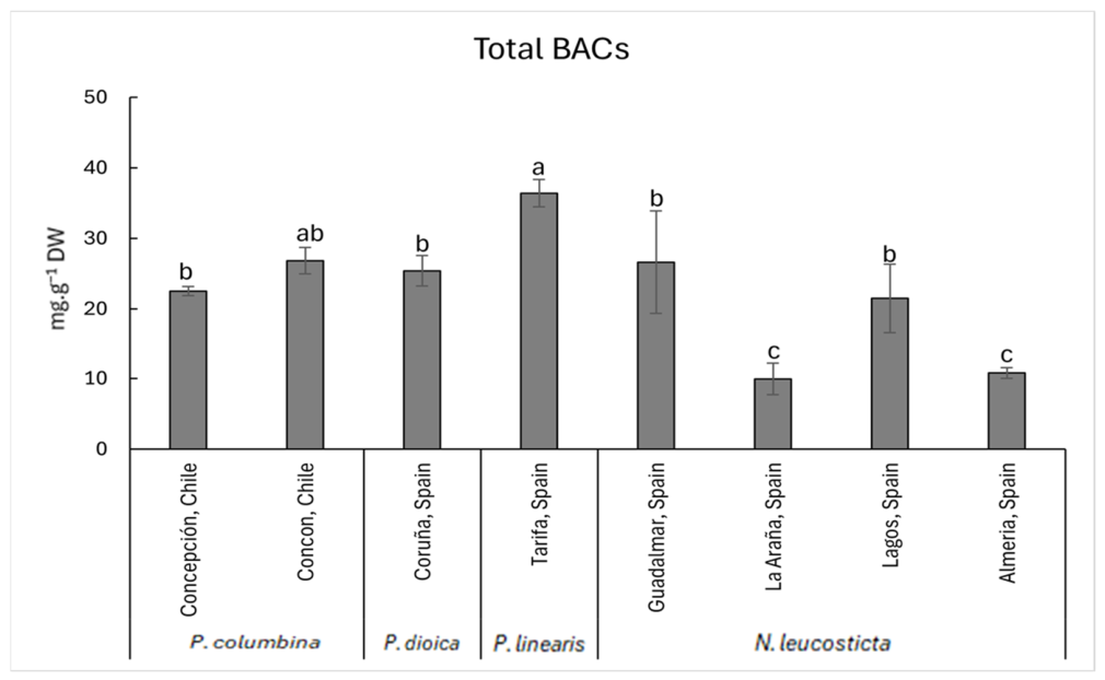
The total BAC index, calculated as the sum of the quantified compound classes, was used as an exploratory descriptor of the overall bioactive compound pool. The content of total bioactive compounds (BACs: MAAs + Polyphenols + Phycobiliproteins) also differed significantly among samples (Figure 2). The highest BAC level was detected in P. linearis from Tarifa (March 2022), whereas the lowest values were observed in N. leucosticta from La Araña (May 2024) and Almeria (February 2025) (Figure 2).

3.2. Temporal Variability of Pyropia elongata in Torre del Pino Beach, Malaga (Spain)

Considering the different collection dates of Pyropia elongata from Torre del Pino Beach, Spain, the antioxidant activity showed significant temporal variability. ABTS values ranged from 2.50 to 16.53 µmol TEAC·g−1 DW, reaching the highest level in May 2025, and the lowest activities occurred in March 2023, January 2024, and March 2024 (Table 4).
The total polyphenols ranged from 3.72 to 10.25 mg·g−1 DW, peaking in May 2022. Samples from June 2022, January 2024, and March 2024 exhibited intermediate polyphenol levels, while the lowest contents were recorded in March 2023, May 2024, and May 2025 (Table 4).
The phycobiliproteins also varied significantly. The PE ranged from 0.25 to 2.19 mg·g−1 DW, with maximum values in March 2024 and May 2022, and the minimum amount was detected in March 2023. The PC ranged from 0.04 to 2.13 mg·g−1 DW, being highest in May 2022, and the lowest value was in March 2023. Consequently, the PE:PC ratio varied markedly, with the highest ratio in March 2023 and the lowest ratio in June 2022 (Table 4).
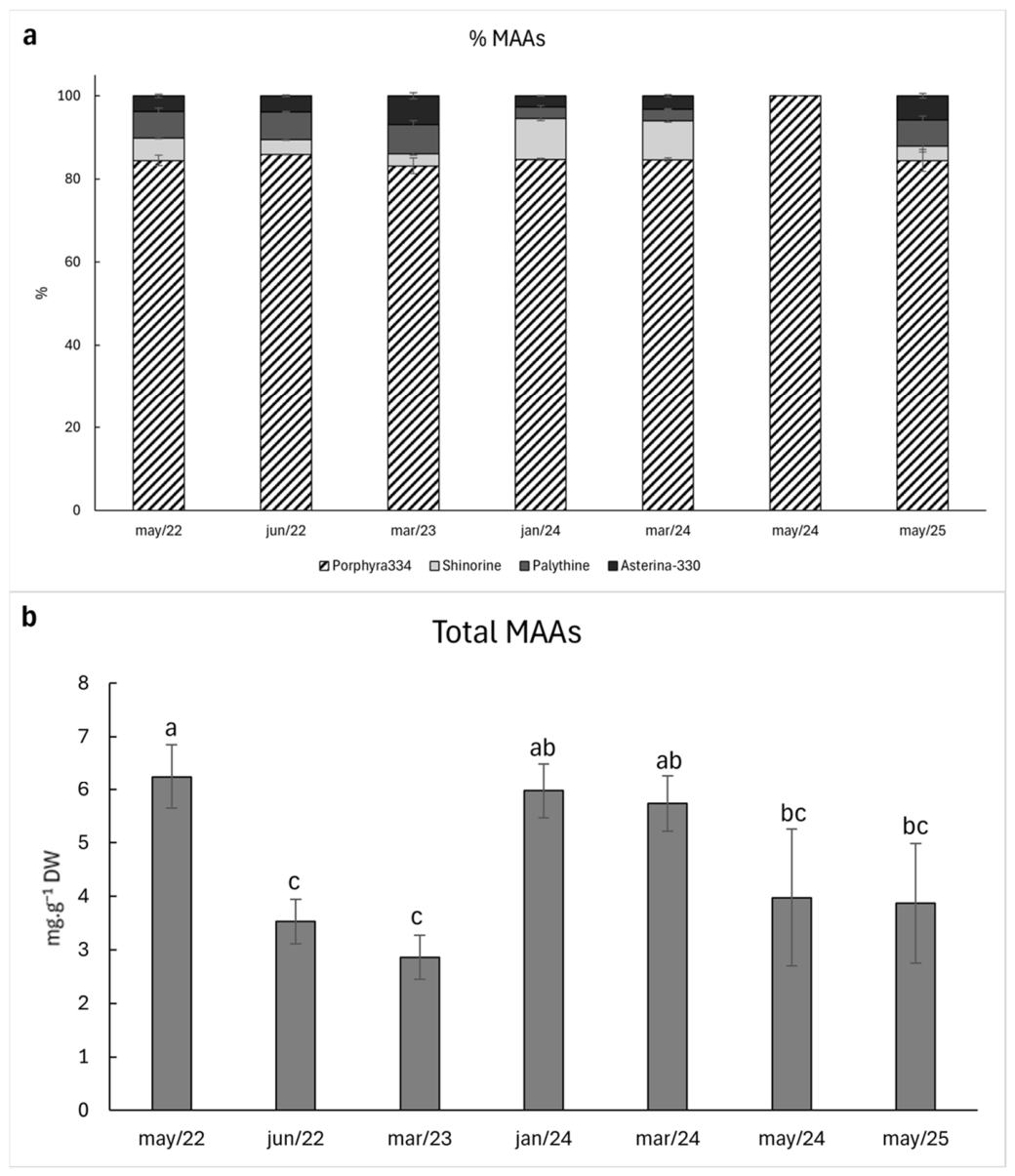
The MAA profile of Pyropia elongata from Torre del Pino Beach, Spain, from different collection dates was consistently dominated by porphyra-334, which represented the largest proportion of the total MAAs in all samples. Shinorine generally contributed a secondary fraction, with a more noticeable relative contribution in January and March 2024 samples, whereas palythine and asterina-330 remained minor components and varied only slightly among dates. Notably, the May 2024 sample was composed almost exclusively of porphyra-334, with negligible contributions from the other MAAs (Figure 3a).
The total MAA content differed significantly among collection dates, ranging approximately from 3 to 6 mg·g−1. The highest total MAAs were observed in May 2022, while January 2024 and March 2024 showed similarly high intermediate values. In contrast, June 2022 and March 2023 presented the lowest total MAA concentrations (Figure 3b). The concentrations of individual MAAs, expressed as mg g−1 DW, are presented in Supplementary Table S2.
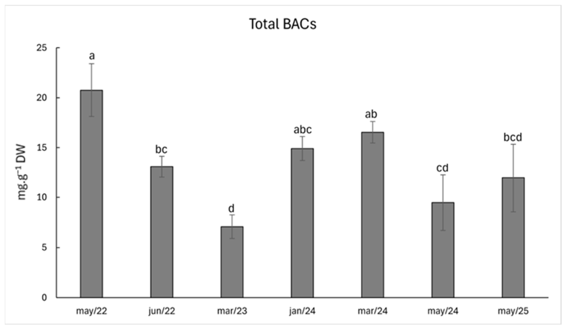
The total BAC content varied significantly among sampling periods. The highest BAC value was observed in May 2022, whereas the lowest value was recorded in March 2023 (Figure 4).

3.3. Development of Cosmeceutical Formulations

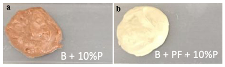
Different creams were designed to confirm the photoprotective potential of Pyropia extracts. The addition of the extracts gives them a brownish color that varies in intensity depending on the extract concentration (Figure 5a). The addition of physical filters bleached the color of the creams, becoming a more yellow-brownish color (Figure 5b).
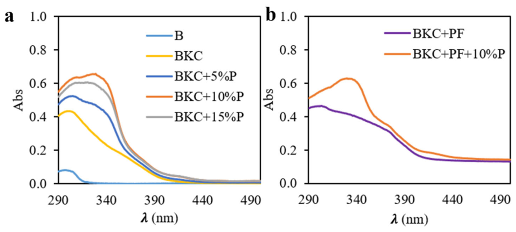
The absorbance spectra of the formulated creams are presented in Figure 6. The base cream with karanja and calophyl oil (BKC) showed higher absorbance values compared with the base cream, particularly in the UV-B region, with a smaller increase also observed in the UV-A range. When physical filters (PF) were incorporated into the formulation, absorbance further increased in the UV-A and visible regions (Figure 6). The inclusion of Pyropia extract produced an additional increase in absorbance mainly between 290 and 350 nm (Figure 6).
The photoprotection factors obtained from the different creams are summarized in Table 5 and Table 6. The base cream that only presented coconut oil (B) showed very low values in the different photoprotection factors (<1.5). The addition of karanja and calophyll oils (BKC) increased the SPF to 4.2 ± 0.7, while the incorporation of physical filters (BKC + PF) resulted in an SPF of 5.8 ± 0.8; however, these differences were not statistically significant. In contrast, the UVAPF increased significantly from 1.0 ± 0.01 in BKC to 4.0 ± 0.5 in BKC + PF. Among the remaining photoprotection parameters, only those associated with UVA effects (elastosis and photoaging) showed significant differences between these two formulations. For the creams with different percentages of Pyropia extract, SPF values ranged from 6.8 ± 0.4 to 9.8 ± 2.3 when 5% and 15% of the extract were incorporated, respectively. No statistically detectable differences were observed among extract percentages for the evaluated photoprotection factors; however, this result does not imply equivalence among extract concentrations. Compared with the BKC formulation, statistically significant increases were observed only in some cases, namely SPF, UVAPF, and elastosis, when 10–15% Pyropia extract was added.
The cream with physical filters and 10% of Pyropia extract did not show significant differences with the cream without physical filters in the photoprotection factors related to UV-B (SPF, photocarcinogenesis and immunosuppression), whereas significant differences were observed in those related to UV-A (UVAPF, elastosis, singlet oxygen formation and photoaging), i.e., the UVAPF increased from 3.17 ± 0.37 to 6.03 ± 0.76 when physical filters were added. Regarding the ratio SPF/UVAPF, it increased when the different extracts were added, and the highest ratio (2.9) was observed in the BKC + 15% Pyropia. Finally, the critical lambda varied from 308 in the base cream, to 363 nm with Pyropia extract, and to 376 with the addition of the physical filter.
The reference sunscreen showed an SPF value that agree with the labeled SPF and the UVAPF was higher than that obtained with our creams (9.87), so the ratio SPF/UVA was also higher (5.07), and the critical lambda was 370 nm.

4. Discussion

After the biochemical analysis of different Porphyra species collected in this study, a geographical variability was observed in the antioxidant and photoprotective profiles of Porphyra sensu lato from Chile and Spain.
Despite the similar latitudinal range of some collection sites in Chile and Spain, the sampled regions may differ in broader oceanographic and atmospheric conditions. For example, the Chilean Pacific coast is influenced by the Humboldt Current and coastal upwelling, whereas the Mediterranean coast of southern Spain is generally characterized by warmer and more oligotrophic waters. In addition, the opposite seasonality between the Northern and Southern Hemispheres may result in different irradiance and temperature histories before collection. These regional contrasts provide plausible hypotheses to explain part of the observed variability in photoprotective compounds. However, because local oceanographic variables such as nutrient availability, seawater temperature, salinity, and emersion history were not directly measured at each sampling site, these explanations should be interpreted as hypotheses rather than confirmed drivers. This variability is consistent with the ecology of Bangiales, an ancient red algal lineage adapted to environmentally heterogeneous coastal and intertidal habitats, where organisms experience strong fluctuations in irradiance, temperature, salinity, hydrodynamics, nutrients and desiccation [18,19]. Solar radiation, and specially UV radiation, play a central role in the induction of photoprotective compounds, i.e., exposure to natural sunlight has been associated with substantially higher MAA levels compared to shaded conditions [30,42,43]. High nitrogen availability increases the growth and content of N-compounds like phycobiliproteins or MAAs; in fact, nitrate concentration appears to be a limiting factor for MAA synthesis in these algae when they are exposed to extreme stress and light [44]. In addition, the nitrogen enrichment has shown a protective role against different environmental stressed conditions, compensating or reducing their negative effects [45,46]. Accordingly, the biochemical differences observed in the present work likely reflect a multifactorial response involving species identity, collection timing, and local environmental conditions.
Antioxidant capacity showed an approximately fourfold variation among sites, indicating that the biochemical value of Porphyra biomass is strongly influenced by collection origin and conditions. Positive correlation was observed between total soluble polyphenols and ABTS pattern, suggesting that phenolics contribute to antioxidant capacity. Phenolic compounds are recognized as important antioxidant constituents in brown seaweeds [47,48] and with less importance in red macroalgae [49] which is consistent with the trend observed here [50].
However, no clear correlation was found between antioxidant activity or total phenolic content and either temperature or global radiation, suggesting that other abiotic factors may be involved in modulating this response; these may include salinity, nutrient availability, hydrodynamics/wave exposure, emersion time and/or desiccation stress. In addition, biotic factors such as herbivory pressure may also contribute, since phenolic compounds can reduce palatability and may increase as part of inducible defense responses in environments with stronger feeding pressure [51].
Phycobiliprotein contents (PE and PC) did not show a clear pattern associated with temperature, global radiation, or species identity in the present study, suggesting that their variability is controlled by multiple interacting factors. Because phycobiliproteins are nitrogen-rich light-harvesting proteins, nitrogen availability remains a biologically plausible driver; in broad oceanographic terms, nitrate concentrations are generally higher along the Chilean Pacific coast than in the Mediterranean, which is consistent with nitrate concentrations reported for the Alboran Sea (Málaga)—around 0.05 µmol·L−1 during the winter [52]. However, this large-scale contrast did not translate into a simple Chile-versus-Spain pattern in PE or PC content. This likely reflects the fact that phycobiliprotein accumulation depends not only on regional nitrate availability, but also on local and short-term nutrient dynamics, uptake capacity, internal nitrogen status, photoacclimation processes, and physiological condition at the time of collection. In addition, the samples were collected at different times, and phycobiliprotein pools are known to be dynamic and responsive to recent environmental history. Therefore, nitrate availability may act as a contributing background factor, but it does not appear to be a sufficient predictor of phycobiliprotein content in these field samples [53].
Porphyra sensu lato has been reported to contain relatively high MAA levels compared with many other macroalgal groups [54,55,56,57]. MAA composition was consistently dominated by porphyra-334, with shinorine typically representing the second most abundant compound and palythine and asterina-330 occurring as minor constituents.
This conserved fingerprint suggests a core MAA profile in these taxa, likely maintained because it provides robust UVA screening and photostability [58,59]. At the same time, the relative increase in shinorine in P. columbina from Concepción, and the higher proportional contribution of palythine and asterina-330 in some N. leucosticta samples, indicate that fine-scale MAA tuning may occur in response to both species identity and environmental conditions, including local irradiance, nutrient context, and short-term stress history. Importantly, total MAA concentration varied significantly among samples, with maxima in P. linearis (Tarifa) and N. leucosticta (Guadalmar) and minima in N. leucosticta from La Araña and Almeria. This pattern supports the view that species identity alone is not sufficient to predict MAA yield, and that certain locations and collection windows may favor high-MAA biomass. The total MAA concentrations observed in the present study also fall within the range previously reported for P. columbina in Chile (7 to 10.5 mg·g−1 DW) [54]. Similarly, ref. [55] reported 6.65 mg·g−1 of total MAAs in Pyropia acanthophora from Santa Catarina, Brazil, a value very similar to that found here for P. dioica from Coruña, Spain. Notably, both sites are located at similar latitudes, suggesting that latitude-related environmental conditions may contribute to this similarity. In addition, nitrate concentrations in these two regions are also comparable [55]. Because MAAs are nitrogen-containing, low-molecular-weight, water-soluble, UV-absorbing secondary metabolites [13], nitrate availability may be one of the factors contributing to variation in their concentrations. However, as highlighted by [54], MAA accumulation is influenced by multiple sources of variation, including species identity, irradiance exposure, collection timing, and tissue-related differences. Therefore, the differences observed should be interpreted as a multifactorial response rather than being attributed to a single abiotic driver. Despite no positive correlation was observed in this study, several authors have described the antioxidant capacity of MAAs [43,60]. Previous primary studies have shown that the antioxidant activity of MAAs is compound- and assay-dependent, varying according to MAA structure, pH, and the radical system evaluated [61,62]. Greater antioxidant activity has been reported under alkaline conditions for some MAAs [61,62]. In particular, ref. [63] reported higher ABTS radical-scavenging capacity for mycosporine-glycine, followed by asterina-330, whereas in the β-carotene bleaching assay, asterina-330 showed the highest activity. Therefore, the relatively higher contribution of asterina-330 in some samples from southern Spain may indicate a potential contribution to antioxidant performance.
After a screening of different Porphyra sensu lato species from the Chilean and Spanish coasts, the study focused on the seasonal variation in one species present on the coast of Malaga. The time-series of P. elongata from Málaga revealed strong intra-site variability, and these results indicate that antioxidant capacity in P. elongata is not explained by polyphenols alone, and may reflect shifts in other antioxidant fractions, as observed in the previous species. Such variability is ecologically plausible because different compound classes respond to different triggers, as mentioned above, and the overall biochemical profile reflects their combined, and potentially interactive, contributions. The MAA profile of P. elongata remained stable and porphyra-334 dominated throughout the year, while shinorine displayed a more noticeable contribution in some winter/early spring samples (January–March 2024). This pattern is compatible with a scenario in which a baseline UVA-screening capacity is maintained throughout the species’ seasonal occurrence, while secondary MAAs are modulated according to changing environmental conditions within that window. From an applied perspective, this variability highlights the importance of harvest timing when targeting specific biochemical endpoints.
Several studies have proposed the possible uses of MAAs as UV screen substances due to their biodegradable and non-toxic character [43]. This interest is largely driven by the growing concern about the environmental impact of conventional sunscreen ingredients. Many organic UV filters used in commercial sunscreens are now considered emerging contaminants in aquatic environments. These compounds have been detected in surface waters [63,64,65,66] and in several aquatic organisms (e.g., mussels, clams, crabs, squids and fishes) [67,68,69]. Their accumulation has been associated with multiple toxicological and ecological impacts, such as the induction of coral bleaching, reduced phytoplankton growth, or hormone disorder in fish or mammals [70,71,72,73,74,75,76,77]. Therefore, some regions with sensitive marine ecosystems have taken regulatory actions to ban some toxic sunscreen ingredients, like the State of Hawaii or the Caribbean Islands [78]. Mineral UV filters are considered to provide the best and safest photoprotection since they can protect against all UVR (known as broad band UV filters), they do not degrade when exposed to UVR and they present limited penetration into the skin. However, they are less used in formulations because they provide a white and chalky texture that consumers do not appreciate. In this context, natural photoprotective compounds such as MAAs are increasingly explored as sustainable alternatives.
As mentioned before Porphyra is one of the species with the highest MAA content. In this context and considering the properties of the Porphyra-derived compounds analyzed in this study, the initial strategy was to develop creams containing natural UV-protective ingredients based on Porphyra sensu lato extracts, due to their content of UV-absorbing compounds (MAAs and polyphenols), antioxidant capacity, and potential anti-aging activity. However, the incorporation of Pyropia extract alone into the base cream resulted in very low photoprotective values [79,80], likely because MAA absorption peaks are relatively narrow (approximately 320–345 nm) and do not cover the full erythemal or persistent pigment darkening (PPD) action spectra used to determine SPF and UVAPF. Similarly, the addition of polyphenols alone to cream formulations has also been associated with low photoprotective values [81,82]. Therefore, Pyropia extracts were combined with other natural ingredients (calophyl and karanja oils) to improve the photoprotective performance of the developed creams, as such combinations may provide broader-band UV absorption while also enhancing overall antioxidant capacity. These oils were selected because previous studies have reported relevant bioactivities, such as antioxidant, antimicrobial, and potential photoprotective effects [83,84,85,86].
Photoprotective ingredients (PINGs) are non-filtering agents that enhance the skin’s intrinsic defenses against solar radiation through mechanisms such as antioxidant activity, DNA repair, immunomodulation, anti-inflammatory effects, and regulation of pigmentation, thereby helping to prevent or repair photodamage [87]. When incorporated into sunscreens, these compounds can provide protection beyond that offered by UV filters alone, making biological photoprotection a promising strategy to address some limitations of traditional sunscreens and reduce dependence on high UV-filter concentrations. A well-known example is the extract of the tropical fern Phlebodium aureum (previously named as Polypodium leucotomos). The company Cantabria Labs has several patents of this fern extract, named as Fernblock®, which is used as a booster in their sunscreen products. This ingredient enhances photoprotection due to the ability to inhibit the generation and release of ROS, protect the DNA and modulate immune and inflammatory responses, among others [88,89,90]. Natural products can also be used as oral supplements to enhance photoprotection. Indeed, the oral administration of Fernblock® has shown molecular and cellular effects associated with long-term inhibition of photoaging and carcinogenesis [88,91].
Several algae-derived ingredients have already reached the cosmetics market, highlighting the commercial potential of marine bioactives. Among them, Helioguard™365, developed by Mibelle Group from Porphyra umbilicalis, is described as a natural UV-screening ingredient rich in MAAs, with reported protective activity against UV-A radiation as well as additional skincare benefits, including reduced lipid peroxidation and improved skin firmness and smoothness. Another example is Helionori®, commercialized by Gelyma, which is also obtained from an MAA-enriched extract of P. umbilicalis. This ingredient has been associated with natural UV-A protection, prevention of sunburn cell formation, preservation of cellular metabolism under UVA exposure, and protection of DNA and cell membranes. In addition, Ronacare® RenouMer, based on extracts of Polysiphonia elongata, has been marketed for its anti-aging properties, particularly its ability to reduce skin roughness, improve hydration, and lessen wrinkle appearance. Algal ingredients are also incorporated into finished sunscreen formulations. For instance, ALGA MARIS® (Laboratoires Biarritz, Biarritz, France) contains Alga-Gorria®, an extract from Gelidium corneum rich in trace elements and antioxidant compounds such as flavonoids and carotenoids. In this formulation, mineral UV filters provide protection against UV-B and UV-A radiation, while the algal extract contributes antioxidant activity by scavenging free radicals and helping to prevent premature skin aging.
In conclusion, this study reinforces Porphyra sensu lato as a flexible natural reservoir of photoprotective and antioxidant compounds, while emphasizing that geography and collection time influence compound yields and biochemical profiles. The formulation results indicate that Porphyra/Pyropia extracts can improve in vitro optical screening performance in natural cosmetic matrices, particularly when combined with broad-spectrum zinc oxide/titanium dioxide filters. However, these findings should be interpreted as preliminary formulation-screening results and require further validation using standardized photoprotection methods, stability testing, safety assessment, and appropriate performance benchmarks before claims of sunscreen efficacy can be made.

Supplementary Materials

The following supporting information can be downloaded at: https://www.mdpi.com/article/10.3390/phycology6020059/s1, Table S1: Design of the different formulations based on natural cosmetic used in the study. The different compounds are separated in different phases, showing the temperatura requiered for each phase.B: Base cream; BKC: Base cream with karanja and calophyll oils, BKC + P: with Pyropia extract; BKC + PF + P: with physical filters.; Table S2: Concentrations of mycosporine-like amino acids (MAAs) (palythine, asterina-330, shinorine, Porphyra-334, and total MAAs (sum of all identified MAAs); mg·g−1 dry weight), in Porphyra species collected from different sites and at different times (n = 3; mean ± SD). Different letters indicate significant differences according to one-way analysis of variance (ANOVA) followed by Tukey’s test (p ≤ 0.05), and the dash (-) indicates that MAA was not detected; Table S3: Concentrations of mycosporine-like amino acids (MAAs) (palythine, asterina-330, shinorine, Porphyra-334, and total MAAs (sum of all identified MAAs); mg·g−1 dry weight), in Pyropia elongata collected from Torre del Pino beach, Spain, at different times (n = 3; mean ± SD). Different letters indicate significant differences according to one-way analysis of variance (ANOVA) followed by Tukey’s test (p ≤ 0.05), and the dash (-) indicates that MAA was not detected.

Author Contributions

Conceptualization, J.V., N.K. and F.L.F.; Methodology, D.T.P., J.V., N.K. and F.L.F.; Investigation, D.T.P., J.V., N.K. and F.L.F.; Writing, D.T.P., J.V., N.K. and F.L.F.; Review, D.T.P., J.V., J.B.B., N.K. and F.L.F.; Editing—original draft, D.T.P., J.V., J.B.B., N.K. and F.L.F.; Project administration, N.K. and F.L.F.; Supervision, F.L.F. All authors have read and agreed to the published version of the manuscript.

Funding

This research was funded by the Ministry of Science, Innovation and University of Spanish government, Project Blue Nutricosmeceutic based on the biorefinery of extracts from algae and plants enriched in biological photoprotectors, CPP2023-011018 (NUCOSBLUE).

Data Availability Statement

Data are available upon request.

Acknowledgments

The authors thank the Laboratory of Fotobiología y Biotecnología de Organismos Acuáticos (RNM-295, FYBOA) and the University Institute of Blue Biotechnology and Development at Grice Hutchinson Research Center (IBYDA-UMA) for providing the facilities to conduct the experiments and analyses.

Conflicts of Interest

The authors declare no conflicts of interest. All authors certify that they have no affiliations with or involvement in any organization or entity with any financial interest or non-financial interest in the subject matter or materials discussed in this manuscript.

References

  1. Sahoo, D.; Tang, X.; Yarish, C. Porphyra–the economic seaweed as a new experimental system. Curr. Sci. 2002, 83, 1313–1316. [Google Scholar]
  2. Blouin, N.A.; Brodie, J.A.; Grossman, A.C.; Xu, P.; Brawley, S.H. Porphyra: A marine crop shaped by stress. Trends Plant Sci. 2011, 16, 29–37. [Google Scholar] [CrossRef] [Scilit]
  3. Cao, J.; Wang, J.; Wang, S.; Xu, X. Porphyra species: A mini-review of its pharmacological and nutritional properties. J. Med. Food 2016, 19, 111–119. [Google Scholar] [CrossRef] [Scilit] [PubMed]
  4. Venkatraman, K.L.; Mehta, A. Health benefits and pharmacological effects of Porphyra species. Plant Foods Hum. Nutr. 2019, 74, 10–17. [Google Scholar] [CrossRef] [Scilit] [PubMed]
  5. Milstein, D.; Medeiros, A.S.; Oliveira, E.C.; Oliveira, M.C. Native or introduced? A re-evaluation of Pyropia species (Bangiales, Rhodophyta) from Brazil based on molecular analyses. Eur. J. Phycol. 2015, 50, 37–45. [Google Scholar] [CrossRef] [Scilit]
  6. Blouin, N.; Xiugeng, F.; Peng, J.; Yarish, C.; Brawley, S.H. Seeding nets with neutral spores of the red alga Porphyra umbilicalis (L.) Kützing for use in integrated multi-trophic aquaculture (IMTA). Aquaculture 2007, 270, 77–91. [Google Scholar] [CrossRef] [Scilit]
  7. Bito, T.; Teng, F.; Watanabe, F. Bioactive compounds of edible purple laver Porphyra sp. (Nori). J. Agric. Food Chem. 2017, 65, 10685–10692. [Google Scholar] [CrossRef] [Scilit]
  8. Pereira, D.T.; Korbee, N.; Vega, J.; Figueroa, F.L. The role of nitrate supply in bioactive compound synthesis and antioxidant activity in the cultivation of Porphyra linearis (Rhodophyta, Bangiales) for future cosmeceutical and bioremediation applications. Mar. Drugs 2024, 22, 222. [Google Scholar] [CrossRef] [Scilit]
  9. Zhou, C.; Yu, X.; Zhang, Y.; He, R.; Ma, H. Ultrasonic degradation, purification and analysis of structure and antioxidant activity of polysaccharide from Porphyra yezoensis Udea. Carbohydr. Polym. 2012, 87, 2046–2051. [Google Scholar] [CrossRef] [Scilit]
  10. Guedes, É.A.; Silva, T.G.D.; Aguiar, J.S.; Barros, L.D.D.; Pinotti, L.M.; Sant’Ana, A.E. Cytotoxic activity of marine algae against cancerous cells. Ver. Bras. Farmacogn. 2013, 23, 668–673. [Google Scholar] [CrossRef] [Scilit]
  11. Mercurio, D.G.; Wagemaker, T.A.L.; Alves, V.M.; Benevenuto, C.G.; Gaspar, L.R.; Campos, P.M. In vivo photoprotective effects of cosmetic formulations containing UV filters, vitamins, Ginkgo biloba and red algae extracts. J. Photochem. Photobiol. B 2015, 153, 121–126. [Google Scholar] [CrossRef] [Scilit]
  12. Morais, T.; Cotas, J.; Pacheco, D.; Pereira, L. Seaweeds compounds: An ecosustainable source of cosmetic ingredients? Cosmetics 2021, 8, 8. [Google Scholar] [CrossRef] [Scilit]
  13. Shick, J.M.; Dunlap, W.C. Mycosporine-like amino acids and related gadusols: Biosynthesis, accumulation, and UV-protective functions in aquatic organisms. Ann. Rev. Physiol. 2002, 64, 223–262. [Google Scholar] [CrossRef] [Scilit]
  14. Raj, S.; Kuniyil, A.M.; Sreenikethanam, A.; Gugulothu, P.; Jeyakumar, R.B.; Bajhaiya, A.K. Microalgae as a source of mycosporine-like amino acids (MAAs); advances and future prospects. Int. J. Environ. Res. Public Health 2021, 18, 12402. [Google Scholar] [CrossRef] [Scilit]
  15. Sen, S.; Mallick, N. Mycosporine-like amino acids: Algal metabolites shaping the safety and sustainability profiles of commercial sunscreens. Algal Res. 2021, 58, 102425. [Google Scholar] [CrossRef] [Scilit]
  16. Figueroa, F.L.; Álvarez-Gómez, F.; Bonomi-Barufi, J.; Vega, J.; Massocato, T.F.; Gómez-Pinchetti, J.L.; Korbee, N. Interactive effects of solar radiation and inorganic nutrients on biofiltration, biomass production, photosynthetic activity and the accumulation of bioactive compounds in Gracilaria cornea (Rhodophyta). Algal Res. 2022, 68, 102890. [Google Scholar] [CrossRef] [Scilit]
  17. Rosic, N.; Climstein, M.; Boyle, G.M.; Thanh Nguyen, D.; Feng, Y. Exploring mycosporine-like amino acid UV-Absorbing natural products for a new generation of environmentally friendly sunscreens. Mar. Drugs 2023, 21, 253. [Google Scholar] [CrossRef] [Scilit]
  18. Chan, C.X.; Zäuner, S.; Wheeler, G.; Grossman, A.R.; Prochnik, S.E.; Blouin, N.A.; Zhuang, Y.; Benning, C.; Berg, G.M.; Yarish, C.; et al. Analysis of Porphyra membrane transporters demonstrates gene transfer among photosynthetic eukaryotes and numerous sodium-coupled transport systems. Plant Physiol. 2012, 158, 2001–2012. [Google Scholar] [CrossRef] [Scilit]
  19. Li, J.; Cui, G.; Liu, Y.; Wang, Q.; Gong, Q.; Gao, X. Effects of desiccation, water velocity, and nitrogen limitation on the growth and nutrient removal of Neoporphyra haitanensis and Neoporphyra dentata (Bangiales, Rhodophyta). Water 2021, 13, 2745. [Google Scholar] [CrossRef] [Scilit]
  20. Liu, C.; Zou, D. Effects of elevated CO2 on the photosynthesis and nitrate reductase activity of Pyropia haitanensis (Bangiales, Rhodophyta) grown at different nutrient levels. Chin. J. Oceanol. Limnol. 2015, 33, 419–429. [Google Scholar] [CrossRef] [Scilit]
  21. Pereira, D.T.; Moreu Badia, I.; Vega, J.; López Palica, F.; López Paniagua, D.; Korbee, N.; Figueroa, F.L. Effects of nitrate source (artificial and fishpond) and UV radiation on physiological, photosynthetic, and biochemical parameters of Porphyra dioica for sustainable integrated multitrophic aquaculture (IMTA). Nitrogen 2025, 6, 108. [Google Scholar] [CrossRef] [Scilit]
  22. Korbee, N.; Figueroa, F.L.; Aguilera, J. Effect of light quality on the accumulation of photosynthetic pigments, proteins and mycosporine-like amino acids in the red alga Porphyra leucosticta (Bangiales, Rhodophyta). J. Photochem. Photobiol. B Biol. 2005, 80, 71–78. [Google Scholar] [CrossRef] [Scilit] [PubMed]
  23. Korbee Peinado, N.; Abdala-Díaz, R.T.; Figueroa, F.L. Ammonium and UV radiation stimulate the accumulation of mycosporine-like amino acids in Porphyra columbina (Rhodophyta) from Patagonia, Argentina. J. Phycol. 2004, 40, 248–259. [Google Scholar] [CrossRef] [Scilit]
  24. Hoyer, K.; Karsten, U.; Sawall, T.; Wiencke, C. Photoprotective substances in Antarctic macroalgae and their variation with respect to depth distribution, different tissues and developmental stages. Mar. Ecol. Prog. Ser. 2001, 211, 117–129. [Google Scholar] [CrossRef] [Scilit]
  25. Pereira, D.T.; García-García, P.; Korbee, N.; Vega, J.; Señoráns, F.J.; Figueroa, F.L. Optimizing the extraction of bioactive compounds from Porphyra linearis (Rhodophyta): Evaluating alkaline and enzymatic hydrolysis for nutraceutical applications. Mar. Drugs 2024, 22, 284. [Google Scholar] [CrossRef] [Scilit] [PubMed]
  26. Beer, S.; Eshel, A. Determining phycoerythrin and phycocyanin concentrations in aqueous crude extracts of red algae. Aust. J. Mar. Freshw. Res. 1985, 36, 785–792. [Google Scholar] [CrossRef] [Scilit]
  27. Singleton, V.L.; Rossi, J.A. Colorimetry of total phenolics with phosphomolybdic-phosphotungstic acid reagents. Am. J. Enol. Vitic. 1965, 16, 144–158. [Google Scholar] [CrossRef] [Scilit]
  28. Chaves-Peña, P.; De La Coba, F.; Figueroa, F.L.; Korbee, N. Quantitative and qualitative HPLC analysis of mycosporine-like amino acids extracted in distilled water for cosmetical uses in four Rhodophyta. Mar. Drugs 2020, 18, 27. [Google Scholar] [CrossRef] [Scilit]
  29. Vega, J.; Bárcenas-Pérez, D.; Fuentes-Ríos, D.; López-Romero, J.M.; Hrouzek, P.; Figueroa, F.L.; Cheel, J. Isolation of mycosporine-like amino acids from red macroalgae and a marine lichen by high-performance countercurrent chromatography: A strategy to obtain biological UV-filters. Mar. Drugs 2023, 21, 357. [Google Scholar] [CrossRef] [Scilit]
  30. Karsten, U.; Sawall, T.; Hanelt, D.; Bischof, K.; Figueroa, F.L.; Flores-Moya, A.; Wiencke, C. An inventory of UV-absorbing mycosporine-like amino acids in macroalgae from polar to warm-temperate regions. Bot. Mar. 1998, 41, 443–453. [Google Scholar] [CrossRef] [Scilit]
  31. Re, R.; Pellegrini, N.; Proteggente, A.; Pannala, A.; Yang, M.; Rice-Evans, C. Antioxidant activity applying an improved ABTS radical cation decolorization assay. Free Radic. Biol. Med. 1999, 26, 1231–1237. [Google Scholar] [CrossRef] [Scilit]
  32. ISO 24443: 2012; Determination of Sunscreen UVA Photoprotection In Vitro. International Organization for Standardization: Geneva, Switzerland, 2012.
  33. McKinlay, A.F.; Diffey, B.L. A reference action spectrum for ultraviolet induced erythema in human skin. CIE J. 1987, 6, 17–22. [Google Scholar]
  34. Pissavini, M.; Tricaud, C.; Wiener, G.; Lauer, A.; Contier, M.; Kolbe, L.; Trullás Cabanas, C.; Boyer, F.; Nollent, V.; Meredith, E.; et al. Validation of an in vitro sun protection factor (SPF) method in blinded ring-testing. Int. J. Cosmet. Sci. 2018, 40, 263–268. [Google Scholar] [CrossRef] [Scilit]
  35. de la Coba, F.; Aguilera, J.; Korbee, N.; de Gálvez, M.V.; Herrera-Ceballos, E.; Álvarez-Gómez, F.; Figueroa, F.L. UVA and UVB photoprotective capabilities of topical formulations containing mycosporine-like amino acids (MAAs) through different biological effective protection factors (BEPFs). Mar. Drugs 2019, 17, 55. [Google Scholar] [CrossRef] [Scilit]
  36. de Gruijl, F.R.; Van der Leun, J.C. Estimate of the wavelength dependency of ultraviolet carcinogenesis in humans and its relevance to the risk assessment of a stratospheric ozone depletion. Health Phys. 1994, 67, 319–325. [Google Scholar] [CrossRef] [Scilit] [PubMed]
  37. De Fabo, E.C.; Kripke, M.L. Wavelength dependence and dose-rate independence of UV radiation-induced immunologic unresponsiveness of mice to a UV-induced fibrosarcoma. Photochem. Photobiol. 1980, 32, 183–188. [Google Scholar] [CrossRef] [Scilit]
  38. Wulf, H.C.; Poulsen, T.; Davies, R.E.; Urbach, F. Narrow-band UV radiation and induction of dermal elastosis and skin cancer. Photo-Dermatology 1989, 6, 44–51. [Google Scholar]
  39. Hanson, K.M.; Simon, J.D. Epidermal trans-urocanic acid and the UV-A-induced photoaging of the skin. Proc. Natl. Acad. Sci. USA 1998, 95, 10576–10578. [Google Scholar] [CrossRef] [Scilit]
  40. Bissett, D.L.; Hannon, D.P.; Orr, T.V. Wavelength dependence of histological, physical, and visible changes in chronically UV-irradiated hairless mouse skin. Photochem. Photobiol. 1989, 50, 763–769. [Google Scholar] [CrossRef] [Scilit]
  41. Diffey, B.L.; Tanner, P.R.; Matts, P.J.; Nash, J.F. In vitro assessment of the broad-spectrum ultraviolet protection of sunscreen products. J. Am. Acad. Dermatol. 2000, 43, 1024–1035. [Google Scholar] [CrossRef] [Scilit] [PubMed]
  42. Bonomi-Barufi, J.; Figueroa, F.L.; Korbee, N.; Momoli, M.M.; Martins, A.P.; Colepicolo, P.; Sluys, M.V.; Oliveira, M.C. How macroalgae can deal with radiation variability and photoacclimation capacity: The example of Gracilaria tenuistipitata (Rhodophyta) in laboratory. Algal Res. 2020, 50, 102007. [Google Scholar] [CrossRef] [Scilit]
  43. Vega, J.; Schneider, G.; Moreira, B.R.; Herrera, C.; Bonomi-Barufi, J.; Figueroa, F.L. Mycosporine-like amino acids from red macroalgae: UV-photoprotectors with potential cosmeceutical applications. Appl. Sci. 2021, 11, 5112. [Google Scholar] [CrossRef] [Scilit]
  44. Guihéneuf, F.; Gietl, A.; Stengel, D.B. Temporal and spatial variability of mycosporine-like amino acids and pigments in three edible red seaweeds from western Ireland. J. Appl. Phycol. 2018, 30, 2573–2586. [Google Scholar] [CrossRef] [Scilit]
  45. Gao, G.; Gao, Q.; Bao, M.; Xu, J.; Li, X. Nitrogen availability modulates the effects of ocean acidification on biomass yield and food quality of a marine crop Pyropia yezoensis. Food Chem. 2019, 271, 623–629. [Google Scholar] [CrossRef] [Scilit]
  46. Barufi, J.B.; Mata, M.T.; Oliveira, M.C.; Figueroa, F.L. Nitrate reduces the negative effect of UV radiation on photosynthesis and pigmentation in Gracilaria tenuistipitata (Rhodophyta): The photoprotection role of mycosporine-like amino acids. Phycologia 2012, 51, 636–648. [Google Scholar] [CrossRef] [Scilit]
  47. Mateos, R.; Pérez-Correa, J.R.; Domínguez, H. Bioactive properties of marine phenolics. Mar. Drugs 2020, 18, 501. [Google Scholar] [CrossRef] [Scilit] [PubMed]
  48. Zheng, H.; Zhao, Y.; Guo, L. A Bioactive substance derived from brown seaweeds: Phlorotannins. Mar. Drugs 2022, 20, 742. [Google Scholar] [CrossRef] [Scilit]
  49. Guinea, M.; Franco, V.; Araujo-Bazán, L.; Rodríguez-Martín, I.; González, S. In vivo UVB-photoprotective activity of extracts from commercial marine macroalgae. Food Chem. Toxicol. 2012, 5, 1109–1117. [Google Scholar] [CrossRef] [Scilit]
  50. Carpena, M.; Garcia-Perez, P.; Garcia-Oliveira, P.; Chamorro, F.; Otero, P.; Lourenço-Lopes, C.; Cao, H.; Simal-Gandara, J.; Prieto, M.A. Biological properties and potential of compounds extracted from red seaweeds. Phytochem. Rev. 2023, 22, 1509–1540. [Google Scholar] [CrossRef] [Scilit] [PubMed]
  51. Amsler, C.D.; Fairhead, V.A. Defensive and sensory chemical ecology of brown algae. Adv. Bot. Res. 2006, 43, 1–91. [Google Scholar] [CrossRef] [Scilit]
  52. Ramírez, T.; Cortés, D.; Mercado, J.M.; Vargas-Yáñez, M.; Sebastián, M.; Liger, E. Seasonal dynamics of inorganic nutrients and phytoplankton biomass in the NW Alboran Sea. Estuar. Coast. Shelf Sci. 2005, 65, 654–670. [Google Scholar] [CrossRef] [Scilit]
  53. Ballestín, S.S.; Bartolomé, M.J.L. Toxicity of different chemical components in sun cream filters and their impact on human health: A review. Appl. Sci. 2023, 13, 712. [Google Scholar] [CrossRef] [Scilit]
  54. Huovinen, P.; Gómez, I.; Figueroa, F.L.; Ulloa, N.; Morales, V.; Lovengreen, C. Ultraviolet-absorbing mycosporine-like aminoacids in red macroalgae from Chile. Bot. Mar. 2004, 47, 21–29. [Google Scholar] [CrossRef] [Scilit]
  55. Briani, B.; Sissini, M.N.; Lucena, L.A.; Batista, M.B.; Costa, I.O.; Nunes, J.M.C.; Schmitz, C.; Ramlov, F.; Maraschin, M.; Korbee, N.K.; et al. Mycosporine like amino acids (MAAs) in red marine algae and their relations with abiotic factors along southern Atlantic coast. J. Phycol. 2018, 50, 380–390. [Google Scholar] [CrossRef] [Scilit]
  56. Schneider, G.; Figueroa, F.L.; Vega, J.; Chaves, P.; Álvarez-Gómez, F.; Korbee, N.; Bonomi-Barufi, J. Photoprotection properties of marine photosynthetic organisms grown in high ultraviolet exposure areas: Cosmeceutical applications. Algal Res. 2020, 49, 101956. [Google Scholar] [CrossRef] [Scilit]
  57. Vega, J.; Bonomi-Barufi, J.; Gómez-Pinchetti, J.L.; Figueroa, F.L. Cyanobacteria and red macroalgae as potential source of antioxidants and UV radiation absorbing compounds for cosmeceutical applications. Mar. Drugs 2020, 18, 659. [Google Scholar] [CrossRef] [Scilit]
  58. Rastogi, R.P.; Sinha, R.P. Biotechnological and industrial significance of cyanobacterial secondary metabolites. Biotechnol. Adv. 2009, 27, 521–539. [Google Scholar] [CrossRef] [Scilit] [PubMed]
  59. Richa; Rastogi, R.P.; Kumari, S.; Singh, K.L.; Kannaujiya, V.K.; Singh, G.; Kesheri, M.; Sinha, R.P. Biotechnological potential of mycosporine-like amino acids and phycobiliproteins of cyanobacterial origin. Biotechnol. Bioinf. Bioeng. 2011, 1, 159–171. [Google Scholar]
  60. Wada, N.; Sakamoto, T.; Matsugo, S. Mycosporine-Like Amino Acids and their derivatives as natural antioxidants. Antioxidants 2015, 4, 603–646. [Google Scholar] [CrossRef] [Scilit] [PubMed]
  61. de la Coba, F.; Aguilera, J.; Figueroa, F.L.; de Gálvez, M.V.; Herrera, E. Antioxidant activity of mycosporine-like amino acids isolated from three red macroalgae and one marine lichen. J. Appl. Phycol. 2009, 21, 161–169. [Google Scholar] [CrossRef] [Scilit]
  62. Torres, P.; Santos, J.P.; Chow, F.; Ferreira, M.J.P.; Santos, D.Y.A.C. Comparative analysis of in vitro antioxidant capacities of mycosporine-like amino acids (MAAs). Algal Res. 2018, 34, 57–67. [Google Scholar] [CrossRef] [Scilit]
  63. Tashiro, Y.; Kameda, Y. Concentration of organic sun-blocking agents in seawater of beaches and coral reefs of Okinawa Island, Japan. Mar. Pollut. Bull. 2013, 77, 333–340. [Google Scholar] [CrossRef] [Scilit]
  64. Tsui, M.M.P.; Leung, H.W.; Wai, T.-C.; Yamashita, N.; Taniyasu, S.; Liu, W.; Lam, P.K.S.; Murphy, M.B. Occurrence, distribution and ecological risk assessment of multiple classes of UV filters in surface waters from different countries. Water Res. 2014, 67, 55–65. [Google Scholar] [CrossRef] [Scilit]
  65. Sánchez Rodríguez, A.; Rodrigo Sanz, M.; Betancort Rodríguez, J.R. Occurrence of eight UV filters in beaches of Gran Canaria (Canary Islands). An approach to environmental risk assessment. Chemosphere 2015, 131, 85–90. [Google Scholar] [CrossRef] [Scilit]
  66. Carve, M.; Nugegoda, D.; Allinson, G.; Shimeta, J. A systematic review and ecological risk assessment for organic ultraviolet filters in aquatic environments. Environ. Pollut. 2021, 268, 115894. [Google Scholar] [CrossRef] [Scilit]
  67. Gago-Ferrero, P.; Díaz-Cruz, M.S.; Barceló, D. An overview of UV-absorbing compounds (organic UV filters) in aquatic biota. Anal. Bioanal. Chem. 2012, 404, 2597–2610. [Google Scholar] [CrossRef] [Scilit] [PubMed]
  68. Vidal-Liñán, L.; Beiras, R. Ecotoxicological evaluation of the UV filters ethylhexyl dimethyl p-aminobenzoic acid and octocrylene using marine organisms Isochrysis galbana, Mytilus galloprovincialis and Paracentrotus lividus. Arch. Environ. Contam. Toxicol. 2017, 72, 606–611. [Google Scholar] [CrossRef] [Scilit] [PubMed]
  69. Prakash, V.; Anbumani, S. A systematic review on occurrence and ecotoxicity of organic UV filters in aquatic organisms. Rev. Environ. Contam. Toxicol. 2021, 257, 121–161. [Google Scholar] [CrossRef] [Scilit]
  70. Weisbrod, C.J.; Kunz, P.Y.; Zenker, A.K.; Fent, K. Effects of the UV filter benzophenone-2 on reproduction in fish. Toxicol. Appl. Pharmacol. 2007, 225, 255–266. [Google Scholar] [CrossRef] [Scilit] [PubMed]
  71. Blüthgen, N.; Zucchi, S.; Fent, K. Effects of the UV filter benzophenone-3 (oxybenzone) at low concentrations in zebrafish (Danio rerio). Toxicol. Appl. Pharmacol. 2012, 263, 184–194. [Google Scholar] [CrossRef] [Scilit]
  72. Downs, C.A.; Kramarsky-Winter, E.; Segal, R.; Fauth, J.; Knutson, S.; Bronstein, O.; Ciner, F.R.; Jeger, R.; Lichtenfeld, Y.; Woodley, C.M.; et al. Toxicopathological effects of the sunscreen UV filter, oxybenzone (benzophenone-3), on coral planulae and cultured primary cells and its environmental contamination in Hawaii and the U.S. Virgin Islands. Arch. Environ. Contam. Toxicol. 2016, 70, 265–288. [Google Scholar] [CrossRef] [Scilit]
  73. Tsui, M.M.P.; Lam, J.C.W.; Ng, T.Y.; Ang, P.O.; Murphy, M.B.; Lam, P.K.S. Occurrence, distribution, and fate of organic UV filters in coral communities. Environ. Sci. Technol. 2017, 51, 4182–4190. [Google Scholar] [CrossRef] [Scilit]
  74. Araújo, M.J.; Rocha, R.J.M.; Soares, A.M.V.M.; Benedé, J.L.; Chisvert, A.; Monteiro, M.S. Effects of UV filter 4-methylbenzylidene camphor during early development of Solea senegalensis Kaup, 1858. Sci. Total Environ. 2018, 628–629, 1395–1404. [Google Scholar] [CrossRef] [Scilit]
  75. Nataraj, B.; Maharajan, K.; Hemalatha, D.; Rangasamy, B.; Arul, N.; Ramesh, M. Comparative toxicity of UV-filter octyl methoxycinnamate and its photoproducts on zebrafish development. Sci. Total Environ. 2020, 718, 134546. [Google Scholar] [CrossRef] [Scilit]
  76. Thorel, E.; Clergeaud, F.; Jaugeon, L.; Rodrigues, A.M.S.; Lucas, J.; Stien, D.; Lebaron, P. Effect of 10 UV filters on the brine shrimp Artemia salina and the marine microalga Tetraselmis sp. Toxics 2020, 8, 29. [Google Scholar] [CrossRef] [Scilit]
  77. Moeller, M.; Pawlowski, S.; Petersen-Thiery, M.; Miller, I.B.; Nietzer, S.; Heisel-Sure, Y.; Kellermann, M.Y.; Schupp, P.J. Challenges in current coral reef protection—Possible impacts of UV filters used in sunscreens, a critical review. Front. Mar. Sci. 2021, 8, 665548. [Google Scholar] [CrossRef] [Scilit]
  78. Levine, A. Reducing the prevalence of chemical UV filters from sunscreen in aquatic environments: Regulatory, public awareness, and other considerations. Integr. Environ. Assess. Manag. 2021, 17, 982–988. [Google Scholar] [CrossRef] [Scilit]
  79. Vega, J.; Álvarez-Gómez, F.; Güenaga, L.; Figueroa, F.L.; Gómez-Pinchetti, J.L. Antioxidant activity of extracts from marine macroalgae, wild-collected and cultivated, in an integrated multi-trophic aquaculture system. Aquaculture 2020, 522, 735088. [Google Scholar] [CrossRef] [Scilit]
  80. Moreira, B.R.; Vega, J.; Sisa, A.D.A.; Bernal, J.S.B.; Abdala-Díaz, R.T.; Maraschin, M.; Figueroa, F.L.; Bonomi-Barufi, J. Antioxidant and anti-photoaging properties of red marine macroalgae: Screening of bioactive molecules for cosmeceutical applications. Algal Res. 2022, 68, 102893. [Google Scholar] [CrossRef] [Scilit]
  81. Gager, L.; Connan, S.; Molla, M.; Couteau, C.; Arbona, J.-F.; Coiffard, L.; Cérantola, S.; Stiger-Pouvreau, V. Active phlorotannins from seven brown seaweeds commercially harvested in Brittany (France) detected by ^1H NMR and in vitro assays: Temporal variation and potential valorization in cosmetic applications. J. Appl. Phycol. 2020, 32, 2375–2386. [Google Scholar] [CrossRef] [Scilit]
  82. Hermund, D.B.; Torsteinsen, H.; Vega, J.; Figueroa, F.L.; Jacobsen, C. Screening for new cosmeceuticals from brown algae Fucus vesiculosus with antioxidant and photo-protecting properties. Mar. Drugs 2022, 20, 687. [Google Scholar] [CrossRef] [Scilit]
  83. Al Muqarrabun, L.M.R.; Ahmat, N.; Ruzaina, S.A.S.; Ismail, N.H.; Sahidin, I. Medicinal uses, phytochemistry and pharmacology of Pongamia pinnata (L.) Pierre: A review. J. Ethnopharmacol. 2013, 150, 395–420. [Google Scholar] [CrossRef] [Scilit]
  84. Gupta, S.; Gupta, P. The genus Calophyllum: Review of ethnomedicinal uses, phytochemistry and pharmacology. In Bioactive Natural Products in Drug Discovery; Springer: Singapore, 2020; pp. 215–242. [Google Scholar] [CrossRef] [Scilit]
  85. Ácsová, A.; Hojerová, J.; Janotková, L.; Bendová, H.; Jedličková, L.; Hamranová, V.; Martiniaková, S. The real UVB photoprotective efficacy of vegetable oils: In vitro and in vivo studies. Photochem. Photobiol. Sci. 2021, 20, 139–151. [Google Scholar] [CrossRef] [Scilit]
  86. Grygier, A.; Chakradhari, S.; Ratusz, K.; Rudzińska, M.; Patel, K.S.; Lazdiņa, D.; Górnaś, P. Seven underutilized species of the Fabaceae family with high potential for industrial application as alternative sources of oil and lipophilic bioactive compounds. Ind. Crops Prod. 2022, 186, 115251. [Google Scholar] [CrossRef] [Scilit]
  87. Krutmann, J.; Brown, A.; Passeron, T.; Granger, C.; Gilaberte, Y.; Trullas, C.; Piquero-Casals, J.; Leone, G.; Schalka, S.; Lim, H.W. PINGing Sunshine: A review of the evidence for adding non-filtering photoprotective ingredients to sunscreens. Photodermatol. Photoimmunol. Photomed. 2025, 41, e70062. [Google Scholar] [CrossRef] [Scilit]
  88. González, S.; Gilaberte, Y.; Philips, N.; Juarranz, A. Fernblock, a nutriceutical with photoprotective properties and potential preventive agent for skin photoaging and photoinduced skin cancers. Int. J. Mol. Sci. 2011, 12, 8466–8475. [Google Scholar] [CrossRef] [Scilit]
  89. Parrado, C.; Mascaraque, M.; Gilaberte, Y.; Juarranz, A.; Gonzalez, S. Fernblock (Polypodium leucotomos Extract): Molecular mechanisms and pleiotropic effects in light-related skin conditions, photoaging and skin cancers, a review. Int. J. Mol. Sci. 2016, 17, 1026. [Google Scholar] [CrossRef] [Scilit]
  90. Aguilera, J.; Vicente-Manzanares, M.; de Gálvez, M.V.; Herrera-Ceballos, E.; Rodríguez-Luna, A.; González, S. Booster effect of a natural extract of Polypodium leucotomos (Fernblock®) that improves the UV barrier function and immune protection capability of sunscreen formulations. Front. Med. 2021, 8, 684665. [Google Scholar] [CrossRef] [Scilit]
  91. Parrado, C.; Philips, N.; Gilaberte, Y.; Juarranz, A.; González, S. Oral photoprotection: Effective agents and potential candidates. Front. Med. 2018, 5, 188. [Google Scholar] [CrossRef] [Scilit]
Figure 1. Percentage of mycosporine-like amino acids (MAAs) (porphyra-334, shinorine, palythine, asterina-330) (a) and total MAAs (sum of all identified MAAs); mg·g−1 dry weight) (b) in Porphyra columbina; Porphyra dioica; Porphyra linearis; and Neopyropia leucosticta collected from different sites (n = 3; mean ± SD). Different letters indicate significant differences according to one-way analysis of variance (ANOVA) followed by Tukey’s test (p ≤ 0.05).
Phycology 06 00059 g001
Figure 2. Total bioactive compounds (BACs: sum of phenolics, phycobiliproteins, and MAAs) present in Porphyra species collected from different sites (n = 3; mean ± SD). Different letters indicate significant differences according to one-way analysis of variance (ANOVA) followed by Tukey’s test (p ≤ 0.05).
Phycology 06 00059 g002
Figure 3. Percentage of mycosporine-like amino acids (MAAs) (porphyra-334, shinorine, palythine, asterina-330) (a) and total MAAs (sum of all identified MAAs); mg·g−1 dry weight) (b) in Pyropia elongata collected from Torre del Pino Beach, Spain, at different times (n = 3; mean ± SD). Different letters indicate significant differences according to one-way analysis of variance (ANOVA) followed by Tukey’s test (p ≤ 0.05).
Phycology 06 00059 g003
Figure 4. Total bioactive compounds (BACs) present in Pyropia elongata collected from Torre del Pino Beach, Spain (n = 3; mean ± SD). Different letters indicate significant differences according to one-way analysis of variance (ANOVA) followed by Tukey’s test (p ≤ 0.05).
Phycology 06 00059 g004
Figure 5. Two of the analyzed creams composed of base cream with coconut oil (B) + 10% of Pyropia extract (P) (a) and base cream with coconut oil (B) + physical filters (PF) + 10% of Pyropia extract (P) (b).
Phycology 06 00059 g005
Figure 6. Absorbance spectra of the formulated creams with different percentages of Pyropia extract (a) and with physical filter (b). Values are expressed as the mean of three replicates (n = 3). Absorbance values were divided by the maximum of all of them in order to obtain values from 0 to 1. B: base cream with coconut oil; BK: base cream with coconut and karanja oil; BKC: base cream with coconut, karanja and calophyl oils; PF: physical filters; P: Pyropia extract.
Phycology 06 00059 g006
Table 1. Species sampled, collection sites, geographic coordinates (latitude and longitude), collection dates, monthly mean of daily average temperature, and monthly mean of daily average global radiation (KJ·m−2) for Porphyra sensu lato specimens collected in Chile (CL) and Spain (ES). The daily temperature and global radiation were calculated from the previous months of the sampling date.
SpecieCollection SiteCoordinatesCollection DateTemperature (°C)Global Radiation (KJ·m−2)
Porphyra columbinaConcepcion (CL)36°46′ S, 73°03′ WMarch 202215.219,500
Concon (CL)32°55′ S, 71°31′ WMarch 202218.024,156
Porphyra dioicaLaxe, Coruña (ES)43°13′ N, 9°00′ WMay 202214.621,401
Porphyra linearisTarifa, Cadiz (ES)36°01′ N, 5°37′ WMarch 202213.913,448
Neopyropia leucostictaGuadalmar, Malaga (ES)36°39′ N, 4°27′ WMay 202219.324,021
La Araña, Malaga (ES) 36°42′ N, 4°19′ WMay 202418.925,504
Lagos, Malaga (ES)36°44′ N, 4°00′ WMay 202219.324,021
Almeria (ES)36°49′ N, 2°26′ WFebruary 202512.412,227
Table 2. Geographical coordinates (latitude and longitude), collection dates, monthly mean of daily average temperature, and monthly mean of daily average global radiation (KJ m−2) for Pyropia elongata collected in “Torre del Pino” Beach, Nerja, Malaga, Spain. The daily temperature and global radiation were calculated from the previous months of the sampling date.
SpeciesCollection SiteCoordinatesCollection DateTemperature (°C)Global Radiation (KJ·m−2)
Pyropia elongataNerja, Malaga (ES)36°44′ N, 3°47′ WMay 202219.324,021
June 202222.727,608
March 202313.916,816
January 202412.29403
March 202414.413,960
May 202418.925,504
May 202519.024,587
Table 3. Concentrations of antioxidant activity (ABTS assay; µmol TEAC·g−1 dry weight), polyphenols (mg·g−1 dry weight), and phycobiliproteins (PE: phycoerythrin; PC: phycocyanin) (mg·g−1 dry weight), and the ratio PE:PC in Porphyra species collected from different sites and at different times (n = 3; mean ± SD). Different letters indicate significant differences according to one-way analysis of variance (ANOVA) followed by Tukey’s test (p ≤ 0.05).
SpecieCollection SiteCollection DateABTS
(µmol TEAC·g−1 DW)
Polyphenols
(mg·g−1 DW)
PE
(mg·g−1 DW)
PC
(mg·g−1 DW)
PE:PC
Porphyra columbinaConcepcion, ChileMarch 202215.36 ± 0.83 ab12.43 ± 0.84 ab1.84 ± 0.60 de1.28 ± 0.45 c1.44
Concon, ChileMarch 202210.62 ± 0.51 de10.83 ± 0.58 b5.40 ± 0.82 b3.22 ± 0.44 ab1.68
Porphyra dioicaCoruña, SpainMay 202214.75 ± 1.89 abc14.56 ± 1.11 a3.04 ± 0.88 cd2.76 ± 0.79 ab1.10
Porphyra linearisTarifa, SpainMarch 202211.77 ± 2.02 cde13.10 ± 1.78 ab8.26 ± 0.09 a3.19 ± 0.24 ab2.60
Neopyropia leucostictaGuadalmar, SpainMay 20228.63 ± 1.06 e11.56 ± 1.93 ab4.72 ± 0.88 bc3.86 ± 0.43 a1.22
La Araña, SpainMay 20243.60 ± 0.56 f4.78 ± 0.90 c1.24 ± 0.34 e0.46 ± 0.19 c2.70
Lagos, SpainMay 202216.82 ± 0.92 a11.74 ± 1.85 ab2.79 ± 0.31 de2.80 ± 0.25 ab1.00
Almeria, SpainFebruary 202512.44 ± 1.66 bcd5.15 ± 0.60 c1.23 ± 0.15 e0.21 ± 0.15 c5.86
Table 4. Concentration of antioxidant activity (ABTS assay; µmol TEAC·g−1 dry weight), polyphenols (mg·g−1 dry weight), and phycobiliproteins (PE: phycoerythrin; PC: phycocyanin) (mg·g−1 dry weight), and the ratio PE:PC in Pyropia elongata collected from Torre del Pino Beach, Spain, at different times (n = 3; mean ± SD). Different letters indicate significant differences according to one-way analysis of variance (ANOVA) followed by Tukey’s test (p ≤ 0.05).
Collection DateABTSPolyphenolsPEPCPE:PC
May 202212.78 ± 1.88 b10.25 ± 1.65 a1.92 ± 0.41 a2.13 ± 0.66 a0.90
June 202213.35 ± 1.39 b7.59 ± 0.15 b0.70 ± 0.20 bc1.25 ± 0.45 b0.56
March 20233.74 ± 0.07 d3.95 ± 1.04 c0.25 ± 0.02 c0.04 ± 0.001 d6.25
January 20242.70 ± 1.61 d6.70 ± 0.40 b1.52 ± 0.36 ab0.72 ± 0.32 bcd2.11
March 20242.50 ± 0.36 d7.53 ± 0.79 b2.19 ± 0.20 a1.08 ± 0.19 bc2.03
May 20248.55 ± 0.93 c3.72 ± 0.73 c1.25 ± 0.77 abc0.52 ± 0.32 bcd2.40
May 202516.53 ± 1.54 a4.71 ± 0.04 c0.71 ± 0.27 bc0.32 ± 0.17 cd2.22
Table 5. Different photoprotection factors (PPFs) of the designed base creams and the reference sunscreen. Sun protection factor (SPF), UVA protection factor (UVAPF), ratio SPF/UVAPF and critical lambda (λc, nm). Values are expressed as mean ± standard deviation (n = 3; mean ± SD). Different letters indicate significant differences according to one-way analysis of variance (ANOVA) followed by Tukey’s test (p ≤ 0.05).
SPFUVAPFSPF/UVAPFλc
BK1.18 ± 0.01 e1.00 ± 0.00 f1.18308
BKC4.24 ± 0.64 d2.00 ± 0.15 e2.12365
BKC + PF5.83 ± 0.80 cd4.04 ± 0.48 b1.46376
BKC + 5% P6.87 ± 0.41 bc2.78 ± 0.11 d2.48363
BKC + 10% P8.64 ± 2.05 ab3.17 ± 0.37 cd2.83363
BKC + 15% P9.84 ± 2.30 a3.40 ± 0.37 c2.90363
BKC + PF + 10% P9.77 ± 1.59 a6.03 ± 0.76 a1.66376
ALGA MARIS SPF5050.83 ± 11.399.87 ± 0.855.07370
BK: base cream with coconut and karanja oil; BKC: base cream with coconut, karanja and calophyl oils; PF: physical filters; P: Pyropia extract.
Table 6. Different biological effective protection factors (BEPFs) related to different biological responses driven by UVR (photocarcinogenesis, immunosuppression, elastosis, singlet oxygen and photoaging). Values are expressed as mean ± standard deviation (n = 3; mean ± SD). Different letters indicate significant differences according to one-way analysis of variance (ANOVA) followed by Tukey’s test (p ≤ 0.05).
PhotocarinogenesisImmunosuppressionElastosisSinglet OxygenPhotoaging
B1.34 ± 0.01 c1.28 ± 0.01 d1.02 ± 0.00 e1.00 ± 0.00 d1.00 ± 0.00 e
BKC5.44 ± 0.99 b5.10 ± 0.90 c2.05 ± 0.15 d2.33 ± 0.22 c2.15 ± 0.18 d
BKC + PF6.42 ± 0.88 b6.29 ± 0.86 cd4.00 ± 0.47 b4.73 ± 0.63 b4.42 ± 0.57 b
BKC + 5% P8.13 ± 0.57 ab8.13 ± 0.57 ab2.73 ± 0.10 c3.63 ± 0.20 b3.24 ± 0.16 bc
BKC + 10% P9.57 ± 2.38 a9.87 ± 2.55 a3.05 ± 0.33 c4.47 ± 0.79 b3.86 ± 0.59 b
BKC + 15% P10.72 ± 2.77 a11.02 ± 2.96 a3.27 ± 0.33 c4.43 ± 0.71 b4.22 ± 0.60 abcde
BKC + PF + 10% P9.32 ± 1.51 a9.52 ± 1.56 a5.72 ± 0.69 a8.83 ± 1.44 a7.13 ± 1.03 a
B: Base cream with coconut oil; BKC: base cream with coconut, karanja and calophyl oils; PF: physical filters; P: Pyropia extract.
Disclaimer/Publisher’s Note: The statements, opinions and data contained in all publications are solely those of the individual author(s) and contributor(s) and not of MDPI and/or the editor(s). MDPI and/or the editor(s) disclaim responsibility for any injury to people or property resulting from any ideas, methods, instructions or products referred to in the content.