
Skip to Content
PhycologyPhycology
  • Article
  • Open Access

27 May 2026

Life in the Underground: Hidden Cyanobacterial Diversity in Cave Lampenflora Assessed by Metabarcoding

,
,
and
Department of Algology and Mycology, Institute of Botany and Botanical Garden “Jevremovac”, Faculty of Biology, University of Belgrade, Takovska 43, 11000 Belgrade, Serbia
*
Author to whom correspondence should be addressed.

Abstract

Recent studies of understudied habitats, particularly caves, have revealed previously unrecognised cyanobacterial diversity. In this study, we used a metabarcoding approach to assess cyanobacterial communities in lampenflora developing in the most visited sections of Stopić Cave, Serbia. Two visually distinct lampenflora types were analysed: aerophytic lampenflora on exposed surfaces and submerged lampenflora within retained water, along with key environmental parameters. A wide range of Cyanobacteria was detected, including cave-adapted, rock-dwelling, atmophytic taxa from various habitats (deserts, thermal and saline environments), as well as species typically associated with freshwater and saline environments. Notably, many of the documented taxa have only recently been described. Dominant Cyanobacteria (>10%) included those assigned to Cyanothece aeruginosa, Loriellopsis cavernicola, Marileptolyngbya sina, Neochroococcus gongqingensis, Pseudocyanosarcina phycocyania, Scytonema hofmanii and Thainema salinarum, while representatives of the genera Chalicogloea, Crocosphaera, Dulcicalothrix, Gloeothece, Kovacikia, Timaviella, and Trichocoleus each contributed ≥1% of the community. In addition, Vampirovibrio chlorellavorus, a representative of Candidatus Melainabacteria, the non-photosynthetic relative of Cyanobacteria and an obligate parasite of Chlorella species, was detected in all aerophytic lampenflora.

1. Introduction

Emerging in the Precambrian, possibly in the Paleoarchean, Cyanobacteria are recognised as the oldest oxygenic phototrophic microorganisms and key drivers of Earth’s early atmospheric oxygenation [1]. Despite their long evolutionary history and ecological significance, their true diversity, ecological breadth, physiological versatility, and metabolic potential remain insufficiently explored, largely due to methodological challenges associated with their isolation, cultivation, and characterisation [2]. Although traditionally studied primarily in freshwater ecosystems, recent surveys targeting extreme and understudied habitats have revealed a vast and previously undetected diversity of these phototrophs (e.g., [3,4,5]). The growing number of identified cyanobacterial taxa in such environments has also been driven by the adoption of novel methodological approaches in taxonomy and systematics. In recent years, microbial classification has undergone profound revision through the use of a polyphasic framework (integration of molecular data with phenotypic characteristics, ecological preferences, physiological traits, etc.) [6]. The adoption of this polyphasic approach has led to extensive taxonomic restructuring, uncovered substantial cryptic diversity, and demonstrated that many traditionally defined genera and species are polyphyletic, resulting in the description of numerous new genera and species.
Cyanobacteria inhabiting cave systems are most commonly found near entrances (the photic zone), in dimly lit areas where limited photosynthetically active radiation penetrates, or in artificially illuminated areas of show caves. In these microhabitats, they are typically components of thick, structured biofilms developing on diverse substrates, including rock surfaces, speleothems, and moist sediments [7]. The distinctive environmental conditions of caves—such as long-term isolation, reduced light availability, high humidity, and relative environmental stability—impose specific ecological pressures that can promote physiological and morphological adaptations in Cyanobacteria and favour the development of highly adapted (e.g., low-light-adapted [8]) and sometimes highly specialised taxa [9]. Numerous cyanobacterial taxa have been described from cave environments and continue to be reported (e.g., Chalicogloea cavernicola [10], Loriellopsis cavernicola [11], and Timaviella karstica [12]), highlighting the ongoing expansion of knowledge in this field. Caves therefore represent valuable natural laboratories for advancing our understanding of microbial evolution and adaptation. Since they are promising reservoirs of microbes, further integrative investigations combining morphological, ultrastructural, and molecular approaches are likely to reveal even greater cyanobacterial diversity than is currently documented.
Several studies have previously been published based on research conducted in Stopić Cave in Serbia. Nikolić et al. [13] investigated and compared the phototrophic community in the entrance zone and lampenflora on cave walls; Stupar et al. [14] examined airborne fungal propagules, while Jakovljević et al. [15] analysed the phototrophic assemblages of the Ponorac Cave stream. This study was prompted by the occurrence of lampenflora in the most frequently visited section of the show cave, particularly in areas characterised by rimstone dams. Therefore, the aim of this study was to assess cyanobacterial diversity in detail within lampenflora developing in Stopić Cave, Serbia, by using a metabarcoding approach. In addition to comprehensive data on Cyanobacteria, environmental data, other phototrophs in the samples, the major bacterial kingdoms and the phylum of particular interest (Candidatus Melainabacteria) were also analysed to provide clearer insight into the Cyanobacteria at the studied sites.

2. Materials and Methods

2.1. Study Area

Stopić Cave is located near the village of Rožanstvo in the Zlatibor district of western Serbia, with its impressive and spacious entrance situated at an altitude of 771.18 metres. The cave is part of a larger speleological system, which, in addition to Stopić Cave, also includes the Trnava Stream Sinkhole and Pećinica (the former sinkhole of the Trnava Stream) [16]. Stopić Cave is a river cave formed by the Trnava Stream, which has both permanent and seasonal flows. It is a branched cave system consisting of five distinct speleomorphological units: the Light Hall (87 m long, including side channels), the Dark Hall (98.5 m), the Hall with Rimstone Dams (30 m), the Canal with Rimstone Dams (587 m), and the River Canal (795 m). The Light Hall and the Dark Hall are part of the Main Canal, a section relatively poor in cave formations. The Hall with Rimstone Dams is located at the entrance to the Canal with Rimstone Dams (occasionally hydrologically active), which extends towards Pećinica. Both the hall and the canal are notable for their numerous rimstone dams of varying widths, lengths, and depths. These striking accumulative formations are the result of chemical erosion in limestone rocks and the deposition of calcium carbonate. They are the cave’s main attraction and are unique in Serbia, as no other cave in the country contains rimstone dams of such size and distinctive characteristics. The largest rimstone basin in the Hall measures 12.5 metres long and 3 metres wide, while one of the most impressive basins in the accessible part of the Canal reaches a depth of 7.2 metres, with a 5.5-metre long and 60 cm wide stalactite suspended above it. The River Canal (constantly hydrologically active) connects Stopić Cave with the Trnava Stream Sinkhole and, unlike the Main Canal, is particularly notable for its abundance and diversity of cave formations. According to age, two horizontal cave channels should be distinguished: the higher and older one includes Pećinica and the Canal with Rimstone Dams, and the lower and younger one includes the Trnava Stream Sinkhole and the River Canal. The Main Canal is the junction of both types of horizontal canals [16,17,18].
Stopić Cave has long been known to the local population and has played an important role in their daily life and customs, particularly in the past. Over time, the cave has attracted many visitors and researchers. The first written record of the cave was published by Radoslav Vasović in 1901 in the report of the Serbian Geologists’ Association, while the earliest speleological research was conducted in 1909 and 1913 by Jovan Cvijić, a prominent scientist and founder of modern scientific speleology in Serbia. A major speleological project focused on the cave’s development was led by Radenko Lazarević in 1984 and 1985. Owing to its unique morphological and hydrological features, Stopić Cave was declared a Natural Monument in 2005 and officially opened to visitors in 2009 [16,17,18].

2.2. Sampling Sites, Ecological Parameters and Sampling Procedure

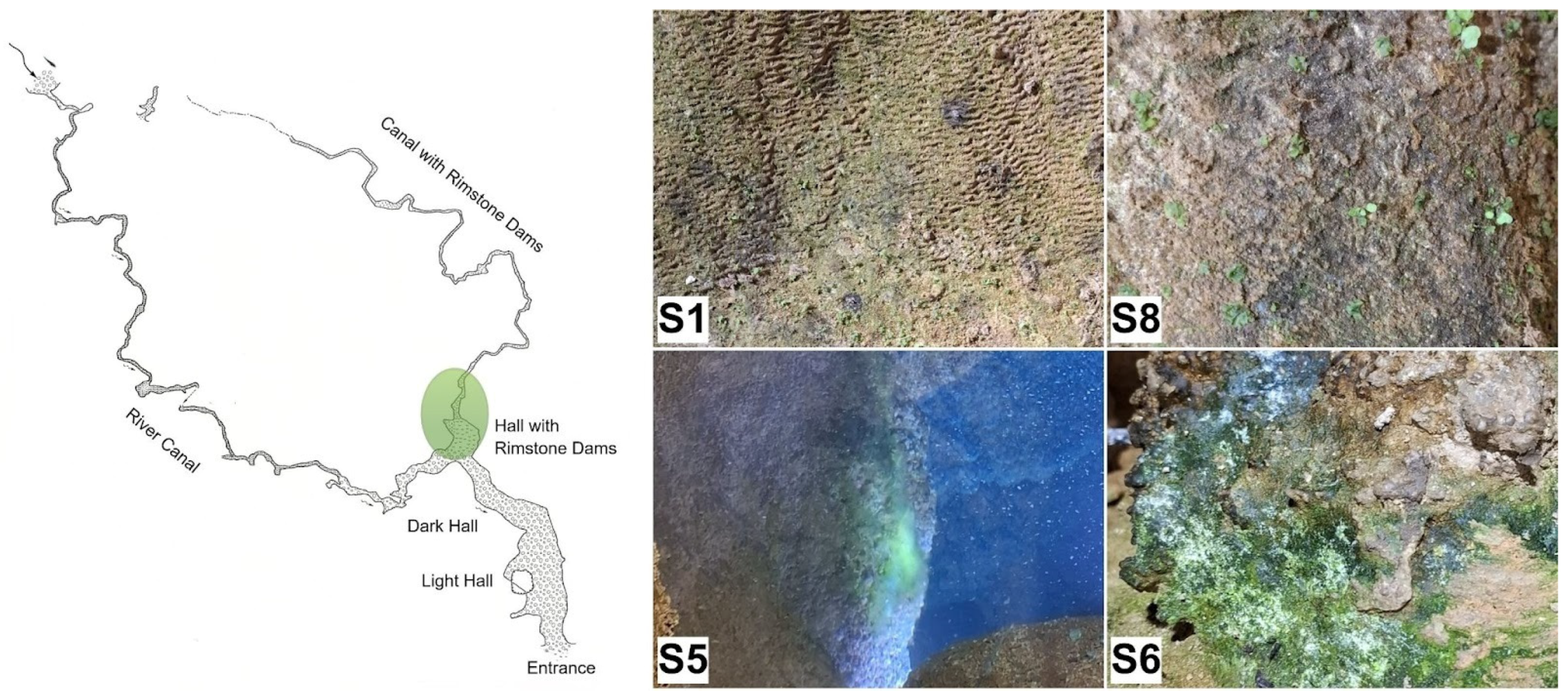
Sampling in Stopić Cave was conducted in 2025 in areas where recent lampenflora (phototrophic biofilm) development was most evident, specifically in the sections most frequently visited by tourists: the Hall with Rimstone Dams and the currently accessible part of the Canal with Rimstone Dams (Figure 1), where it appeared on and in the basins. In each section, two sampling sites were selected. Sites S1 and S8 were located at the beginning of the Hall, directly on the rimstone dams, in areas that are periodically submerged when this part of the cave becomes hydrologically active. Lampenflora at these two sites was subaerophytic at the time of sampling, similar in appearance, and ranged from endolithic to epilithic growth forms; it was observed that these lampenflora were easily detached, as portions of the underlying substrate frequently separated together with the lampenflora during sampling. Sites S5 and S6 were situated in the accessible part of the Canal, near the end of the existing tourist trail. Site S5 was located in water within the prominent rimstone basin, which is 7.2 metres deep and above which hangs a 5.5-metre-long stalactite. Site S6 was positioned near the aforementioned basin, out of the water, and predominantly epilithic lampenflora at this site developed in a subaerophytic environment in a highly humid area that is only occasionally wetted by water.
Before sampling, the distance (m) from each sampling site to the light source was measured using a laser rangefinder (Uni-Trend Technology, Dongguan, Guangdong Province, China). Light intensity (LI) was measured with a Velleman DMV 1300 lux metre (Velleman, Gavere, Belgium) and values were recorded in lux (lx). The values in lux were also converted to photosynthetic photon flux density (PPFD, μmol m−2 s−1) using an appropriate conversion factor for cool white LED light. In addition, temperature (T, °C) and relative humidity (RH, %) were measured with an Extech temperature–humidity metre (Extech, Nashua, NH, USA), the moisture content of the lampenflora-covered substrate (SM, %) was determined with an Extech MO55 moisture metre (Extech, Nashua, NH, USA), and pH was measured with a calibrated pH metre (Thermo Fisher Scientific, Waltham, MA, USA).
For microscopy observations, lampenflora material was sampled using a non-destructive adhesive tape method [19] (except at S5) and was also collected in sterile tubes. Samples for molecular analyses were aseptically collected using sterile instruments and placed in sterile tubes. All samples were transported to the laboratory on ice and, upon arrival, processed for eDNA extraction.
Figure 1. Map of Stopić Cave showing the area highlighted in green where sampling was conducted (the Hall with Rimstone Dams and the initial section of the Canal with Rimstone Dams) and the appearance of sampling sites (S1, S5, S6, and S8). The base map is adapted from the Speleological Atlas of Serbia [20] and modified accordingly.

2.3. Microscopic Analyses

Samples were examined under a Carl Zeiss Axio Imager M.1 light microscope equipped with AxioVision Release 4.8 software, using multiple magnifications. Selected samples were additionally examined using the Nikon SMZ 745T stereomicroscope equipped with a Dual Sight 1000 camera (Nikon, Tokyo, Japan). Cyanobacteria were assessed according to standard taxonomic keys [21,22,23], and algae were identified using John et al. [24].

2.4. DNA Extraction, Library Preparation, and NGS Sequencing

Samples collected from all sampling points were stored at 4 °C for further analyses. eDNA extraction was performed using the Quick-DNA Fungal/Bacterial Miniprep Kit (Zymo Research, Irvine, CA, USA) according to the manufacturer’s protocol. A total of 100 μL of pure eDNA was eluted from each sample and stored at −20 °C until amplification. The quantity and quality of extracted DNA were assessed using Quant-IT PicoGreen (Invitrogen, Molecular Probes, Eugene, OR, USA). Amplicon libraries were prepared and sequenced by Macrogen Europe (Amsterdam, The Netherlands). The bacterial 16S rRNA gene V3–V4 hypervariable region was amplified using universal primers [25,26] (341F: 5′-CCTACGGGNGGCWGCAG-3′ and 805R: 5′-GACTACHVGGGTATCTAATCC-3′) in a PCR-based library preparation protocol compatible with Illumina sequencing. In a second limited-cycle PCR step, Illumina sequencing adapters and dual-index barcodes were incorporated to enable multiplexing of samples. To verify the size of PCR-enriched fragments, library size distribution was assessed using an Agilent Technologies 2100 Bioanalyzer with a DNA 1000 chip. Library quantification was performed to ensure optimal cluster density during sequencing. Prepared Illumina libraries were quantified by quantitative PCR (qPCR) following the Illumina qPCR Quantification Protocol Guide.
PCR products were purified, quantified, normalised, and pooled in equimolar concentrations prior to sequencing. High-throughput sequencing was performed using paired-end 300 bp chemistry (2 × 300 bp) on an Illumina MiSeq platform, generating approximately 100,000 reads per sample (50,000 paired-end reads). Raw sequencing data were demultiplexed based on sample-specific indices, and FASTQ files were provided for downstream bioinformatic analyses.

2.5. Sequence Data Processing and Taxonomy Annotation

Raw sequencing data generated by the Illumina MiSeq were processed using a standardised amplicon metagenomics bioinformatics pipeline. Adapter and primer sequences were trimmed using Cutadapt v5.1 [27], followed by quality filtering using fastp v1.0.1 [28]. Quality control statistics were assessed using FastQC v0.11.9 [29]. Denoising, error correction, read merging, and chimaera removal were performed using the DADA2 algorithm q2-dada2, v2024.10.0 [30], implemented in QIIME 2 v2024.10.1 [31], resulting in high-resolution amplicon sequence variants (ASVs). Representative ASV sequences and abundance tables were generated for downstream analyses. Taxonomic assignment was performed using BLAST+ (2.17.0+, [32]) sequence alignment against curated reference databases (NCBI taxonomy database), and full taxonomic lineages were retrieved. Sequence alignment and phylogenetic tree construction were conducted using MAFFT v7 and FastTree v2.1.

2.6. Statistical Analyses

All diversity analyses were performed using the Macrogen bioinformatics pipeline and QIIME 2. Alpha-diversity was evaluated using observed features: ASV richness (a direct measure of species richness reflecting the total number of unique sequences), the Chao1 richness estimator (an estimate of total species richness accounting for rare taxa), Good’s coverage (the proportion of total diversity captured by sequencing), the Shannon diversity index (reflecting both species richness and evenness in the sample), and the Gini–Simpson index (measuring the probability that two randomly selected individuals belong to different species). Beta-diversity was assessed using UniFrac distance metrics and visualised using hierarchical clustering (UPGMA). Distance matrices and ordination analyses were used to compare microbial community structures among samples. Abundance tables and taxonomic profiles were generated for downstream statistical and ecological interpretation.

3. Results

3.1. Environmental Parameters and Lampenflora

Sampling sites for lampenflora in Stopić Cave are presented in Table 1, together with information on the cave section where sampling was conducted, sample type, distance from the nearest light source, and measured ecological parameters. Air temperature was highest at site S1, located at the very beginning of the Hall with Rimstone Dams (start of the staircase), and lowest at sites S5 and S6 in the Canal with Rimstone Dams. In contrast, relative air humidity showed the opposite trend: the lowest value was recorded at S1, while higher humidity levels were measured at all other sampling sites. Light intensity varied considerably among sites, with the highest value recorded at S1 and the lowest recorded at S8. For the remaining measured parameters, substrate moisture (SM) exceeded 50% at all sampling sites, with the highest value recorded at S6, while the pH ranged from 7.0 to 7.7.
Table 1. Sampling sites (S1, S5, S6 and S8), cave section where sampling was conducted, sample type, ecological parameters (Air T—air temperature; RH—relative humidity; LI—light intensity; SM—substrate moisture; pH—pH value of biofilm-covered substrate) and distance from the nearest light source.

3.2. Alpha- and Beta-Diversity

A total of 518,066 raw sequences were obtained from 16S rRNA gene library sequencing of the V3–V4 region (ranging from 109,404 (S8) to 159,234 (S1) per sample). After quality control steps, the number of classifiable paired-end sequence reads ranged from 28,591 (S6) to 70,968 (S1).
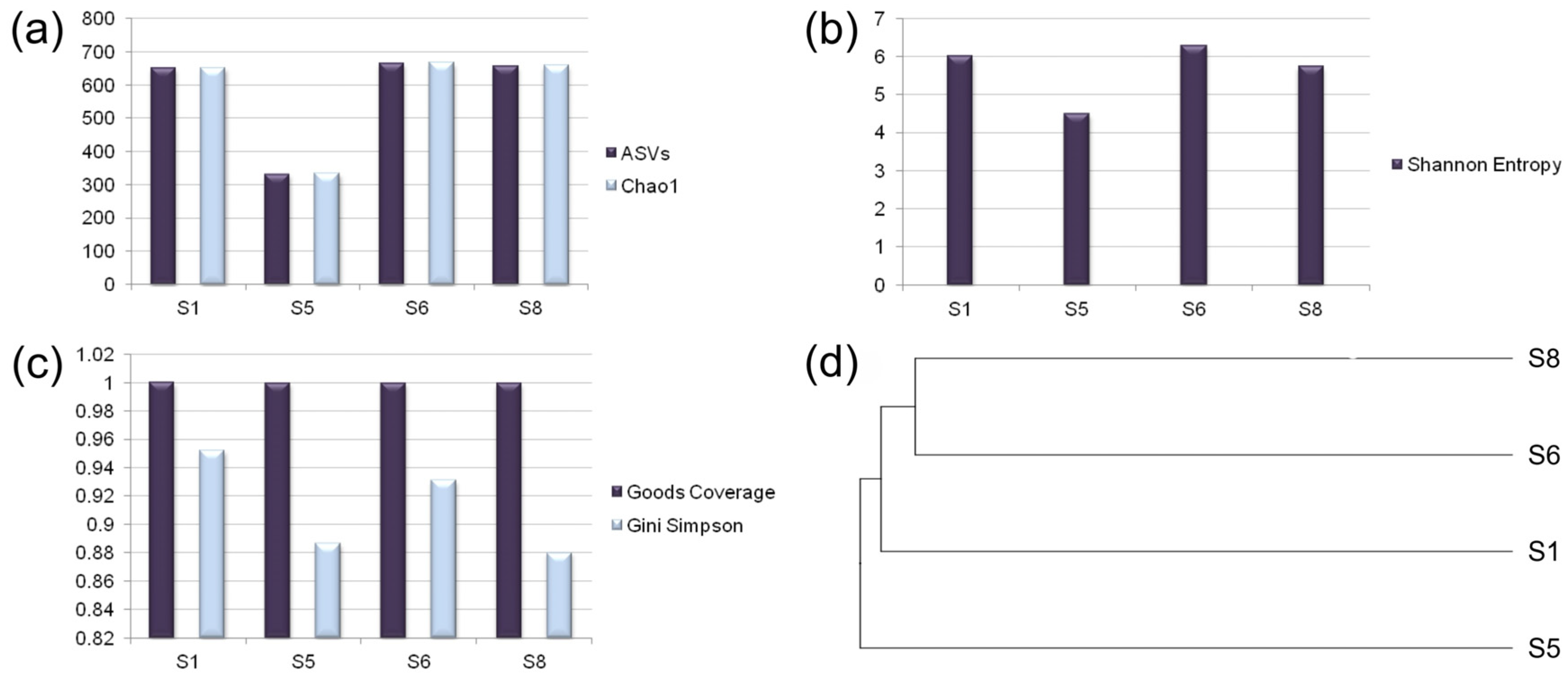
As shown in Figure 2a–c, which refers to the alpha-diversity of all bacteria, the number of distinct Amplicon Sequence Variants (ASVs), Chao1 and the Shannon index exhibited similar patterns. All three indices showed comparable values across sampling sites, with the lowest values at S5 and higher values at S1, S6, and S8. According to Figure 2c, the Gini–Simpson index generally followed the same trend as the other indices, except at S8, where its value was even lower than that at S5. Good’s coverage showed similar values (values from S1 to S8 were 1, 0.9999, 0.9998 and 0.9997, respectively).
Figure 2. Alpha- and beta-diversity of microbial communities of lampenflora in Stopić Cave. (a) ASVs and Chao1; (b) Shannon index; (c) Gini–Simpson index and Good’s coverage; (d) dendrogram based on UPGMA (unweighted pair-group method with arithmetic averages) analysis (clustering is based on genetic dissimilarities between microbial communities of lampenflora from sampling sites S1, S5, S6, and S8 in Stopić Cave, while distance was calculated using the UniFrac distance metric).
According to the clustering pattern of the UPGMA dendrogram (beta-diversity, Figure 2d), S6 and S8 were the most phylogenetically similar, forming a primary cluster that subsequently groups with S1. In comparison, site S5 was positioned separately and represented the most phylogenetically distinct sample among those analysed.

3.3. Taxonomic Analysis

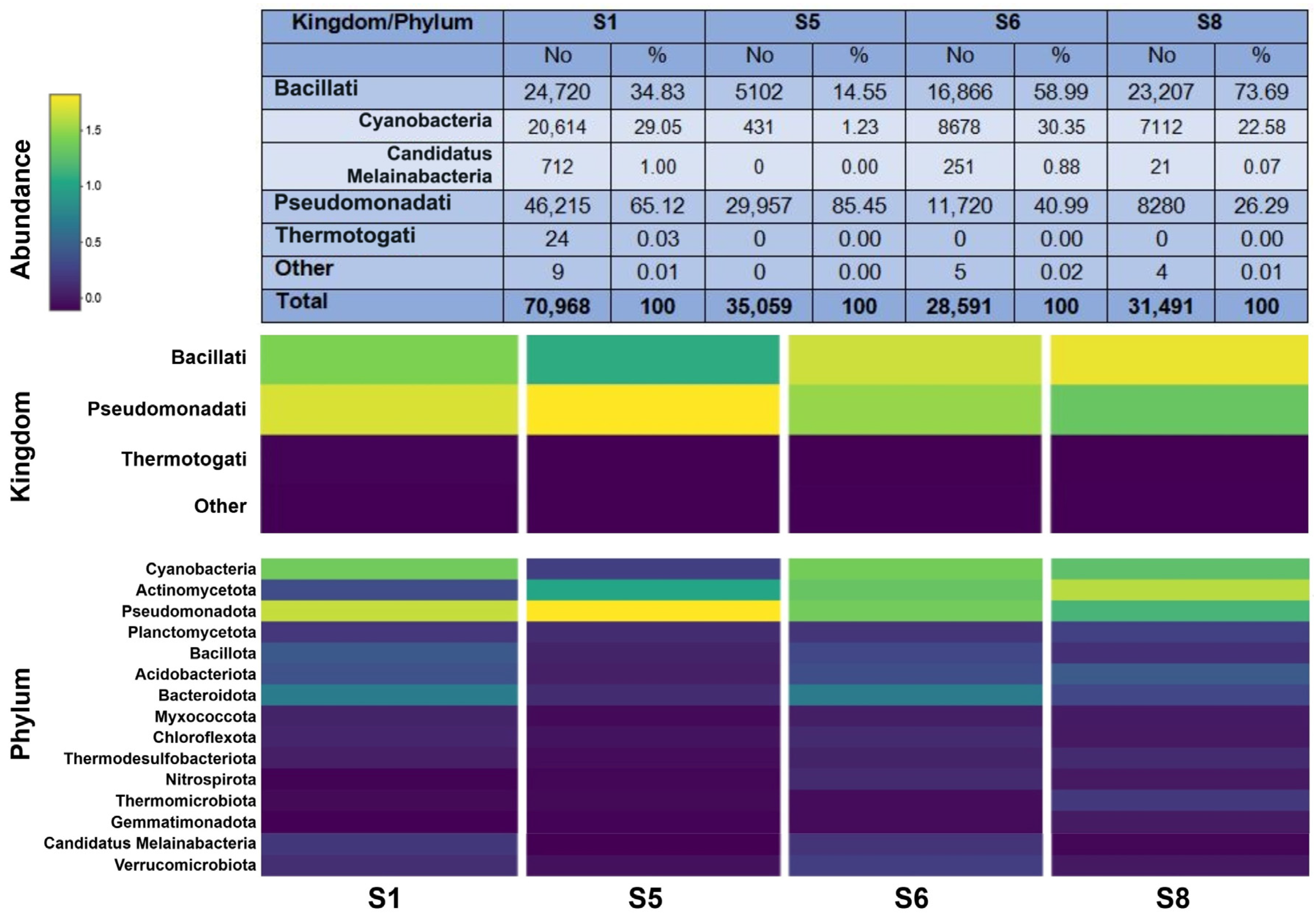
The most abundant bacterial groups detected in Stopić Cave were Pseudomonadati (65.12%) and Bacillati (34.83%). Bacillati were present at all sampling sites and were particularly dominant at sites S6 and S8. Further analysis showed that a substantial proportion of both the total bacterial community and the Bacillati group belonged to the phylum Cyanobacteria (Cyanobacteriota), especially at sites S1 and S6. At S1, Cyanobacteria accounted for 20,614 reads (29.05%) out of a total of 70,968 reads, representing 83% of all Bacillati at this site. At S6, 8678 (30.35%) out of 28,591 total reads were assigned to Cyanobacteria, comprising 51% of the Bacillati community. At S8, 7112 (22.58%) out of 31,491 reads corresponded to Cyanobacteria, representing 31% of Bacillati. In contrast, site S5 exhibited the lowest abundance of Cyanobacteria: of the 35,059 total reads recorded at this site, 431 (1.23%) were assigned to these microorganisms, accounting for only 8% of the Bacillati community. The phylum Candidatus Melainabacteria was detected in Stopić Cave at S1, S6, and S8, accounting for 1%, 0.88% and 0.07% of total reads, or 3%, 1.48%, and 0.09% of all Bacillati, respectively (Figure 3).
Figure 3. Abundance taxonomy counts and relative abundance taxonomy counts (%) at the kingdom level with the addition of the phyla Cyanobacteria and Candidatus Melainabacteria; Heatmap of relative abundance of bacterial sequences at the kingdom and phylum levels: only phyla with a total percentage abundance above 0.3% in at least one sample were included.
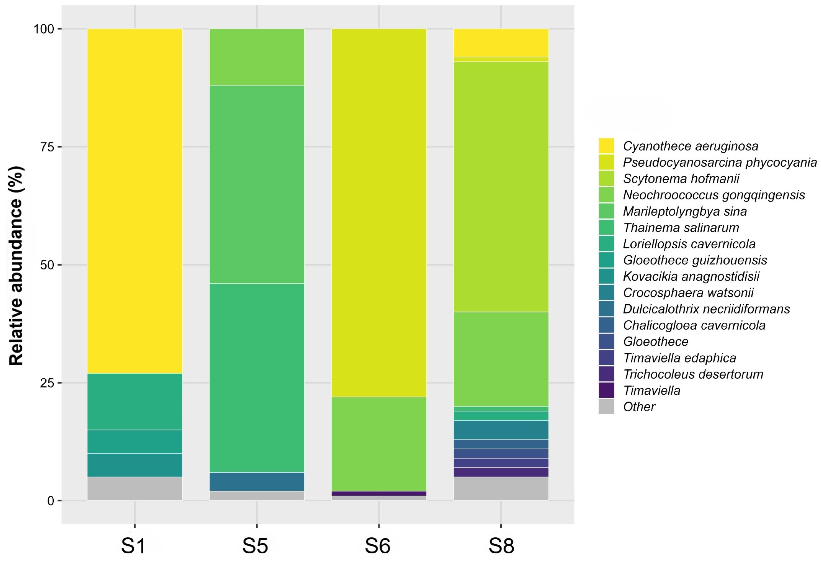
The relative abundance of ASVs assigned to cyanobacterial taxa (≥1% of total cyanobacterial reads) at each sampling site was visualised in Figure 4 and using sunburst diagrams derived from Krona plots (Figure S1), allowing representation of community composition.
Figure 4. The relative abundance of cyanobacterial taxa (≥1% of total cyanobacterial reads) at sampling sites S1, S5, S6 and S8.
At site S1, the Cyanobacteria community was strongly dominated by Cyanothece aeruginosa (Nägeli) Komárek (73%). Subdominant taxa included Loriellopsis cavernicola Hernández-Mariné & Canals (12%), Gloeothece guizhouensis J.Chen & F.Cai (5%), and Kovacikia anagnostidisii Kaštovský, J.R.Johansen & R. Hauerová (5%), together accounting for the remaining major fraction of the assemblage.
The community structure at site S5 was more evenly distributed between two predominant taxa, with Marileptolyngbya sina W.G.Zhou & J.Ling representing 42% and Thainema salinarum Rasouli-Dogaheh & Hauer 40% of the total cyanobacterial abundance. Additional contributors included Neochroococcus gongqingensis R.Geng & G.Yu (12%) and Dulcicalothrix necriidiformans Saraf, Suradkar, Dawda, Gaysina, Gabidullin, Kumat, I.Behere, Kotulkar, Batule & Prashant Singh (4%).
Cyanobacteria at site S6 were overwhelmingly dominated by Pseudocyanosarcina phycocyania P.Jung, Büdel & Lakatos, comprising 78% of the community, followed by Neochroococcus gongqingensis R.Geng & G.Yu (20%), while members of the genus Timaviella Sciuto & Moro accounted for approximately 1% of the total abundance.
The highest cyanobacterial richness was recorded at site S8. The lampenflora community at this site was dominated by Scytonema hofmanii C.Agardh ex Bornet & Flahault (53%), followed by Neochroococcus gongqingensis (20%). Other taxa exceeding 1% of total cyanobacterial reads included Cyanothece aeruginosa (6%), Crocosphaera watsonii Zehr, Rachel A.Foster, Waterbury & E.Webb (4%), Loriellopsis cavernicola (2%), Chalicogloea cavernicola M.Roldán, M.Hernández-Mariné & Komárek (2%), members of the genus Gloeothece Nägeli (2%), Trichocoleus desertorum Mühlsteinová, J.R.Johansen & N.Pietrasiak (2%), Timaviella edaphica (Elenkin) O.N.Vinogradova & Mikhailyuk (2%), Thainema salinarum Rasouli-Dogaheh & Hauer (1%) and Pseudocyanosarcina phycocyania (1%). Although its relative abundance was below 1%, Altericista variichlora Averina, E. Polyakova, Senatskaya & Pinevich is also noteworthy at this site and merits mention because of its ecological relevance (Figure S1).
Philym Candidatus Melainabacteria that was detected at all sites except S5 was represented by Vampirovibrio chlorellavorus Gromov & Mamkayeva, a predatory bacterium and an obligate parasite that attaches to the cell wall of green algae of the genus Chlorella Beyerinck [Beijerinck].
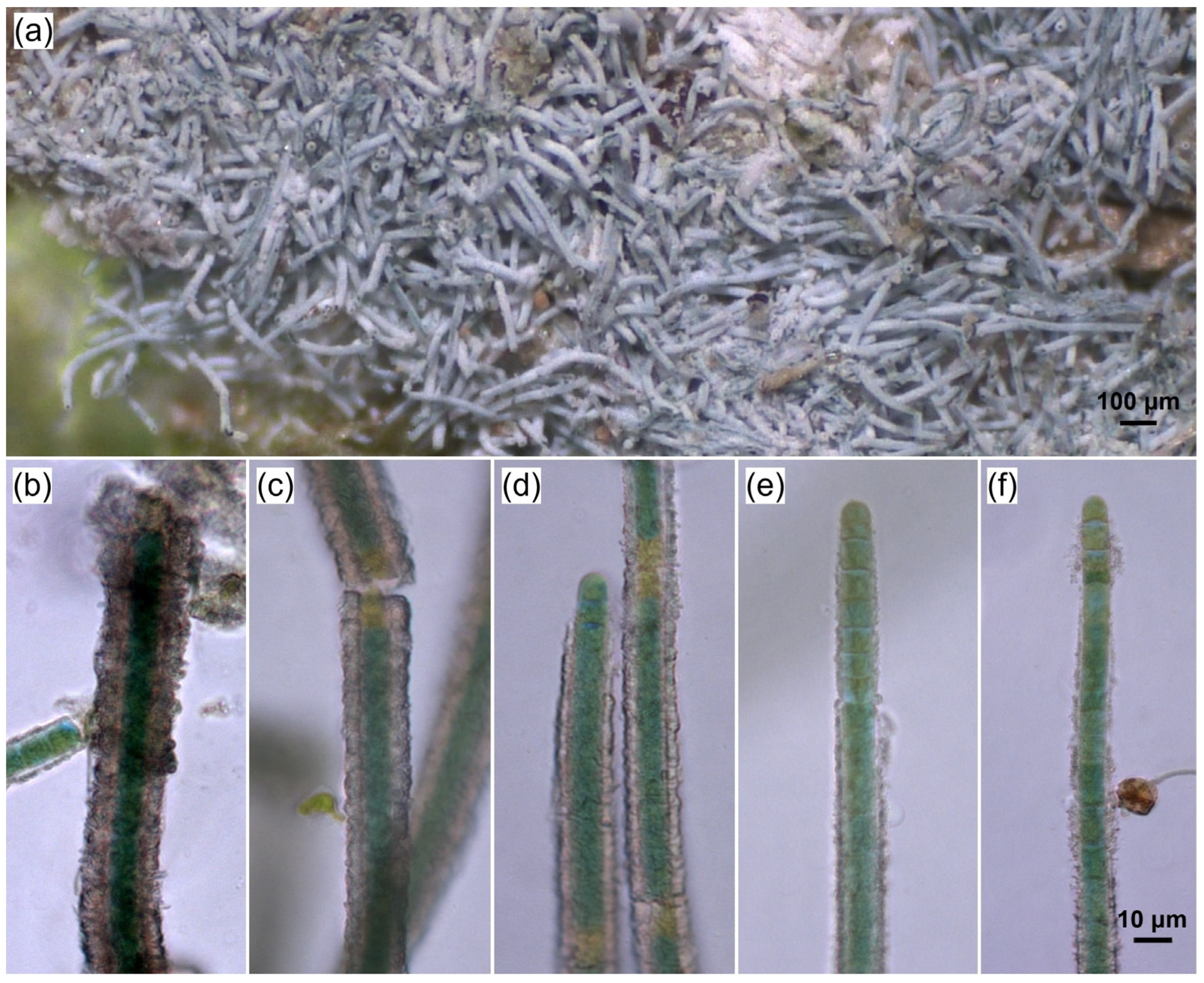
Microscopic analyses generally revealed lower diversity of Cyanobacteria and showed that Cyanobacteria were scarce in the investigated samples, appearing only sporadically, with the exception of site S8. The highest morphotype diversity was recorded at sites S1 and S8. At S1, both coccoid and trichal morphotypes were present; at site S6 coccoid and simple trichal forms were observed, while S8 contained both simple trichal and heterocytous representatives. No morphotypes of Cyanobacteria were detected microscopically at site S5. Site S8 was particularly distinctive, as the lampenflora biomass consisted predominantly of calcified cyanobacterial representatives—Scytonema C.Agardh ex Bornet & Flahault (Figure 5)—highlighting their major structural contribution to the biofilm at this site.
Figure 5. Scytonema with calcified sheaths from site S8, Stopić Cave; (a) in lampenflora; (bf) Scytonema filaments.
Apart from Cyanobacteria, microscopic analyses revealed that algae were abundant in the biomass of all samples except S8. Sample S1 was characterised by the presence of coccal green algae and cf. Vaucheria A.P.de Candolle. Rhizoclonium Kützing sp. was documented at S5. The lampenflora at S6 contained Klebsormidium flaccidum (Kützing) P.C.Silva, Mattox & W.H.Blackwell, Mesotaenium Nägeli sp., cf. Vaucheria, and coccal green algae, whereas the lampenflora at S8 consisted of Chlorella sp. and Stichococcus chlorelloides Grintzesco & Ș.Péterfi.

4. Discussion

Considering the ecological parameters measured in the cave, air temperature (T) was higher closer to the cave entrance and decreased towards the inner sections, while relative humidity (RH) exhibited the opposite trend. Stopić Cave is a hydrologically active system, and both its large entrance and permanent water flow significantly affect the microclimatic conditions within the cave. The effect of the external climate is particularly evident in the Main Canal, the Hall with Rimstone Dams, and the initial section of the Canal with Rimstone Dams. During summer, temperatures in these sections typically exceed 10 °C, while RH often falls below 95%, conditions characteristic of more open cave ecosystems [16]. The impact of the large entrance on the cave microclimate has also been discussed by Nikolić et al. (2020), Jakovljević et al. (2024) and Stupar et al. (2023) [13,14,15]. Regarding light availability, many sites within the cave are illuminated by one or a few light sources, receiving both direct and indirect light. Consequently, light intensity (LI) values were site-specific and reflected a combination of factors, including site position, exposure, distance from the light source, type of illumination (direct or indirect), and the number of light sources.
All sampled lampenflora differed in appearance, structure, colour, and community composition, reflecting adaptive responses to local microenvironmental conditions, resource availability, and physical constraints. Environmental factors such as moisture (water availability), immersion versus subaerial exposure, nutrient supply, pH, temperature, and light regime strongly influence community assembly [33,34,35]. Light availability determines not only growth rates but also pigment composition and vertical stratification within biofilms. The water regime influences oxygen diffusion, nutrient transport, and mechanical stability, while substrate characteristics affect organism attachment and the potential for endolithic growth. Over time, these factors select for organisms with specific physiological and ecological adaptations, resulting in distinct biofilm phenotypes even within the same ecosystem. Community assemblages may differ even over very small spatial distances, despite occurring on seemingly identical substrates (e.g., S1 and S8). Conversely, sites that differ significantly in hydrological conditions may support markedly different communities; in contrast to intermittently wetted or subaerial biofilms which tend to include more desiccation-tolerant Cyanobacteria and aerophytic algae, permanently submerged biofilms often favour taxa adapted to stable aquatic environments (e.g., S5). The biofilm at S5 exhibited the most distinct characteristics among the sampled sites, as it formed underwater and remained permanently submerged until sampling. This distinctiveness was also reflected in both the alpha- and beta-diversity of the bacterial community. Alpha-diversity, specifically the number of ASVs, as well as the Chao1, Shannon, and Gini–Simpson indices, was lower at S5, indicating reduced bacterial diversity. Good’s coverage was high and values were similar across all sites, suggesting that a large proportion of the total community diversity was captured by sequencing (i.e., most species present in the community were represented in the sample). Furthermore, analysis of differences in species composition among sites (beta-diversity), using UPGMA tree clustering, showed that the S5 community was phylogenetically the most distant from those at the other sampled sites.
A substantial proportion of the total bacterial community belonged to the phylum Cyanobacteria, particularly at sites S1 and S6, followed by S8, whereas S5 exhibited the lowest abundance of Cyanobacteria and lacked Candidatus Melainabacteria. The data on ASVs assigned to cyanobacterial taxa indicate pronounced heterogeneity in their community composition among the sampling sites, reflected in site-specific dominance patterns and variable taxonomic richness. The primer pair 341F/805R used in this study is a universal 16S rRNA gene primer set widely applied in environmental microbiology because of its broad coverage of bacterial diversity and efficient detection of cyanobacterial lineages across diverse environments. An additional advantage of this primer set is its widespread use, enabling direct comparison with numerous previously published metabarcoding datasets. It has been successfully applied for cyanobacterial characterisation [36] and for the analysis of bacterial communities, including lampenflora-associated cyanobacteria [37]. However, as a universal primer set, it may exhibit certain taxonomic biases and limitations in cyanobacterial taxonomic resolution. For example, Kang et al. (2022) [38] reported higher cyanobacterial diversity using cyanobacteria-specific primers; however, some genera, such as Planktothricoides and Xenococcus, were detected exclusively with universal primers. In addition, although the V3–V4 region of the 16S rRNA gene is widely used for cyanobacterial community profiling, its discriminatory power at the species level remains limited for certain taxa. Compared with microscopy, metabarcoding revealed substantially higher taxonomic diversity, although microscopy enabled detection of all major cyanobacterial morphological groups present in the samples, including coccoid, simple trichal, and heterocytous forms. Similar discrepancies between the two approaches were also reported by Kezlya et al. (2025) [39] and Wang et al. (2024) [40]. This difference is largely attributable to the high sensitivity of metabarcoding, which is particularly effective in detecting tiny, morphologically similar, fragile, and rare taxa [40,41], including organisms represented only by fragmented or non-living material in environmental samples. Consequently, many taxa detected through metabarcoding could not be reliably observed microscopically because of their extremely low abundance and sporadic occurrence. This was especially evident in sample S5, which contained the lowest proportion of cyanobacterial reads relative to the total bacterial community, substantially reducing the probability of microscopic detection.
Some representatives such as Loriellopsis cavernicola and Chalicogloea cavernicola are generally associated with cave habitats. Loriellopsis cavernicola, characterised by obligatory true branching (predominantly T-type and, rarely, V-type), the presence of heterocysts, and reproduction via hormocysts and akinetes, was isolated from a cave in Spain and described as a new species by Lamprinou et al. (2011) [11], who simultaneously established the new genus Loriellopsis. It has recently attracted attention as a promising Cyanobacterium for various research applications [42,43]. Chalicogloea cavernicola, the type and currently only species of the genus Chalicogloea (morphologically similar to Chroococcus), is a unicellular Cyanobacterium isolated from low-light aerophytic environments. It was originally found growing on calcite speleothems illuminated by white fluorescent lamps in a show cave in Collbató (Barcelona, Spain) [10]. More recently, Jung et al. (2025) [4] also documented this taxon in caves of northern Spain and associated its presence with calcification processes at the site where it was recorded. In addition to the two mentioned taxa, representatives of the genus Timaviella were recorded in this study. The genus Timaviella was separated from the morphologically similar genus Leptolyngbya by Sciuto et al. (2017) [12] and was first described from a cave environment; specifically, Timaviella circinata and Timaviella karstica were isolated from lampenflora in the Giant Cave in Italy. Timaviella karstica, together with Chalicogloea cavernicola, has been recognised as a typical cave-inhabiting species by Jung et al. (2025) [4]. In contrast, another species of this genus was recorded in Stopić Cave: Timaviella edaphica (formerly Leptolyngbya edaphica), a common soil-dwelling Cyanobacterium [44]. Representatives of the genus Timaviella have also been reported from another cave environment (corresponding more to a cave entrance zone environment) in Serbia, where they were identified as the most abundant Cyanobacteria in blue-green deposits on stone walls [45].
Several Cyanobacteria are commonly found on rock surfaces, such as representatives of the genera Scytonema and Cyanothece. The heterocytous Scytonema hofmanii, the type species of the genus Scytonema, is commonly reported from cave environments (e.g., [46,47,48]). It typically occurs in moist, illuminated zones near cave entrances or in areas influenced by artificial lighting, where it forms lampenflora. At site S8 in this study, the presence of CaCO3 precipitates prevented full observation of the dominant Cyanobacteria by light microscopy; however, their morphology suggested affiliation with the genus Scytonema. This identification was subsequently confirmed by molecular analyses, which revealed the taxon to be Scytonema hofmanii (Figure 5). The ecological role of these calcified structures remains uncertain and is still debated, but they may contribute to light trapping, detoxification processes, protection from predators, or biofilm formation through the creation of heterogeneous micro-niches [4,8]. Besides S. hofmanii, a few other species of Scytonema are known to form calcium carbonate encrustations [49] and in cave environments, the calcifying Scytonema julianum is among the most frequently reported representatives of the genus [50]. Cyanothece aeruginosa, the most abundant Cyanobacterium at site S1, is the type species of the genus Cyanothece. It was originally isolated from wet rock surfaces and has been thoroughly characterised using a polyphasic taxonomic approach [49,51]. Species of Cyanothece are morphologically, ultrastructurally, and genomically diverse unicellular Cyanobacteria that occupy a wide range of ecological niches and have been recorded from numerous habitats. Members of this genus are metabolically versatile and have therefore been studied extensively in a wide range of ecological and physiological contexts [52].
Several representatives detected in the samples were first described from aerophytic or atmophytic habitats developing on wet soil or atmophytic sites in environments such as deserts, thermal areas, saline ecosystems or as photobionts. Trichocoleus desertorum was originally isolated from desert soils and described by Muhlsteinová et al. (2014) [53]. A cyanobacterial strain showing high similarity to T. desertorum was later isolated from the Sahara Desert in Algeria [54]. Subsequently, another strain of this species was obtained from Negev Desert petroglyphs in a study investigating its potential involvement in geochemical cycles and rock weathering processes [55]. Although this species is primarily associated with soils in arid environments worldwide, it has been suggested that it can tolerate a broad range of environmental conditions [55]. Supporting this view, a strain of T. desertorum was also isolated from municipal wastewater in Pakistan [56]. Kovacikia anagnostidisii, one of the most abundant Cyanobacteria at site S1, was originally described from Clearwater Springs in Yellowstone National Park (WY, USA), where it occurred as an atmophytic organism in massive rope-like crusts near water with temperatures of approximately 85 °C [57]. The genus Kovacikia is considered to have a cosmopolitan distribution, with many representatives showing a strong association with thermal environments [58]. Physiological studies of several Kovacikia species have demonstrated their ability to perform far-red light photoacclimation (FaRLiP), an adaptation enabling photosynthesis under far-red-enriched light conditions typical of shaded or low-light habitats such as caves. This capability has been experimentally confirmed for Kovacikia euganea. It has been suggested that other Kovacikia species from both thermal and freshwater environments may also possess this trait; however, further studies encompassing a broader range of taxa are needed to determine whether FaRLiP represents a universal characteristic of the genus [59]. Pseudocyanosarcina phycocyania, the sole species of the genus Pseudocyanosarcina, was the most abundant Cyanobacterium at site S6. Jung et al. (2021) [60] described this species after isolating it as a photobiont from the saxicolous lichen Peltula clavata (Kremp.) Wetmore, collected from seepage rock above a river in Queensland, Australia. A distinctive feature of P. phycocyania is the presence of blue phycocyanin granules within the cells [60]. A species associated with saline environments is the simple trichal Cyanobacterium Thainema salinarum, the sole species of the genus Thainema. It was originally isolated from green filamentous mats (where it was mixed with other Cyanobacteria) on a wet soil surface in a solar saltern evaporation basin in Petchaburi Province, central Thailand, under salinities ranging from 90 to 250‰ [61]. More recently, a strain of T. salinarum was reported from the Sundarbans, the largest intertidal mangrove forest in the world [62]. Another one, also described from wet soil in Guizhou Province, China, is the recently described Gloeothece guizhouensis [63].
Several Cyanobacteria representing more than 1% of the total abundance were primarily associated with taxa from aquatic environments, including both freshwater and marine habitats. Notably, three of these aquatic taxa were recorded at site S5 in submerged lampenflora, distinguishing this site from the other investigated locations. Two species were mainly associated with freshwater ecosystems: Neochroococcus gongqingensis and Dulcicalothrix necriidiformans. The coccoid Cyanobacterium Neochroococcus gongqingensis, which was abundant in three samples (S5: 12%; S6: 20%; S8: 20%), is the sole species of the genus Neochroococcus. It was originally isolated from a watercourse at the Poyang Lake Model Research Base in Jiangxi Province, eastern China. Based on its colonial form and cellular spatial arrangement, this species is morphologically similar to representatives of the genera Eucapsis and Chroococcus [64]. The heterocytous Cyanobacterium Dulcicalothrix necriidiformans is the type species of the genus Dulcicalothrix, which is morphologically similar to Calothrix, Rivularia, and Macrochaete. D. necriidiformans was first isolated from an oligotrophic pond in Shrirampur taluka, Ahmednagar District, Maharashtra, India, and was distinguished from closely related taxa by its unique structural feature: the ability to form necridia [65]. Altericista variichlora was recently described by Averina et al. (2021) [66] when the new genus Altericista (similar to Synechocystis) was established. This freshwater, non-halotolerant species is characterised by the production of accessory chlorophylls d and f in cultures illuminated by far-red light, an attribute rarely observed in unicellular strains, as reported by the authors. Its ability to photoacclimate (several types of complementary chromatic adaptation exist) is of significant ecological importance in low-light environments. Some taxa were primarily associated with marine or saline environments. Marileptolyngbya sina, the most abundant Cyanobacterium at site S5 (42%), is the sole species of the genus Marileptolyngbya. It was originally isolated from Xincun Bay, Hainan Island, China, where it occurred epiphytically on Thalassia hemprichii, and is considered a marine species [67]. Another taxon of marine origin recorded in this study is Crocosphaera watsonii, described by Mareš et al. (2019) [68] and currently regarded as a synonym of Cyanobium waterburyi [49]. This unicellular Cyanobacterium contributes substantially to biologically fixed nitrogen in oligotrophic subtropical and tropical regions of the Atlantic and Pacific Oceans. This diazotrophic organism has been extensively studied under both laboratory culture conditions and in situ [69,70].
Vampirovibrio chlorellavorus was detected at all sampling sites except S5, and its occurrence consistently coincided with coccal green algae and Chlorella sp. at these sites, which is one of the most frequently reported phototrophs in lampenflora worldwide [71]. Originally described by Gromov and Mamkaeva (1972) [72] as Bdellovibrio chlorellavorus, the organism was noted for its predatory interaction with Chlorella vulgaris Beijerinck; however, owing to its distinctive mode of predation, it was later reclassified as Vampirovibrio chlorellavorus [73]. More recently, Soo et al. (2015) [74] identified this species as the first cultivable representative of the non-photosynthetic cyanobacterial lineage Melainabacteria, as well as the first recognised predatory cyanobacterium. V. chlorellavorus is known to infect multiple Chlorella species, including C. kessleri Fott & Nováková, C. sorokiniana Shihira & R.W.Krauss, and C. vulgaris, as well as other members of the family Chlorellaceae, such as Micractinium inermum R.Hoshina & Y.Fujiwara [75]. The organism exhibits a pleomorphic life cycle, existing as coccoid cells in its free-living stage and adopting a vibrioid morphology upon attachment to its host. It feeds on living algal cells, and its predatory cycle has been divided into five stages: prey location, attachment and formation of a secretion apparatus, ingestion, binary division, and release [74]. During infection, algal cells undergo visible changes, transitioning from dark green to yellow-brown, and eventually forming empty “ghost” cells. In natural environments, V. chlorellavorus plays an important ecological role by regulating populations of widespread terrestrial and freshwater microalgae [76].

5. Conclusions

Recent advances in cyanobacterial taxonomy, particularly the adoption of polyphasic approaches integrating morphological, molecular, and ecological data, have significantly improved our understanding of their diversity. Consequently, numerous new genera and species have been described in recent years, revealing that this group is much more diverse than previously recognised. Results from the lampenflora community in Stopić Cave support this trend. Analyses revealed high diversity of Cyanobacteria and largely unique, previously undocumented microbial communities, including several taxa that have been recently described. These findings underscore the importance of investigating specialised and relatively understudied environments, where studies of cyanobacterial diversity are essential for building a more accurate picture of microbial biodiversity and ecological functioning. Documenting cyanobacterial taxa from cave ecosystems contributes not only to taxonomic and phylogenetic knowledge but also to a broader understanding of microbial adaptation to variable environmental conditions.

Supplementary Materials

The following supporting information can be downloaded at: https://www.mdpi.com/article/10.3390/phycology6020058/s1, Figure S1: A sunburst diagrams derived from Krona plots showing the cyanobacterial taxonomic composition at sampling sites (a) S1, (b) S5, (c) S6 and (d) S8. The concentric circles start from the phylum level (innermost circle) to the spe-cies level (outermost circle). Associated abundances are based on the number of identified spectra.

Author Contributions

Conceptualisation, S.P.; methodology, S.P., Ž.S., M.S. and O.J.; software, S.P. and Ž.S.; validation, S.P., Ž.S., M.S. and O.J.; formal analysis, S.P. and Ž.S.; investigation, S.P.; resources, S.P., Ž.S., M.S. and O.J.; data curation, S.P. and Ž.S.; writing—original draft preparation, S.P.; writing—review and editing, S.P., Ž.S., M.S. and O.J.; visualisation, S.P., Ž.S., M.S. and O.J.; supervision, S.P. and Ž.S.; project administration, S.P.; funding acquisition, S.P., Ž.S., M.S. and O.J. All authors have read and agreed to the published version of the manuscript.

Funding

This research was funded by the Ministry of Science, Technological Development, and Innovation of the Republic of Serbia, grant numbers 451-03-33/2026-03/200178.

Institutional Review Board Statement

Not applicable.

Data Availability Statement

The data and information generated and analysed during this study are included in the article. Further inquiries can be directed to the corresponding author.

Acknowledgments

The authors gratefully acknowledge the Ministry of Science, Technological Development and Innovation of the Republic of Serbia for financial support under Contract No. 451-03-33/2026-03/200178. We also extend our sincere thanks to the representatives of the Tourist Organization Zlatibor for their assistance during sampling in Stopić Cave and for providing essential information. Finally, we express our gratitude to Vanja Milovanović for analysing sample S8 using a stereomicroscope.

Conflicts of Interest

The authors declare no conflicts of interest.

References

  1. Schirrmeister, B.E.; Gugger, M.; Donoghue, P.C.J. Cyanobacteria and the Great Oxidation Event: Evidence from genes and fossils. Palaeontology 2015, 58, 769–785. [Google Scholar] [CrossRef] [Scilit]
  2. Schmelling, N.M.; Bross, M. What is holding back cyanobacterial research and applications? A survey of the cyanobacterial research community. Nat. Commun. 2024, 15, 6758. [Google Scholar] [CrossRef] [Scilit] [PubMed]
  3. Czerwik-Marcinkowska, J.; Massalski, A. Diversity of cyanobacteria on limestone caves. In Cyanobacteria; Tiwari, A., Ed.; Chapter 3; IntechOpen: London, UK, 2018. [Google Scholar] [CrossRef] [Scilit]
  4. Jung, P.; Briegel-Williams, L.; Nürnberg, D.J.; Wolf, T.; Guillen, A.; Leira, M.; Lakatos, M. A glimpse into darkness: Diversity of culturable cyanobacteria, green algae and fungi from subaerial cave biofilms. J. Phycol. 2025, 61, 1699–1717. [Google Scholar] [CrossRef] [Scilit] [PubMed]
  5. Prescott, R.D.; Zamkovaya, T.; Donachie, S.P.; Northup, D.E.; Medley, J.J.; Monsalve, N.; Saw, J.H.; Decho, A.W.; Chain, P.S.G.; Boston, P.J. Islands within islands: Bacterial phylogenetic structure and consortia in Hawaiian lava caves and fumaroles. Front. Microbiol. 2022, 13, 934708. [Google Scholar] [CrossRef] [Scilit] [PubMed]
  6. Komárek, J. A polyphasic approach for the taxonomy of cyanobacteria: Principles and applications. Eur. J. Phycol. 2016, 51, 346–353. [Google Scholar] [CrossRef] [Scilit]
  7. Kosznik-Kwaśnicka, K.; Golec, P.; Jaroszewicz, W.; Lubomska, D.; Piechowicz, L. Into the unknown: Microbial communities in caves, their role, and potential use. Microorganisms 2022, 10, 222. [Google Scholar] [CrossRef] [Scilit]
  8. Jung, P.; Harion, F.; Wu, S.; Nürnberg, D.J.; Bellamoli, F.; Guillen, A.; Leira, M.; Lakatos, M. Dark blue-green: Cave-inhabiting cyanobacteria as a model for astrobiology. Front. Astron. Space Sci. 2023, 10, 1107371. [Google Scholar] [CrossRef] [Scilit]
  9. Hershey, O.S.; Barton, H.A. The microbial diversity of caves. In Cave Ecology; Moldovan, O.T., Kováč, L., Halse, S., Eds.; Springer: Cham, Switzerland, 2018; pp. 69–90. [Google Scholar] [CrossRef] [Scilit]
  10. Roldán, M.; Ramírez, M.; Del Campo, J.; Hernández-Mariné, M.; Komárek, J. Chalicogloea cavernicola gen. nov., sp. nov. (Chroococcales, Cyanobacteria), from low-light aerophytic environments: Combined molecular, phenotypic and ecological criteria. Int. J. Syst. Evol. Microbiol. 2013, 63, 2326–2333. [Google Scholar] [CrossRef] [Scilit]
  11. Lamprinou, V.; Hernández-Mariné, M.; Canals, T.; Kormas, K.; Economou-Amilli, A.; Pantazidou, A. Morphology and molecular evaluation of Iphinoe spelaeobios gen. nov., sp. nov. and Loriellopsis cavernicola gen. nov., sp. nov., two stigonematalean cyanobacteria from Greek and Spanish caves. Int. J. Syst. Evol. Microbiol. 2011, 61, 2907–2915. [Google Scholar] [CrossRef] [Scilit]
  12. Sciuto, K.; Moschin, E.; Moro, I. Cryptic cyanobacterial diversity in the Giant Cave (Trieste, Italy): A new genus Timaviella (Leptolyngbyaceae). Cryptogam. Algol. 2017, 38, 285–323. [Google Scholar] [CrossRef] [Scilit]
  13. Nikolić, N.; Zarubica, N.; Gavrilović, B.; Predojević, D.; Trbojević, I.; Subakov Simić, G.; Popović, S. Lampenflora and the entrance biofilm in two show caves: Comparison of microbial community, environmental, and biofilm parameters. J. Cave Karst Stud. 2020, 82, 69–81. [Google Scholar] [CrossRef] [Scilit]
  14. Stupar, M.; Savković, Ž.; Popović, S.; Subakov Simić, G.; Ljaljević Grbić, M. Speleomycology of air in Stopića Cave (Serbia). Microb. Ecol. 2023, 86, 2021–2031. [Google Scholar] [CrossRef] [Scilit]
  15. Jakovljević, O.; Predojević, D.; Knežević, J.; Karadžić, V.; Krizmanić, J.; Subakov Simić, G.; Popović, S. First insight into cyanobacterial and algal communities from a cave stream (Stopić Cave, Serbia). Aquat. Sci. 2024, 86, 10. [Google Scholar] [CrossRef] [Scilit]
  16. Lazarević, R. Stopića Pećina; Ženid and Tourist Organization Zlatibor: Belgrade, Serbia, 2012. (In Serbian) [Google Scholar]
  17. Kličković, M. Stopića pećina—Nova turistička pećina Srbije [Stopića Cave—New show cave of Serbia]. Naš Krš 2014, 47, 65–76. (In Serbian) [Google Scholar]
  18. Tourist Organization Zlatibor. Available online: https://www.zlatibor.org.rs/ (accessed on 4 February 2026).
  19. Urzì, C.; De Leo, F. Sampling with adhesive tape strips: An easy and rapid method to monitor microbial colonization on monument surfaces. J. Microbiol. Methods 2001, 44, 1–11. [Google Scholar] [CrossRef] [Scilit]
  20. Đurović, P. Speleološki Atlas Srbije [Speleological Atlas of Serbia]; Srpska Akademija Nauka i Umetnosti: Beograd, Serbia, 1998. [Google Scholar]
  21. Komárek, J.; Anagnostidis, K. Cyanoprokaryota 1. Teil: Chroococcales. In Süßwasserflora von Mitteleuropa; Ettl, H., Gärtner, G., Heynig, H., Mollenhauer, D., Eds.; Gustav Fischer: Jena, Germany, 1998; pp. 1–548. [Google Scholar]
  22. Komárek, J.; Anagnostidis, K. Cyanoprokaryota 2. Teil: Oscillatoriales. In Süßwasserflora von Mitteleuropa; Büdel, B., Gärtner, G., Krienitz, L., Schagerl, M., Eds.; Elsevier: Heidelberg, Germany, 2005; pp. 1–759. [Google Scholar]
  23. Komárek, J. Cyanoprokaryota 3. Teil: Heterocytous Genera. In Süßwasserflora von Mitteleuropa; Büdel, B., Gärtner, G., Krienitz, L., Schagerl, M., Eds.; Springer: Berlin, Germany, 2013; pp. 1–1130. [Google Scholar]
  24. John, D.M.; Whitton, B.A.; Brook, A.J. The Freshwater Algal Flora of the British Isles: An Identification Guide to Freshwater and Terrestrial Algae; Cambridge University Press: Cambridge, UK, 2003; 702p. [Google Scholar]
  25. Caporaso, J.G.; Lauber, C.L.; Walters, W.A.; Berg-Lyons, D.; Lozupone, C.A.; Turnbaugh, P.J.; Knight, R. Global patterns of 16S rRNA diversity at a depth of millions of sequences per sample. Proc. Natl. Acad. Sci. USA 2011, 108, 4516–4522. [Google Scholar] [CrossRef] [Scilit]
  26. Klindworth, A.; Pruesse, E.; Schweer, T.; Peplies, J.; Quast, C.; Horn, M.; Glöckner, F.O. Evaluation of general 16S ribosomal RNA gene PCR primers for classical and next-generation sequencing-based diversity studies. Nucleic Acids Res. 2013, 41, e1. [Google Scholar] [CrossRef] [Scilit] [PubMed]
  27. Martin, M. Cutadapt removes adapter sequences from high-throughput sequencing reads. EMBnet J. 2011, 17, 10–12. [Google Scholar] [CrossRef] [Scilit]
  28. Chen, S.; Zhou, Y.; Chen, Y.; Gu, J. fastp: An ultra-fast all-in-one FASTQ preprocessor. Bioinformatics 2018, 34, i884–i890. [Google Scholar] [CrossRef] [Scilit]
  29. Andrews, S. FastQC: A Quality Control Tool for High Throughput Sequence Data, 2010. Available online: https://www.bioinformatics.babraham.ac.uk/projects/fastqc/ (accessed on 4 February 2026).
  30. Callahan, B.J.; McMurdie, P.J.; Rosen, M.J.; Han, A.W.; Johnson, A.J.A.; Holmes, S.P. DADA2: High-resolution sample inference from Illumina amplicon data. Nat. Methods 2016, 13, 581–583. [Google Scholar] [CrossRef] [Scilit]
  31. Bolyen, E.; Rideout, J.R.; Dillon, M.R.; Bokulich, N.A.; Abnet, C.C.; Al-Ghalith, G.A.; Alexander, H.; Alm, E.J.; Arumugam, M.; Asnicar, F.; et al. Reproducible, interactive, scalable and extensible microbiome data science using QIIME 2. Nat. Biotechnol. 2019, 37, 852–857. [Google Scholar] [CrossRef] [Scilit] [PubMed]
  32. Camacho, C.; Coulouris, G.; Avagyan, V.; Ma, N.; Papadopoulos, J.; Bealer, K.; Madden, T.L. BLAST+: Architecture and applications. BMC Bioinform. 2009, 10, 421. [Google Scholar] [CrossRef] [Scilit]
  33. Gorbushina, A.A. Life on the rocks. Environ. Microbiol. 2007, 9, 1613–1631. [Google Scholar] [CrossRef] [Scilit]
  34. Fuentes, E.; Vázquez-Nion, D.; Prieto, B. Laboratory development of subaerial biofilms commonly found on buildings: A methodological review. Build. Environ. 2022, 223, 109451. [Google Scholar] [CrossRef] [Scilit]
  35. Nowicka-Krawczyk, P.; Komar, M.; Gutarowska, B.; Żelazna-Wieczorek, J.; Jesionowska, I.A.; Glińska, S.; Balcerzak, Ł. Growth Strategy of Aerial Green Algae on Building Materials in the Temperate Climate Zone and Its Relevance to Substrate Biodeterioration. Sci. Rep. 2026, 16, 2167. [Google Scholar] [CrossRef] [Scilit]
  36. Sudianto, E.; Shlafstein, M.D.; Durieu, B.; Harmel, M.; Cornet, L.; Saw, J.H. Taxonomic Description of Cyanobacteria from Extreme Habitats through Genome-Based Classification. Front. Microbiol. 2026, 17, 1824103. [Google Scholar] [CrossRef] [Scilit] [PubMed]
  37. Addesso, R.; Baldantoni, D.; Cubero, B.; De La Rosa, J.M.; Gutierrez-Patricio, S.; Tiago, I.; Caldeira, A.T.; De Waele, J.; Miller, A.Z. Unveiling the Menace of Lampenflora to Underground Tourist Environments. Sci. Rep. 2024, 14, 20789. [Google Scholar] [CrossRef] [Scilit] [PubMed]
  38. Kang, J.; Mohamed, H.F.; Liu, X.; Pei, L.; Huang, S.; Lin, X.; Zheng, X.; Luo, Z. Combined Culture and DNA Metabarcoding Analysis of Cyanobacterial Community Structure in Response to Coral Reef Health Status in the South China Sea. J. Mar. Sci. Eng. 2022, 10, 1984. [Google Scholar] [CrossRef] [Scilit]
  39. Kezlya, E.; Mironova, E.; Voyakina, E.; Kravchenko, S.; Mironov, A.; Kuzmin, V.; Chernova, E.; Iurmanov, A.; Maltsev, Y.; Kulikovskiy, M. Cyanobacteria in Waterbodies of the Biggest Anthropogenic Agglomeration: Combined DNA Metabarcoding, Microscopy, and Culture Analysis. Phycology 2025, 5, 88. [Google Scholar] [CrossRef] [Scilit]
  40. Wang, C.; Gu, J.; Li, W.; Wang, J.; Wang, Z.; Lin, Q. Metabarcoding Reveals a High Diversity and Complex Eukaryotic Microalgal Community in Coastal Waters of the Northern Beibu Gulf, China. Front. Microbiol. 2024, 15, 1403964. [Google Scholar] [CrossRef] [Scilit]
  41. Salonen, I.S.; Chronopoulou, P.M.; Leskinen, E.; Koho, K.A. Metabarcoding Successfully Tracks Temporal Changes in Eukaryotic Communities in Coastal Sediments. FEMS Microbiol. Ecol. 2019, 95, fiy226. [Google Scholar] [CrossRef] [Scilit]
  42. Ikhane, A.O.; Osunsanmi, F.O.; Mosa, R.A.; Opoku, A.R. Antibacterial potential of crude extracts from Cylindrospermum alatosporum NR125682 and Loriellopsis cavernicola NR117881. Microorganisms 2025, 13, 211. [Google Scholar] [CrossRef] [Scilit]
  43. Sithole, S.Z.; Ikhane, A.O.; Osunsanmi, F.O.; Mosa, R.A.; Opoku, A.R. In vitro photoprotective and skin aging-related enzyme inhibitory activities of Cylindrospermum alatosporum (NR125682) and Loriellopsis cavernicola (NR117881) extracts. Appl. Sci. 2025, 15, 9718. [Google Scholar] [CrossRef] [Scilit]
  44. Mikhailyuk, T.; Vinogradova, O.; Holzinger, A.; Glaser, K.; Akimov, Y.; Karsten, U. Timaviella dunensis sp. nov. from sand dunes of the Baltic Sea, Germany, and emendation of Timaviella edaphica (Elenkin) O.M. Vinogr. & Mikhailyuk (Synechococcales, Cyanobacteria) based on an integrative approach. Phytotaxa 2022, 532, 192–208. [Google Scholar] [CrossRef] [Scilit]
  45. Dimkić, I.; Ćopić, M.; Petrović, M.; Stupar, M.; Savković, Ž.; Knežević, A.; Subakov Simić, G.; Ljaljević Grbić, M.; Unković, N. Bacteriobiota of the Cave Church of Sts. Peter and Paul in Serbia—Culturable and non-culturable communities’ assessment in the bioconservation potential of a peculiar fresco painting. Int. J. Mol. Sci. 2023, 24, 1016. [Google Scholar] [CrossRef] [Scilit] [PubMed]
  46. Mazina, S.E.; Kozlova, E.V. Lampenflora of Lipska Cave, Montenegro. Cave Karst Sci. 2018, 45, 128–132. [Google Scholar]
  47. Mulec, J.; Vaupotic, J.; Walochnik, J. Prokaryotic and eukaryotic airborne microorganisms as tracers of microclimatic changes in the underground (Postojna Cave, Slovenia). Microb. Ecol. 2012, 64, 654–667. [Google Scholar] [CrossRef] [Scilit]
  48. Poulíčková, A.; Hašler, P. Aerophytic diatoms from caves in central Moravia (Czech Republic). Preslia 2007, 79, 185–204. [Google Scholar]
  49. Guiry, M.D.; Guiry, G.M. AlgaeBase. World-Wide Electronic Publication, National University of Ireland, Galway, Ireland. Available online: https://www.algaebase.org (accessed on 4 February 2026).
  50. Jurado, V.; Hernández-Mariné, M.; Rogerio-Candelera, M.A.; Ruano, F.; Aguilar, C.; Aguilar, J.; Saiz-Jimenez, C. Cleaning of phototrophic biofilms in a show cave: The case of Tesoro Cave, Spain. Appl. Sci. 2022, 12, 7357. [Google Scholar] [CrossRef] [Scilit]
  51. Komárek, J.; Kaštovský, J.; Mareš, J.; Johansen, J.R. Taxonomic classification of cyanoprokaryotes (cyanobacterial genera) 2014 using a polyphasic approach. Preslia 2014, 86, 295–335. [Google Scholar]
  52. Bandyopadhyay, A.; Elvitigala, T.; Welsh, E.; Stöckel, J.; Liberton, M.; Min, H.; Sherman, L.A.; Pakrasi, H.B. Novel Metabolic Attributes of the Genus Cyanothece, Comprising a Group of Unicellular Nitrogen-Fixing Cyanobacteria. mBio 2011, 2, 5. [Google Scholar] [CrossRef] [Scilit]
  53. Mühlsteinová, R.; Johansen, J.R.; Pietrasiak, N.; Martin, M.P.; Osorio-Santos, K.; Warren, S.D. Polyphasic characterization of Trichocoleus desertorum sp. nov. (Pseudanabaenales, cyanobacteria) from desert soils and phylogenetic placement of the genus Trichocoleus. Phytotaxa 2014, 163, 241–261. [Google Scholar] [CrossRef] [Scilit]
  54. Mehda, S.; Muñoz-Martín, M.; Oustani, M.; Hamdi-Aïssa, B.; Perona, E.; Mateo, P. Microenvironmental conditions drive the differential cyanobacterial community composition of biocrusts from the Sahara Desert. Microorganisms 2021, 9, 487. [Google Scholar] [CrossRef] [Scilit]
  55. Irit, N.; Barak, H.; Rabbachin, L.; Kahn, A.; Pavan, M.; Kramarsky-Winter, E.; Piñar, G.; Sterflinger, K.; Kushmaro, A. Trichocoleus desertorum isolated from Negev desert petroglyphs: Characterization, adaptation and bioerosion potential. Sci. Total Environ. 2023, 904, 166739. [Google Scholar] [CrossRef] [Scilit]
  56. Khan, F.; Malik, S.; Shahid, A.; Siddiqui, A.J.; Musharraf, S.G.; Zhu, H.; Alkhattabi, N.A.; Gull, M.; Mehmood, M.A. Characterization of a newly isolated cyanobacterium Trichocoleus desertorum BERC08 as a potential feedstock for the algal biorefinery. Biomass Conv. Bioref. 2023, 13, 5283–5294. [Google Scholar] [CrossRef] [Scilit]
  57. Kaštovský, J.; Johansen, J.R.; Hauerová, R.; Akagha, M.U. Hot Is Rich—An Enormous Diversity of Simple Trichal Cyanobacteria from Yellowstone Hot Springs. Diversity 2023, 15, 975. [Google Scholar] [CrossRef] [Scilit]
  58. Tang, J.; Shah, M.R.; Yao, D.; Jiang, Y.; Du, L.; Zhao, K.; Li, L.; Li, M.; Waleron, M.M.; Waleron, M.; et al. Polyphasic identification and genomic insights of Leptothermofonsia sichuanensis gen. sp. nov., a novel thermophilic cyanobacterium within Leptolyngbyaceae. Front. Microbiol. 2022, 13, 765105. [Google Scholar] [CrossRef] [Scilit]
  59. Zampieri, R.M.; Bizzotto, E.; Campanaro, S.; Caldara, F.; Bellucci, M.; La Rocca, N. Kovacikia euganea sp. nov. (Leptolyngbyaceae, Cyanobacteria), a new chlorophyll f-producing cyanobacterium from the Euganean Thermal District (Italy). Front. Microbiol. 2025, 16, 1545008. [Google Scholar] [CrossRef] [Scilit]
  60. Jung, P.; Brust, K.; Schultz, M.; Büdel, B.; Donner, A.; Lakatos, M. Opening the gap: Rare lichens with rare cyanobionts—Unexpected cyanobiont diversity in cyanobacterial lichens of the order Lichinales. Front. Microbiol. 2021, 12, 728378. [Google Scholar] [CrossRef] [Scilit]
  61. Rasouli-Dogaheh, S.; Komárek, J.; Chatchawan, T.; Hauer, T. Thainema gen. nov. (Leptolyngbyaceae, Synechococcales): A new genus of simple trichal cyanobacteria isolated from a solar saltern environment in Thailand. PLoS ONE 2022, 17, e0261682. [Google Scholar] [CrossRef] [Scilit]
  62. Roy, A.R.; Lefler, F.W.; Laughinghouse, H.D.; Chakraborty, S.; de Los Santos Villalobos, S.; Mukherjee, J. Genome sequences of Almyronema epifaneia and Thainema salinarum isolated from an intertidal mangrove forest. J. Phycol. 2026, 62, 17–24. [Google Scholar] [CrossRef] [Scilit]
  63. Chen, J.; Li, S.; Li, R.; Yu, G.; Cai, F. The description of a novel Gloeothece species (Chroococcales, Cyanobacteria) from China, based on a polyphasic approach. Phytotaxa 2024, 662, 53–66. [Google Scholar] [CrossRef] [Scilit]
  64. Geng, R.; Wang, Y.; Cai, F.; Zhang, Y.; Yang, P.; Dai, G.; Li, R.; Yu, G. Neochroococcus gongqingensis gen. et sp. nov., a new member of coccoid cyanobacteria from a watercourse, eastern China. Fottea 2021, 21, 44–52. [Google Scholar] [CrossRef] [Scilit]
  65. Saraf, A.; Suradkar, A.; Dawda, H.G.; Gaysina, L.A.; Gabidullin, Y.; Kumat, A.; Behere, I.; Kotulkar, M.; Batule, P.; Singh, P. Phylogenetic complexities of the members of Rivulariaceae with the re-creation of the family Calotrichaceae and description of Dulcicalothrix necriidiformans gen. nov., sp. nov., and reclassification of Calothrix desertica. FEMS Microbiol. Lett. 2019, 366, fnz219. [Google Scholar] [CrossRef] [Scilit]
  66. Averina, S.; Polyakova, E.; Senatskaya, E.; Pinevich, A. A new cyanobacterial genus Altericista and three species, A. lacusladogae sp. nov., A. violacea sp. nov., and A. variichlora sp. nov., described using a polyphasic approach. J. Phycol. 2021, 57, 1517–1529. [Google Scholar] [CrossRef] [Scilit]
  67. Zhou, W.-G.; Ding, D.-W.; Yang, Q.-S.; Ahmad, M.; Zhang, Y.-Z.; Lin, X.-C.; Zhang, Y.-Y.; Ling, J.; Dong, J.-D. Marileptolyngbya sina gen. nov., sp. nov. and Salileptolyngbya diazotrophicum gen. nov., sp. nov. (Synechococcales, Cyanobacteria), species of cyanobacteria isolated from a marine ecosystem. Phytotaxa 2018, 383, 75–92. [Google Scholar] [CrossRef] [Scilit]
  68. Mareš, J.; Johansen, J.R.; Hauer, T.; Zima, J., Jr.; Ventura, S.; Cuzman, O.; Tiribilli, B.; Kaštovský, J. Taxonomic resolution of the genus Cyanothece (Chroococcales, Cyanobacteria), with a treatment on Gloeothece and three new genera, Crocosphaera, Rippkaea, and Zehria. J. Phycol. 2019, 55, 578–610. [Google Scholar] [CrossRef] [Scilit]
  69. Inomura, K.; Deutsch, C.; Wilson, S.T.; Masuda, T.; Lawrenz, E.; Bučinská, L.; Sobotka, R.; Gauglitz, J.M.; Saito, M.A.; Prášil, O.; et al. Quantifying oxygen management and temperature and light dependencies of nitrogen fixation by Crocosphaera watsonii. mSphere 2019, 4, e00531-19. [Google Scholar] [CrossRef] [Scilit]
  70. Masuda, T.; Mareš, J.; Shiozaki, T.; Inomura, K.; Fujiwara, A.; Prášil, O. Crocosphaera watsonii—A widespread nitrogen-fixing unicellular marine cyanobacterium. J. Phycol. 2024, 60, 604–620. [Google Scholar] [CrossRef] [Scilit]
  71. Popović, S.; Nikolić, N.; Subakov Simić, G. Lampenflora: Biofilm diversity in world caves. In Aerophytic Algae and Cyanobacteria; Blanco, S., Ed.; Academic Press: London, UK, 2026; pp. 301–336. [Google Scholar] [CrossRef] [Scilit]
  72. Gromov, B.V.; Mamkaeva, K.A. Electron microscopic study of parasitism by Bdellovibrio chlorellavorus bacteria on cells of the green alga Chlorella vulgaris. Tsitologiia 1972, 14, 256–260. [Google Scholar]
  73. Gromov, B.V.; Mamkaeva, K.A. New genus of bacteria, Vampirovibrio, parasitizing Chlorella and previously assigned to the genus Bdellovibrio. Mikrobiologiia 1980, 49, 165–167. [Google Scholar]
  74. Soo, R.M.; Woodcroft, B.J.; Parks, D.H.; Tyson, G.W.; Hugenholtz, P. Back from the dead: The curious tale of the predatory cyanobacterium Vampirovibrio chlorellavorus. PeerJ 2015, 3, e968. [Google Scholar] [CrossRef] [Scilit]
  75. Hovde, B.T.; Steichen, S.A.; Starkenburg, S.R.; Brown, J.K. Vampirovibrio chlorellavorus Draft Genome Sequence, Annotation, and Preliminary Characterization of Pathogenicity Determinants. Phycol. Res. 2020, 68, 23–29. [Google Scholar] [CrossRef] [Scilit]
  76. Ganuza, E.; Sellers, C.E.; Bennett, B.W.; Lyons, E.M.; Carney, L.T. A Novel Treatment Protects Chlorella at Commercial Scale from the Predatory Bacterium Vampirovibrio chlorellavorus. Front. Microbiol. 2016, 7, 848. [Google Scholar] [CrossRef] [Scilit]
Disclaimer/Publisher’s Note: The statements, opinions and data contained in all publications are solely those of the individual author(s) and contributor(s) and not of MDPI and/or the editor(s). MDPI and/or the editor(s) disclaim responsibility for any injury to people or property resulting from any ideas, methods, instructions or products referred to in the content.

Article Metrics

Citations

Article Access Statistics

Multiple requests from the same IP address are counted as one view.