Article

Isolation, Molecular Identification, and Biochemical Profiling of Native Microalgae from the Santa Elena Peninsula (Ecuador) as a Basis for Sustainable Aquaculture


by
Janeth I. Galarza
1,*,
Jimmy Villón
1,
Claudio A. Álvarez
2,
Bryan Pillacela
1,
María Soledad Romero
3,
Macarena Mellado
4,
Alexis Hernández-Pérez
5,
Rosario Díaz
5 and
Gonzalo Álvarez
2,*
1
Centro de Investigaciones Biológicas y Prácticas Académicas, Facultad de Ciencias del Mar, Universidad Estatal Península de Santa Elena, La Libertad 240204, Ecuador
2
Departamento de Acuicultura, Facultad de Ciencias del Mar, Universidad Católica del Norte, Larrondo 1281, Coquimbo 1780000, Chile
3
Departamento de Biología Marina, Facultad de Ciencias del Mar, Universidad Católica del Norte, Larrondo 1281, Coquimbo 1780000, Chile
4
Centro Integrativo de Biología y Química Aplicada (CIBQA), Facultad de Ciencias de la Salud, Universidad Bernardo O’Higgins, Santiago 8370854, Chile
5
Programa de Doctorado en Acuicultura, Universidad Católica del Norte, Larrondo 1281, Coquimbo 1780000, Chile
*
Authors to whom correspondence should be addressed.
Phycology 2026, 6(2), 60; https://doi.org/10.3390/phycology6020060
Submission received: 4 April 2026 / Revised: 14 May 2026 / Accepted: 25 May 2026 / Published: 31 May 2026

Abstract

Microalgae are valuable biotechnological resources due to their high productivity and their capacity to synthesize compounds with nutritional and antioxidant functions. However, in the Santa Elena Peninsula (Ecuador), their use in aquaculture is limited to commercial strains. In this study, native microalgae were isolated and evaluated for their nutritional value in aquaculture. Samples were collected at five coastal sites, cultivated under controlled conditions, and characterized using optical microscopy and SEM, identified at the molecular level through the 28S rRNA gene, and their biochemical profiles were analyzed, including carotenoid quantification. The isolates were identified as PM-UPSE-006 (Tetradesmus obliquus), PM-UPSE-007 (Conticribra weissflogii), PM-UPSE-016 (Halamphora coffeiformis), PM018 (Dunaliella sp.), and PM-UPSE-022 (Chlorella vulgaris), with T. obliquus and H. coffeiformis being recorded for the first time in the peninsula. The highest growth rates were observed in T. obliquus, Dunaliella sp., and C. vulgaris, while Dunaliella sp. and C. vulgaris stood out for their protein content (57.28% DM and 55.37% DM), T. obliquus for carbohydrates (40.5% DM), and H. coffeiformis, Dunaliella sp., and C. vulgaris for carotenoids (0.53–1.60% DM). These results demonstrate their ex situ adaptability, competitive growth, and noteworthy biochemical profiles, establishing them as promising biotechnological resources for sustainable aquaculture.

1. Introduction

Microalgae are among the most versatile groups of photosynthetic microorganisms, widely recognized for their high productivity, metabolic diversity, and remarkable ability to adapt to various environmental conditions [1,2]. These characteristics make them strategic resources for biotechnological applications, particularly in aquaculture, where their inclusion in functional diets has shown benefits by providing high-quality proteins, polyunsaturated lipids, carbohydrates, and pigments with antioxidant properties, including carotenoids [3,4,5]. In this context, pigments such as β-carotene, lutein, astaxanthin, and zeaxanthin have been associated with improvements in the growth, immune response, and pigmentation of farmed aquatic organisms [6,7]. The growing demand for natural and sustainable ingredients has intensified the search for new microalgal strains that can be cultivated under controlled conditions and contribute to more resilient and environmentally responsible aquaculture [8,9]. In this context, microalgae have emerged as promising candidates because of their rapid growth rates and adaptability to controlled culture systems. These characteristics position them as versatile resources in aquaculture and across a wide range of biotechnological applications, including nutraceutical supplements, bioremediation, biofuel production, functional food ingredients, cosmetics, pharmaceuticals, natural pigments, and antioxidants [10,11,12].
Aquaculture in Ecuador is strongly dominated by the culture of marine shrimp (Penaeus vannamei), which accounts for more than 95% of the national production [13,14,15], while other species, such as tilapia and rainbow trout, have experienced notable growth in recent years [16,17]. In 2024, the country exported more than 1.23 million metric tons of shrimp, generating over 6 billion dollars in revenue and consolidating its position as the world’s leading exporter in key markets, such as the United States (52.6%), Europe (21.8%), and Asia (18.2%) [18].
However, this industry faces limitations caused by infectious diseases, such as white spot syndrome virus and IHHNV, which are responsible for high mortality rates and significant economic losses [19,20]. Simultaneously, rainbow trout farming systems in the high Andean regions are affected by bacterial and fungal pathologies, such as bacterial kidney disease, saprolegniasis, and Flavobacterium columnare, which can cause mortality of up to 80% in the early stages [8,21,22]. These issues highlight the need to develop innovative nutritional strategies based on natural and sustainable ingredients that enhance the growth and health of cultured organisms and reduce dependence on conventional inputs and the exclusive use of commercial microalgae [11,23,24,25].
The predominant use of commercial microalgal species has limited the evaluation of native strains with high biotechnological potential [26,27]. However, the use of native microalgae strains can promote more stable and efficient growth than commercial strains by adapting to local environmental conditions [28,29,30]. Furthermore, the introduction of exogenous strains may pose ecological risks by disrupting natural biodiversity, whereas the isolation and preservation of native microalgae contribute to the conservation of local genetic resources and ensure their long-term availability [30,31]. This situation is especially relevant in megadiverse regions such as the Ecuadorian Pacific coast, where there is still insufficient knowledge about the diversity and biochemical functionality of microalgal communities, the exploration of which is key to strengthening ecological resilience in the face of climate change.
The Santa Elena Peninsula (Ecuador), characterized by its marine-coastal environments, represents a strategic reservoir of microalgal biodiversity that could contribute to the development of local inputs to strengthen aquaculture sustainability [32,33]. However, the limited taxonomic and functional characterization of some species in these communities has hindered their integration into ex situ cultivation programs and incorporation into nutritional formulations. However, accurate species identification is an essential step prior to evaluating physiological and biochemical profiles. While morphological taxonomy has traditionally been used, its usefulness is restricted by the phenotypic plasticity of many microalgal species. In this sense, molecular tools based on the analysis of conserved regions of ribosomal DNA (18S rRNA, 28S rRNA, ITS, or 16S rRNA) have proven to be more robust for confirming taxonomic identity and establishing phylogenetic relationships, ensuring reliable classification for subsequent studies [34,35]. In this context, the present study aimed to isolate, identify, and evaluate the biochemical profile (proteins, carbohydrates, ether extracts, and carotenoids) of native microalgae from the Santa Elena Peninsula (Ecuador) cultivated ex situ, to determine their potential as a nutritional source in sustainable aquaculture.

2. Materials and Methods

2.1. Biological Material

The samples were collected between April and October 2021 at five sites along the coast of the Santa Elena Peninsula: Comuna Dos Mangas (1°49′27.8″ S 80°41′16.0″ W); Mar Bravo (2°13′38.1″ S 80°58′08.3″ W), Comuna Palmar (2°01′42.3″ S 80°44′12.0″ W), La Caleta (2°13′13.2″ S 80°54′54.8″ W), Muelle de Salinas (2°12′16.6″ S 80°58′06.7″ W). Sampling was conducted in two types of environments: shallow surf zones along the seashore and intertidal channels located in land areas near the coastline, using horizontal sampling.
In the intertidal channels, sampling was performed in small pools using a net 30 cm in length and 10 cm in diameter. Collection was carried out manually using back-and-forth movements, maintaining the same technical specifications as the previous net [36,37,38]. The sampling points were georeferenced using a handheld GPS device (eTrex 10, Garmin, Olathe, KS, USA), and the salinity was measured in situ using a handheld refractometer (ORA 90BE, KERN & SOHN GmbH, Balingen, Germany). They were then kept in cool temperature (4 °C) and transported to the laboratory at the Centro de Investigaciones Biológicas y Prácticas Académicas (CIBPA-UPSE) for processing [36].

2.2. Microalgae Strain Culture

Microalgal species were isolated by serial decimal dilutions (1:10; ranging from 10−1 to 10−4), followed by the inoculation of 100 µL aliquots on solid media. Agar plates were prepared using three culture media: f/2 [39], Conway [40], and inorganic fertilizer (NPK at 2%) (Marchfol, Marchfol S.A., Guayaquil, Ecuador), selected due to their compositional differences in nutrients and their widespread use in microalgal cultivation [41]. Given the environmental heterogeneity of the sampling sites, the media were prepared across salinity gradients of 35, 30, and 25 PSU, as well as under freshwater conditions.
The plates were incubated at a constant temperature of 24 ± 1 °C under an irradiance of 40 µmol m−2 s−1 measured using a photometer (LI-COR Light Meter LI-138B, Lincoln, NE, USA) and a photoperiod of 12:12 h (light:dark). Colony development was monitored periodically, and well-defined colonies were aseptically transferred to test tubes containing 10 mL of the same medium in which they developed, while maintaining the previous experimental conditions. The complete isolation and laboratory acclimation process took between 6 and 8 months. Chlorophyta strains acclimated more rapidly to laboratory conditions than diatoms, which required a comparatively longer adaptation period. From these initial cultures, purified isolates were obtained and subsequently transferred to 200 mL Erlenmeyer flasks containing 100 mL of culture medium to form strain cultures.

2.3. Morphological Analyses

Microscopic observations were performed using an Olympus IX71 light microscope (Olympus Corporation, Tokyo, Japan) under phase contrast at 40× (magnification). Photomicrographs were captured using an Axiocam 208 color digital camera (Carl Zeiss Microscopy GmbH, Jena, Germany) and calibrated using ZEN Lite software (Carl Zeiss Microscopy GmbH, Jena, Germany).
Scanning electron microscopy (SEM) observations were made following the recommendations of [42,43]. Cells preserved in 3% glutaraldehyde were retained on 0.45 µm polyamide membrane filters (Sartorius AG, Goettingen, Germany) and washed with Milli-Q ultra-pure water (Merck Millipore KGaA, Darmstadt, Germany) to remove salts and preservatives. The samples were then dehydrated in a graded ethanol series (20, 40, 60, 80, 95, and 100%) for 15 min, followed by 100% hexamethyldisilazane. After dehydration, the samples were dried using a critical point dryer (Samdri-780A, Tousimis Research Corporation, Rockville, MD, USA), mounted on bronze stubs, and coated with gold-palladium using a JFC-1100 sputter coater (Jeol, Tokyo, Japan). Finally, the samples were observed using a Hitachi SU3500 scanning electron microscope (Hitachi High Technologies Corp., Tokyo, Japan).

2.4. Molecular Identification of Isolates Using the 28S rRNA Gene

Genomic DNA was extracted following the protocol described by Maddocks and Jenkins [44], with some modifications. Three milliliters of cell culture was centrifuged at 3500 rpm for 5 min. The cell pellet was resuspended in 400 µL lysis buffer (50 mM Tris-HCl (pH 8.0), 5 mM EDTA, 100 mM NaCl, and 2% Triton X-100). Next, 2 µL of proteinase K (20 mg mL−1) was added, and the mixture was incubated at 37 °C for 1 h. After digestion, 200 µL of phenol and 200 µL of a chloroform:isoamyl alcohol mixture (24:1, v/v) were added for protein extraction. The samples were centrifuged, and the aqueous phase was then recovered. DNA was precipitated in cold by adding 3 M sodium acetate (C2H3NaO2) for neutralization. Subsequently, 95% and 75% ethanol were added to promote complete DNA precipitation, respectively. The resulting pellets were air-dried at room temperature and resuspended in nuclease-free water for further analyses. The concentration and purity of the DNA were determined using a NanoDrop 2000 spectrophotometer (Thermo Fisher Scientific, Waltham, MA, USA).
Molecular identification was performed by amplifying conserved regions of the 28S rRNA ribosomal gene (large ribosomal subunit, LSU) using polymerase chain reaction (PCR). The primers used were designed by Bolch and Percy [45]: D1R-F-5′-ACCCGCTGAATTTAAGCATA-3′ and D2C-R-5′ CCTTGGTCCGTGTTTCAAGA-3′. PCR rounds were performed in a final volume of 15 µL, containing 1 µL of genomic DNA (100 ng/µL), 0.125 mM of each primer, 0.2 mM dNTPs (Invitrogen, Thermo Fisher Scientific, Waltham, MA, USA), 3 mM MgCl2 (Invitrogen, Thermo Fisher Scientific, Waltham, MA, USA), 1 U Taq DNA polymerase (InvitrogenTM), and 1X reaction buffer (Invitrogen, Thermo Fisher Scientific, Waltham, MA, USA) (InvitrogenTM). The mixture and reaction conditions were prepared according to the manufacturer’s instructions (Invitrogen). Amplification conditions consisted of an initial denaturation at 95 °C for 10 min, followed by 35 cycles of denaturation at 94 °C for 30 s, annealing at 58 °C for 30 s, and extension at 72 °C for 1 min 30 s, and a final extension at 72 °C for 5 min. All reactions were performed using a T100 thermal cycler (Bio-Rad Laboratories, Hercules, CA, USA). Amplified products were separated using 1% agarose gel electrophoresis with a 1 Kb molecular weight marker (Invitrogen, Thermo Fisher Scientific, Waltham, MA, USA) (InvitrogenTM). Finally, the products were purified and sent for sequencing at Macrogen Inc. (Seoul, Republic of Korea).

2.5. Phylogenetic Analysis

Sequence identification began with similarity searches in the GenBank database using BLASTn (NCBI BLAST, National Center for Biotechnology Information, Bethesda, MD, USA). Subsequently, phylogenetic analysis was performed in R (version 4.4.1) [46] for sequence processing, distance matrix calculations and phylogenetic tree construction. The nucleotide sequences used for inference were obtained from GenBank (NCBI), and their accession numbers are shown in the resulting tree figure. The sequences were then imported into R and subjected to further analysis. Multiple sequence alignment was performed using MUSCLE [47] to ensure positional homology among all sequences, and the final alignment was converted to DNAb in format (msa package, version 1.38.0) [48]. The shortest sequence (PM-UPSE-022) was used as a reference. To refine the alignment, unreliable positions were removed by discarding columns containing gaps in the reference sequence and retaining only sites with nucleotide information. The filtered alignment was converted into a phyDat object (phangorn package version 2.12.1) [49]. This distance matrix was calculated using the raw model (APE package, version 5.8) [50], which estimated the proportion of divergent nucleotide positions between comparable sites. Using this distance matrix, an initial phylogenetic tree was constructed using the neighbor-joining (NJ) method. Subsequently, a test of nucleotide substitution models (JC, K80, F81, HKY, SYM, and GTR) was performed, and the best-fitting model was selected based on the Bayesian Information Criterion (BIC). The model with the lowest BIC (GTR + Γ, with four rate categories) was selected to optimize the tree topology and branch lengths through maximum likelihood inference, incorporating stochastic rearrangements to maximize the likelihood (phangorn package) [49]. One thousand bootstrap replicates were generated to assess the statistical support of the optimized maximum likelihood tree (phangorn package). Finally, the tree was rooted using Porphyra umbilicalis as an outgroup (APE package, version 5.8) [50], and a graphical representation of the phylogenetic tree was produced using the gg tree package (version 3.14.0) [51].

2.6. Growth Experiments

The cultures were carried out in triplicate in 500 mL Erlenmeyer flasks, each containing 250 mL of culture media (Table 1), adjusted to pH 7.5, and maintaining the salinity required by each microalga for its growth. The cultures were maintained at a constant temperature of 24 ± 1 °C, irradiance of 60 µmol m−2 s−1, and a photoperiod of 12:12 h (light:dark), using artificial illumination provided by white light lamps (LED 18 W, 6500 K) and manual agitation thrice a day.
The growth of microalgal species was examined in triplicate cultures of each strain maintained for 11–13 days, as described above. Two-mL aliquots were taken daily, fixed with 10% Lugol, and the cell densities (cells mL−1) were determined using a Neubauer improved counting chamber (Marienfeld, Germany; depth 0.100 mm). and microscope (OMAX Corporation, Orange County, CA, USA). Growth curves were plotted, and growth parameters (maximum cell density, growth rate, and conditioning time to growth) were obtained by approximating the integrated logistic model (Equation (1)) [52].
The non-linear least-squares (quasi-Newton) method from the macro ‘Solver’ of the Microsoft Excel spreadsheet was employed for numerical modeling of the microalgae strains growth data [53].
X = X 0   e μ   ( t t l a g ) 1 X 0 X   1 e μ   ( t t l a g )  
where X is the cell density (cells mL−1), X is the maximum cell density (cells mL−1), μ is the growth rate, t is time (d), and tlag (d) is the time required to adapt to the growth conditions.
In the case of strain PM-UPSE-016, daily productivity samples of 250 mL were concentrated by filtration using GF/F (47 mm diameter). The filters were dried at 105 °C for 24 h in an oven (VMTECH 101-3AB, Wincom Company Ltd., Changsha, China) and then weighed using an analytical balance (BOECO BAS 31 plus, 0.0001 g; BOECO, Hamburg, Germany). Biomass was expressed as μg mL−1.

2.7. Extraction and Quantification of Total Carotenoids and Biochemical Analysis

Total carotenoid extraction was performed as described by Arredondo-Vega and Voltolina [54], with some modifications. Thirty milligrams of dry biomass were weighed and transferred to capped glass tubes. One milliliter of saline solution (1%) was added and homogenized using vortex. Subsequently, the samples were centrifuged at 3500 rpm for 4 min, the supernatant was discarded, and the washing process was repeated to remove residual salts. The resulting cell pellet was resuspended in 3 mL of cold 90% acetone (v/v), vortexed, and sonicated for 3 min to extract the pigments. The samples were stored at −20 °C for 24 h. After this period, the samples were subjected to a second round of sonication for 3 min, followed by centrifugation at 2500 rpm for 5 min at 10 °C. The supernatant containing the extracted carotenoids was transferred to a 25 mL volumetric flask. This extraction process was repeated two or three times until the cell pellet was completely colorless [55,56]. All extracts were pooled in the same flask, and the final volume was adjusted to 10 mL using a cold 90% acetone (v/v) solution. The absorbance of the final extract was measured immediately using a GENESYS™ 40/50 (Thermo Scientific™) UV-Vis spectrophotometer with 3 mL glass cells at 480, 664, and 667 nm. The concentration of total carotenoids (μg·mL−1) was calculated using the formula proposed by [57] based on the registered absorbance values (Equation (2)). All measurements were performed in triplicate under low-light conditions and on ice to minimize oxidative degradation of the pigments.
C a r o t e n o i d s   μ g L = 1000   ×   A 480 1.12   ×   A 664 ( 34.07   ×   A 647 )   214
The biochemical analyses were carried out at the Laboratorio de Nutrición Animal de la Universidad Central de Ecuador. The samples, previously dried at 37 °C for 24 h, were packaged using a triple-packaging system to protect them from environmental humidity and oxidation and were then sent for analysis. The analysis report indicated that the methods applied were as follows: crude protein: Kjeldahl NTE INEN-ISO 5983-1, 2014) [58]; ether extract (total lipids) (NTE INEN-ISO 11085:2013) [59]; total carbohydrates were estimated by difference, according to the standard method proposed by the AOAC [60]. Crude fiber was determined by acid and alkaline hydrolysis with intermediate filtration (NTE INEN-ISO 6865, 2014) [61]. Each analysis was performed in triplicate.

3. Results

3.1. Morphological Characterization of Isolates

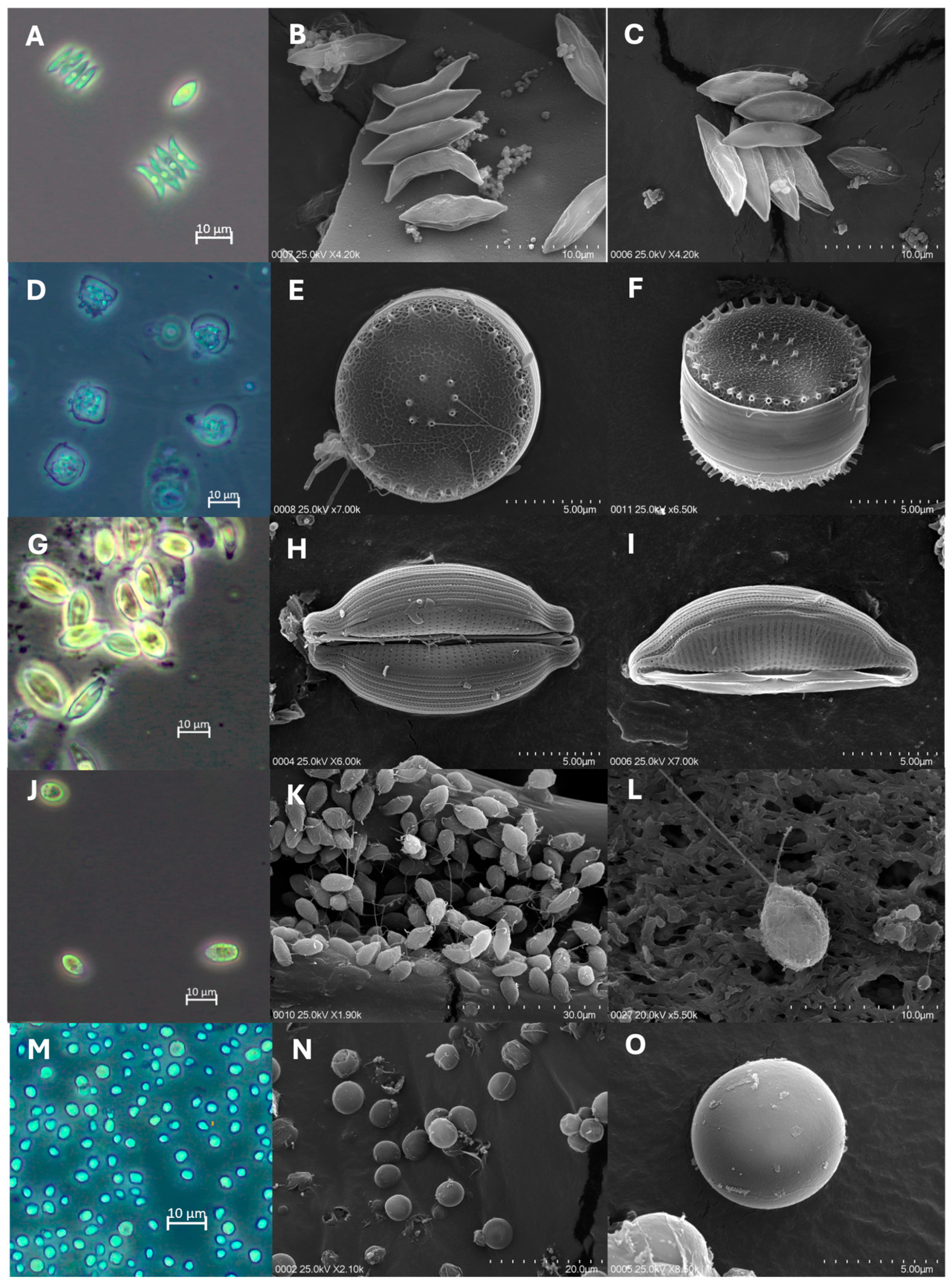
Morphological examination of the cultures carried out with microscopic observations and SEM showed that the cells from the culture strain PM-UPSE-006 fit the description of Tetradesmus obliquus. The cells were spindle-shaped with an acute apex and contained one pyrenoid (Figure 1A,B). Coenobia were arranged on a flat plane in two rows, conformed by 4–8 cells (Figure 1A–C). The length of the cells was 11.82 ± 1.27 μm, and their width was 3.80 ± 0.98 μm.
Morphological examination of the cells from strain PM-UPSE-007 showed good overall agreement with the descriptions made for Conticriba weissflogii. The cells have a circular valve with a flat surface (12.61 ± 0.84 μm in diameter) (Figure 1D,E). In the SEM analyses, the external view of the cells showed valves with a silicified net and siliceous granules (Figure 1E). Two fultoportulae rings are distributed in the valve, one central with 5–7 fultoportulae and the other marginal with 30–35 fultoportulae (Figure 1D,F).
Observations of strain PM-UPSE-016 revealed morphological characteristics consistent with Halamphora coffeiformis (Figure 1G). The cells have a semi-lanceolate to semi-elliptical shape, with ends protracted into rostrate poles (Figure 1H). The pervalvar axis was strongly curved (Figure 1I). The raphe was straight and located close to the ventral margin (Figure 1I). The valve length was 16.27 ± 1.51 μm and the width is 7.94 ± 1.03 μm.
Morphological examination of the cells from strain PM018 resembled the descriptions made for the genus Dunaliella. The cells were nearly spherical to ellipsoidal in shape and appeared green in color (Figure 1J). The cell length was 9.22 ± 1.49 μm, and the width was 5.73 ± 0.41 μm. The cells have two motile flagella approximately equal to or slightly longer than the cell length (Figure 1K,L).
The cells of strain PM-UPSE-022 were similar to those of Chlorella vulgaris. The cells were spherical or slightly ellipsoidal (3.50 ± 0.47 μm in diameter) and had a green coloration (Figure 1M). Cells lacking flagella have smooth surfaces and rigid cell walls (Figure 1N,O).

3.2. Molecular Identification of Isolates Based on the 28S rRNA Gene

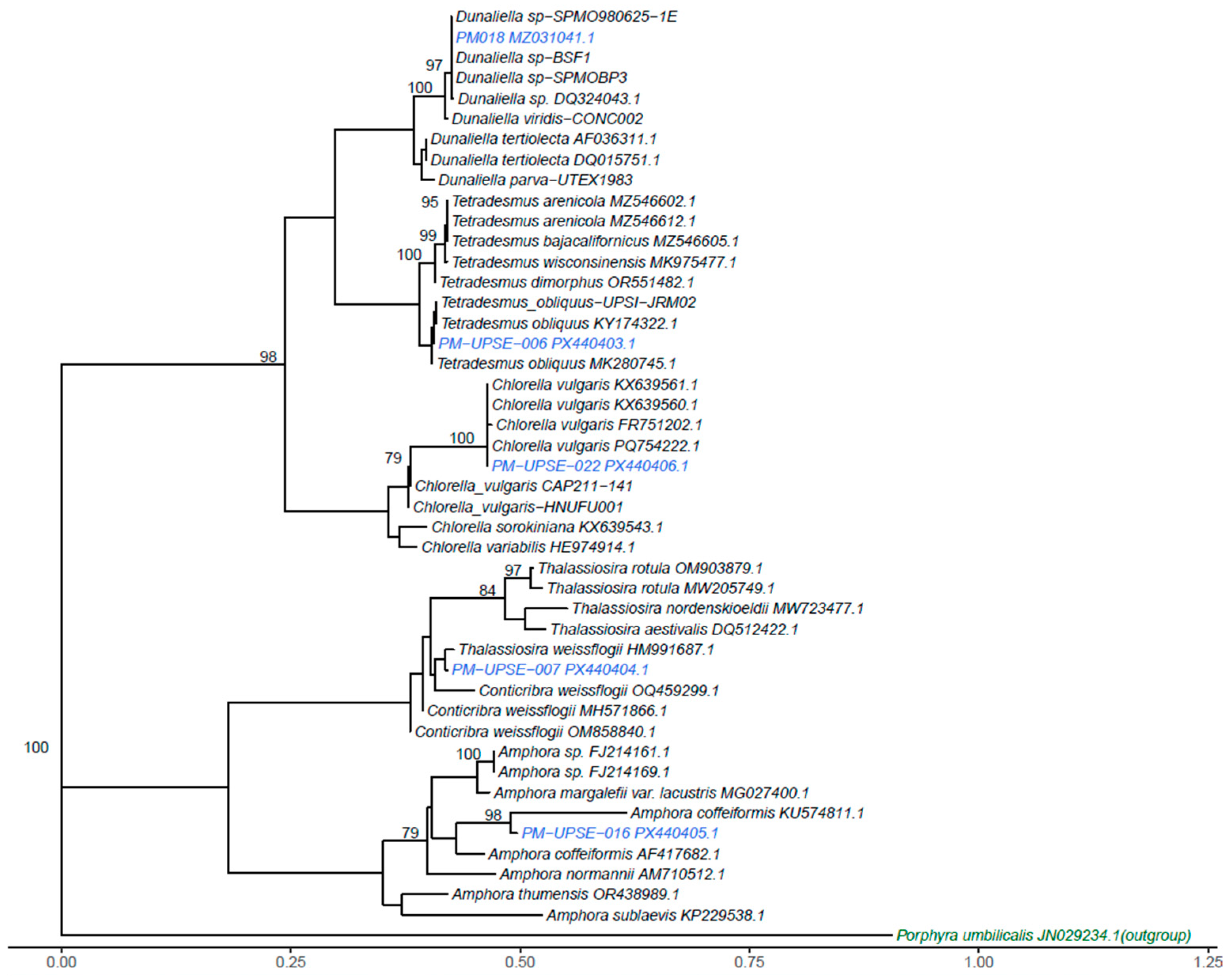
Molecular analysis of the 28S rRNA ribosomal gene confirmed the taxonomic identity of the five evaluated strains. The strain PM-UPSE-006 (GenBank accession number PX440403.1) showed 99.03% similarity to Tetradesmus obliquus and was grouped within the clade corresponding to this species.
The strain PM-UPSE-007 (GenBank accession number PX440404.1) showed 99.35% similarity to Conticribra weissflogii and was consistently placed within its clade, whereas strain PM-UPSE-016 (GenBank accession number PX440405.1) revealed 98.44% similarity to Halamphora coffeiformis (previously reported as Amphora coffeiformis) and formed a monophyletic clade with previously reported strains.
The analyses of strain PM018 (GenBank accession number MZ031041.1) exhibited 100.0% similarity with different strains of the genus Dunaliella sp. and clustered in a well-supported clade with representatives of this genus, although further characterization is required to accurately determine its species-level identification.
Finally, strain PM-UPSE-022 (GenBank accession number PX440406.1) presented 92.45% similarity with Chlorella vulgaris, and despite this relatively low identity, both morphological evidence and its placement in a clade closely related to strains of this species strongly support its classification as C. vulgaris.
In all cases, the phylogenetic analyses showed a topology consistent with the molecular similarities and a bootstrap support of 100%, which provides high robustness to the taxonomic delimitation and evolutionary interpretation of the isolates (Figure 2).

3.3. Growth Performance of Microalgal Isolates

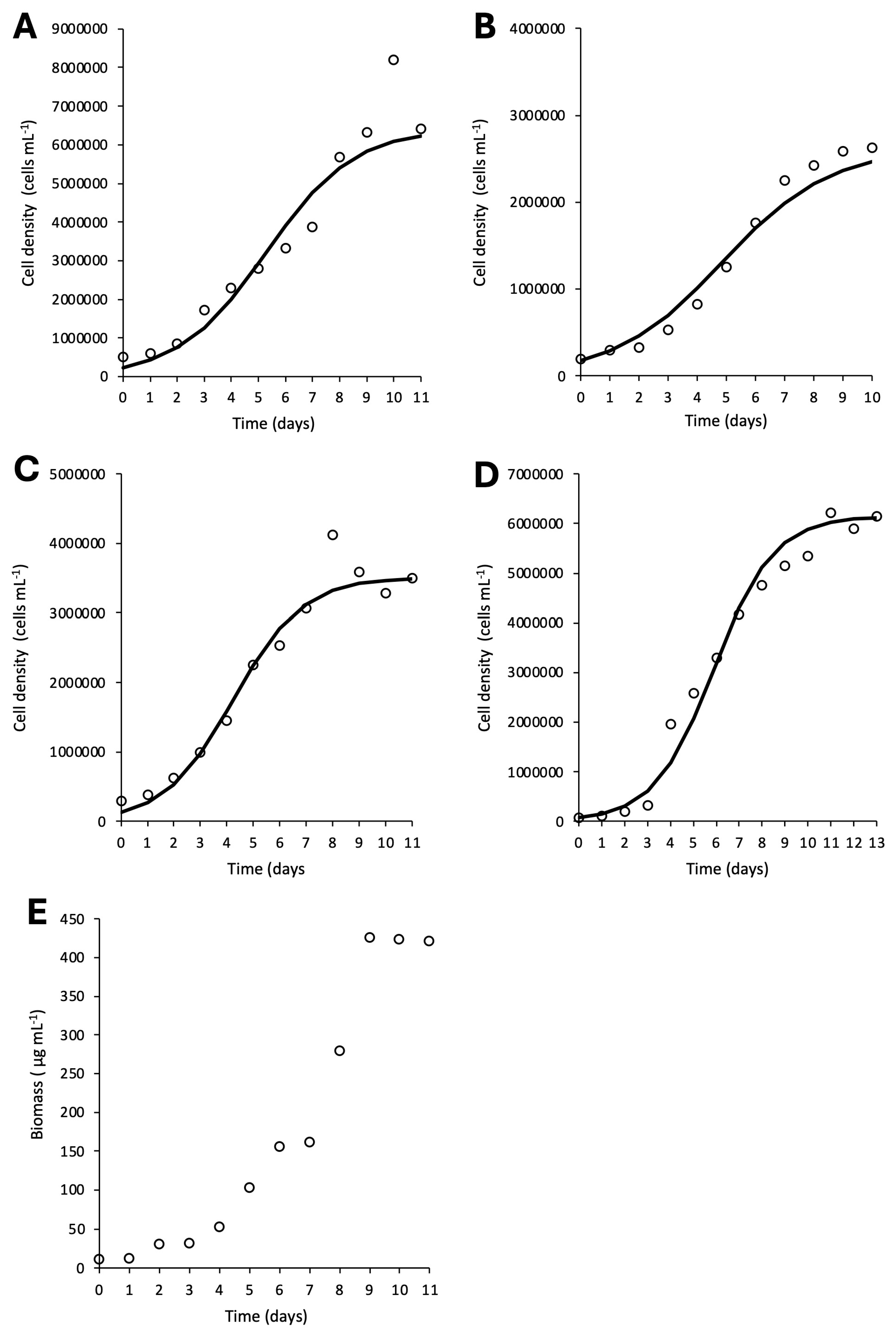
Tetradesmus obliquus (Figure 3A) exhibited an exponential phase between days 2 and 10, with a maximum experimental cell density of 8,191,667 cells mL−1. The modeling of growth kinetics showed a lower theoretical maximum cell density of 6,235,078 cells mL−1 (R2 = 0.75) and a growth rate of 0.62 d−1 (Table 1).
Conticribra weissflogii (Figure 3B) showed a shorter exponential phase between days 2 and 7, reaching a maximum experimental cell density of 2,625,000 cells mL−1, which is similar to the maximum theoretical cell density of 2,469,200 cells mL−1 (R2 0.92). For this strain, the growth rate was 0.54 d−1 (Table 1).
Dunaliella sp. (Figure 3C). revealed an exponential phase between days 2 and 7, with a maximum experimental cell density of 4,125,000 cells mL−1. In this culture, the maximum theoretical cell density was lower, with a value of 3,488,305 cells mL−1 (R2 = 0.90), while the growth rate corresponded to 0.76 d−1 (Table 1).
Chlorella vulgaris (Figure 3D) showed an exponential phase between days 3 and 8, with a maximum experimental cell density of 6,225,000 cells mL−1, which was very similar to that estimated by theoretical modeling (6,121,990 cells mL−1) (R2 = 0.94). For this strain, the growth rate was 0.76 d−1 (Table 1).
Halamphora coffeiformis (Figure 3E) productivity curve showed an exponential phase between days 3 and 8, followed by stabilization of the biomass concentration. The maximum biomass obtained corresponds to 425.8 μg mL−1.

3.4. Total Carotenoid Content and Biochemical Composition

Tetradesmus obliquus (PM-UPSE-006) provided 49.4% protein and 40.5% carbohydrates, with an elevated ether extract fraction (18.8% dw), positioning it as a balanced energy and protein source for shrimp feed.
For its part, Conticribra weissflogii (PM-UPSE-007) showed an acceptable protein content (44.9% dw) and the highest ether extract value in the group (20.3% dw), which are valuable characteristics for enriching larval feeds.
Halamphora coffeiformis (PM-UPSE-016) had the lowest protein content (13.3% dw) but presented a high proportion of carotenes (0.53% dw) and ether extract (18.8% dw), suggesting its secondary potential as a source of pigments and energy in specialized formulations (Table 2).
Among the microalgae analyzed, Dunaliella sp. (PM018) stood out for its high protein content (57.28% dw), significant proportion of carbohydrates (32.19% dw), and the highest level of total carotenes (1.60% dw), highlighting its high value as a functional ingredient in aquaculture diets.
Finally, Chlorella vulgaris (PM-UPSE-022) also displayed a notable protein profile (55.37% dw), complemented by an ether extract content (11.02% dw) and carotenoids (0.76% dw), which were higher than those of most of the other species.
The crude fiber content was low in most of the evaluated microalgae, ranging from 0.00 to 5.54% dry weight, which may favor their digestibility and potential use in aquaculture diets.

4. Discussion

The isolate PM-UPSE-006, identified as Tetradesmus obliquus, represents the first confirmed record for the Santa Elena Peninsula and possibly for the Ecuadorian coast. To date, its distribution has only been documented in biodiversity studies in the Andean region and the eastern part of the country [34,62].
Morphologically, the isolate exhibited characteristics consistent with the classic descriptions of the genus. The cells measured (11.82 ± 1.27 µm length; 3.80 ± 0.98 µm width), placing them at the upper end of the UTEX 3031 strain range (8.1–12.3 µm length; 2.9–4.2 µm width) [63] and slightly above BR003 (10 µm length; 3–5 µm width) [64]. Compared to the FACHB–276 strain (7–9 µm × 4–5 µm); reported by [65], the cells from this study were longer and slightly slimmer, a trend consistent with the dependence of the phenotype on the medium and physicochemical conditions.
In terms of growth, the PM-UPSE-006 strain showed significant kinetic performance in the f/2 medium. The isolate exhibited a specific growth rate of μ = 0.62 d−1, which is higher than the μ = 0.13 d−1 reported by [66] in BBM medium using a digestate and CO2 application, and much higher than the μ= 0.004 d−1 reported by Ahiahonu, et al. [67] in BG11 medium with ambient CO2 capture for lipid accumulation.
These differences indicate an ex situ adaptation in the f/2 medium, which provides a more suitable nutritional balance for the strain than the media designed for other microbial groups. From a biochemical perspective, PM-UPSE-006 had 49.4% protein (dry weight), which is higher than that reported for strain UJEA_AD (38% protein and 31.2% lipids) in BG11 and comparable to the 47.8% described by Akgül [68] under nitrogen deficiency. These results confirm that our standard conditions favor protein accumulation, whereas media such as BG11 and stress treatments tend to increase the lipid content [64]. Thus, the native strain is a stable protein source, with the possibility of manipulating lipid metabolism in future studies. This finding significantly expands the known geographic range of the species and is especially important because it positions T. obliquus as a local biotechnological resource with potential applications in bioremediation, biofuel production, and obtaining biomass with a high protein content [65,69].
In contrast to the previous species, the isolate PM-UPSE-007 Conticribra weissflogii has already been recorded in the Santa Elena Peninsula and along the Ecuadorian coast, as documented by studies of the Oceanographic Institute of the Ecuadorian Navy (INOCAR), under its former name, Thalassiosira weissflogii [70].
Morphologically, it presents valves measuring 12.61 ± 0.84 µm in diameter, which fall within the range described by [71] for the ND-8 strain (10–18 µm), although below the values reported by [72] (14.2–17 µm) and [73] (10–35 µm). Likewise, the reduction in the number of central fultoportulae compared to that in the studies previously mentioned does not necessarily indicate taxonomic differences. The identification was confirmed molecularly through analysis of the 28S rRNA gene, which placed this isolate within the C. weissflogii clade, corroborating that the observed morphological features correspond to the species. In Conway medium (salinity 25 PSU), the isolate reached a density of 2,460,000 cells mL−1 and µ = 0.54 d−1, surpassing the 800,000 cells mL−1 and µ = 0.2 d−1 reported by Zhang, et al. [74] NMB3, and comparable to the 1,940,000 cells mL−1 obtained in modified f/2 by [75]. Compared with cultures with constant aeration and lower irradiance [76], the results of this study remain favorable, demonstrating the effectiveness of the ex situ adaptation protocol employed. Biochemically, C. weissflogii showed 44.9% protein and 20.3% ether extract, values that match the ranges described by Millán-Almaraz, et al. [77].
The total carotenoid content was modest (0.23% dw), but the literature shows that adjustments in silicate and salinity increase pigment production [75,76]. This ability to modulate pigmentation and lipoprotein composition makes this diatom a promising candidate for applications in aquaculture feeding and pigmentation, provided that targeted stress strategies are applied.
The isolate PM-UPSE-016 (Halamphora coffeiformis) also represents the first record for the Santa Elena Peninsula. However, the genus Halamphora has been reported in biodiversity studies in the Los Ilinizas Ecological Reserve and Lake Quilotoa-Cotopaxi in the Andean region of Ecuador [78]. The cells of H. coffeiformis strain PM-UPSE-016 (16.27 ± 1.51 µm length; 7.94 ± 1.03 µm width) were smaller than those of other South American strains isolated from Argentina (9.5–42 µm length; 12–17.7 µm width) [79] and were similar to those reported by Zidarova, et al. [80] in the South Shetland Islands, Antarctica (13.5–20.5 µm length; 3.5–4.5 µm width). This intermediate position demonstrates marked morphological plasticity that complements the known morphometric spectrum of the species. In terms of molecular analysis, the PM-UPSE-016 isolate grouped with sequences deposited as Amphora coffeiformis, a species currently reclassified under the genus Halamphora [81]. However, the 28S rDNA gene sequences still appear in GenBank under the old name, which requires their use for alignment and phylogenetic tree construction. The productivity of H. coffeiformis PM-UPSE-016 reached 425.8 μg mL−1 in f/2 medium at 35 UPS, surpassing the value reported for Amphora aff. amoena (216.25 µg mL−1) under white light [82], despite the use of a higher irradiance (200 μmol m2 s−1). In contrast, A. coffeaeformis (C. Agardh) Kützing showed higher productivity (1130 µg mL−1) when the medium was supplemented with magnesium nitrate [83]. These results indicate that our isolate exhibits competitive performance and potential for biotechnological applications, although its productivity could be increased by optimizing nutrients and irradiance. The biochemical profile of H. coffeiformis (PM-UPSE-016) was characterized by a low protein content (13.3% dw) and a notable proportion of carbohydrates (31.2% dw). These values agree with those reported by Juárez-Gómez, et al. [82] (13.4% dw) and differ from those described by Cui, et al. [84], where nitrogen deficiency increased protein content by up to 44.0%. Similarly, Beekrum and Amonsou [85] reported that Amphora sp. WCA23.2 has a low protein content (~15%) and a lipid profile rich in unsaturated fatty acids and high antioxidant capacity. This finding confirms that, under standard conditions, the native strain directs its metabolism towards carbohydrates and pigments (0.53% dw), which suggests its usefulness as an energy or antioxidant supplement rather than as a protein source, although it has the potential to produce bioactive compounds or fatty acids of interest when specific stress conditions are applied.
On the Santa Elena Peninsula, Dunaliella viridis has been recorded and isolated from the Ecuasal lagoons [86], and its classification was based on taxonomic keys without a detailed morphological description. Likewise, Dunaliella acidophila has been reported as part of the biodiversity of the Ecuadorian Andean region [78]. The morphological characteristics of PM018 matched those described for the genus Dunaliella; however, its dimensions (9.22 ± 1.49 µm length; 5.73 ± 0.41 µm width) were considerably smaller than those reported for D. salina (15.34–17.8 µm length; 9.18–11.01 µm width) [87], but fell within the broad range reported by Tempesta, et al. [88] (5.6–15.4 µm length; 3.8–12.6 µm width) and were similar to those observed in Dunaliella sp. strain DSTA20 (9.3–14 µm length; 7.8–10 µm width) [89]. These morphometric differences, together with phylogenetic analysis, placed the isolate within the Dunaliella sp. clade, but not specifically within D. salina, suggesting that it is a morphological variant of the genus adapted to local conditions. Regarding its growth, Dunaliella PM018 reached a cell density of nearly 3,500,000 cells mL−1 and a specific growth rate of µ = 0.76 d−1. This value surpasses that reported by Araj-Shirvani, et al. [90] (µ = 0.53 d−1) and is close to the range reported by Ahmed, et al. [91], who observed higher densities (c. 7,000,000 cells mL−1) under hypersaline conditions (2 M NaCl) and a temperature of 25 °C, used to induce the accumulation of lipids and carotenoids. In contrast, [89] obtained considerably lower densities (24,000 cells mL−1) in f/2 medium with 0.5 M NaCl at 20 °C, demonstrating the marked influence of salinity and temperature on the physiology of the genus. In this context, the PM018 isolate showed rapid and sustained growth under moderate salinity conditions, which is advantageous for efficient biomass production. In biochemical analysis, it reached a notable protein content (57.3% dw) and a total carotenoid concentration of 1.60% dw, greatly exceeding the values reported by Araj-Shirvani, et al. [90] for D. salina (22.5% protein and 7.41 mg·L−1 of carotenoids). In contrast, studies focusing on carotenoid synthesis induced by high salinity and high irradiance (~100 to 300 μmol m2 s−1) have documented much higher concentrations of β-carotene, as observed by Jo, et al. [89] and Khatoon, et al. [92]. Similarly, Nguyen, et al. [93] and Ahmed, et al. [91] reported that strains such as D. salina and D. bardawil can reach high levels of lipids and pigments under conditions of extreme osmotic stress. Saha, et al. [94] identified β-carotene, lutein, and neoxanthin in Dunaliella, confirming the existence of a diverse and versatile carotenogenic pathway with biotechnological potential. These results indicate that under non-stressful conditions, the native PM018 strain directs its metabolism towards protein accumulation, making it especially attractive for applications in aquaculture nutrition. However, its ability to modulate pigment synthesis suggests remarkable metabolic plasticity that could be leveraged for the targeted production of carotenoids and other high-value antioxidant compounds.
The genus Chlorella is one of the most frequently reported microalgal genera in biodiversity studies. Morales, et al. [95] recorded Chlorella sp. for the first time on the Santa Elena Peninsula, while Cabrera Gavilanes, et al. [96] identified Chlorella sorokiniana in the oxidation ponds of the Engabao Commune, Guayas province, both located on the Ecuadorian coast. Additionally, Chlorella vulgaris has been recorded in the Amazon ecosystems of Ecuador. Refs. [34,35,78] documented its presence in the Cayambe-Coca National Reserve in eastern Ecuador. Taken together, these reports confirm the wide distribution of this genus and highlight the importance of integrating morphological and molecular evidence for accurate identification. The PM-UPSE-022 isolate showed spherical cells with an average diameter of 3.50 ± 0.47 µm, matching the ranges described by Li, et al. [97] (3–5 µm), Romero, et al. [98] (3.79 ± 0.24 µm), and Jo, et al. [99] (4–6 µm). Unlike flagellate species, Chlorella vulgaris maintains a highly conserved morphology with little dimensional variability. Phylogenetic analysis of the 28S rRNA gene strongly corroborated the identity of the isolate as C. vulgaris. The PM-UPSE-022 isolate exhibited a competitive specific growth rate (μ) of 0.76 d−1 using NPK fertilizer (2%) in freshwater. This value is comparable to that recorded by Iba, et al. [100] for C. vulgaris (0.83 d−1) grown with organic fertilizer at 40 UPS, and higher than that reported by Hidayati, et al. [101] for Chlorella sp. (0.246 d−1) in Walne medium supplemented with urea. However, the cell density reached (6,120,000 cells mL−1) was surpassed by the strain CV-2714A (25,900,000 cells mL−1) of Orozco, et al. [102], who also used NPK but supplemented with micronutrients and CO2 injection. This difference may be attributed to the availability of inorganic carbon, which is a key factor in the biomass productivity of Chlorella. Although the results are promising considering the low cost of the medium used, they underscore the need for further experimental adjustments (CO2, micronutrients, and continuous aeration) to cement its use in bioprospecting and pilot-scale aquaculture production. Finally, Chlorella vulgaris (PM-UPSE-022) showed a protein content of 55.4% d.w., accompanied by 11% ether extract and 0.76% carotenoids. These values are consistent with those reported by Becker [26] and Molino, et al. [103], who highlighted C. vulgaris as one of the green microalgae with the highest protein proportion. Similarly, the results align with those of Wolkers, et al. [104], who reported a high protein profile in autotrophic cultures. Moreover, recent studies have shown that it is possible to induce the synthesis of additional carotenoids in Chlorella through oxidative stress or vitamin supplementation [105], reinforcing its value as a source of bioactive compounds for improving the quality of aquaculture diets.
In a global context where food security is a pressing challenge, microalgae represent a strategic resource in Ecuadorian aquaculture because of their ability to provide essential nutrients and bioactive compounds. Their protein, lipid, and pigment profiles allow not only for the partial replacement of conventional ingredients such as fish meal and oil, but also for improved growth, digestibility, and immune response in aquaculture species, as demonstrated by Ahmad, et al. [106]. Additionally, the low crude fiber content observed in most microalgae may favor their digestibility and suitability as aquafeed ingredients, as biomass composition is a key factor influencing nutrient utilization in aquaculture [107]. Concrete examples include supplementation with Chlorella vulgaris in Macrobrachium rosenbergii postlarvae, which significantly increased growth and resistance to bacterial infections [108], and the use of Halamphora coffeaeformis extracts to counteract the toxic effects of arsenic in Clarias gariepinus, which showed antioxidant, anti-inflammatory, and protective properties [109]. Similarly, supplementation with Dunaliella in shrimp diets (Litopenaeus vannamei) has shown immunostimulant and protective effects against pathogens of major sanitary concern [110,111]. In addition, López-Elías, et al. [112] described that the addition of Dunaliella carotenoids at 1% and 2% boosted key immune parameters, such as lysozyme, agglutinin, and phenoloxidase activity, translating to greater resistance to viral infections. Given that microalgae belong to different functional groups, aquaculture organisms may exhibit some degree of selective feeding; however, their use, either individually or in consortia, has been widely documented, enabling the exploitation of complementary nutritional profiles [26,77,106]. This is especially important because the Ecuadorian shrimp and fish farming industries have suffered significant losses due to bacterial and viral infections [19,21]. From a sustainability perspective, the microalgae studied present several advantageous characteristics, such as rapid growth, ability to be cultivated in areas that do not compete with agricultural land, and an active role in bioremediation and nutrient recycling, positioning them as key components in future food systems [11]. Notably, commercial-scale microalgal production is already an established practice in shrimp hatcheries in Ecuador, demonstrating the technical feasibility of integrating these organisms into the existing aquaculture infrastructure. In this context, the microalgae isolated from natural environments in this study demonstrated good adaptation to laboratory conditions (up to 20 L), further supporting their viability for incorporation into operational production systems. Thus, the native isolates from the Santa Elena Peninsula, characterized for the first time in this study, represent promising alternatives as aquaculture feed ingredients and functional food sources, contributing to food security in a scenario that increasingly demands sustainability and resilience.

5. Conclusions

The present study demonstrates that native microalgae from the Santa Elena Peninsula—Tetradesmus obliquus, Conticribra weissflogii, Halamphora coffeiformis, Dunaliella sp., and Chlorella vulgaris—have a high capacity for ex situ adaptation, efficient growth, and distinct biochemical profiles, establishing them as promising biotechnological resources for sustainable aquaculture. The present study focused on the isolation, taxonomic identification, and proximate biochemical characterization of these strains; however, future research is clearly needed. A more comprehensive nutritional profile, including fatty acids, amino acids, and vitamin content, would further strengthen the assessment of their nutritional potential. Additionally, feeding trials with aquaculture species such as Penaeus vannamei can be one of the most immediate future research areas to confirm their potential to partially replace conventional inputs, optimize diet quality, and contribute to the productive resilience of Ecuadorian aquaculture. Taken together, these findings lay the groundwork for harnessing coastal microalgal biodiversity in the development of bioprocesses and functional foods aimed at a blue bioeconomy and sustainable food security.

Author Contributions

Conceptualization, J.I.G., J.V., C.A.Á. and G.Á.; methodology, J.I.G., J.V., B.P., M.S.R. and M.M.; validation, G.Á. and C.A.Á.; formal analysis, G.Á., A.H.-P., R.D. and M.M.; investigation, B.P., J.V., J.I.G., A.H.-P. and M.S.R.; resources, J.I.G., G.Á. and C.A.Á.; data curation, B.P., M.S.R. and R.D.; writing—original draft preparation, all authors; writing—review and editing, J.I.G., G.Á. and C.A.Á.; visualization, M.M. and G.Á.; supervision, J.I.G. and G.Á.; project administration, J.I.G. and J.V.; funding acquisition, J.I.G., J.V., G.Á. and C.A.Á. All authors have read and agreed to the published version of the manuscript.

Funding

This research was funded by the Universidad Estatal Península de Santa Elena (UPSE), Ecuador, under CUP codes: 91870000.0000.384095 and 91870000.0000.389574 (J.G. and J.V.). Gonzalo Álvarez was funded by ANID-FONDECYT 1240184, Chile.

Data Availability Statement

The data supporting the findings of this study are included within the article. Additional datasets are under institutional control of the Ministry of Environment, Water, and Ecological Transition of Ecuador (MAATE) under permits MAE-DNB-CM-2018-0099 and MAATE-DBI-CM-2022-0264. Access to these data is restricted and subject to ongoing administrative authorization processes.

Acknowledgments

The authors thank Dennis Tomalá and Laura Reyes for their valuable technical assistance in maintaining the microalgae isolates and conducting supporting analyses at UPSE. The authors also gratefully acknowledge the support provided by FONDEQUIP Grant 150109, Universidad Católica del Norte, Chile.

Conflicts of Interest

The authors declare no conflicts of interest.

References

  1. Borowitzka, M.A. High-value products from microalgae-their development and commercialisation. J. Appl. Phycol. 2013, 25, 743–756. [Google Scholar] [CrossRef] [Scilit]
  2. Barkia, I.; Saari, N.; Manning, S.R. Microalgae for high-value products towards human health and nutrition. Mar. Drugs 2019, 17, 304. [Google Scholar] [CrossRef] [Scilit] [PubMed]
  3. Safafar, H.; Uldall Nørregaard, P.; Ljubic, A.; Møller, P.; Løvstad Holdt, S.; Jacobsen, C. Enhancement of Protein and Pigment Content in Two Chlorella Species Cultivated on Industrial Process Water. J. Mar. Sci. Eng. 2016, 4, 84. [Google Scholar] [CrossRef] [Scilit]
  4. García-Encinas, J.P.; Ruiz-Cruz, S.; Juárez, J.; Ornelas-Paz, J.D.; Del Toro-Sánchez, C.L.; Márquez-Ríos, E. Proteins from Microalgae: Nutritional, Functional and Bioactive Properties. Foods 2025, 14, 921. [Google Scholar] [CrossRef] [Scilit]
  5. Lu, X.; Yang, S.; He, Y.; Zhao, W.; Nie, M.; Sun, H. Nutritional Value and Productivity Potential of the Marine Microalgae Nitzschia laevis, Phaeodactylum tricornutum and Isochrysis galbana. Mar. Drugs 2024, 22, 386. [Google Scholar] [CrossRef] [Scilit]
  6. Pérez-Legaspi, I.A.; Valadez-Rocha, V.; Ortega-Clemente, L.A.; Jiménez-García, M.I. Microalgal pigment induction and transfer in aquaculture. Rev. Aquac. 2020, 12, 1323–1343. [Google Scholar] [CrossRef] [Scilit]
  7. Shah, M.M.R.; Liang, Y.; Cheng, J.J.; Daroch, M. Astaxanthin-Producing Green Microalga Haematococcus pluvialis: From Single Cell to High Value Commercial Products. Front. Plant Sci. 2016, 7, 531. [Google Scholar] [CrossRef] [Scilit]
  8. FAO. The State of World Fisheries and Aquaculture. 2022. Available online: https://www.fao.org/3/cc0461en/cc0461en (accessed on 14 March 2026).
  9. López-Hernández, J.-F.; Kean-Meng, T.; Asencio-Alcudia, G.-G.; Asyraf-Kassim, M.; Alvarez-González, C.-A.; Márquez-Rocha, F.-J. Sustainable Microalgae and Cyanobacteria Biotechnology. Appl. Sci. 2022, 12, 6887. [Google Scholar] [CrossRef] [Scilit]
  10. García-Silva, I.; Vimolmangkang, S.; Rosales-Mendoza, S. Microalgae-made biopharmaceuticals and their potential role in the One Health approach. Trends Biotechnol. 2026, 44, 1243–1261. [Google Scholar] [CrossRef] [Scilit]
  11. Song, Y.; Hu, Z.; Liu, S.; Luo, S.; He, R.; Yang, X.; Li, S.; Yang, X.; An, Y.; Lu, Y. Utilization of Microalgae and Duckweed as Sustainable Protein Sources for Food and Feed: Nutritional Potential and Functional Applications. J. Agric. Food Chem. 2025, 73, 4466–4482. [Google Scholar] [CrossRef] [Scilit]
  12. Tang, D.Y.Y.; Ooi, C.; Show, P.L. Blue bioeconomy and biotechnology: Towards a sustainably growing microalgae industry. Blue Biotechnol. 2025, 2, 13. [Google Scholar] [CrossRef] [Scilit]
  13. Cámara Nacional de Acuacultura. Camarón—Reporte de Exportaciones Ecuatorianas Totales; Cámara Nacional de Acuacultura: Guayaquil, Equador, 2023. [Google Scholar]
  14. Ibarra Velasquez, A.A. Análisis de las exportaciones de camarón blanco (Litopenaeus vannamei) hacia el mercado de China. Centrosur Agrar. 2022, 1, 13. Available online: https://centrosuragraria.com/index.php/revista/article/view/169 (accessed on 10 May 2025).
  15. Delgado, J.M.V.; Pólit, P.A.; Panta-Vélez, R.P.; Rodríguez-Díaz, J.M.; Dapena, J.D.; Lozano, A.L.; Maddela, N.R. Identification and Composition of Cyanobacteria in Ecuadorian Shrimp Farming Ponds—Possible Risk to Human Health. Curr. Microbiol. 2024, 81, 237. [Google Scholar] [CrossRef] [Scilit]
  16. FAO. Ecuador. Text by Schwarz, L.; In Fisheries and Aquaculture; FAO: Rome, Italy, 2026; Available online: https://www.fao.org/fishery/countrysector/ec/en/en?lang=en (accessed on 12 May 2026).
  17. Preston, D.L.; Lamb, R.W. Effects of trout aquaculture on water chemistry of tropical montane streams in Ecuador. River Res. Appl. 2021, 37, 1562–1566. [Google Scholar] [CrossRef] [Scilit]
  18. Cámara Nacional de Acuacultura. Estadísticas del Sector Camaronero Ecuatoriano. 2024. Available online: https://www.cna-ecuador.com/estadisticas/ (accessed on 14 March 2026).
  19. Shin, H.S.; Montachana Chimborazo, M.E.; Escobar Rivas, J.M.; Lorenzo-Felipe, Á.; Martínez Soler, M.; Zamorano Serrano, M.J.; Fernández Martín, J.; Ramírez Artiles, J.S.; Peñate Sánchez, A.; Lorenzo Navarro, J.; et al. Genetic parameters for growth and morphological traits of the Pacific white shrimp Penaeus vannamei from a selective breeding programme in the industrial sector of Ecuador. Aquac. Rep. 2023, 31, 101649. [Google Scholar] [CrossRef] [Scilit]
  20. Aranguren Caro, L.F.; Gomez-Sanchez, M.M.; Piedrahita, Y.; Mai, H.N.; Cruz-Flores, R.; Alenton, R.R.R.; Dhar, A.K. Current status of infection with infectious hypodermal and hematopoietic necrosis virus (IHHNV) in the Peruvian and Ecuadorian shrimp industry. PLoS ONE 2022, 17, e0272456. [Google Scholar] [CrossRef] [Scilit] [PubMed]
  21. Romero Talavera, A.M.; Mendoza Alvarez, M.R.; Aquino Tacza, A.; Quispe Torre, K.E. Efecto de curcuminoides y carotenoides en el crecimiento y respuesta inmune de alevines de trucha arcoíris (Oncorhynchus mykiss. Cienc. Lat. Rev. Científica Multidiscip. 2023, 7, 10572–10586. [Google Scholar] [CrossRef] [Scilit]
  22. Ulloa-Stanojlovic, F.M.; Caballero-Celli, R.; Smith, C.; Orezzoli, M.M.G.S. Distribution and epidemiology of the infectious pancreatic necrosis virus (IPNV) in rainbow trout (Oncorhynchus mykiss) in Peru. Lat. Am. J. Aquat. Res. 2022, 50, 553–561. [Google Scholar] [CrossRef] [Scilit]
  23. Castillo-Juárez, H.; Campos-Montes, G.R.; Caballero-Zamora, A.; Montaldo, H.H. Genetic improvement of Pacific white shrimp (Penaeus (Litopenaeus) vannamei): Perspectives for genomic selection. Front. Genet. 2015, 5, 93. [Google Scholar] [CrossRef] [Scilit]
  24. Mendes, A.R.; Spínola, M.P.; Lordelo, M.; Prates, J.A.M. Chemical Compounds, Bioactivities, and Applications of Chlorella vulgaris in Food, Feed and Medicine. Appl. Sci. 2024, 14, 10810. [Google Scholar] [CrossRef] [Scilit]
  25. Martínez Soler, M.; Shin, H.S.; Lorenzo-Felipe, Á.; Zamorano Serrano, M.J.; Ginés Ruiz, R.; Pachón Mesa, L.C.; González, D.; Fernández Martín, J.; Ramírez Artiles, J.S.; Peñate Sánchez, A.; et al. Genetic parameters of meat quality, external morphology, and growth traits in Pacific white shrimp (Penaeus vannamei) from an Ecuadorian population. Aquaculture 2024, 593, 741228. [Google Scholar] [CrossRef] [Scilit]
  26. Becker, W. Microalgae for Aquaculture: The Nutritional Value of Microalgae for Aquaculture. In Handbook of Microalgal Culture; Wiley: Hoboken, NJ, USA, 2003; pp. 380–391. [Google Scholar] [CrossRef] [Scilit]
  27. Ferreira, M.; Teixeira, C.; Abreu, H.; Silva, J.; Costas, B.; Kiron, V.; Valente, L.M.P. Nutritional value, antimicrobial and antioxidant activities of micro- and macroalgae, single or blended, unravel their potential use for aquafeeds. J. Appl. Phycol. 2021, 33, 3507–3518. [Google Scholar] [CrossRef] [Scilit]
  28. Cheregi, O.; Ekendahl, S.; Engelbrektsson, J.; Strömberg, N.; Godhe, A.; Spetea, C. Microalgae biotechnology in Nordic countries – the potential of local strains. Physiol. Plant. 2019, 166, 438–450. [Google Scholar] [CrossRef] [Scilit]
  29. Kurpan, D.; Cardeiras, R.; Bagnoud-Velásquez, M.; Dubois, S.; Lecoultre, N.; Gindro, K.; Wahl, F.; Brahier, A.B. Isolation, characterization, and maintenance of native Swiss microalgae for biotechnological prospection. Sci. Rep. 2025, 15, 25573. [Google Scholar] [CrossRef] [Scilit]
  30. Bao, B.; Thomas-Hall, S.R.; Schenk, P.M. Fast-Tracking Isolation, Identification and Characterization of New Microalgae for Nutraceutical and Feed Applications. Phycology 2022, 2, 86–107. [Google Scholar] [CrossRef] [Scilit]
  31. Schenk, P.M.; Thomas-Hall, S.R.; Stephens, E.; Marx, U.C.; Mussgnug, J.H.; Posten, C.; Kruse, O.; Hankamer, B. Second generation biofuels: High-efficiency microalgae for biodiesel production. BioEnergy Res. 2008, 1, 20–43. [Google Scholar] [CrossRef] [Scilit]
  32. Céspedes, M.; Ruiz, J.; Soto, R. Potencial biotecnológico de microalgas marinas nativas del litoral ecuatoriano. Rev. De Biotecnol. Mar. 2021, 8, 15–27. [Google Scholar]
  33. Izaguirre, D.; León-Yánez, S.; Varela, D.A. Microalgas nativas del Ecuador: Diversidad, cultivo y potencial biotecnológico. Acta Nova, Ecuad. 2019, 9, 259–276. [Google Scholar]
  34. Ballesteros, I.; Terán, P.; Guamán-Burneo, C.; González, N.; Cruz, A.; Castillejo, P. DNA barcoding approach to characterize microalgae isolated from freshwater systems in Ecuador. Neotrop. Biodivers. 2021, 7, 170–183. [Google Scholar] [CrossRef] [Scilit]
  35. Tirado, J.O.; Flores, F.; Maldonado, S.; Mihae, R.; Naranjo, B.; Muñoz, D.; Manjunatha, B.; Selvanayagam, M.; Rajeswari, B. Molecular characterization and antioxidant potential of Andean chlorophytes from Ecuador. J. Appl. Pharm. Sci. 2017, 7, 56–60. [Google Scholar] [CrossRef] [Scilit]
  36. Andersen, R.A. Algal Culturing Techniques, 1st ed.; Elsevier Academic Press: Oxford, UK, 2005. [Google Scholar]
  37. Richmond, A. Handbook of Microalgal Culture: Biotechnology and Applied Phycology; Blackwell Science: Oxford, UK, 2004; p. 566. [Google Scholar]
  38. Wahl, M.; Goecke, F.; Labes, A.; Dobretsov, S.; Weinberger, F. The second skin: Ecological role of epibiotic biofilms on marine organisms. Front. Microbiol. 2012, 3, 292. [Google Scholar] [CrossRef] [Scilit] [PubMed]
  39. Guillard, R.R.L.; Ryther, J.H. Studies of marine planktonic diatoms. I. Cyclotella nana Hustedt and Detonula confervacea Cleve. Can. J. Microbiol. 1962, 8, 229–239. [Google Scholar] [CrossRef] [Scilit] [PubMed]
  40. Day, J.G.; Brown, U.; Brown, S.; Warren, A. Cultivation of algae and protozoa. In Manual of Environmental Microbiology; Wiley: Hoboken, NJ, USA, 2007; pp. 79–92. [Google Scholar] [CrossRef] [Scilit]
  41. Tavčar Verdev, P.; Dolinar, M. A Pipeline for the Isolation and Cultivation of Microalgae and Cyanobacteria from Hypersaline Environments. Microorganisms 2025, 13, 603. [Google Scholar] [CrossRef] [Scilit]
  42. Watt, I.M. The Principles and Practice of Electron Microscopy; Cambridge University Press: Cambridge, UK; London, UK; New York, NY, USA; Melbourne, Australia; Sydney, Australia, 1985. [Google Scholar]
  43. Bozzola, J.J.; Russell, L.D. Electron Microscopy: Principles and Techniques for Biologists, 1st ed.; Jones & Bartlett Publishers: Boston, MA, USA, 1992. [Google Scholar]
  44. Maddocks, S.; Jenkins, R. Designing and Ordering Your Polymerase Chain Reaction Primers. In Understanding PCR; Elsevier: Amsterdam, The Netherlands, 2017; pp. 11–30. [Google Scholar] [CrossRef] [Scilit]
  45. Bolch, C.; Percy, L. Linking dinoflagellate life-cycle stages using single-cell isolation and molecular typing. In Biological and Geological Perspectives of Dinoflagellates; Lewis, J.M., Marret, F., Bradley, L., Eds.; The Micropalaeontological Society, Special Publications; Geological Society: London, UK, 2013; pp. 351–358. [Google Scholar] [CrossRef] [Scilit]
  46. R Core Team. R: A Language and Environment for Statistical Computing; R Foundation for Statistical Computing: Vienna, Austria, 2024; Available online: https://www.R-project.org/ (accessed on 8 January 2026).
  47. Edgar, R.C. MUSCLE: Multiple sequence alignment with high accuracy and high throughput. Nucleic Acids Res. 2004, 32, 1792–1797. [Google Scholar] [CrossRef] [Scilit]
  48. Bodenhofer, U.; Bonatesta, E.; Horejš-Kainrath, C.; Hochreiter, S. Msa: An R package for multiple sequence alignment. Bioinformatics 2015, 31, 3997–3999. [Google Scholar] [CrossRef] [Scilit]
  49. Schliep, K.P. Phangorn: Phylogenetic analysis in R. Bioinformatics 2011, 27, 592–593. [Google Scholar] [CrossRef] [Scilit]
  50. Paradis, E.; Claude, J.; Strimmer, K. APE: Analyses of Phylogenetics and Evolution in R language. Bioinformatics 2004, 20, 289–290. [Google Scholar] [CrossRef] [Scilit]
  51. Yu, G.; Smith, D.K.; Zhu, H.; Guan, Y.; Ggtree, T.Y.L. An R package for visualization and annotation of phylogenetic trees with their covariates and other associated data. Methods Ecol. Evol. 2017, 8, 28–36. [Google Scholar] [CrossRef] [Scilit]
  52. Nevarez-Alvarez, L.; Carrillo-Pérez, E.; López-Elías, J.; Vargas-López, J.; Noriega-Rodríguez, J. Cinética de crecimiento de la microalga chaetoceros muelleri en un fotobiorreactor. Biotecnia 2016, 19, 14–18. [Google Scholar] [CrossRef] [Scilit]
  53. Mendes, M.C.d.Q.; Nunes, J.M.C.; Menezes, M.; Fraga, S.; Rodríguez, F.; Vázquez, J.A.; Blanco, J.; Franco, J.M.; Riobó, P. Toxin production, growth kinetics and molecular characterization of Ostreopsis cf. ovata isolated from Todos os Santos Bay, tropical southwestern Atlantic. Toxicon 2017, 138, 18–30. [Google Scholar] [CrossRef] [Scilit]
  54. Arredondo-Vega, B.; Voltolina, D. Determinación de pigmentos totales por métodos espectrofotométricos. In Métodos y Herramientas Analíticas en la Evaluación de la Biomasa Microalgal; Arredondo, B.O., Voltolina, D., Zenteno, T., Arce, M., Gómez, G.A., Eds.; Centro de Investigaciones Biológicas del Noroeste: Ensenada, Mexico, 2017; p. 143. [Google Scholar]
  55. Galarza, J.I.; Arredondo Vega, B.O.; Villón, J.; Henríquez, V. Deesterification of astaxanthin and intermediate esters from Haematococcus pluvialis subjected to stress. Biotechnol. Rep. 2019, 23, e00351. [Google Scholar] [CrossRef] [Scilit]
  56. Galarza, J.I.; Gimpel, J.A.; Rojas, V.; Arredondo-Vega, B.O.; Henríquez, V. Over-accumulation of astaxanthin in Haematococcus pluvialis through chloroplast genetic engineering. Algal Res. 2018, 31, 291–297. [Google Scholar] [CrossRef] [Scilit]
  57. Lichtenthaler, H.; Wellburn, A. Determination of total carotenoids and chlorophylls A and B of leaf in different solvents. Biochem. Soc. Trans. 1983, 11, 591–592. [Google Scholar] [CrossRef] [Scilit]
  58. ISO 5983-1:2005; Animal Feeding Stuffs—Determination of Nitrogen Content and Calculation of Crude Protein Content—Part 1: Kjeldahl Method. International Organization for Standardization: Geneva, Switzerland, 2005.
  59. ISO 11085:2013; Cereals, Cereals Based Products and Animal Feeding Stuffs—Determination of Crude Fat and Total Fat Content—Randall Extraction Method. International Organization for Standardization: Geneva, Switzerland, 2013.
  60. AOAC International. Official Methods of Analysis of AOAC International. Carbohydrates by Difference; AOAC International: Rockville, MD, USA, 2019. [Google Scholar]
  61. ISO 6865:2014; Animal Feeding Stuffs—Determination of Crude Fibre Content—Method with Intermediate Filtration. International Organization for Standardization: Geneva, Switzerland, 2014.
  62. Cevallos-Rivera, M.; Zárate-Baca, S.; Ortega-Andrade, S.; Chimbo-Yépez, G.; Ramírez-Itás, D.; Novoa-Rivera, J.; Sandoval-Guano, C. Exploring freshwater microalgae diversity in Yahuarcocha and San Pablo highland lakes for biotechnological applications in the Ecuadorian Andean. Heliyon 2025, 11, e43064. [Google Scholar] [CrossRef] [Scilit]
  63. Cho, H.S.; Lee, J.M. Taxonomic reinvestigation of the genus Tetradesmus (Scenedesmaceae; Sphaeropleales) based on morphological characteristics and chloroplast genomes. Front. Plant Sci. 2024, 15, 1303175. [Google Scholar] [CrossRef] [Scilit]
  64. do Carmo Cesário, C.; Soares, J.; Cossolin, J.F.S.; Almeida, A.V.M.; Bermudez Sierra, J.J.; de Oliveira Leite, M.; Nunes, M.C.; Serrão, J.E.; Martins, M.A.; dos Reis Coimbra, J.S. Biochemical and morphological characterization of freshwater microalga Tetradesmus obliquus (Chlorophyta: Chlorophyceae). Protoplasma 2022, 259, 937–948. [Google Scholar] [CrossRef] [Scilit] [PubMed]
  65. Zhang, H.; Chen, G.; Sun, Z.; Zhang, X.; Jiang, Z.; Zhang, X. The Microstructures of Tetradesmus obliquus Biofilms Formed Under Different Nutrient Supplies. Appl. Sci. 2025, 15, 5104. [Google Scholar] [CrossRef] [Scilit]
  66. Sánchez-Quintero, Á.; Parsy, A.; Adrien, A.; Spitzer, L.; Jiménez-Lamana, J.; Fernandes, S.C.M.; Beigbeder, J.B. Effects of CO2 and liquid digestate concentrations on the growth performance and biomass composition of Tetradesmus obliquus and Chlorella vulgaris microalgal strains. Front. Bioeng. Biotechnol. 2024, 12, 1459756. [Google Scholar] [CrossRef] [Scilit]
  67. Ahiahonu, E.K.; Anku, W.W.; Roopnarain, A.; Green, E.; Govender, P.P.; Serepa-Dlamini, M.H. Bioresource potential of Tetradesmus obliquus UJEA_AD: Critical evaluation of biosequestration rate, biochemical and fatty acid composition in BG11 media. J. Chem. Technol. Biotechnol. 2022, 97, 689–697. [Google Scholar] [CrossRef] [Scilit]
  68. Akgül, R. Growth performance and biochemical composition of Tetradesmus obliquus (Turpin) M.J. Wynne in media with different nitrogen concentrations. Ciência Rural 2024, 54, e20230269. [Google Scholar] [CrossRef]
  69. Oliveira, C.Y.B.; Oliveira, C.D.L.; Prasad, R.; Ong, H.C.; Araujo, E.S.; Shabnam, N.; Gálvez, A.O. A multidisciplinary review of Tetradesmus obliquus: A microalga suitable for large-scale biomass production and emerging environmental applications. Rev. Aquac. 2021, 13, 1594–1618. [Google Scholar] [CrossRef] [Scilit]
  70. Instituto Oceanográfico de la Armada. Acta Oceanográfica del Pacifico, 6th ed.; Instituto Oceanografico de la Armada: Guayaquil, Ecuador, 2012. [Google Scholar]
  71. Su, J.; Guo, K.; Huang, M.; Liu, Y.; Zhang, J.; Sun, L.; Li, D.; Pang, K.L.; Wang, G.; Chen, L.; et al. Fucoxanthin, a marine xanthophyll isolated from Conticribra weissflogii ND-8: Preventive anti-inflammatory effect in a mouse model of sepsis. Front. Pharmacol. 2019, 10. [Google Scholar] [CrossRef] [Scilit]
  72. Hevia-Orube, J.; Orive, E.; David, H.; Díez, A.; Laza-Martínez, A.; Miguel, I.; Seoane, S. Molecular and morphological analyses of solitary forms of brackish Thalassiosiroid diatoms (Coscinodiscophyceae), with emphasis on their phenotypic plasticity. Eur. J. Phycol. 2016, 51, 11–30. [Google Scholar] [CrossRef] [Scilit]
  73. Genkal, S.I.; Kulikovskiy, M.S. To the morphology, ecology and distribution of Thalassiosira weissflogii (Bacillariophyta). Povolzhskiy Ecol. Zhurnal 2009, 3, 183–189. [Google Scholar]
  74. Zhang, L.; Han, J.; Ma, S.; Zhang, Y.; Wang, Y.; Xu, J. Comprehensive evaluation of growth characteristics, nitrogen removal capacity, and nutritional properties of three diet microalgae. Front. Mar. Sci. 2023, 10, 1117043. [Google Scholar] [CrossRef] [Scilit]
  75. Li, F.; Rui, X.; Amenorfenyo, D.K.; Pan, Y.; Huang, X.; Li, C. Effects of Temperature, Light and Salt on the Production of Fucoxanthin from Conticribra weissflogii. Mar. Drugs 2023, 21, 495. [Google Scholar] [CrossRef] [Scilit]
  76. Amenorfenyo, D.K.; Li, F.; Rui, X.; Huang, X.; Li, C. Influence of Silicate Concentrations on Growth, Carotenoid, and Fatty Acid Profiles of the Marine Diatom Conticribra weissflogii. Mar. Drugs 2024, 22, 504. [Google Scholar] [CrossRef] [Scilit]
  77. Millán-Almaraz, M.I.; López-Peraza, D.J.; Nieves-Soto, M.; Peraza-Yee, M.M. Effect of 4 microalgal diets on the proximal composition, chlorophyll concentration, and total carotenoid content in Artemia franciscana. Cienc. Mar. 2023, 49, 27. [Google Scholar] [CrossRef] [Scilit]
  78. Guamán-Burneo, M.C.; González-Romero, N.; Santillán-Sarmiento, A. Microalgal Diversity in the Ecuadorian Tropical Andes and Its Association with Abiotic Factors. Hydrobiology 2025, 4, 28. [Google Scholar] [CrossRef] [Scilit]
  79. Sala, S.E.; Vouilloud, A.A.; Popovich, C.A.; Sanchez-Puerta, M.V.; Almandoz, G.O.; Coy, B.M.; Montoya, N.G.; Leonardi, P. Molecular, morphological, and toxinological characterizations of an Argentinean strain of Halamphora coffeaeformis with potential biotechnological applications. J. Appl. Phycol. 2021, 33, 799–806. [Google Scholar] [CrossRef] [Scilit]
  80. Zidarova, R.; Ivanov, P.; Dzhembekova, N.; de Haan, M.; Van de Vijver, B. Two new Halamphora (Bacillariophyta) species from the marine coasts off Livingston Island, Antarctica. PhytoKeys 2022, 195, 161–174. [Google Scholar] [CrossRef] [Scilit]
  81. Stepanek, J.G.; Kociolek, J.P. Molecular phylogeny of the diatom genera Amphora and Halamphora (Bacillariophyta) with a focus on morphological and ecological evolution. J. Phycol. 2019, 55, 442–456. [Google Scholar] [CrossRef] [Scilit] [PubMed]
  82. Juárez-Gómez, M.L.; Hayano-Kanashiro, C.; Fimbres-Olivarría, D. Effect of wavelengths on the growth and chemical-proximal composition of diatoms Amphora aff. amoena and Chaetoceros muelleri. Lat. Am. J. Aquat. Res. 2024, 52, 837–844. [Google Scholar] [CrossRef] [Scilit]
  83. Marzouk, M.A.; El-Sayed, A.E.K.; El-Gamal, A.D. Differential growth of Amphora coffeaeformis under different treatments of nitrogen sources. Egypt. J. Phycol. 2021, 22, 31–47. [Google Scholar] [CrossRef] [Scilit]
  84. Cui, Y.; Wang, K.; Zhou, X.; Meng, C.; Gao, Z. Lipid accumulation mechanism of Amphora coffeaeformis under nitrogen deprivation and its application as a feed additive in Carassius auratus aquaculture. Biotechnol. Biofuels Bioprod. 2023, 16, 189. [Google Scholar] [CrossRef] [Scilit]
  85. Beekrum, L.S.; Amonsou, E.O. Nutritional profile, bioactive compounds, and antioxidant activity of microalgal strain, Amphora sp., isolated from the Cape coastal waters, South Africa. Food Sci. Technol. 2023, 43, e76222. [Google Scholar] [CrossRef] [Scilit]
  86. García, L.; Moncayo, S.; De Los, M.; Pastuzo, Á. Characterization of pigments by spectrophotometry in a strain of Dunaliella viridis, isolated from the Ecuasal salt ponds. Rev. Científica Cienc. Nat. Ambient. 2016, 10, 76–81. [Google Scholar] [CrossRef] [Scilit]
  87. Borovkov, A.B.; Gudvilovich, I.N.; Memetshaeva, O.A.; Avsiyan, A.L.; Lelekov, A.S.; Novikova, T.M. Kovalevsky Institute of Biology of the Southern Seas of RAS, 2 Nakhimov av. Ecol. Montenegrina 2019, 22, 157–165. [Google Scholar] [CrossRef] [Scilit]
  88. Tempesta, S.; Paoletti, M.; Pasqualetti, M. Morphological and molecular identification of a strain of the unicellular green alga Dunaliella sp. isolated from Tarquinia Salterns. Transitional Waters Bull. 2010, 4, 60–70. [Google Scholar] [CrossRef]
  89. Jo, C.R.; Cho, K.; An, S.M.; Do, J.M.; Hong, J.W.; Kim, J.H.; Kim, S.Y.; Jeong, H.G.; Kang, N.S. Taxonomical, Physiological, and Biochemical Characteristics of Dunaliella salina DSTA20 from Hypersaline Environments of Taean Salt Pond, Republic of Korea. Microorganisms 2024, 12, 2467. [Google Scholar] [CrossRef] [Scilit]
  90. Araj-Shirvani, M.; Honarvar, M.; Jahadi, M.; Mizani, M. Biochemical profile of Dunaliella isolates from different regions of Iran with a focus on pharmaceutical and nutraceutical potential applications. Food Sci. Nutr. 2024, 12, 4914–4926. [Google Scholar] [CrossRef] [Scilit] [PubMed]
  91. Ahmed, R.A.; He, M.; Aftab, R.A.; Zheng, S.; Nagi, M.; Bakri, R.; Wang, C. Bioenergy application of Dunaliella salina SA 134 grown at various salinity levels for lipid production. Sci. Rep. 2017, 7, 8118. [Google Scholar] [CrossRef] [Scilit] [PubMed]
  92. Khatoon, H.; Yuan, G.T.G.; Mahmud, A.I.; Rahman, M.R. Growth and carotenoid production of Dunaliella salina (Dunal) teodoresco, 1905 cultured at different salinities. Asian Fish. Sci. 2020, 33, 207–212. [Google Scholar] [CrossRef] [Scilit]
  93. Nguyen, P.T.H.; Nguyen, D.T.; Vo, T. Determination of Carotenoid and Beta-Carotene Content in Dunaliella Microalgae Powder. Eur. J. Med. Health Res. 2025, 3, 21–26. [Google Scholar] [CrossRef] [Scilit]
  94. Saha, S.K.; Kazipet, N.; Murray, P. The Carotenogenic Dunaliella salina CCAP 19/20 Produces Enhanced Levels of Carotenoid under Specific Nutrients Limitation. BioMed Res. Int. 2018, 2018, 7532897. [Google Scholar] [CrossRef] [Scilit] [PubMed]
  95. Morales, E.D.; Luna, V.; Navarro, L.; Santana, V.; Gordillo, A.; Arévalo, A. Diversidad de microalgas y cianobacterias en muestras provenientes de diferentes provincias del Ecuador, destinadas a una colección de cultivos. Rev. Ecuat. Med. Cienc. Biológicas 2013, 34, 129–149. [Google Scholar] [CrossRef] [Scilit]
  96. Cabrera Gavilanes, D.M.C.; Ordóñez, E.S.; Ríofrío, G.P. Aislamiento y caracterización molecular de cepas fitoplanctónicas obtenidas en Engabao, Guayas-Ecuador. Rev. Científica Cienc. Nat. Ambient. 2021, 15, 259–277. [Google Scholar] [CrossRef] [Scilit]
  97. Li, T.; Xu, J.; Gao, B.; Xiang, W.; Li, A.; Zhang, C. Morphology, growth, biochemical composition and photosynthetic performance of Chlorella vulgaris (Trebouxiophyceae) under low and high nitrogen supplies. Algal Res. 2016, 16, 481–491. [Google Scholar] [CrossRef] [Scilit]
  98. Romero, N.; Visentini, F.F.; Márquez, V.E.; Santiago, L.G.; Castro, G.R.; Gagneten, A.M. Physiological and morphological responses of green microalgae Chlorella vulgaris to silver nanoparticles. Environ. Res. 2020, 189, 109857. [Google Scholar] [CrossRef] [Scilit]
  99. Jo, S.W.; Do, J.M.; Kang, N.S.; Park, J.M.; Lee, J.H.; Kim, H.S.; Hong, J.W.; Yoon, H.S. Isolation, identification, and biochemical characteristics of a cold-tolerant Chlorella vulgaris knua007 isolated from king george island, antarctica. J. Mar. Sci. Eng. 2020, 8, 935. [Google Scholar] [CrossRef] [Scilit]
  100. Iba, W.; Utami, C.; Balubi, M. The Growth of Chlorella vulgaris Cultured in Liquid Organic Fertilizer of Water Hyacint H (Eichhornia crassipes) at Different Salinities. Aquac. Indones. 2019, 20, 117. [Google Scholar] [CrossRef] [Scilit]
  101. Hidayati, N.A.; Anggraini, I.D.; Marno, S.; Fikri, R.A.; Lathifah, W.; Putri, N.; Fitriani, I.N.; Prabowo, B.; Fitriani, R. Development of fertilizer-based medium for the growth rate improvement of Chlorella sp. in the laboratory scale. In Proceedings of IOP Conference Series: Earth and Environmental Science; IOP Publishing: Bristol, UK, 2022. [Google Scholar]
  102. Orozco, M.C.A.; Reyes, O.E.S.; Patiño, L.V. Efecto de la dosificación de CO2 en la cinética de crecimiento de microalgas Chlorella vulgaris y Scenedesmus obliquuss. Rev. Investig. Agrar. Ambient. 2021, 12, 89–100. [Google Scholar] [CrossRef] [Scilit]
  103. Molino, A.; Iovine, A.; Casella, P.; Mehariya, S.; Chianese, S.; Cerbone, A.; Rimauro, J.; Musmarra, D. Microalgae characterization for consolidated and new application in human food, animal feed and nutraceuticals. Int. J. Environ. Res. Public Health 2018, 15, 2436. [Google Scholar] [CrossRef] [Scilit]
  104. Wolkers, H.; Barbosa, M.; Kleinegris, D.M.; Bosma, R.; Wijffels, R.H.; Harmsen, P.F.H. Microalgae: The Green Gold of the Future; Wageningen UR: Wageningen, The Netherlands, 2011. [Google Scholar]
  105. Bazarnova, J.; Smyatskaya, Y.; Shlykova, A.; Balabaev, A.; Ðurović, S. Obtaining Fat-Soluble Pigments—Carotenoids from the Biomass of Chlorella Microalgae. Appl. Sci. 2022, 12, 3246. [Google Scholar] [CrossRef] [Scilit]
  106. Ahmad, A.; Hassan, S.W.; Banat, F. An overview of microalgae biomass as a sustainable aquaculture feed ingredient: Food security and circular economy. Bioengineered 2022, 13, 9521–9547. [Google Scholar] [CrossRef] [Scilit]
  107. Ma, M.; Hu, Q. Microalgae as feed sources and feed additives for sustainable aquaculture: Prospects and challenges. Rev. Aquac. 2024, 16, 818–835. [Google Scholar] [CrossRef] [Scilit]
  108. Maliwat, G.C.; Velasquez, S.; Robil, J.L.; Chan, M.; Traifalgar, R.F.; Tayamen, M.; Ragaza, J.A. Growth and immune response of giant freshwater prawn Macrobrachium rosenbergii (De Man) postlarvae fed diets containing Chlorella vulgaris (Beijerinck). Aquac. Res. 2017, 48, 1666–1676. [Google Scholar] [CrossRef] [Scilit]
  109. Mekkawy, I.A.; Mahmoud, U.M.; Moneeb, R.H.; Sayed, A.E.D.H. Significance assessment of Amphora coffeaeformis in arsenic-induced hemato-biochemical alterations of african catfish (Clarias gariepinus). Front. Mar. Sci. 2020, 7, 191. [Google Scholar] [CrossRef] [Scilit]
  110. Medina Félix, D.; Campa Córdova, Á.I.; López Elías, J.A.; Martínez Córdova, L.R.; Preciado, G.F.; Cortés Jacinto, E.; Luna González, A.; Mendoza Cano, F.; Huerta Aldaz, N. Dosage and frequency effects of the microalgae Dunaliella sp. on the diet of Litopenaeus vannamei challenged with Vibrio parahaemolyticus. J. Invertebr. Pathol. 2019, 161, 14–22. [Google Scholar] [CrossRef] [Scilit]
  111. Medina-Félix, D.; López-Elías, J.A.; Martínez-Córdova, L.R.; López-Torres, M.A.; Hernández-López, J.; Rivas-Vega, M.E.; Mendoza-Cano, F. Evaluation of the productive and physiological responses of Litopenaeus vannamei infected with WSSV and fed diets enriched with Dunaliella sp. J. Invertebr. Pathol. 2014, 117, 9–12. [Google Scholar] [CrossRef] [Scilit]
  112. López-Elías, J.A.; Medina-Félix, D.; Campa-Córdova, Á.I.; Martínez-Córdova, L.R.; Hernández-López, J.; Mendoza-Cano, J.F.; Rivas-Vega, M.E. Optimización de la supervivencia y respuesta inmune de Litopenaeus vannamei alimentado con dietas ricas en carotenos e infectado con el Síndrome de Mancha Blanca. Lat. Am. J. Aquat. Res. 2016, 44, 305–312. [Google Scholar] [CrossRef] [Scilit]
Figure 1. Photomicrographs of Tetradesmus obliquus (AC); Conticribra weissflogii (DF); Halamphora coffeiformis (GI); Dunaliella sp. (JL) and Chlorella vulgaris (MO).
Phycology 06 00060 g001
Figure 2. Phylogenetic tree based on 28S rRNA gene sequences showing the relationships among the five microalgae strains identified in this study. The analysis was conducted in R (v4.4.1) using MUSCLE for multiple sequence alignment and the GTR + Γ model for maximum likelihood inference. Bootstrap values (1000 replicates) indicate branch support. Porphyra umbilicalis was used as an outgroup in the phylogenetic analysis. GenBank accession numbers are indicated on branches. Sequences corresponding to the isolates obtained in this study are highlighted in blue, whereas the outgroup sequence is highlighted in green.
Phycology 06 00060 g002
Figure 3. Growth curves of Tetradesmus obliquus (A); Conticriba weissflogii (B); Dunaliella sp. (C), Chlorella vulgaris (D), and productivity curve of Halamphora coffeiformis (E).
Phycology 06 00060 g003
Table 1. Microalgal growth parameters estimated using the logistic model.
StrainCulture MediumXtlagμtdR2
Tetradesmus obliquusf/26,235,0781.280.622.030.75
Conticribra weissflogiiConway2,469,2000.220.540.690.92
Dunaliella sp.f/23,488,3051.160.761.970.90
Chlorella vulgarisNPK 2%6,121,9900.10.760.690.94
Table 2. Proximal biochemical composition of microalgal isolates expressed as mean ± SD (% dry weight, n = 3).
Isolated
Microalgae
Protein
(% DW)
Carbohydrates
(% DW)
Ether Extract Content
(% DW)
Total
Carotenoids
(% DW)
Crude
Fiber
(% DW)
Tetradesmus obliquus PM00649.4 ± 0.2040.57 ± 0.6518.89 ± 0.590.22 ± 0.021.97 ± 0.01
Conticribra weissflogii
PM007
44.93 ± 0.0616.72 ± 0.6820.37 ± 0.040.24 ± 0.02ND
Halamphora coffeiformis
PM016
13.33 ±0.0631.21 ± 1.1518.82 ± 0.400.54 ± 0.010.00 ±0.00
Dunaliella sp.
PM018
57.28 ± 0.0432.2 ± 0.229.16 ± 0.241.67 ± 0.010.00 ± 0.00
Chlorella vulgaris
PM022
55.37 ± 0.0510.99 ± 0.3911.03 ± 0.180.76 ± 0.025.54 ± 0.10
ND: Not determined.
Disclaimer/Publisher’s Note: The statements, opinions and data contained in all publications are solely those of the individual author(s) and contributor(s) and not of MDPI and/or the editor(s). MDPI and/or the editor(s) disclaim responsibility for any injury to people or property resulting from any ideas, methods, instructions or products referred to in the content.