Review

Exploring Oak-Derived Phenolics to Control Quorum Sensing and Lipase-Mediated Spoilage in Pseudomonas fluorescens


by
Elsa Daniela Othón-Díaz
,
Brenda A. Silva-Espinoza
,
Gustavo A. González-Aguilar
,
Karina D. García-Orozco
,
Cristóbal J. González-Pérez
,
Minerva Edith Beltrán-Martínez
and
J. Fernando Ayala-Zavala
*
Centro de Investigación en Alimentación y Desarrollo A.C., Carretera Gustavo Enrique Astiazarán Rosas 46, Hermosillo 83304, Sonora, Mexico
*
Author to whom correspondence should be addressed.
Compounds 2026, 6(2), 30; https://doi.org/10.3390/compounds6020030
Submission received: 19 March 2026 / Revised: 2 May 2026 / Accepted: 13 May 2026 / Published: 18 May 2026
(This article belongs to the Special Issue Phenolic Compounds: Extraction, Chemical Profiles, and Bioactivity)

Abstract

Pseudomonas fluorescens is a major psychrotrophic bacterium responsible for spoilage in refrigerated foods, particularly dairy products, where deterioration is driven by biofilm formation, quorum sensing (QS) regulation, and the secretion of thermostable lipases. Conventional control strategies reduce bacterial loads but often fail to prevent enzymatic spoilage. Plant-derived phenolic compounds have been widely reported as QS inhibitors and lipase modulators in various biological systems; however, evidence specifically addressing their effects on P. fluorescens regulatory networks and bacterial lipases remains limited. This review critically examines current knowledge on QS-mediated biofilm formation and lipase production in P. fluorescens and analyzes the reported inhibitory activity of phenolic compounds, with emphasis on oak (Quercus spp.)-derived metabolites. While flavonoids and phenolic acids such as quercetin, gallic acid, and p-coumaric acid have demonstrated QS inhibition and antilipolytic activity in other Pseudomonas species and pancreatic models, direct mechanistic validation in P. fluorescens lipases is scarce. Moreover, most studies rely on crude plant extracts without comprehensive metabolomic characterization, and the potential contribution of additional oak metabolites, including terpenoids, remains largely unexplored. Identifying these gaps is essential for advancing toward integrative approaches that combine enzymology, molecular modeling, and validation in food-relevant systems.

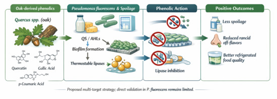
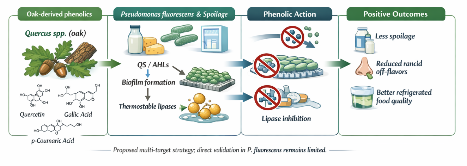
Graphical Abstract

1. Introduction

Pseudomonas fluorescens is a psychrotrophic bacterium widely recognized as a major cause of spoilage in refrigerated foods, particularly dairy products. Its spoilage capacity is not solely associated with microbial growth but with the coordinated production of virulence-associated traits, including quorum sensing (QS)-regulated biofilm formation and the secretion of heat-resistant extracellular enzymes such as lipases and proteases [1]. These enzymes catalyze the hydrolysis of triglycerides and proteins, releasing free fatty acids and peptides that generate rancid off-flavors, gelation, and textural deterioration [2,3]. The persistence of enzymatic activity even after thermal treatments represents a technological challenge, as spoilage may continue despite effective bacterial inactivation. Consequently, P. fluorescens contributes substantially to food waste and economic losses in refrigerated supply chains [4].
Conventional control strategies, including sanitation, surface disinfection, and thermal processing, primarily target bacterial cells but often fail to address the regulatory and enzymatic mechanisms underlying spoilage [5,6]. Biofilm formation enhances persistence on processing surfaces, while thermostable lipases remain active after pasteurization and ultra-high-temperature treatments [7,8]. Moreover, repeated exposure to disinfectants may select for tolerant populations and contribute to environmental and material degradation [6]. These limitations suggest that strategies focused solely on microbial elimination may be insufficient and that targeting QS networks and enzymatic activity could offer complementary approaches. However, the molecular links between QS regulation and lipase expression remain incompletely characterized, and the extent to which interference with signaling pathways effectively reduces enzymatic spoilage remains unclear.
Plant-derived phenolic compounds have emerged as promising candidates capable of modulating bacterial communication and enzymatic activity. These secondary metabolites exhibit diverse bioactivities, including membrane disruption, transcriptional modulation, and enzyme inhibition [9,10,11]. In several Pseudomonas species, flavonoids and phenolic acids such as quercetin, catechin, baicalein, gallic acid, p-coumaric acid, and chlorogenic acid have been reported to reduce QS gene expression and inhibit biofilm formation [12,13,14,15]. In parallel, numerous studies have demonstrated lipase inhibition by phenolic-rich extracts and isolated compounds, primarily using mammalian pancreatic models [16,17].
Nevertheless, direct extrapolation of these findings to spoilage-associated bacterial lipases warrants caution. Although mammalian and bacterial lipases share a conserved α/β-hydrolase fold, differences in secretion systems, thermostability, and regulatory mechanisms may influence inhibitor susceptibility. To date, systematic enzymatic or structural evaluations of phenolic compounds against P. fluorescens lipases remain scarce. Furthermore, most studies focus on isolated phenolic acids or flavonoids. In contrast, complex plant matrices contain additional metabolites, such as terpenoids and other secondary compounds, whose potential contribution to QS interference and enzymatic modulation has been largely overlooked.
Among phenolic-rich taxa, oaks (Quercus spp.) represent a chemically diverse genus widely distributed in temperate ecosystems. Extracts from Quercus species have demonstrated QS inhibition and antilipolytic activity in model organisms and mammalian systems [18,19]. However, many reports rely on crude extracts without comprehensive metabolomic characterization, limiting mechanistic interpretation and reproducibility. Moreover, their specific effects on QS-regulated lipase production in P. fluorescens have not been systematically analyzed.
Given the fragmented nature of current evidence, an integrative assessment of QS regulation, lipase biology, and plant-derived inhibitors in P. fluorescens is needed. This review critically examines the molecular mechanisms underlying QS-mediated spoilage and lipase production in P. fluorescens, evaluates existing evidence on phenolic inhibition, and identifies key gaps that must be addressed to advance toward rational, food-relevant applications of oak-derived metabolites.

2. Lipolysis as a Reaction Affecting Food Quality

Lipolysis is a major biochemical reaction associated with food deterioration across diverse matrices, including dairy products, meat, fish, and fresh produce. Microbial lipases hydrolyze triglycerides into free fatty acids and glycerol, generating off-flavors, textural alterations, and reduced shelf life [20,21,22]. Among lipolytic spoilage bacteria, P. fluorescens is particularly relevant due to its psychrotrophic growth, widespread environmental distribution, and ability to secrete extracellular thermostable lipases active at refrigeration temperatures.

2.1. Pseudomonas fluorescens as a Cause of Food Spoilage

P. fluorescens is a Gram-negative, psychrotrophic bacterium frequently isolated from refrigerated and high-moisture foods with near-neutral pH, including ready-to-eat products, meats, vegetables, and especially dairy matrices [2,21]. This microorganism is consistently identified as one of the main causes of spoilage in milk and dairy derivatives, where its enzymatic activity promotes rancidity and sensory deterioration [23,24]. Its persistence in food systems is driven by several adaptive traits: tolerance to low temperatures (3–7 °C), resistance to heat, and a strong ability to form surface biofilms [25,26]. Most critically, P. fluorescens secretes persistent extracellular enzymes, including proteases, pectinases, and lipases, that remain active under processing and storage conditions and are directly responsible for undesirable physical and chemical changes in foods [2,25,27].
The manifestations of P. fluorescens spoilage vary by product type. In dairy foods, its enzymatic activity leads to bitter and rancid flavors in milk and cheese, gelation of UHT milk, accelerated rancidity in butter, and the development of fruity or off-flavors in yogurt [21,28]. Some strains also synthesize a blue indigo-derived pigment, responsible for blue discolorations in fresh cheeses such as mozzarella and Burgos [2,23]. In meats and fish, P. fluorescens contributes to the formation of surface slime, unpleasant odors, and texture degradation [12,20,22]. In vegetables, the bacterium alters texture and is implicated in browning reactions [21]. Collectively, these effects reduce the sensory quality, shelf life, and economic value of foods across diverse product categories.
Contamination by P. fluorescens can occur throughout the production chain. In dairy systems, sources include the cow’s udder, milking personnel, and milking equipment [7,27]. The organism can also persist in production environments, transport tanks, and processing pipelines by forming biofilms that resist cleaning and sanitation [24,29]. Environmental reservoirs, such as soil, water, and air, further facilitate contamination. Dust or untreated water are frequent vectors of recontamination in pasteurized products [3,6,8]. In meat processing, contamination often arises during slaughtering, cutting, or packaging [20]. In contrast, for plant products, contamination is influenced by harvesting and post-harvest handling conditions [21].
The persistence of P. fluorescens in processing environments and its production of heat-resistant lipases represent major challenges for food manufacturers. Understanding the mechanisms underlying biofilm formation, lipase stability, and regulation is needed to design new strategies to prevent rancidity and extend food shelf life.

2.2. Biofilms Regulated by Quorum Sensing and Their Relation to Lipolysis in Pseudomonas fluorescens

2.2.1. Structure and Composition of P. fluorescens Biofilms

Biofilm formation in P. fluorescens is closely related to its ability to persist and cause food spoilage. These biofilms consist of extracellular matrices composed mainly of exopolysaccharides (EPS), extracellular DNA (eDNA), proteins, fats, and minerals [1,3]. Through the biofilm, bacterial cells adhere to the surfaces of equipment in constant contact with food, as well as to the food itself, reducing its shelf life and causing organoleptic changes. In addition, this adherence can cause corrosion of metal surfaces [30,31]. Biofilms also act as reservoirs of bacterial enzymes, including lipases, whose production is increased compared to that in planktonic cells [1]. These complex matrices can harbor multiple bacterial species, including pathogens, and provide them with a protected environment, making them difficult to kill and conferring resistance against antibiotics and other antimicrobial agents [3,32,33]. Thus, biofilms not only protect P. fluorescens and its enzymes from adverse environmental factors but also prolong the presence of bacterial communities during food production and storage, contributing significantly to food spoilage [1,3,34,35].
The extracellular matrix of biofilms produced by P. fluorescens is composed of polysaccharides such as alginate, poly-N-acetyl-glucosamine (PNGA), levan, glucuronic, and guluronic acid [36,37]. This polymeric matrix accounts for 50–90% of the total organic carbon, forming rigid, poorly soluble structures. In addition, biofilms contain eDNA, which can come from the lysis of the bacterial cells themselves [1]. This eDNA plays a key role in both cell adhesion in the early stages of biofilm formation and in the structure of mature biofilms [38,39].
Non-fimbrial adhesion megaproteins are another essential component of P. fluorescens biofilms [38]. These adhesins belong to the RTX family and are transported by the type I secretion system. They play a key role in cell attachment to surfaces. Among them, LapA and MapA proteins stand out [36,40]. Likewise, FAP amyloid fibers are present in P. fluorescens biofilms, contributing to the matrix structure [39]. Other components, although to a lesser extent, include β-cyclic glucans, lipoproteins (LPS), and membrane vesicles, which increase the complexity and functionality of biofilms [38,39]. These structural and functional elements make P. fluorescens biofilms robust and adaptive systems, favoring their persistence in food processing environments and their resistance to conventional control treatments.

2.2.2. Stages of Biofilm Development

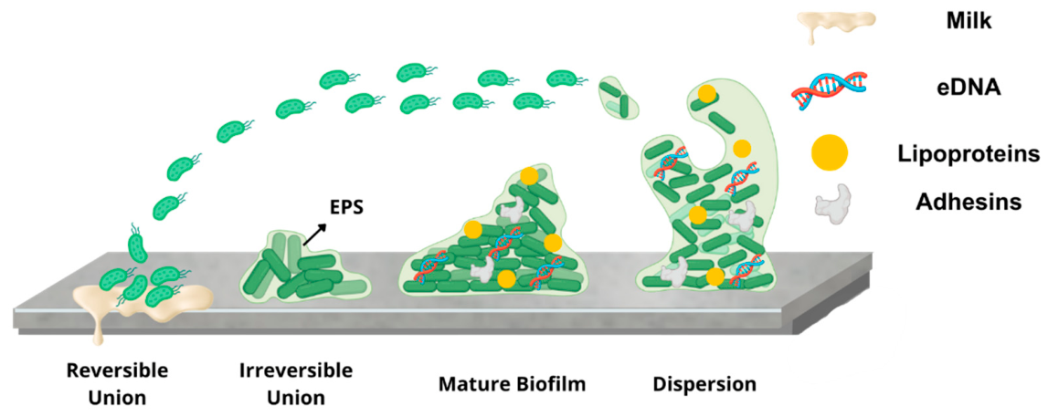
Biofilm formation in P. fluorescens follows a series of distinctive stages, including initial attachment of cells to the surface (1), adhesion and secretion of EPS (2), maturation of the biofilm (3), and, finally, dispersal of bacterial cells to form new biofilms (4) (Figure 1) [33]. In the first stage, surface conditioning occurs, where physicochemical properties such as hydrophobicity, electrostatic charges, and surface free energy are modified. In dairy processing, this conditioning occurs through contact with organic molecules present in the milk, resulting in the formation of a conditioning film [31]. Planktonic cells approach this conditioned surface through mechanisms such as convection, Brownian motion, or sedimentation, and are weakly bound by Van der Waals forces, as well as electrostatic and hydrophobic interactions. The binding is reversible at this initial stage [33,41].
In the second stage, cell adhesion becomes irreversible. This process involves hydrogen bonds, covalent bonds, ionic bonds, and dipole–dipole interactions between the hydrophobic cell surface and the interface [33,41]. Bacterial adhesion structures, both fimbrial (flagella, pili, and fimbriae) and non-fimbrial (LapA and MapA), are also involved. The former facilitates cell movement and adhesion to the surface, while non-fimbrial structures mediate the transition from a transient to a permanent bond. In turn, adherent bacteria synthesize and excrete EPS, forming ionic bridges with the surface conditioning film [31,33,42].
The third stage corresponds to biofilm maturation. Bacteria proliferate within the EPS matrix, forming microcolonies and generating three-dimensional mushroom-like structures. As more EPS is secreted, the matrix expands and acts as a glue between the embedded cells. During this phase, water channels are also created that distribute nutrients to the community and remove waste [33,42]. During this phase, QS-mediated gene regulation is observed, as discussed later.
Finally, stage 4 involves the biofilm dispersal process, considered an evolutionary strategy of bacteria to form new biofilms [42,43]. The dispersal mechanism is divided into three phases: separating cells from microcolonies, their movement to new surfaces, and their subsequent adhesion [33,42]. Two types of dispersal are recognized. Passive detachment occurs due to mechanical factors (grazing, abrasion, erosion). At the same time, in active dispersal, embedded cells actively escape from the matrix, primarily mediated by signals originating from within the biofilm (native) or from outside (environment) [33,44]. Together, these biofilm-forming steps enable P. fluorescens to establish resilient, adaptive bacterial communities on diverse surfaces, especially in industrial and food environments.

2.2.3. Environmental and Molecular Factors Influencing Biofilm Formation

Biofilm formation in P. fluorescens is governed by environmental cues and intracellular regulatory networks (Table 1). Temperature, nutrient availability, osmotic conditions, and divalent cations significantly influence the transition from planktonic to sessile growth [31,42,45]. Enhanced biofilm production has been reported at low temperatures (10 °C) and under nutrient limitation, conditions typical of refrigerated food systems [3,34]. Calcium ions (Ca2+), naturally present in dairy matrices, further promote adhesion and matrix stabilization by modulating surface proteins and gene expression [45,46].
At the molecular level, the cyclic di-guanosine monophosphate (c-di-GMP) pathway and the GacA/GacS small RNA (sRNA) system act as central regulators of biofilm development [3,47,49,50]. c-di-GMP promotes adhesion protein expression (LapA and MapA) while repressing motility [40,48,49], whereas the GacA/GacS system modulates virulence-associated traits and influences the balance between motile and sessile states [48,50]. Together, these interconnected regulatory mechanisms can underscore the complexity of biofilm control in P. fluorescens.

2.2.4. Quorum Sensing and Its Connection to Spoilage Activity in P. fluorescens

The third reported signaling mechanism controlling biofilm formation in P. fluorescens is QS. QS refers to the intercellular communication system mediated by the production and detection of signaling molecules known as autoinducers (AIs), which depend on cell density and can regulate the collective behavior of bacterial communities [3,15,51]. This system controls various stages of biofilm formation and regulates the synthesis of biosurfactants, antibiotics, virulence factors (motility), efflux pumps, and extracellular enzymes (proteases, lipases, chitinases, pectinases), as well as pigments such as pyoverdines, which contribute to food spoilage [3,52].
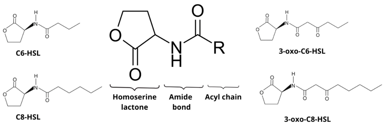
In P. fluorescens, the QS system is based on the autoinducer molecules N-acyl-homoserine lactones (AHLs), which are produced by the LuxI-type synthase and recognized by the LuxR-type transcriptional regulator [3,51,53]. AHLs comprise a fatty acyl chain and lactonized homoserine, with structures that can have between 4 and 18 carbons (Figure 2) [3,54]. Their synthesis occurs from S-adenosylmethionine (SAM) molecules, serving as an amino donor, and an acylated carrier protein (ACP) as a precursor to the acyl side chain [54,55]. Once synthesized, AHLs leave the cell by diffusion, forming a complex with LuxR from other cells when the density is high, to finally bind to the promoters of the target genes that will activate specific pathways related to biofilm formation, production of virulence factors, and extracellular enzymes, granting its damaging capacity to P. fluorescens [3,52,55].
Several QS systems have been reported for P. fluorescens, which may vary depending on the strain [3,53]. There are LuxI/R homologs, such as mpuI/R, which regulates mupirocin biosynthesis in P. fluorescens NCIMB 10586 [56], as well as the phzI/R system in P. fluorescens 2–79 [57]. PcoI/R is another LuxI/R-type system reported for the P. fluorescens 2P24 strain, isolated from wheat roots, whose role has been associated with regulating biofilm formation, colonization, and biocontrol activity [58]. Other QS systems are not based on LuxI/R, such as the HdtS protein in P. fluorescens F113, which only produces AHLs, and PsoR, a protein also known as LuxR alone, which can respond to AHLs produced by neighboring cells but lacks a LuxI-type synthase [3,59]. Each system synthesizes and recognizes different types of AHLs (Table 2), becoming a complex and diverse system within P. fluorescens.
Biofilm formation and QS-mediated regulation in P. fluorescens are key processes that enhance its ability to adhere to surfaces, resist antimicrobial treatments, and produce extracellular enzymes, such as lipases, responsible for food spoilage. These mechanisms hinder the control of this bacterium in industrial settings, thereby accelerating food spoilage and negatively impacting its quality and shelf life. Therefore, the design of strategies to inhibit biofilm formation and QS signaling represents a promising approach to minimizing the impact of P. fluorescens in the food industry.

2.3. Pseudomonas fluorescens Lipases and Their Relationship with Food Spoilage

2.3.1. Biochemical Properties of P. fluorescens Lipases

Among the primary factors contributing to P. fluorescens’s spoilage capacity is the production of extracellular hydrolytic enzymes, particularly lipases. Lipases (triacylglycerol acylhydrolases, E.C.3.1.1.3) are enzymes that hydrolyze triglycerides present in food, releasing glycerol and fatty acids that impart unpleasant flavors and odors [21]. These enzymes release short-chain (between carbon atoms C4 and C8) and medium-chain unsaturated fats; the former is associated with rancid flavors, while the latter contributes to a bitter, soapy taste [2,3]. P. fluorescens lipases exhibit lipolytic activity at pH between 7 and 10, with increased activity at temperatures between 4 and 7 °C, which is why they are so effective in refrigerated foods [2,3,62]. They are also heat-resistant, surviving thermal processes such as pasteurization and UHT (Ultra-High Temperature) and remaining stable at temperatures up to 120 °C [7,8,63]. This thermostability distinguishes them from most other psychrotrophic enzymes, highlighting their role in the deterioration of refrigerated products, particularly dairy items.

2.3.2. Structural Features of P. fluorescens Lipases

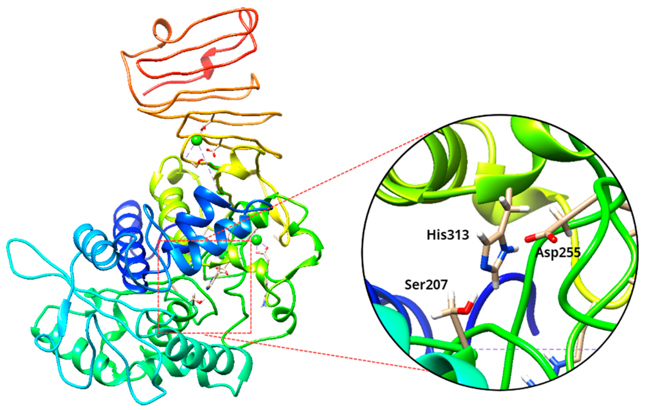
P. fluorescens lipases belong to the α/β-hydrolase fold superfamily and are classified within the serine hydrolase group [64,65]. They include members of subfamilies I.1 and I.3, with molecular weights ranging from 30 kDa (I.1) to 50–65 kDa (I.3), the latter being the most abundant [63,66]. The catalytic center (Figure 3) is composed of the conserved triad of serine, aspartic (or glutamic) acid, and histidine residues, forming the enzyme’s active site located near a hydrophobic pocket on the protein surface [65,67,68].
An oxyanion hole stabilizes the tetrahedral intermediate formed during catalysis [68], while disulfide bonds provide conformational stability. A flexible oligopeptide domain or “lid” covers the active site, controlling substrate access and interfacial activation [68,69]. Certain P. fluorescens strains possess lipases with two such lids, which can shift conformation without losing activity, contributing to their resilience [70]. These structural features explain the enzyme’s robustness under the fluctuating thermal and pH conditions typical of food processing environments.

2.3.3. Genetic Organization and Regulation of Lipase Expression

Lipase production in P. fluorescens typically occurs during the late exponential to early stationary growth phases and is modulated by environmental parameters such as oxygen availability, temperature, iron and calcium concentrations, and QS [1,3,71]. Lipolytic activity begins to rise when cell density reaches approximately 106 CFU/mL and increases further when bacteria are embedded within biofilms, from which lipases can diffuse without cell detachment [1,3].
Several lipases have been characterized, including LipA, LipB, LipM, and phospholipase C [7,8,72]. LipA, produced by P. fluorescens B52, is considered the principal enzyme responsible for milk deterioration [8,73]. The aprXlipA operon, comprising eight genes, encodes both the metalloprotease AprX and the lipase LipA located at opposite ends of the locus, along with a protease inhibitor (inh), a type I secretion system (aprDEF), and two serine protease homologs (prtAB) [8,73,74]. The expression of this operon is influenced by suboptimal growth temperatures and the presence of calcium and iron, which act as transcriptional modulators [73,74].

2.3.4. Regulation by Quorum Sensing and Other Signaling Pathways

The production of P. fluorescens lipases is intricately connected to QS and additional regulatory systems. Although evidence remains limited, certain AHL molecules, notably C4-HSL, have been linked to increased lipase synthesis in P. psychrophila under pasteurized milk conditions, suggesting a similar regulatory mechanism in P. fluorescens [3,75]. QS signaling may therefore enhance the bacterium’s lipolytic potential by upregulating lipase genes in response to population density or environmental stress.
Other pathways, including the EnvZ–OmpR two-component system and the RhlI/R and Gac regulatory systems described in related Pseudomonas species, have also been implicated in lipase regulation [3,74,76]. Lipase secretion into the extracellular environment is mediated by a type I secretion system (ABC transporter), which recognizes a C-terminal targeting motif in the lipase sequence via the membrane fusion protein component [8,73,76].
Importantly, QS not only regulates lipase production but also controls biofilm development and the expression of adhesion factors in P. fluorescens. This dual regulatory role suggests that lipolytic activity and biofilm formation are functionally interconnected processes rather than independent spoilage mechanisms. Within biofilms, high cell density and coordinated gene expression may potentiate lipase secretion, while the biofilm matrix itself facilitates enzyme retention and diffusion across dairy matrices. Consequently, interference with QS signaling could simultaneously disrupt biofilm stability and lipase synthesis, representing a particularly attractive target for controlling enzymatic spoilage in refrigerated foods.

2.3.5. Lipase-Mediated Spoilage in Dairy Products

P. fluorescens lipases compromise dairy product quality through the hydrolysis of milk triglycerides, releasing free fatty acids responsible for rancid and soapy off-flavors [31]. Due to their marked resistance to pasteurization and UHT treatments, these enzymes remain active during storage and induce a range of technological defects depending on the dairy matrix (Table 3). In UHT milk, residual lipases promote gelation during refrigeration, whereas in butter and cheeses they accelerate lipid degradation and rancidity. Likewise, products formulated from milk powder containing residual lipases may develop lipolytic defects throughout shelf life [7,8,27].
The persistence of enzymatic activity despite thermal processing represents a major technological challenge. Because lipases survive treatments designed to inactivate vegetative cells, controlling bacterial contamination alone is insufficient to prevent spoilage. Consequently, effective mitigation strategies must address not only microbial growth but also lipase production, secretion, and catalytic activity.
Despite advances in the structural and functional characterization of these enzymes, significant gaps remain regarding the precise molecular mechanisms regulating their expression and activation under food-processing conditions. Further investigation of regulatory pathways, particularly those linked to QS and other signaling systems, is essential for designing targeted interventions to mitigate enzymatic spoilage. A deeper understanding of these mechanisms provides the foundation for identifying molecules that can disrupt lipase regulation or activity, thereby offering new opportunities to maintain the quality and shelf-life of refrigerated foods. In this context, growing attention has focused on natural compounds, particularly plant-derived phenolics, that can interfere with QS, biofilm formation, and lipase activity in P. fluorescens.

3. Alternative Methods for Inhibiting Deterioration Caused by Pseudomonas fluorescens in Foods

3.1. Emerging Control Strategies and the Shift Toward Natural Compounds

The limited effectiveness of conventional methods for controlling P. fluorescens has driven research toward new preservation technologies and natural antimicrobial alternatives. In food-processing environments, routine cleaning-in-place (CIP) and chemical sanitization reduce microbial loads but often fail to eliminate biofilm-associated cells or inactivate heat-stable enzymes that survive pasteurization and ultra-high-temperature (UHT) treatments [7,8]. Moreover, repeated exposure to disinfectants can promote microbial tolerance and damage processing surfaces, increasing maintenance costs and compromising equipment integrity [5,6].
To overcome these challenges, several non-thermal and biological strategies have been developed. Physical interventions such as modified-atmosphere packaging, ohmic and microwave heating, pulsed electric fields, irradiation, and high-pressure processing (HPP) have shown potential to reduce microbial loads while preserving food quality [23,77,78]. Biological approaches, including bacteriophage application, have also been explored for their ability to selectively target spoilage bacteria [79].
Despite these advances, these methods primarily act at the cellular level and seldom address the biochemical and regulatory mechanisms that sustain spoilage. Consequently, increasing attention has turned toward natural bioactive compounds with broad antimicrobial spectra, structural versatility, and lower risk of resistance development [9,26,80]. Within this context, phenolic compounds, plant secondary metabolites with well-documented antioxidant and antimicrobial properties, are emerging as promising candidates for controlling P. fluorescens by interfering with QS, biofilm formation, and lipolytic activity.

3.2. Phenolic Compounds and Their Role in the Inhibition of Quorum Sensing, Biofilms, and Lipases of Pseudomonas fluorescens

Phenolic compounds represent one of the largest families of plant metabolites, with over 10,000 structures identified [11,81]. They include phenolic acids, flavonoids, stilbenes, lignans, and tannins, molecules characterized by one or more hydroxyl groups attached to aromatic rings [82,83]. Beyond their antioxidant and anti-inflammatory properties, these metabolites exhibit antimicrobial activity through various mechanisms, including disruption of membrane integrity, interference with nucleic acid synthesis, inhibition of metabolic enzymes, and modulation of bacterial signaling pathways [84,85]. Moreover, several phenolic compounds have been shown to reduce bacterial adhesion and biofilm formation by interfering with QS regulation [3,83,84]. In addition, they can potentiate the action of antibiotics by reversing resistance mechanisms and restoring microbial susceptibility [86], making them viable candidates to combat bacterial persistence and spoilage in food systems.
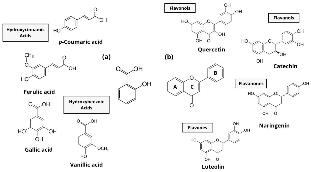
Among the major classes of these metabolites, phenolic acids (e.g., gallic, p-coumaric, and caffeic) and flavonoids (e.g., quercetin, catechin, and baicalein) are particularly relevant due to their structural diversity, which underlies their antimicrobial and enzyme-inhibitory potential. In these molecules, the number and position of hydroxyl groups (Figure 4) are key determinants of binding affinity and inhibitory efficiency toward microbial targets [87,88].
Owing to these structural and functional characteristics, phenolic compounds can simultaneously target multiple microbial pathways. In Pseudomonas fluorescens, their capacity to interfere with QS, biofilm development, and lipase activity positions them as promising molecules for mitigating enzymatic spoilage. The following sections describe the current evidence supporting these inhibitory effects.

3.2.1. Inhibition of Quorum Sensing and Biofilm Formation

The ability of phenolic compounds to disrupt QS systems was first demonstrated by Huber et al. [89] in Pseudomonas putida, where epigallocatechin gallate, ellagic acid, and tannic acid reduced QS-regulated gene expression by up to 40%. Since then, multiple studies have confirmed similar effects in Pseudomonas species associated with food spoilage (Table 4).
Quercetin has been shown to inhibit the LasR receptor of P. aeruginosa, resulting in an 80% reduction in biofilm formation and a 73% decrease in exopolysaccharide (EPS) production [15]. In P. fluorescens, flavonoids such as catechin, naringenin, and epicatechin suppressed AHL synthesis, reduced luxI expression by 42%, and inhibited biofilm formation by up to 88% [13]. Similarly, baicalein interfered with c-di-GMP signaling and RpoS-regulated genes, resulting in decreased biofilm production [12].
Phenolic acids have also demonstrated significant QS inhibitory effects. p-Coumaric and gallic acids reduced AHL synthesis and flgA expression, thereby diminishing surface colonization in P. fluorescens KM120 [14]. Chlorogenic acid immobilized in chitosan matrices inhibited flagellar motility and removed over 70% of mature biofilms [90]. Collectively, these studies highlight that phenolic compounds act at multiple regulatory levels, binding to QS receptors, downregulating QS-associated genes, and disrupting the synthesis of signaling molecules, thereby limiting the formation and persistence of biofilms in Pseudomonas species.
Importantly, QS in P. fluorescens is tightly linked to biofilm maturation and extracellular enzyme secretion. Therefore, interference at the level of AHL synthesis or receptor binding may indirectly modulate downstream pathways, including c-di-GMP signaling, motility regulation, and lipase gene expression. This integrative perspective suggests that phenolic compounds do not merely inhibit isolated virulence factors but may disrupt interconnected regulatory networks that sustain spoilage.

3.2.2. Inhibition of Lipolytic Activity

Beyond their interference with QS-regulated pathways, phenolic compounds may also directly target the enzymatic machinery responsible for lipid hydrolysis. Given that lipase production in P. fluorescens is partially modulated by QS and stress-response regulators, dual inhibition at both the regulatory and catalytic levels represents a particularly attractive strategy for controlling spoilage.
Experimental evidence supporting this catalytic interference is summarized in Table 5, where several phenolic extracts and isolated compounds have demonstrated antilipolytic activity against both mammalian and microbial lipases. Extracts from Vitis rotundifolia, Eryngium bornmuelleri, Lens culinaris, and Intsia palembanica, all rich in flavonoids, have demonstrated strong inhibitory effects on pancreatic lipases, with IC50 values below 9 mg/mL [91,92,93,94]. These results suggest that the combined presence of flavonoids and phenolic acids in complex matrices can generate synergistic inhibition, potentially through multiple binding interactions at the enzyme surface. Among isolated compounds, quercetin, kaempferol, and p-coumaric acid exhibited particularly low IC50 values, indicating that specific structural motifs, such as catechol or hydroxylated benzoyl groups, play a decisive role in catalytic inhibition [16,17].
When comparing pure compounds and plant extracts, differences in potency highlight the contribution of molecular diversity: extracts containing multiple phenolics may exert additive or complementary effects through interactions with distinct structural regions, whereas single molecules target specific catalytic residues. Structure–activity analyses reveal that a greater number of hydroxyl groups and the presence of conjugated double bonds between C2–C3 in the flavone backbone enhance lipase inhibition by stabilizing hydrogen bonds and hydrophobic π–π interactions within the active site [16,88,95]. These structural features may facilitate competitive binding within the catalytic pocket or induce conformational shifts in the lid domain of lipases, potentially restricting substrate accessibility.
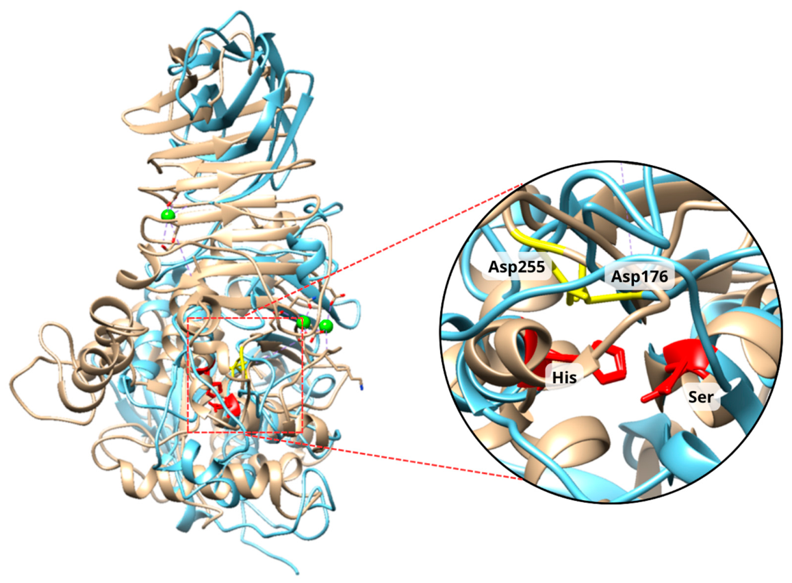
To contextualize the relevance of phenolic inhibition against pancreatic lipase models versus the potential to inhibit P. fluorescens, a structural comparison was performed between the human pancreatic lipase (HPL; PDB: 1N8S) and a predicted structural model of P. fluorescens LipB. The LipB model was generated using AlphaFold from the amino acid sequence of P. fluorescens LipB (UniProt: P41773), using the crystallographic structure of Pseudomonas sp. MIS38 lipase (PML; PDB: 2ZVD) as a structural template, a well-characterized family I.3 bacterial lipase whose catalytic triad Ser207–Asp255–His313 has been confirmed by both crystallography and MD simulation analysis [96]. Sequence alignment revealed that two of the three catalytic triad residues are positionally conserved: Ser152 of HPL aligns with Ser207 of LipB, and His263 aligns with His313, corresponding to the nucleophilic serine and the catalytic histidine, respectively (Figure 5). Structural superposition using the Matchmaker tool in UCSF Chimera, https://www.rbvi.ucsf.edu/chimera/download.html (accessed on 30 April 2026) [97] yielded an RMSD of 1.063 Å over 31 pruned Cα atom pairs, despite a low global sequence identity of 15.1%, suggesting local structural conservation within the catalytic region, despite the low overall sequence identity.
The catalytic aspartate (Asp176 in HPL) does not occupy an equivalent sequential position in LipB; however, Asp255, the functionally equivalent residue in family I.3 lipases, is present in the LipB model, consistent with the conservation of this catalytic role across Pseudomonas lipases [68,96]. Taken together, this structural comparison supports the possibility that phenolic compounds with demonstrated inhibitory activity against HPL may interact with geometrically similar features in P. fluorescens LipB, particularly within the catalytic pocket and adjacent structural motifs involved in substrate stabilization. However, this hypothesis requires direct biochemical validation.
However, direct experimental validation in bacterial systems remains limited. Despite extensive work with pancreatic enzymes, studies focusing on bacterial lipases remain scarce. Only a few reports, such as those using Propionibacterium acnes lipase, have confirmed that phenolic compounds, such as quercetin and myricetin, inhibited microbial lipolytic activity in a concentration-dependent manner [91]. Given that P. fluorescens lipases differ from mammalian counterparts in both their secretion mechanisms, via a type I secretion system, and their remarkable thermostability [8,76,98], direct extrapolation of inhibition data from pancreatic models may be limited. Addressing this gap through targeted in silico and enzymatic analyses will determine the inhibitory capacity of phenolic compounds against P. fluorescens lipases. In this context, complex plant extracts rich in diverse phenolic structures represent promising candidates for further exploration.

3.3. Plant Extracts Rich in Phenolic Compounds: The Case of Oaks (Quercus spp.)

Among phenolic-rich plant taxa investigated for antimicrobial potential, species of the genus Quercus (family Fagaceae) have attracted increasing attention due to their remarkable ecological dominance and widespread distribution across temperate and subtropical regions. With more than 500 species distributed throughout the Northern Hemisphere and approximately 150 species reported in Mexico alone, Quercus represents one of the most diverse woody genera globally [82,99].
Likewise, Quercus species constitute a widely available botanical resource within forestry, cooperage, and agri-food systems. Oak wood is widely used in barrel production for wine and spirits aging, as well as in the timber and furniture industries, generating bark, chips, sawdust, and other lignocellulosic residues [100,101,102]. Seasonal pruning also produces substantial amounts of leaves that remain underexploited. In addition, acorn, traditionally consumed in various cultures as a food ingredient or animal feed, represents another plant matrix derived from this genus [103,104,105].
From a phytochemical perspective, these diverse oak-derived materials are characterized by a high phenolic load, including abundant phenolic acids and flavonoids such as gallic, ellagic, p-coumaric, vanillic, caffeic, syringic, and ferulic acids, as well as rutin, quercetin, catechin, epicatechin, kaempferol, and naringenin [10,82,106]. These metabolites, distributed across bark, leaves, acorns, and galls, confer antioxidant, antimicrobial, and anti-inflammatory activities [107], supporting the exploration of oak extracts as multifunctional sources of bioactive compounds for food preservation applications.
Beyond the presence of individual phenolic compounds, the rationale for focusing on oak-derived extracts should be understood in terms of their overall compositional architecture rather than the uniqueness of isolated metabolites. Many of the compounds discussed in this review, such as quercetin, catechin, gallic acid, and p-coumaric acid, are widely distributed across plant taxa. However, their simultaneous occurrence within phenolic matrices enriched in hydrolyzable tannins may distinguish Quercus spp. from other widely studied botanical sources. For example, Camellia sinensis is dominated by condensed tannins (catechins, particularly epigallocatechin gallate), Vitis vinifera is rich in anthocyanins, proanthocyanidins, and stilbenes, while Rosmarinus officinalis is distinguished by phenolic diterpenes such as carnosic acid and carnosol [108,109,110]. In contrast, Quercus species are frequently characterized by phenolic profiles enriched in hydrolyzable tannins, particularly ellagitannins and galloylated derivatives, coexisting with flavonols and phenolic acids across different organs and tissues [82,111,112]. This compositional arrangement may favor complementary modes of action; however, the contribution of each phenolic subclass to QS and lipase modulation remains to be experimentally clarified. Where low-molecular-weight phenolics interact with regulatory proteins involved in QS, while multivalent tannins establish strong interactions with extracellular enzymes, potentially modulating enzymatic activity [13,15,17,113]. Such diversity in molecular size and structure suggests that oak extracts may exert multi-target effects rather than acting through isolated inhibitory mechanisms, an aspect particularly relevant for QS-regulated lipase production in P. fluorescens. A comparative overview of representative phenolic architectures across selected plant matrices is presented in Table 6.

3.3.1. Evidence of QS and Biofilm Inhibition by Oak Extracts

Several studies have demonstrated that Quercus extracts can interfere with QS-regulated processes in Gram-negative bacteria. Gall extracts of Q. infectoria contained protocatechuic, chlorogenic, and gallic acids, as well as flavonoids such as quercetin, astragalin, and luteolin, which significantly reduced the expression of QS-related genes in P. aeruginosa. This resulted in a decrease in the production of proteases, pyocyanin, and biofilms [18].
In another study, bark extracts from Quercus sp. inhibited AHL synthesis and the production of extracellular enzymes in Pectobacterium carotovorum, also suppressing the expR/I regulatory system [121]. Likewise, Q. robur bark extract exhibited strong QS inhibitory activity in Chromobacterium violaceum CV026, reducing violacein pigment production without exerting significant bactericidal effects, an indication of specific QS interference [122]. A subsequent comparative analysis by Inchagova et al. [123] evaluated several plant extracts against C. violaceum ATCC 31532 and found Q. cortex extract to be the most effective at inhibiting violacein biosynthesis, confirming the high QS inhibitory potential of the genus.
Although these studies have focused mainly on model organisms or pathogenic Pseudomonas species, the consistent inhibitory trends suggest that oak-derived phenolics may act through multiple molecular targets, including AHL synthases (LuxI-type proteins) and regulatory receptors (LuxR homologs). Given that P. fluorescens employs analogous QS signaling molecules and homologous regulatory networks, these QS inhibitory findings are particularly relevant for exploring oak extracts as inhibitors of spoilage-associated QS and biofilm development.

3.3.2. Antilipolytic Potential of Oak-Derived Phenolics

In addition to QS inhibitory effects, oak extracts have shown promising activity against lipolytic enzymes; however, most research has been conducted in mammalian systems. Extracts from Q. infectoria inhibited pancreatic lipases by up to 85%, reflecting strong antilipolytic potential [19,124]. These effects were attributed to the presence of hydrolysable tannins and galloyl derivatives, although the specific mechanisms and binding sites remain to be clarified.
Structurally, major phenolic compounds identified in Quercus species, such as quercetin, catechin, and p-coumaric acid, possess multiple hydroxyl groups and conjugated double bonds that facilitate hydrogen bonding and π–π stacking interactions with catalytic residues of lipases [87,125]. In silico analyses support this mechanism: quercetin has been predicted to bind near the lipase active site, inducing conformational changes that reduce substrate affinity [125]. Given the structural similarities among bacterial and mammalian lipase catalytic domains, these observations suggest that oak-derived phenolics could likewise interfere with P. fluorescens lipases, potentially limiting enzymatic spoilage in dairy systems.
Taken together, the dual capacity of oak-derived phenolics to interfere with QS and inhibit lipolytic activity positions Quercus species as representative examples of phenolic-rich plant systems with potential to disrupt interconnected spoilage mechanisms. Although direct studies on P. fluorescens remain limited, the mechanistic parallels observed across Gram-negative bacteria and mammalian lipase models provide a strong rationale for further investigation under food-relevant conditions. Such integrative approaches may clarify whether complex oak extracts can simultaneously attenuate regulatory signaling and enzymatic degradation in refrigerated food matrices.

3.3.3. Potential Application Strategies of Oak-Derived Phenolics in Food Systems

Although most studies on oak-derived phenolics have focused on mechanistic and in vitro evaluations, their practical implementation in food systems may follow several complementary approaches. One strategy involves the direct incorporation of phenolic-rich oak extracts into food matrices at sub-inhibitory concentrations that modulate QS and lipase activity without compromising sensory quality. By targeting regulatory pathways rather than exerting strong bactericidal effects, such applications may reduce enzymatic spoilage while minimizing selective pressure for resistance [126].
Notably, oak extracts have already been evaluated in food systems, including milk and lipid-rich products, for their capacity to reduce oxidative rancidity and extend shelf life [127,128,129]. While oxidative lipid deterioration differs mechanistically from enzyme-mediated lipolysis, both processes ultimately compromise lipid stability and sensory quality [130]. Therefore, existing evidence of the incorporation of oak extract into dairy matrices supports their technological feasibility and consumer acceptability. This prior application provides a practical foundation for exploring their additional functionality as modulators of QS-regulated lipase production and activity.
Beyond direct incorporation, oak-derived phenolics can also be incorporated into edible coatings or active packaging systems, enabling the controlled release of bioactive compounds at the food–surface interface, where biofilm formation and enzyme secretion are most critical [131,132,133]. Another promising strategy involves their use as natural adjuncts in cleaning-in-place (CIP) protocols, targeting biofilm-associated cells and interfering with QS signaling before substantial lipase accumulation occurs. Given their multifunctional properties, oak phenolics could be incorporated into multi-hurdle preservation strategies alongside mild thermal treatments, modified-atmosphere packaging, or high-pressure processing [29].
However, translation from in vitro inhibition to industrial application requires further investigation. Critical aspects include determining effective concentrations in real food matrices, assessing potential sensory impacts, evaluating compound stability during processing and storage, and addressing regulatory and safety considerations. Bridging these gaps will be essential to fully exploit oak-derived phenolics as sustainable tools for controlling enzymatic spoilage in refrigerated dairy products.

4. Conclusions

P. fluorescens remains one of the most persistent spoilage microorganisms in refrigerated foods, largely due to its ability to form biofilms, communicate through QS, and secrete persistent lipases that resist conventional sanitization and heat treatments. Traditional control strategies, focused on bacterial elimination, have proven inadequate to mitigate enzymatic spoilage, underscoring the need for molecular approaches that disrupt the underlying regulatory and catalytic mechanisms driving spoilage.
In this context, plant-derived phenolic compounds emerge as promising natural agents capable of interfering with multiple virulence pathways in P. fluorescens. Evidence indicates that phenolic acids and flavonoids can inhibit QS signaling, impair biofilm development, and reduce lipolytic activity, thereby addressing both the regulatory and enzymatic dimensions of spoilage. Their structural diversity, particularly the presence of hydroxyl and conjugated groups, underlies these multifunctional effects and provides a rational basis for their targeted use in food preservation.
Among natural sources, oaks (Quercus spp.) constitute a rich reservoir of phenolic metabolites, such as quercetin, p-coumaric, and gallic acids, whose antimicrobial, antioxidant, and antilipolytic properties make them especially attractive for application in food systems. Despite encouraging findings from studies in related Pseudomonas species and pancreatic models, the direct evaluation of oak-derived phenolics against P. fluorescens lipases and QS networks remains limited.
Future research should focus on (i) elucidating the molecular mechanisms by which phenolics modulate QS and lipase activity, (ii) characterizing and comparing the phenolic profiles of different Quercus species, and (iii) validating their efficacy in real food matrices under industrial conditions. Integrating these natural compounds into multi-hurdle preservation strategies could offer sustainable and consumer-acceptable solutions to extend the shelf life and quality of refrigerated foods.

Author Contributions

Conceptualization, E.D.O.-D. and J.F.A.-Z.; methodology, E.D.O.-D.; investigation, E.D.O.-D.; writing—original draft preparation, E.D.O.-D.; writing—review and editing, B.A.S.-E., G.A.G.-A., K.D.G.-O., C.J.G.-P., M.E.B.-M. and J.F.A.-Z.; supervision, B.A.S.-E. and J.F.A.-Z.; project administration, B.A.S.-E.; funding acquisition, B.A.S.-E. All authors have read and agreed to the published version of the manuscript.

Funding

This research was funded by the Ministry of Science, Humanities, Technology and Innovation (SECIHTI), Mexico, under Grant CBF-2025-I-4295. (Basic and Frontier Science Initiative). The APC was funded by the same grant.

Data Availability Statement

No new data were created or analyzed in this study. Data sharing is not applicable to this article.

Acknowledgments

The authors acknowledge the doctoral scholarship granted to Elsa Daniela Othón-Díaz by the Ministry of Science, Humanities, Technology and Innovation (SECIHTI), Mexico. Grammarly AI-assisted writing tools were used to improve the English language, grammar, punctuation, and readability of the manuscript, https://www.grammarly.com/ (accessed on 30 April 2026). All AI-assisted edits were reviewed and approved by the authors, who take full responsibility for the final content of the manuscript.

Conflicts of Interest

The authors declare no conflicts of interest.

Abbreviations

The following abbreviations are used in this manuscript:
AHLAcyl-homoserine lactone
CIPCleaning-in-place
EPSExtracellular polymeric substances
HPPHigh-pressure processing
IC50Half maximal inhibitory concentration
QSQuorum sensing
UHTUltra-high-temperature

References

  1. Teh, K.H.; Flint, S.; Palmer, J.; Andrewes, P.; Bremer, P.; Lindsay, D. Biofilm—An unrecognised source of spoilage enzymes in dairy products? Int. Dairy J. 2014, 34, 32–40. [Google Scholar] [CrossRef]
  2. Kumar, H.; Franzetti, L.; Kaushal, A.; Kumar, D. Pseudomonas fluorescens: A potential food spoiler and challenges and advances in its detection. Ann. Microbiol. 2019, 69, 873–883. [Google Scholar] [CrossRef]
  3. Quintieri, L.; Caputo, L.; Brasca, M.; Fanelli, F. Recent Advances in the Mechanisms and Regulation of QS in Dairy Spoilage by Pseudomonas spp. Foods 2021, 10, 88. [Google Scholar] [CrossRef] [PubMed]
  4. Corrêa, A.P.; Daroit, D.J.; Velho, R.V.; Brandelli, A. Hydrolytic potential of a psychrotrophic Pseudomonas isolated from refrigerated raw milk. Braz. J. Microbiol. 2011, 42, 1479–1484. [Google Scholar] [CrossRef][Green Version]
  5. Marino, M.; Maifreni, M.; Baggio, A.; Innocente, N. Inactivation of Foodborne Bacteria Biofilms by Aqueous and Gaseous Ozone. Front. Microbiol. 2018, 9, 2024. [Google Scholar] [CrossRef]
  6. Tirpanci-Sivri, G.; Abdelhamid, A.G.; Kasler, D.R.; Yousef, A.E. Removal of Pseudomonas fluorescens biofilms from pilot-scale food processing equipment using ozone-assisted cleaning-in-place. Front. Microbiol. 2023, 14, 1141907. [Google Scholar] [CrossRef]
  7. Hu, Z.; Meng, X.-C.; Liu, F. Isolation and characterisation of lytic bacteriophages against Pseudomonas spp., a novel biological intervention for preventing spoilage of raw milk. Int. Dairy J. 2016, 55, 72–78. [Google Scholar] [CrossRef]
  8. Machado, S.G.; Baglinière, F.; Marchand, S.; Van Coillie, E.; Vanetti, M.C.D.; De Block, J.; Heyndrickx, M. The Biodiversity of the Microbiota Producing Heat-Resistant Enzymes Responsible for Spoilage in Processed Bovine Milk and Dairy Products. Front. Microbiol. 2017, 8, 302. [Google Scholar] [CrossRef]
  9. Gonçalves, S.; Romano, A. Inhibitory Properties of Phenolic Compounds Against Enzymes Linked with Human Diseases. In Phenolic Compounds; Marcos, S.-H., Mariana, P.-T., Maria del Rosario, G.-M., Eds.; IntechOpen: Rijeka, Croatia, 2017; Chapter 6. [Google Scholar]
  10. Morales, D. Oak trees (Quercus spp.) as a source of extracts with biological activities: A narrative review. Trends Food Sci. Technol. 2021, 109, 116–125. [Google Scholar] [CrossRef]
  11. Tanase, C.; Coșarcă, S.; Muntean, D.-L. A Critical Review of Phenolic Compounds Extracted from the Bark of Woody Vascular Plants and Their Potential Biological Activity. Molecules 2019, 24, 1182. [Google Scholar] [CrossRef]
  12. Cen, C.; Wang, X.; Li, H.; Chen, J.; Wang, Y. An inhibitor of the adaptability of Pseudomonas fluorescens in a high-salt environment. Phenomenon and mechanism of inhibition. Int. J. Food Microbiol. 2024, 412, 110553. [Google Scholar] [CrossRef] [PubMed]
  13. Ding, T.; Li, T.; Li, J. Virtual screening for quorum-sensing inhibitors of Pseudomonas fluorescens P07 from a food-derived compound database. J. Appl. Microbiol. 2019, 127, 763–777. [Google Scholar] [CrossRef]
  14. Myszka, K.; Schmidt, M.T.; Białas, W.; Olkowicz, M.; Leja, K.; Czaczyk, K. Role of gallic and p-coumaric acids in the AHL-dependent expression of flgA gene and in the process of biofilm formation in food-associated Pseudomonas fluorescens KM120. J. Sci. Food Agric. 2016, 96, 4037–4047. [Google Scholar] [CrossRef]
  15. Gopu, V.; Meena, C.K.; Shetty, P.H. Quercetin Influences Quorum Sensing in Food Borne Bacteria: In-Vitro and In-Silico Evidence. PLoS ONE 2015, 10, e0134684. [Google Scholar] [CrossRef] [PubMed]
  16. Martinez-Gonzalez, A.I.; Alvarez-Parrilla, E.; Díaz-Sánchez, Á.G.; de la Rosa, L.A.; Núñez-Gastélum, J.A.; Vazquez-Flores, A.A.; Gonzalez-Aguilar, G.A. In vitro Inhibition of Pancreatic Lipase by Polyphenols: A Kinetic, Fluorescence Spectroscopy and Molecular Docking Study. Food Technol. Biotechnol. 2017, 55, 519–530. [Google Scholar] [CrossRef]
  17. Moreno-Córdova, E.N.; Arvizu-Flores, A.A.; Valenzuela-Soto, E.M.; García-Orozco, K.D.; Wall-Medrano, A.; Alvarez-Parrilla, E.; Ayala-Zavala, J.F.; Domínguez-Avila, J.A.; González-Aguilar, G.A. Gallotannins are uncompetitive inhibitors of pancreatic lipase activity. Biophys. Chem. 2020, 264, 106409. [Google Scholar] [CrossRef]
  18. Ahmed, A.A.; Salih, F.A. Quercus infectoria gall extracts reduce quorum sensing-controlled virulence factors production and biofilm formation in Pseudomonas aeruginosa recovered from burn wounds. BMC Complement. Altern. Med. 2019, 19, 177. [Google Scholar] [CrossRef]
  19. Gholamhoseinian, A.; Shahouzehi, B.; Sharifi-Far, F. Inhibitory effect of some plant extracts on pancreatic lipase. Int. J. Pharmacol. 2010, 6, 18–24. [Google Scholar] [CrossRef]
  20. de Andrade Cavalari, C.M.; Imazaki, P.H.; Pirard, B.; Lebrun, S.; Vanleyssem, R.; Gemmi, C.; Antoine, C.; Crevecoeur, S.; Daube, G.; Clinquart, A.; et al. Carnobacterium maltaromaticum as bioprotective culture against spoilage bacteria in ground meat and cooked ham. Meat Sci. 2024, 211, 109441. [Google Scholar] [CrossRef]
  21. Raposo, A.; Pérez, E.; de Faria, C.T.; Ferrús, M.A.; Carrascosa, C. Food Spoilage by Pseudomonas spp.—An Overview. In Foodborne Pathogens and Antibiotic Resistance; Singh, O.V., Ed.; John Wiley & Sons, Inc.: Hoboken, NJ, USA, 2016; pp. 41–71. [Google Scholar] [CrossRef]
  22. Wang, Y.; Feng, L.; Lu, H.; Zhu, J.; Kumar, V.; Liu, X. Transcriptomic analysis of the food spoilers Pseudomonas fluorescens reveals the antibiofilm of carvacrol by interference with intracellular signaling processes. Food Control 2021, 127, 108115. [Google Scholar] [CrossRef]
  23. Martin, N.H.; Torres-Frenzel, P.; Wiedmann, M. Invited review: Controlling dairy product spoilage to reduce food loss and waste. J. Dairy Sci. 2021, 104, 1251–1261. [Google Scholar] [CrossRef]
  24. Scatamburlo, T.M.; Yamazi, A.K.; Cavicchioli, V.Q.; Pieri, F.A.; Nero, L.A. Spoilage potential of Pseudomonas species isolated from goat milk. J. Dairy Sci. 2015, 98, 759–764. [Google Scholar] [CrossRef]
  25. Karanth, S.; Feng, S.; Patra, D.; Pradhan, A.K. Linking microbial contamination to food spoilage and food waste: The role of smart packaging, spoilage risk assessments, and date labeling. Front. Microbiol. 2023, 14, 1198124. [Google Scholar] [CrossRef]
  26. Quintieri, L.; Zühlke, D.; Fanelli, F.; Caputo, L.; Liuzzi, V.C.; Logrieco, A.F.; Hirschfeld, C.; Becher, D.; Riedel, K. Proteomic analysis of the food spoiler Pseudomonas fluorescens ITEM 17298 reveals the antibiofilm activity of the pepsin-digested bovine lactoferrin. Food Microbiol. 2019, 82, 177–193. [Google Scholar] [CrossRef]
  27. Ledenbach, L.H.; Marshall, R.T. Microbiological Spoilage of Dairy Products. In Compendium of the Microbiological Spoilage of Foods and Beverages; Sperber, W.H., Doyle, M.P., Eds.; Springer: New York, NY, USA, 2009; pp. 41–67. [Google Scholar] [CrossRef]
  28. Tayyarcan, E.K.; Boyaci, I.H. Isolation, characterization, and application of bacteriophage cocktails for the biocontrol of Pseudomonas fluorescens group strains in whole and skimmed milk. Braz. J. Microbiol. 2023, 54, 3061–3071. [Google Scholar] [CrossRef]
  29. Quintieri, L.; Fanelli, F.; Caputo, L. Antibiotic Resistant Pseudomonas Spp. Spoilers in Fresh Dairy Products: An Underestimated Risk and the Control Strategies. Foods 2019, 8, 372. [Google Scholar] [CrossRef]
  30. Zarei, M.; Yousefvand, A.; Maktabi, S.; Pourmahdi Borujeni, M.; Mohammadpour, H. Identification, phylogenetic characterisation and proteolytic activity quantification of high biofilm-forming Pseudomonas fluorescens group bacterial strains isolated from cold raw milk. Int. Dairy J. 2020, 109, 104787. [Google Scholar] [CrossRef]
  31. Marchand, S.; De Block, J.; De Jonghe, V.; Coorevits, A.; Heyndrickx, M.; Herman, L. Biofilm Formation in Milk Production and Processing Environments; Influence on Milk Quality and Safety. Compr. Rev. Food Sci. Food Saf. 2012, 11, 133–147. [Google Scholar] [CrossRef]
  32. Rossi, C.; Serio, A.; Chaves-López, C.; Anniballi, F.; Auricchio, B.; Goffredo, E.; Cenci-Goga, B.T.; Lista, F.; Fillo, S.; Paparella, A. Biofilm formation, pigment production and motility in Pseudomonas spp. isolated from the dairy industry. Food Control 2018, 86, 241–248. [Google Scholar] [CrossRef]
  33. Zhao, A.; Sun, J.; Liu, Y. Understanding bacterial biofilms: From definition to treatment strategies. Front. Cell. Infect. Microbiol. 2023, 13, 1137947. [Google Scholar] [CrossRef] [PubMed]
  34. Rossi, C.; Chaves-López, C.; Serio, A.; Goffredo, E.; Cenci Goga, B.T.; Paparella, A. Influence of incubation conditions on biofilm formation by Pseudomonas fluorescens isolated from dairy products and dairy manufacturing plants. Ital. J. Food Saf. 2016, 5, 5793. [Google Scholar] [CrossRef] [PubMed]
  35. Zarei, M.; Rahimi, S.; Saris, P.E.J.; Yousefvand, A. Pseudomonas fluorescens group bacterial strains interact differently with pathogens during dual-species biofilm formation on stainless steel surfaces in milk. Front. Microbiol. 2022, 13, 1053239. [Google Scholar] [CrossRef]
  36. Blanco-Romero, E.; Garrido-Sanz, D.; Rivilla, R.; Redondo-Nieto, M.; Martín, M. In Silico Characterization and Phylogenetic Distribution of Extracellular Matrix Components in the Model Rhizobacteria Pseudomonas fluorescens F113 and Other Pseudomonads. Microorganisms 2020, 8, 1740. [Google Scholar] [CrossRef]
  37. Kives, J.; Orgaz, B.; SanJosé, C. Polysaccharide differences between planktonic and biofilm-associated EPS from Pseudomonas fluorescens B52. Colloids Surf. B Biointerfaces 2006, 52, 123–127. [Google Scholar] [CrossRef]
  38. Mann, E.E.; Wozniak, D.J. Pseudomonas biofilm matrix composition and niche biology. FEMS Microbiol. Rev. 2012, 36, 893–916. [Google Scholar] [CrossRef]
  39. Moshynets, O.V.; Pokholenko, I.; Iungin, O.; Potters, G.; Spiers, A.J. eDNA, Amyloid Fibers and Membrane Vesicles Identified in Pseudomonas fluorescens SBW25 Biofilms. Int. J. Mol. Sci. 2022, 23, 15096. [Google Scholar] [CrossRef]
  40. Collins, A.J.; Pastora Alexander, B.; Smith, T.J.; O’Toole George, A. MapA, a Second Large RTX Adhesin Conserved across the Pseudomonads, Contributes to Biofilm Formation by Pseudomonas fluorescens. J. Bacteriol. 2020, 202, e00277-20. [Google Scholar] [CrossRef]
  41. Carniello, V.; Peterson, B.W.; van der Mei, H.C.; Busscher, H.J. Physico-chemistry from initial bacterial adhesion to surface-programmed biofilm growth. Adv. Colloid Interface Sci. 2018, 261, 1–14. [Google Scholar] [CrossRef] [PubMed]
  42. Muhammad, M.H.; Idris, A.L.; Fan, X.; Guo, Y.; Yu, Y.; Jin, X.; Qiu, J.; Guan, X.; Huang, T. Beyond Risk: Bacterial Biofilms and Their Regulating Approaches. Front. Microbiol. 2020, 11, 928. [Google Scholar] [CrossRef]
  43. Guilhen, C.; Forestier, C.; Balestrino, D. Biofilm dispersal: Multiple elaborate strategies for dissemination of bacteria with unique properties. Mol. Microbiol. 2017, 105, 188–210. [Google Scholar] [CrossRef]
  44. Rumbaugh, K.P.; Sauer, K. Biofilm dispersion. Nat. Rev. Microbiol. 2020, 18, 571–586. [Google Scholar] [CrossRef]
  45. Yuan, L.; Zhang, Y.; Mi, Z.; Zheng, X.; Wang, S.; Li, H.; Yang, Z. Calcium-mediated modulation of Pseudomonas fluorescens biofilm formation. J. Dairy Sci. 2024, 107, 1950–1966. [Google Scholar] [CrossRef] [PubMed]
  46. Wang, T.; Flint, S.; Palmer, J. Magnesium and calcium ions: Roles in bacterial cell attachment and biofilm structure maturation. Biofouling 2019, 35, 959–974. [Google Scholar] [CrossRef] [PubMed]
  47. Collins, A.J.; Smith, T.J.; Sondermann, H.; O’Toole, G.A. From Input to Output: The Lap/c-di-GMP Biofilm Regulatory Circuit. Annu. Rev. Microbiol. 2020, 74, 607–631. [Google Scholar] [CrossRef]
  48. Fazli, M.; Almblad, H.; Rybtke, M.L.; Givskov, M.; Eberl, L.; Tolker-Nielsen, T. Regulation of biofilm formation in Pseudomonas and Burkholderia species. Environ. Microbiol. 2014, 16, 1961–1981. [Google Scholar] [CrossRef]
  49. Liang, F.; Zhang, B.; Yang, Q.; Zhang, Y.; Zheng, D.; Zhang, L.-Q.; Yan, Q.; Wu, X. Cyclic-di-GMP Regulates the Quorum-Sensing System and Biocontrol Activity of Pseudomonas fluorescens 2P24 through the RsmA and RsmE Proteins. Appl. Environ. Microbiol. 2020, 86, e02016-20. [Google Scholar] [CrossRef] [PubMed]
  50. Martínez-Gil, M.; Ramos-González María, I.; Espinosa-Urgel, M. Roles of Cyclic Di-GMP and the Gac System in Transcriptional Control of the Genes Coding for the Pseudomonas putida Adhesins LapA and LapF. J. Bacteriol. 2014, 196, 1484–1495. [Google Scholar] [CrossRef]
  51. Li, T.; Wang, D.; Liu, N.; Ma, Y.; Ding, T.; Mei, Y.; Li, J. Inhibition of quorum sensing-controlled virulence factors and biofilm formation in Pseudomonas fluorescens by cinnamaldehyde. Int. J. Food Microbiol. 2018, 269, 98–106. [Google Scholar] [CrossRef]
  52. Li, T.; Wang, D.; Ren, L.; Mei, Y.; Ding, T.; Li, Q.; Chen, H.; Li, J. Involvement of Exogenous N-Acyl-Homoserine Lactones in Spoilage Potential of Pseudomonas fluorescens Isolated From Refrigerated Turbot. Front. Microbiol. 2019, 10, 2716. [Google Scholar] [CrossRef]
  53. Ting, D.; Yong, L. Quorum sensing inhibitory effects of vanillin on the biofilm formation of Pseudomonas fluorescens P07 by transcriptome analysis. J. Food Sci. Technol. 2020, 5, 275–292. [Google Scholar] [CrossRef]
  54. Galloway, W.R.J.D.; Hodgkinson, J.T.; Bowden, S.D.; Welch, M.; Spring, D.R. Quorum Sensing in Gram-Negative Bacteria: Small-Molecule Modulation of AHL and AI-2 Quorum Sensing Pathways. Chem. Rev. 2011, 111, 28–67. [Google Scholar] [CrossRef]
  55. Tang, R.; Zhu, J.; Feng, L.; Li, J.; Liu, X. Characterization of LuxI/LuxR and their regulation involved in biofilm formation and stress resistance in fish spoilers Pseudomonas fluorescens. Int. J. Food Microbiol. 2019, 297, 60–71. [Google Scholar] [CrossRef]
  56. El-Sayed, A.K.; Hothersall, J.; Thomas, C.M. Quorum-sensing-dependent regulation of biosynthesis of the polyketide antibiotic mupirocin in Pseudomonas fluorescens NCIMB 10586. The GenBank accession numbers for the sequences determined in this work are AF318063 (mupA), AF318064 (mupR) and AF318065 (mupI). Microbiology 2001, 147, 2127–2139. [Google Scholar] [CrossRef]
  57. Gray, K.M.; Garey, J.R. The evolution of bacterial LuxI and LuxR quorum sensing regulators. Microbiology 2001, 147, 2379–2387. [Google Scholar] [CrossRef]
  58. Wei, H.-L.; Zhang, L.-Q. Quorum-sensing system influences root colonization and biological control ability in Pseudomonas fluorescens 2P24. Antonie Leeuwenhoek 2006, 89, 267–280. [Google Scholar] [CrossRef]
  59. Patel, H.K.; Suárez-Moreno, Z.R.; Degrassi, G.; Subramoni, S.; Gonzalez, J.F.; Venturi, V. Bacterial LuxR solos have evolved to respond to different molecules including signals from plants. Front. Plant Sci. 2013, 4, 447. [Google Scholar] [CrossRef]
  60. Khan Sharik, R.; Mavrodi Dmitri, V.; Jog Geetanjali, J.; Suga, H.; Thomashow Linda, S.; Farrand Stephen, K. Activation of the phz Operon of Pseudomonas fluorescens 2-79 Requires the LuxR Homolog PhzR, N-(3-OH-Hexanoyl)-l-Homoserine Lactone Produced by the LuxI Homolog PhzI, and a cis-Acting phz Box. J. Bacteriol. 2005, 187, 6517–6527. [Google Scholar] [CrossRef]
  61. Subramoni, S.; Gonzalez Juan, F.; Johnson, A.; Péchy-Tarr, M.; Rochat, L.; Paulsen, I.; Loper Joyce, E.; Keel, C.; Venturi, V. Bacterial Subfamily of LuxR Regulators That Respond to Plant Compounds. Appl. Environ. Microbiol. 2011, 77, 4579–4588. [Google Scholar] [CrossRef]
  62. Woods, R.G.; Burger, M.; Beven, C.-A.; Beacham, I.R. The aprX–lipA operon of Pseudomonas fluorescens B52: A molecular analysis of metalloprotease and lipase production. The GenBank accession numbers for the sequences reported in this paper are AF216700, AF216701 and AF216702. Microbiology 2001, 147, 345–354. [Google Scholar] [CrossRef]
  63. Chen, L.; Daniel, R.M.; Coolbear, T. Detection and impact of protease and lipase activities in milk and milk powders. Int. Dairy J. 2003, 13, 255–275. [Google Scholar] [CrossRef]
  64. Chandra, P.; Enespa, E.; Singh, R.; Arora, P.K. Microbial lipases and their industrial applications: A comprehensive review. Microb. Cell Factories 2020, 19, 169. [Google Scholar] [CrossRef] [PubMed]
  65. Javed, S.; Azeem, F.; Hussain, S.; Rasul, I.; Siddique, M.H.; Riaz, M.; Afzal, M.; Kouser, A.; Nadeem, H. Bacterial lipases: A review on purification and characterization. Prog. Biophys. Mol. Biol. 2018, 132, 23–34. [Google Scholar] [CrossRef]
  66. Machado, S.G.; da Silva, F.L.; Bazzolli, D.M.S.; Heyndrickx, M.; Costa, P.M.d.A.; Vanetti, M.C.D. Pseudomonas spp. and Serratia liquefaciens as Predominant Spoilers in Cold Raw Milk. J. Food Sci. 2015, 80, M1842–M1849. [Google Scholar] [CrossRef] [PubMed]
  67. Faouzi, L.; Fatimazahra, E.B.; Moulay, S.; Adel, S.; Wifak, B.; Soumya, E.; Iraqui, M.; Saad, K.I. Higher tolerance of a novel lipase from Aspergillus flavus to the presence of free fatty acids at lipid/water interface. Afr. J. Biochem. Res. 2015, 9, 9–17. [Google Scholar] [CrossRef]
  68. Rios, N.S.; Pinheiro, B.B.; Pinheiro, M.P.; Bezerra, R.M.; dos Santos, J.C.S.; Barros Gonçalves, L.R. Biotechnological potential of lipases from Pseudomonas: Sources, properties and applications. Process Biochem. 2018, 75, 99–120. [Google Scholar] [CrossRef]
  69. Boscolo, B.; Trotta, F.; Ghibaudi, E. High catalytic performances of Pseudomonas fluorescens lipase adsorbed on a new type of cyclodextrin-based nanosponges. J. Mol. Catal. B Enzym. 2010, 62, 155–161. [Google Scholar] [CrossRef]
  70. Rodrigues, R.C.; Fernandez-Lafuente, R. Lipase from Rhizomucor miehei as an industrial biocatalyst in chemical process. J. Mol. Catal. B Enzym. 2010, 64, 1–22. [Google Scholar] [CrossRef]
  71. Wong, J.X.; Ramli, S.; Son, R. A review: Characteristics and prevalence of psychrotolerant food spoilage bacteria in chill-stored meat, milk and fish. Food Res. 2023, 7, 23–32. [Google Scholar] [CrossRef]
  72. Martins, M.L.; Pinto, U.M.; Riedel, K.; Vanetti, M.C.D. Milk-deteriorating exoenzymes from Pseudomonas fluorescens 041 isolated from refrigerated raw milk. Food Microbiol. 2015, 46, 207–217. [Google Scholar] [CrossRef]
  73. Zhang, C.; Bijl, E.; Svensson, B.; Hettinga, K. The Extracellular Protease AprX from Pseudomonas and its Spoilage Potential for UHT Milk: A Review. Compr. Rev. Food Sci. Food Saf. 2019, 18, 834–852. [Google Scholar] [CrossRef]
  74. McCarthy, C.N.; Woods, R.G.; Beacham, I.R. Regulation of the aprX–lipA operon of Pseudomonas fluorescens B52: Differential regulation of the proximal and distal genes, encoding protease and lipase, by ompR–envZ. FEMS Microbiol. Lett. 2004, 241, 243–248. [Google Scholar] [CrossRef]
  75. Bai, A.J.; Rai Vittal, R. Quorum Sensing Regulation and Inhibition of Exoenzyme Production and Biofilm Formation in the Food Spoilage Bacteria Pseudomonas psychrophila PSPF19. Food Biotechnol. 2014, 28, 293–308. [Google Scholar] [CrossRef]
  76. Rosenau, F.; Jaeger, K.-E. Bacterial lipases from Pseudomonas: Regulation of gene expression and mechanisms of secretion. Biochimie 2000, 82, 1023–1032. [Google Scholar] [CrossRef]
  77. Gschwendtner, S.; Alatossava, T.; Kublik, S.; Fuka, M.M.; Schloter, M.; Munsch-Alatossava, P. N2 Gas Flushing Alleviates the Loss of Bacterial Diversity and Inhibits Psychrotrophic Pseudomonas during the Cold Storage of Bovine Raw Milk. PLoS ONE 2016, 11, e0146015. [Google Scholar] [CrossRef]
  78. Stratakos, A.C.; Inguglia, E.S.; Linton, M.; Tollerton, J.; Murphy, L.; Corcionivoschi, N.; Koidis, A.; Tiwari, B.K. Effect of high pressure processing on the safety, shelf life and quality of raw milk. Innov. Food Sci. Emerg. Technol. 2019, 52, 325–333. [Google Scholar] [CrossRef]
  79. Johno, D.; Zhang, Y.; Mohammadi, T.N.; Zhao, J.; Lin, Y.; Wang, C.; Lu, Y.; Abdelaziz, M.N.S.; Maung, A.T.; Lin, C.-Y.; et al. Characterization of selected phages for biocontrol of food-spoilage pseudomonads. Int. Microbiol. 2024, 27, 1333–1344. [Google Scholar] [CrossRef] [PubMed]
  80. Seukep, A.J.; Nembu, N.E.; Mbuntcha, H.G.; Kuete, V. Chapter Two—Bacterial drug resistance towards natural products. In Advances in Botanical Research; Kuete, V., Ed.; Academic Press: Cambridge, MA, USA, 2023; Volume 106, pp. 21–45. [Google Scholar] [CrossRef]
  81. Gutiérrez-del-Río, I.; Fernández, J.; Lombó, F. Plant nutraceuticals as antimicrobial agents in food preservation: Terpenoids, polyphenols and thiols. Int. J. Antimicrob. Agents 2018, 52, 309–315. [Google Scholar] [CrossRef]
  82. Burlacu, E.; Nisca, A.; Tanase, C. A Comprehensive Review of Phytochemistry and Biological Activities of Quercus Species. Forests 2020, 11, 904. [Google Scholar] [CrossRef]
  83. Santos, C.A.; Lima, E.M.F.; Franco, B.D.G.d.M.; Pinto, U.M. Exploring Phenolic Compounds as Quorum Sensing Inhibitors in Foodborne Bacteria. Front. Microbiol. 2021, 12, 735931. [Google Scholar] [CrossRef]
  84. Bouyahya, A.; Chamkhi, I.; Balahbib, A.; Rebezov, M.; Shariati, M.A.; Wilairatana, P.; Mubarak, M.S.; Benali, T.; El Omari, N. Mechanisms, Anti-Quorum-Sensing Actions, and Clinical Trials of Medicinal Plant Bioactive Compounds against Bacteria: A Comprehensive Review. Molecules 2022, 27, 1484. [Google Scholar] [CrossRef]
  85. Donadio, G.; Mensitieri, F.; Santoro, V.; Parisi, V.; Bellone, M.L.; De Tommasi, N.; Izzo, V.; Dal Piaz, F. Interactions with Microbial Proteins Driving the Antibacterial Activity of Flavonoids. Pharmaceutics 2021, 13, 660. [Google Scholar] [CrossRef]
  86. Shamsudin, N.F.; Ahmed, Q.U.; Mahmood, S.; Ali Shah, S.A.; Khatib, A.; Mukhtar, S.; Alsharif, M.A.; Parveen, H.; Zakaria, Z.A. Antibacterial Effects of Flavonoids and Their Structure-Activity Relationship Study: A Comparative Interpretation. Molecules 2022, 27, 1149. [Google Scholar] [CrossRef]
  87. Martinez-Gonzalez, A.I.; Díaz-Sánchez, Á.G.; de la Rosa, L.A.; Vargas-Requena, C.L.; Bustos-Jaimes, I.; Alvarez, P.E. Polyphenolic Compounds and Digestive Enzymes: In Vitro Non-Covalent Interactions. Molecules 2017, 22, 669. [Google Scholar] [CrossRef]
  88. Wu, X.; Feng, Y.; Lu, Y.; Li, Y.; Fan, L.; Liu, L.; Wu, K.; Wang, X.; Zhang, B.; He, Z. Effect of phenolic hydroxyl groups on inhibitory activities of phenylpropanoid glycosides against lipase. J. Funct. Foods 2017, 38, 510–518. [Google Scholar] [CrossRef]
  89. Huber, B.; Eberl, L.; Feucht, W.; Polster, J. Influence of Polyphenols on Bacterial Biofilm Formation and Quorum-sensing. Z. Für Naturforschung C 2003, 58, 879–884. [Google Scholar] [CrossRef]
  90. Yang, X.; Lan, W.; Xie, J. Inhibitory effect of chlorogenic acid-grafted chitosan on seafood isolates Pseudomonas fluorescens and its biofilm. Lett. Appl. Microbiol. 2023, 76, ovad050. [Google Scholar] [CrossRef]
  91. Batubara, I.; Kuspradini, H.; Muddathir, A.M.; Mitsunaga, T. Intsia palembanica wood extracts and its isolated compounds as Propionibacterium acnes lipase inhibitor. J. Wood Sci. 2014, 60, 169–174. [Google Scholar] [CrossRef]
  92. Dalar, A.; Türker, M.; Zabaras, D.; Konczak, I. Phenolic Composition, Antioxidant and Enzyme Inhibitory Activities of Eryngium bornmuelleri leaf. Plant Foods Hum. Nutr. 2014, 69, 30–36. [Google Scholar] [CrossRef]
  93. You, Q.; Chen, F.; Wang, X.; Jiang, Y.; Lin, S. Anti-diabetic activities of phenolic compounds in muscadine against alpha-glucosidase and pancreatic lipase. LWT—Food Sci. Technol. 2012, 46, 164–168. [Google Scholar] [CrossRef]
  94. Zhang, B.; Deng, Z.; Ramdath, D.D.; Tang, Y.; Chen, P.X.; Liu, R.; Liu, Q.; Tsao, R. Phenolic profiles of 20 Canadian lentil cultivars and their contribution to antioxidant activity and inhibitory effects on α-glucosidase and pancreatic lipase. Food Chem. 2015, 172, 862–872. [Google Scholar] [CrossRef] [PubMed]
  95. Li, M.-M.; Chen, Y.-T.; Ruan, J.-C.; Wang, W.-J.; Chen, J.-G.; Zhang, Q.-F. Structure-activity relationship of dietary flavonoids on pancreatic lipase. Curr. Res. Food Sci. 2023, 6, 100424. [Google Scholar] [CrossRef] [PubMed]
  96. Angkawidjaja, C.; Matsumura, H.; Koga, Y.; Takano, K.; Kanaya, S. X-ray Crystallographic and MD Simulation Studies on the Mechanism of Interfacial Activation of a Family I.3 Lipase with Two Lids. J. Mol. Biol. 2010, 400, 82–95. [Google Scholar] [CrossRef] [PubMed]
  97. Meng, E.C.; Pettersen, E.F.; Couch, G.S.; Huang, C.-C.; Ferrin, T.E. Tools for integrated sequence-structure analysis with UCSF Chimera. BMC Bioinform. 2006, 7, 339. [Google Scholar] [CrossRef]
  98. Jaeger, K.-E.; Kovacic, F. Determination of Lipolytic Enzyme Activities. In Pseudomonas Methods and Protocols; Filloux, A., Ramos, J.-L., Eds.; Springer: New York, NY, USA, 2014; pp. 111–134. [Google Scholar] [CrossRef]
  99. Martínez-Yrizar, A.; Felger, R.S.; Búrquez, A. Los ecosistemas terrestres: Un diverso capital natural. In Diversidad Biológica de Sonora; Molina Freaner, F., Van Devender, T.R., Eds.; Universidad Nacional Autónoma de México: Mexico City, Mexico; Comisión Nacional para el Conocimiento y Uso de la Biodiversidad: Mexico City, Mexico, 2010; p. 496. [Google Scholar]
  100. Carpena, M.; Pereira, A.G.; Prieto, M.A.; Simal-Gandara, J. Wine Aging Technology: Fundamental Role of Wood Barrels. Foods 2020, 9, 1160. [Google Scholar] [CrossRef] [PubMed]
  101. Lunguleasa, A.; Spirchez, C. Briquettes Obtained from Lignocellulosic Hemp (Cannabis sativa spp.) Waste, Comparative to Oak (Quercus robur L.) Ones. Appl. Sci. 2025, 15, 11284. [Google Scholar] [CrossRef]
  102. Zhang, B.; Cai, J.; Duan, C.-Q.; Reeves, M.J.; He, F. A Review of Polyphenolics in Oak Woods. Int. J. Mol. Sci. 2015, 16, 6978–7014. [Google Scholar] [CrossRef]
  103. Górnaś, P.; Rudzińska, M.; Grygier, A.; Ying, Q.; Mišina, I.; Urvaka, E.; Rungis, D. Sustainable valorization of oak acorns as a potential source of oil rich in bioactive compounds. Process Saf. Environ. Prot. 2019, 128, 244–250. [Google Scholar] [CrossRef]
  104. Pencák, T.; Dordevic, D.; Tremlová, B. Utilization of Oak (genus Quercus) tree parts in food industry: A review. MASO Int.-J. Food Sci. Technol. 2023, 13, 25–30. [Google Scholar] [CrossRef]
  105. Yin, P.; Yang, L.; Li, K.; Fan, H.; Xue, Q.; Li, X.; Sun, L.; Liu, Y. Bioactive components and antioxidant activities of oak cup crude extract and its four partially purified fractions by HPD-100 macroporous resin chromatography. Arab. J. Chem. 2016, 12, 249–261. [Google Scholar] [CrossRef]
  106. Othón-Díaz, E.D.; Fimbres-García, J.O.; Flores-Sauceda, M.; Silva-Espinoza, B.A.; López-Martínez, L.X.; Bernal-Mercado, A.T.; Ayala-Zavala, J.F. Antioxidants in Oak (Quercus sp.): Potential Application to Reduce Oxidative Rancidity in Foods. Antioxidants 2023, 12, 861. [Google Scholar] [CrossRef]
  107. Valencia-Avilés, E.; García-Pérez, M.E.; Garnica-Romo, M.G.; Figueroa-Cárdenas, J.D.D.; Meléndez-Herrera, E.; Salgado-Garciglia, R.; Martínez-Flores, H.E. Antioxidant Properties of Polyphenolic Extracts from Quercus Laurina, Quercus Crassifolia, and Quercus Scytophylla Bark. Antioxidants 2018, 7, 81. [Google Scholar] [CrossRef]
  108. del Baño, M.J.; Lorente, J.; Castillo, J.; Benavente-García, O.; del Río, J.A.; Ortuño, A.; Quirin, K.-W.; Gerard, D. Phenolic Diterpenes, Flavones, and Rosmarinic Acid Distribution during the Development of Leaves, Flowers, Stems, and Roots of Rosmarinus officinalis. Antioxidant Activity. J. Agric. Food Chem. 2003, 51, 4247–4253. [Google Scholar] [CrossRef]
  109. Flamini, R.; Mattivi, F.; Rosso, M.D.; Arapitsas, P.; Bavaresco, L. Advanced Knowledge of Three Important Classes of Grape Phenolics: Anthocyanins, Stilbenes and Flavonols. Int. J. Mol. Sci. 2013, 14, 19651–19669. [Google Scholar] [CrossRef]
  110. Meyer, B.R.; White, H.M.; McCormack, J.D.; Niemeyer, E.D. Catechin Composition, Phenolic Content, and Antioxidant Properties of Commercially-Available Bagged, Gunpowder, and Matcha Green Teas. Plant Foods Hum. Nutr. 2023, 78, 662–669. [Google Scholar] [CrossRef] [PubMed]
  111. Banc, R.; Rusu, M.E.; Filip, L.; Popa, D.-S. Phytochemical Profiling and Biological Activities of Quercus sp. Galls (Oak Galls): A Systematic Review of Studies Published in the Last 5 Years. Plants 2023, 12, 3873. [Google Scholar] [CrossRef] [PubMed]
  112. Bertić, M.; Schroeder, H.; Kersten, B.; Fladung, M.; Orgel, F.; Buegger, F.; Schnitzler, J.-P.; Ghirardo, A. European oak chemical diversity—From ecotypes to herbivore resistance. New Phytol. 2021, 232, 818–834. [Google Scholar] [CrossRef]
  113. Noorolahi, Z.; Sahari, M.A.; Barzegar, M.; Ahmadi Gavlighi, H. Tannin fraction of pistachio green hull extract with pancreatic lipase inhibitory and antioxidant activity. J. Food Biochem. 2020, 44, e13208. [Google Scholar] [CrossRef] [PubMed]
  114. Alañón, M.E.; Castro-Vázquez, L.; Díaz-Maroto, M.C.; Hermosín-Gutiérrez, I.; Gordon, M.H.; Pérez-Coello, M.S. Antioxidant capacity and phenolic composition of different woods used in cooperage. Food Chem. 2011, 129, 1584–1590. [Google Scholar] [CrossRef]
  115. Lavado, G.; Ladero, L.; Cava, R. Cork oak (Quercus suber L.) leaf extracts potential use as natural antioxidants in cooked meat. Ind. Crops Prod. 2021, 160, 113086. [Google Scholar] [CrossRef]
  116. Phung, T.; Khang, D.T.; Thu Ha, P.T.; Hai, T.N.; Elzaawely, A.A.; Xuan, T.D. Antioxidant Capacity and Phenolic Contents of Three Quercus Species. Int. Lett. Nat. Sci. 2016, 54, 85–99. [Google Scholar] [CrossRef]
  117. Zhao, C.-N.; Tang, G.-Y.; Cao, S.-Y.; Xu, X.-Y.; Gan, R.-Y.; Liu, Q.; Mao, Q.-Q.; Shang, A.; Li, H.-B. Phenolic Profiles and Antioxidant Activities of 30 Tea Infusions from Green, Black, Oolong, White, Yellow and Dark Teas. Antioxidants 2019, 8, 215. [Google Scholar] [CrossRef]
  118. Goufo, P.; Singh, R.K.; Cortez, I. A Reference List of Phenolic Compounds (Including Stilbenes) in Grapevine (Vitis vinifera L.) Roots, Woods, Canes, Stems, and Leaves. Antioxidants 2020, 9, 398. [Google Scholar] [CrossRef]
  119. Andrade, J.M.; Faustino, C.; Garcia, C.; Ladeiras, D.; Reis, C.P.; Rijo, P. Rosmarinus officinalis L.: An update review of its phytochemistry and biological activity. Future Sci. OA 2018, 4, FSO283. [Google Scholar] [CrossRef]
  120. Baptista, A.; Menicucci, F.; Brunetti, C.; dos Santos Nascimento, L.B.; Pasquini, D.; Alderotti, F.; Detti, C.; Ferrini, F.; Gori, A. Unlocking the Hidden Potential of Rosemary (Salvia rosmarinus Spenn.): New Insights into Phenolics, Terpenes, and Antioxidants of Mediterranean Cultivars. Plants 2024, 13, 3395. [Google Scholar] [CrossRef]
  121. Vasilchenko, A.S.; Poshvina, D.V.; Sidorov, R.Y.; Iashnikov, A.V.; Rogozhin, E.A.; Vasilchenko, A.V. Oak bark (Quercus sp. cortex) protects plants through the inhibition of quorum sensing mediated virulence of Pectobacterium carotovorum. World J. Microbiol. Biotechnol. 2022, 38, 184. [Google Scholar] [CrossRef]
  122. Deryabin, D.G.; Tolmacheva, A.A. Antibacterial and Anti-Quorum Sensing Molecular Composition Derived from Quercus cortex (Oak bark) Extract. Molecules 2015, 20, 17093–17108. [Google Scholar] [CrossRef]
  123. Inchagova, K.S.; Abdrahmanova, R.A.; Duskaev, G.K.; Khisamov, R.R. Quorum Sensing Suppression of Chromobacterium Violaceum when exposed to combinations of dry plant extracts. IOP Conf. Ser. Earth Environ. Sci. 2021, 659, 012104. [Google Scholar] [CrossRef]
  124. Thubthimthed, S.; Laovitthayanggoon, S.; Siriarchavatana, P.; Chaithongsri, K.; Banchonglikitkul, C. Anti-Lipase Activity of Quercus infectoria G. Olivier Extract. Thai J. Pharm. Sci. 2013, 38, 106–108. [Google Scholar] [CrossRef]
  125. Zhou, J.-F.; Wang, W.-J.; Yin, Z.-P.; Zheng, G.-D.; Chen, J.-G.; Li, J.-E.; Chen, L.-L.; Zhang, Q.-F. Quercetin is a promising pancreatic lipase inhibitor in reducing fat absorption in vivo. Food Biosci. 2021, 43, 101248. [Google Scholar] [CrossRef]
  126. Alum, E.U.; Gulumbe, B.H.; Izah, S.C.; Uti, D.E.; Aja, P.M.; Igwenyi, I.O.; Offor, C.E. Natural product-based inhibitors of quorum sensing: A novel approach to combat antibiotic resistance. Biochem. Biophys. Rep. 2025, 43, 102111. [Google Scholar] [CrossRef]
  127. Başyiğit, B.; Sağlam, H.; Köroğlu, K.; Karaaslan, M. Compositional analysis, biological activity, and food protecting ability of ethanolic extract of Quercus infectoria gall. J. Food Process. Preserv. 2020, 44, e14692. [Google Scholar] [CrossRef]
  128. Ferreira, V.C.S.; Morcuende, D.; Hérnandez-López, S.H.; Madruga, M.S.; Silva, F.A.P.; Estévez, M. Antioxidant Extracts from Acorns (Quercus ilex L.) Effectively Protect Ready-to-Eat (RTE) Chicken Patties Irrespective of Packaging Atmosphere. J. Food Sci. 2017, 82, 622–631. [Google Scholar] [CrossRef]
  129. Ranjbar-Nedamani, E.; Sadeghi-Mahoonak, A.; Ghorbani, M.; Kashaninejad, M. Evaluation of antioxidant interactions in combined extracts of green tea (Camellia sinensis), rosemary (Rosmarinus officinalis) and oak fruit (Quercus branti). J. Food Sci. Technol. 2015, 52, 4565–4571. [Google Scholar] [CrossRef]
  130. Kasaai, M.R. Oxidative and hydrolytic deteriorations of lipids and several alternative pathways for their protections: An overview. Food Nutr. Chem. 2025, 3, 238. [Google Scholar] [CrossRef]
  131. Dembińska, K.; Shinde, A.H.; Pejchalová, M.; Richert, A.; Swiontek Brzezinska, M. The Application of Natural Phenolic Substances as Antimicrobial Agents in Agriculture and Food Industry. Foods 2025, 14, 1893. [Google Scholar] [CrossRef]
  132. Singh, A.K.; Kim, J.Y.; Lee, Y.S. Phenolic Compounds in Active Packaging and Edible Films/Coatings: Natural Bioactive Molecules and Novel Packaging Ingredients. Molecules 2022, 27, 7513. [Google Scholar] [CrossRef]
  133. Vallejo-Torres, C.; Estévez, M.; Sánchez-Terrón, G.; Ventanas, S.; Morcuende, D. Alginate-Based Edible Coating Impregnated with Phenolic-Rich Extract from Acorns Improves Oxidative Stability and Odor Liking in Ready-to-Eat Chicken Patties. Food Sci. Anim. Resour. 2025, 45, 1585–1601. [Google Scholar] [CrossRef]
Figure 1. The biofilm formation process of P. fluorescens in dairy processing environments.
Compounds 06 00030 g001
Figure 2. Basic structure of AHL signaling molecules, with some examples.
Compounds 06 00030 g002
Figure 3. The active site of the P. fluorescens lipase LipB, composed of the amino acids serine, histidine, and aspartic acid. Green spheres represent calcium ions (Ca2+) (predictive model obtained from UniProt P41773, https://www.uniprot.org/uniprotkb/P41773/entry, accessed on 30 April 2026).
Compounds 06 00030 g003
Figure 4. Basic structure and examples of (a) phenolic acids, which are composed of an aromatic ring, a carboxylic group, and one or more hydroxyl groups. (b) flavonoids, composed of 2 aromatic rings (A and B) and an oxygenated heterocyclic ring (C).
Compounds 06 00030 g004
Figure 5. Structural superposition of human pancreatic lipase (HPL; PDB: 1N8S, blue ribbon) and the AlphaFold-predicted model of P. fluorescens LipB (UniProt: P41773; tan ribbon), performed using the Matchmaker tool in UCSF Chimera. The figure shows a close-up view of the active site region. Residues shown in red (Ser and His) represent positionally conserved catalytic triad members: Ser152 (HPL)/Ser207 (LipB) and His263 (HPL)/His313 (LipB). Residues shown in yellow (Asp176 in HPL and Asp255 in LipB) are the catalytic aspartates, which do not occupy equivalent sequential positions in the alignment despite fulfilling analogous catalytic roles in their respective enzymes. Green spheres represent calcium ions present in the LipB structure. RMSD = 1.063 Å over 31 pruned Cα atom pairs; global sequence identity = 15.1%.
Compounds 06 00030 g005
Table 1. Environmental and molecular factors influencing biofilm formation in P. fluorescens.
CategoryFactor/PathwayEffect on Biofilm FormationMechanismReferences
EnvironmentalLow temperature (10 °C)↑ Biofilm productionEnhanced EPS and adhesion[3,34]
EnvironmentalLow nutrient availability↑ Biofilm formationStress-induced sessile transition[3]
EnvironmentalCa2+↑ Adhesion and EPS productionModifies adhesins, regulates gene expression[45,46]
Molecularc-di-GMPPromotes sessile stateRegulates LapA/MapA, represses motility[40,47,48,49]
MolecularGacA/GacS (sRNA pathway)Regulates biofilm & motilityControls virulence and QS-related genes[48,49,50]
↑ indicates increase or enhancement.
Table 2. AHLs produced by the different QS systems found in P. fluorescens.
StrainQS SystemAHLsReference
P. fluorescens NCIMB 10586mpuI
mpuR
NR[56]
P. fluorescens 2–79PhzI
PhzR
N-(3-hydroxyhexanoyl)-l-homoserine lactone (3-OH-C6-HSL)
N-(3-hydroxy-octanoyl)-l-homoserine lactone (3-OH-C8-HSL)
N-(3-hydroxy-decanoyl)-l-homoserine lactone (3-OH-C10-HSL)
Alkanoyl hexanoyl-homoserine lactone (C6-HSL)
Octanoyl-homoserine lactone (C8-HSL)
[60]
P. fluorescens 2P24PcoI
PcoR
3-acyl-HSL
3-oxoacyl-HSL
C6-HSL
C8-HSL
Oxo-C6-HSL
Oxo-C8-HSL
[58]
P. fluorescens F113HdtSN-hexanoyl-HSL
N-(3-hydroxy-7-cis-tetradecenoyl)-HSL
N-decanoyl-HSL
[53]
P. fluorescens Pf-5 and CHA0PsoRNR[61]
NR = Not reported.
Table 3. Lipase-mediated spoilage effects of P. fluorescens in dairy products.
Dairy ProductLipase ActivitySpoilage EffectStorage/Processing ContextReferences
UHT milkHydrolysis of milk triglyceridesGelation during refrigerationPost-UHT storage[7,8]
ButterHydrolysis of short-chain fatty acidsRancid and soapy flavorsFrozen storage[31]
Cheese (neutral pH)Continued lipid hydrolysisAccelerated rancidityRipening/storage[31]
Milk powder-based productsResidual lipase activityOff-flavor developmentShelf-life storage[7,27]
Table 4. Reported effects of phenolic compounds on the QS of Pseudomonas spp.
Phenolic CompoundsBacteriaQS Inhibitory EffectReferences
(−)-Epigallocatechin gallate, ellagic acid and tannic acidP. putida- ↓ 40% reduction in the expression of luxAB and gfp.[89]
QuercetinP. aeruginosa↓ 80% reduction in biofilm formation.
↓ 73.52% reduction in EPS production.
↓ 65% reduction in alginate production.
Inhibition of motility.
[15]
Catechin, naringenin, and epicatechinP. fluorescens P07Inhibition of the production of extracellular enzymes.
Inhibition of swimming motility.
↓ 88.24% reduction in biofilm formation.
↓ Reduction in EPS production.
Inhibition of AHL production.
↓ 42.65% reduction in the expression of luxI.
[13]
BaicaleinP. fluorescens P08Inhibition of c-di-GMP production.
Interference with transport and secretion systems.
↓ Reduction in polysaccharide production.
Effect on biofilm formation.
Interaction with RpoS.
[12]
Gallic acid and p-coumaric acidP. fluorescens KM120Inhibition of AHL production.
↓ Reduction in the expression of flgA.
↓ Reduction in the colonization of steel surfaces.
[14]
Chlorogenic acid in chitosanP. fluorescensInhibition of flagellar motility.
Inhibition of biofilm formation.
Removal of 71.4% of mature biofilms.
↓ 60.72% reduction in EPS production.
[90]
(−) indicates the levorotatory optical isomer. “↓” indicates reduction relative to the untreated control.
Table 5. Antilipolytic activity of some phenolic compounds.
Phenolic CompoundPlant SourceType of LipaseIC50 Active CompoundIC50 ExtractReferences
(+) CatechinVitis rotundifoliaPancreatic lipase2500.98 µg/mL8.63 mg/mL[93]
Quercetin33.21 µg/mL
RutinEryngium bornmuelleriPancreatic lipaseNR5.01 mg/mL[92]
Kaempferol-3-glucoside (Astragalin)Lens culinarisPancreatic lipase31.79 µg/mLNR[94]
Kaempferol33.02 µg/mL
Epicatechin>200 µg/mL
Quercetin22.54 µg/mL
Quercetin-arabinose20.81 µg/mL
Tannic acidStandardPancreatic lipase22.40 μMNR[17]
Penta-O-galoyl-β-d-glucose64.69 μM
Mangiferin144.33 μM
Chlorogenic acid>400 μM
Gallic acid>400 μM
Protocatechuic acid>500 μM
Vanillic acid>500 μM
Caffeic acidStandardPancreatic lipase401.5 μMNR[16]
p-Coumaric acid 170.2 μM
Quercetin6.1 μM
QuercetinIntsia palembanicaBacterial lipase (Propionibacterium acnes)127.26 µg/mL4.10 µg/mL[91]
Myricetin107.40 µg/mL
NR = Not reported. IC50 = Concentration required to inhibit lipolytic activity by 50%. (+) indicates the dextrorotatory optical isomer.
Table 6. Comparative phenolic profiles of selected plant sources with emphasis on hydrolyzable tannin content.
PlantDominant Phenolic ClassMain Compounds IdentifiedPresence of Hydrolyzable TanninsReferences
Quercus spp.
Q. salicina (leaves/bark)
Q. robur (wood)
Q. suber (leaves)
Hydrolyzable tannins (ellagitannins, gallotannins).
Flavonols.
Hydroxycinnamic and hydroxybenzoic acids.
Ellagic acid, gallic acid, protocatechuic acid, chlorogenic acid, vanillic acid, syringic acid, ferulic acid, p-coumaric acid, caffeic acid; quercetin, rutin, myricetin, catechin, epicatechin, kaempferol; syringaldehyde, coniferaldehyde.Predominant[106,114,115,116]
Camellia sinensis
(green tea, leaves)
Condensed tannins (flavan-3-ols/catechins).EGCG, ECG, EGC, epicatechin, gallocatechin, gallocatechin gallate; trace flavonols (quercetin, kaempferol, myricetin).Not characteristic[110,117]
Vitis vinifera
(grape skin, seed, pomace)
Condensed tannins.
Anthocyanins.
Stilbenes.
Proanthocyanidins (catechin, epicatechin oligomers); malvidin-, cyanidin-, peonidin-3-glucosides; trans-resveratrol, pterostilbene; gallic acid, caffeic acid, caftaric acid.Not characteristic[109,118]
Rosmarinus officinalis
(rosemary, leaves, and aerial parts)
Phenolic diterpenes.
Hydroxycinnamic acids.
Carnosic acid, carnosol, rosmanol (abietane-type diterpenes); rosmarinic acid; minor flavones (luteolin, apigenin, genkwanin).Not characteristic[108,119,120]
EGCG = epigallocatechin gallate; ECG = epicatechin gallate; EGC = epigallocatechin.
Disclaimer/Publisher’s Note: The statements, opinions and data contained in all publications are solely those of the individual author(s) and contributor(s) and not of MDPI and/or the editor(s). MDPI and/or the editor(s) disclaim responsibility for any injury to people or property resulting from any ideas, methods, instructions or products referred to in the content.