Article

Phenolic Profile, Antioxidant and Antiproliferative Activity, and Acute Toxicity of Bursera hindsiana Engl


by
Julio César López-Romero
1,*,
Heriberto Torres-Moreno
1,*,
José Luis Montijo-Montijo
1,
Maribel Plascencia-Jatomea
2,
Mónica Alejandra Villegas-Ochoa
3,
Norma Julieta Salazar-López
4 and
Gustavo Adolfo González Aguilar
3
1
Departamento de Ciencias Químico Biológicas y Agropecuarias, Universidad de Sonora, Campus Caborca, Ave. Universidad e Irigoyen, H. Caborca 83600, Sonora, Mexico
2
Departamento de Investigación y Posgrado en Alimentos, Universidad de Sonora, Blvd. Luis Encinas y Rosales, Hermosillo 83000, Sonora, Mexico
3
Coordinación de Tecnología de Alimentos de Origen Vegetal, Centro de Investigación en Alimentación y Desarrollo, Carretera Gustavo Enrique Astiazarán Rosas No. 46, Hermosillo 83304, Sonora, Mexico
4
Lic. en Nutrición, Facultad de Medicina de Mexicali, Universidad Autónoma de Baja California, Dr. Humberto Torres Sanginés S/N, Centro Cívico, Mexicali 21000, Baja California, Mexico
*
Authors to whom correspondence should be addressed.
Compounds 2026, 6(3), 40; https://doi.org/10.3390/compounds6030040

Submission received: 10 May 2026 / Revised: 11 June 2026 / Accepted: 25 June 2026 / Published: 1 July 2026
(This article belongs to the Special Issue Phenolic Compounds: Extraction, Chemical Profiles, and Bioactivity)

Abstract

The aim of this study was to determine the phenolic compound profile, antioxidant activity, antiproliferative activity, and toxicity of B. hindsiana. Ethanolic extractions of B. hindsiana leaves and stems were performed. The content of phenolic compounds was determined by the Folin–Ciocalteu method, while the phenolic compound profile was determined by UPLC-DAD. The antioxidant activity was evaluated using the DPPH, ABTS, ORAC, and FRAP methods. Antiproliferative activity was determined by the MTT method against HeLa, A549, and ARPE-19 cell lines. Acute toxicity was determined in Artemia salina. The results showed that the B. hindsiana leaf extract had the highest concentration of phenolic compounds, with quercetin-3-β-glucoside, rutin, and chlorogenic acid being the major compounds. Regarding antioxidant activity, the leaf extract showed a greater capacity (p < 0.05) to stabilize free radicals and reduce metals. For antiproliferative activity, the leaf extract also showed a greater capacity (p < 0.05) to inhibit the proliferation cancer cell lines. Finally, the B. hindsiana extracts presented an LC50 value greater than 100 µg/mL in A. salina. Overall, the B. hindsiana extracts show promising biological potential, which may be associated with the phenolic compounds present, with low toxicity. This research is the first study reporting the phenolic compound profile and the leaf and stem biological activities from B. hindsiana.

1. Introduction

Bursera hindsiana is a medicinal plant used in traditional Mexican medicine that has not been characterized. B. hindsiana is a shrub that can grow up to 4 m tall, its bark is reddish and resinous, and it thrives in semi-warm, semi-dry, and dry climates [1]. In Mexico, this plant is found in the states of Baja California, Baja California Sur, and Sonora, where it is known as copal or torote prieto [2]. The constituents of this plant such as stems and fruits through infusions are used in traditional Sonoran medicine to treat various health conditions such as shortness of breath, inflammation, and cough [1]. Currently, the biological effects of this plant have been little explored, and there is no information on the profile of chemical compounds present or data on the plant’s toxicity. Obtaining this information could help us understand the traditional uses of this plant and open the door to generating information on new biological effects, as well as identifying the chemical compounds associated with these effects and providing data on its safety.
Currently, it is well established that oxidative stress is a process associated with the development of several pathological conditions in humans. In this regard, the human body naturally owns endogenous antioxidant systems that help neutralize free radicals, thus maintaining homeostatic balance. However, when there is an overproduction of free radicals, the antioxidants are unable to neutralize them, generating a process known as oxidative stress [3]. This process can allow free radicals to react with the constituents of eukaryotic cells, such as lipids, proteins, and genetic material, which can lead to mutations [4]. In turn, these can develop into disorders such as Parkinson’s disease, Alzheimer’s disease, and cancer, the latter having the most repercussions [5,6].
Cancer is a major global public health problem, with high morbidity and mortality rates that have increased significantly in recent years, making it the second leading cause of death worldwide [7]. It is estimated that cancer causes approximately 10 million deaths annually, and this figure is projected to rise to over 18 million by 2050 [8]. This trend is associated with current high-risk factors, as well as the lack of timely detection. Treating this disease is becoming increasingly complex due to the declining effectiveness of current treatments, coupled with the adverse effects they can produce [9,10].
Given these complications, new alternatives for the treatment and management of cancer are required. In this regard, medicinal plants have been a strategy for managing health conditions since the dawn of humanity [11]. Currently, medicinal plants continue to be a widely used strategy worldwide for treating diseases [12]. Numerous studies focus on examining the biological potential of medicinal plants, which has resulted in the discovery of effective drugs used in the clinic [13]. Furthermore, studying the safety of medicinal plants is essential for advancing the development of new drugs.
Based on the above, the objective of the present research is to determine the phenolic profile of leaf and stem extracts of B. hindsiana, its antioxidant and antiproliferative activity, and its toxicity.

2. Materials and Methods

2.1. Plant Material and Extract Elaboration

Bursera hindsiana leaves and stems were collected in September of 2019 in Bahia de Kino (collection coordinates: 28.864415, −112.022894), Sonora, México, and were identified in the Herbarium of the Universidad de Sonora. Leaves and stems were cleaned to remove dust and soil particles. Afterward, leaves (whole) and stems (cut into 4–5 cm segments) were extracted with ethanol at a 1:10 w/v ratio for 4 days with occasional stirring. The ethanolic extracts were filtered (Whatman No. 4 filter paper), and the solvent was removed under vacuum at 40 °C. The obtained extracts were stored at −20 °C until their use.

2.2. Total Phenolic Content

B. hindsiana extracts at 1 mg/mL (10 µL) were mixed with 80 µL of distilled water, 40 µL of Folin–Ciocalteu reagent (0.25 N), 60 µL of sodium carbonate (5% in distilled water), and 80 µL of distilled water. The reactions were incubated at 25 °C for 1 h in darkness. The absorbance was measured at 750 nm using a microplate reader (Thermo Scientific Multiskan GO, Osterode, Germany). Obtained results are expressed as mg of gallic acid equivalent (GAE)/g dried weight (d.w.) [14].

2.3. UPLC-DAD Analysis

The phenolic compound profile of B. hindsiana extracts were identified and quantified through an UPLC™ system (Acquity, Waters Co., Milford, MA, USA) equipped with a photodiode array extended λ (PDA eλ) detector, an Acquity UPLC™ BEH C18 VanGuard precolumn (130 Å, 1.7 µm, 2.1 mm × 5 mm), and a UPLC™ BEH C18 column (1.7 µm, 3.0 × 100 mm) with a column temperature of 60 °C and an auto sampler set at 5 °C. The mobile phase was constituted by 0.5% formic acid in water (solvent A) and methanol (solvent B). The elution was performed under different conditions: 0 min, flow 0.4 mL/min, 80% solvent A; 0.25 min, flow 0.150 mL/min, 80% solvent A; 5 min, flow 0.200 mL/min, 80% solvent A; 12 min, flow 0.180 mL/min, 55% solvent A; 25 min, flow 0.100 mL/min, 0% solvent A; 26 min, flow 0.200 mL/min, 60% solvent A; 27 min, flow 0.400 mL/min, 80% solvent A; 30 min, flow 0.400 mL/min, 80% solvent A. The detection of phenolic compounds was analyzed using the standard retention time. The absorption spectrum at 240 nm (protocatechuic acid, catechin, and chlorogenic acid), 280 nm (vanillic acid, p-coumaric acid, gallic acid, ellagic acid, catechol, cinnamic acid, and epigallocatechin gallate), 320 nm (caffeic acid, apigenin, and chlorogenic acid), 360 nm (rutin, kaempferol-3-β-glucoside, quercetin-3-β-glucoside, and kaempferol) and 380 nm (quercetin) was analyzed. Quantification of phenolic compounds was analyzed using standard calibration curves. Results are reported as µg/g d.w [15].

2.4. DPPH Assays

The DPPH assay was analyzed using the modified method described by Usia et al. [16]. B. hindsiana extracts (100 μL) were combined with DPPH solution at 300 μm (100 μL) in a microplate. The microplates were incubated at 25 °C for 30 min in the dark. The samples were read on a microplate reader at 517 nm (ThermoScientific Multiskan GO, Osterode, Germany). Results are expressed as IC50 (µg/mL). The inhibition percentage was calculated using the following formula:
DPPH scavenging activity (%) = (A0 − A1)/A0 × 100
where A0 is the absorbance of the control, and A1 is the absorbance of the sample.

2.5. TEAC Assay

The TEAC assay was performed according to the methodology previously described by Re et al. [17] with some modifications. Initially, ABTS·+ was performed by reacting ABTS, 7 mm (5 mL), with potassium persulfate, 0.139 mm (88 µL), for 16 h in darkness. Afterward, the radical absorbance was adjusted to 0.700 ± 0.02 at 754 nm. Subsequently, 5 µL of B. hindsiana extract was reacted with the adjusted radical (245 µL). Finally, samples were incubated for 5 min, and the absorbance was measured at 754 nm (Fluostar Omega microplate reader, BMG Labtech, Ortenberg, Germany). Results are expressed as IC50 (µg/mL). The inhibition percentage was calculated using the following formula:
ABTS scavenging activity (%) = (A0 − A1)/A0 × 100
where A0 is the absorbance of the control, and A1 is the absorbance of the sample.

2.6. ORAC Assay

The ORAC assay was performed using the modified method described by Ou et al. [18]. The AAPH reagent generated peroxyl radicals, and fluorescein was used as the fluorescent indicator. Initially, we mixed fluorescein at 10 nm (150 μL), phosphate buffer (75 mm, pH 7.4) (25 μL) as a blank, and B. hindsiana extracts at 50 μg/mL (25 μL). Subsequently, AAPH was added at 240 mm and incubated (37 °C). Finally, fluorescence was read every 90 s for 1.5 h at an excitation wavelength of 485 nm and an emission wavelength of 530 nm using a microplate reader (Fluostar Omega, BMG Labtech, Ortenberg, Germany). Results are reported as μmol Trolox Equivalent (TE)/g dried weight (d.w.).

2.7. FRAP Assay

The FRAP assay was performed as described by Benzie and Strain [19]. Initially, FRAP reagent was elaborated by reacting acetate buffer pH 3.6 (300 mm), TPTZ (dissolved in 40 mM HCl), and ferric chloride (20 mm) in a proportion 10:1:1. Afterward, B. hindsiana extract (20 µL) was reacted with FRAP reagent (280 µL). The samples were incubated at 25 °C for 30 min in the dark, and absorbance was measured at 630 nm using a microplate reader (Thermo Scientific Multiskan GO, Osterode, Germany). Results are expressed as µmol of Fe(II)/g d.w.

2.8. Cell Cultures

Cell lines were maintained in Dulbecco’s Modified Eagle Medium (DMEM) supplemented with 5% heat-inactivated serum of fetal bovine serum (By Productos S.A. de C.V., Guadalajara, Jalisco, Mexico) and penicillin (Sigma-Aldrich, Saint Louis, MO, USA) (100 U/mL) in 25 cm2 culture dishes. The cultures were stored in an Isoterm incubator (5% of CO2 at 37 °C and 95% of relative humidity) (Fischer Scientific, Pittsburgh, PA, USA). Non-small-cell lung cancer cells (A549) and human cervical cancer, HPV-positive (HeLa), were purchased from the American Type Culture Collection (ATCC; Rockville, MD, USA).

2.9. Antiproliferative Assay

The MTT assay was used to determine the antiproliferative activity of B. hindsiana extracts [20,21]. Initially, on a microplate (Costar, Corning, NY, USA), a cell suspension (2 × 10⁵ cells/mL, 50 µL) was incubated for 24 h. Afterwards, in the microplates, the B. hindsiana extracts at different concentrations (50 µL) were incubated (48 h). The B. hindsiana extracts were dissolved in DMSO and re-suspended in DMEM to obtain a final DMSO concentration of 0.25% (which showed no interference with the growth of the cell lines). Subsequently, the cells were washed with PBS and incubated with MTT (5 mg/mL) for 4 h. The formazan crystals were resuspended in acidic isopropanol, and absorbance was measured at 570 and 630 nm in a microplate reader (iMark microplate absorbance reader, Bio-Rad Laboratories, Mexico City, Mexico). The obtained results are expressed as IC50 (µg/mL) values.

2.10. Acute Toxicity in Artemia salina

Acute toxicity was assessed according to the method previously described by Chávez-Magdaleno et al. [22]. An Erlenmeyer flask was adapted to a system of aeration and artificial lighting. Once the equipment was set up, 250 mL of sterile seawater was placed in the flask, along with 30 g of A. salina eggs (White Mountain, Great Salt Lake, Utah, EUA). These were incubated at 25 °C for 24 h to allow the nauplii to hatch. Afterwards, the A. salina nauplii were divided into groups of 10 and placed into test tubes containing sterile seawater and the B. hindsiana extracts at different concentrations. As controls, seawater and DMSO were used. The tubes were kept under light for 24 h. Finally, the number of surviving nauplii was counted, and the results are reported as a survival percentage (%).

2.11. Statistical Analysis

The obtained results were analyzed by one-way ANOVA using the NCSS sofware (2007). Differences among treatments were determined with the Tukey–Kramer test (p < 0.05).

3. Results and Discussion

3.1. Phenolic Compounds

The study of the chemical composition of understudied medicinal plants plays an important role in natural product chemistry, since it allows one to understand the biological effects these plants may exhibit, as well as the possible mechanisms of action involved for each of the biological activities observed. Some of the most widely distributed chemical compounds in plants are phenolic compounds, which have been attributed with a broad spectrum of biological activities [23].
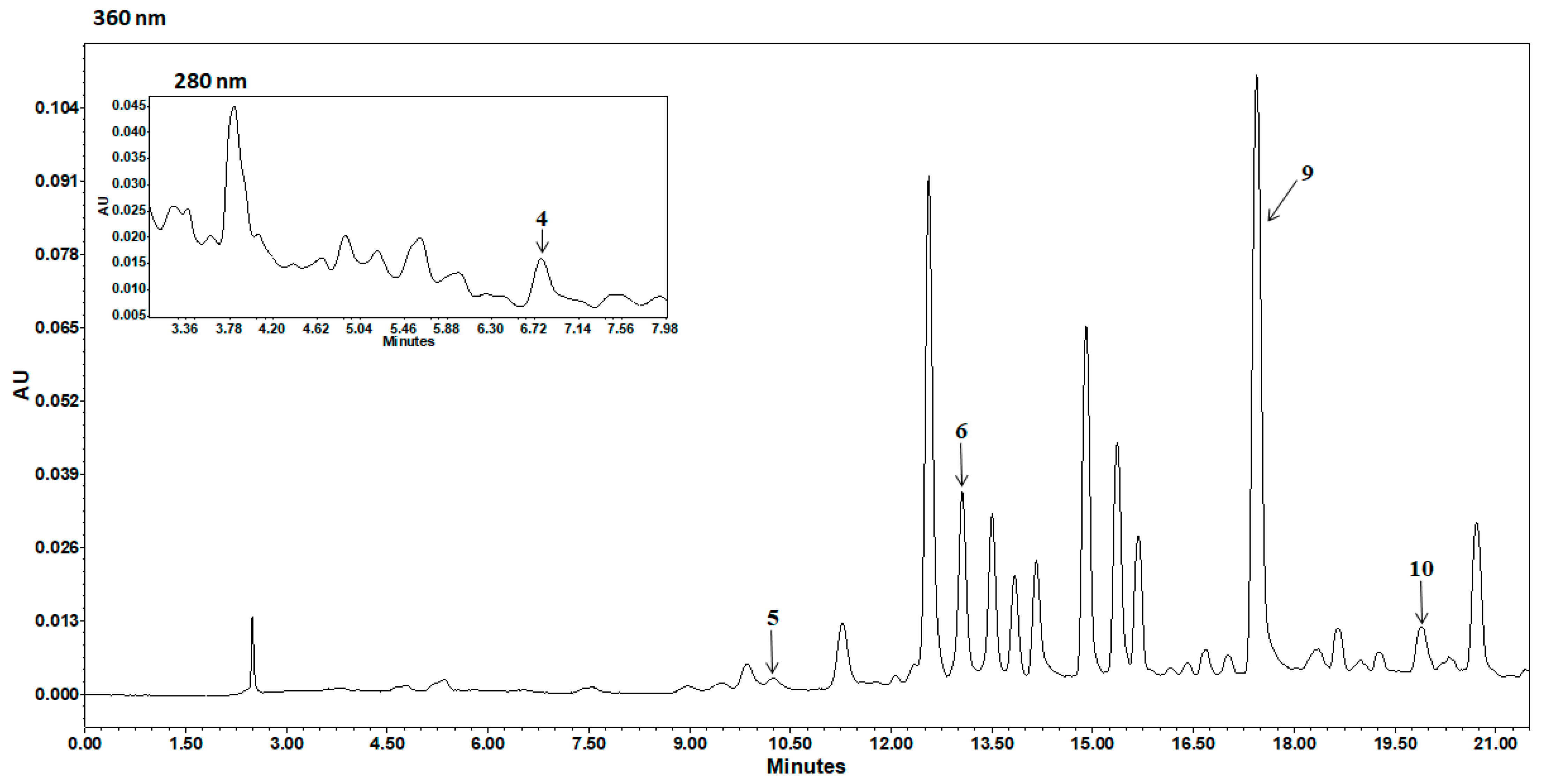
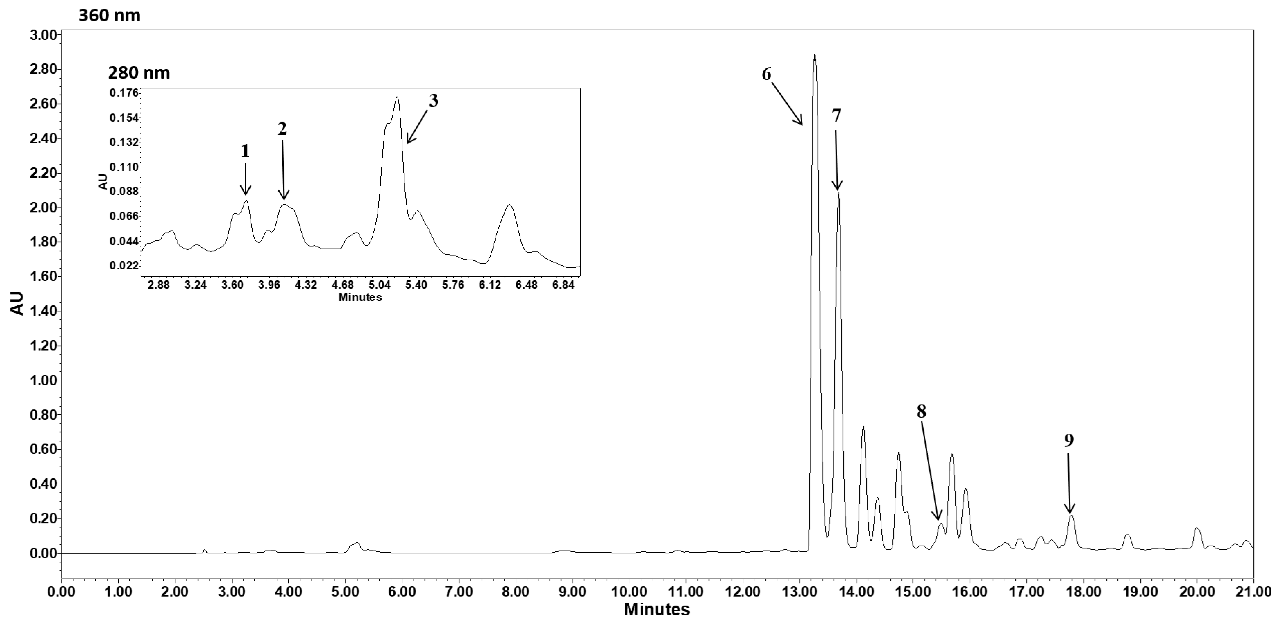
The phenolic compound content of leaf and stem extracts of B. hindsiana is shown in Table 1. The leaf extract showed a concentration 2.09 times higher (p < 0.05) than that of the stem extract. Subsequently, UPLC-DAD analysis was performed to identify the specific phenolic compounds present in the extracts (Table 2, Figure 1 and Figure 2). Overall, the total concentration of identified phenolic compounds is 97% higher in the leaf extract compared to the stem extract. Seven compounds were identified in the leaf extract, with quercetin-3-β-glucoside, rutin, and chlorogenic acid being the most abundant, with concentrations of 47%, 38%, and 9%, respectively. Furthermore, five compounds were identified in the stem extract, with quercetin being the most abundant, followed by quercetin-3-β-glucoside and vinylic acid. These compounds represented 54%, 26%, and 14% of the total compounds identified.
These results show a significantly higher concentration of phenolic compounds in the leaf extract compared to the stem extract. This difference could be attributed to the location of each component within the plant. Leaves are naturally located on the outermost part of the plant, covering the stems. Solar radiation is known to be a key factor in the synthesis of phenolic compounds, as these act as a protective agent against radiation [24,25]. Therefore, leaves may be exposed to greater radiation, which would explain the results observed in this study.
This research is the first study to report the presence of flavonoids and phenolic acids in B. hindsiana, with a total of 10 compounds. This information is relevant because it could help us understand and explain the biological effects this plant may exhibit.

3.2. Antioxidant Activity

One of the most widely explored biological activities in natural products is their antioxidant activity due to its practicality and reproducibility. The antioxidant activity of the leaf and stem extracts of B. hindsiana is shown in Table 1. In the DPPH assay, the leaf extract showed an IC50 value 3.7 times lower (p < 0.05) than that of the stem extract. A similar result was observed in the ABTS assay, where the leaf extract required a concentration 3.8 times lower (p < 0.05) to inhibit 50% of the radical compared to the stem extract. Regarding the capacity to stabilize the peroxyl (ORAC assay), the leaf extract was 3.2 times more effective (p < 0.05) than the stem extract was. Finally, regarding the reducing effect, it was observed that the leaf extract showed a reducing effect 2.8 times greater (p < 0.05) than that shown by the stem extract.
The results described above demonstrated that bioactive compounds in B. hindsiana extracts were able to transfer hydrogen atoms and electrons, providing an antioxidant effect. Through DPPH and ABTS methods, it is possible to prove the capability of compounds to transfer hydrogen or electron and remove DPPH· and ABTS·+, respectively [26]. Additionally, the capability of compounds to transfer hydrogen due to peroxyl radical removal (ORAC assay) is proven [27]. Furthermore, extract compounds showed a reducing potential by electrons transferring through ferric ion reduction to ferrous by FRAP [26]. These last findings confirm that extract compounds are suitable to react with free radicals and metals, suggesting their potential application as therapeutics agents.
This study reveals novel information about the biological potential of B. hindsiana, which can be considered of high antioxidant potential based on its capacity to stabilize free radicals and reduce metals [28,29]. The antioxidant potential of leaves and stems of B. hindsiana had not been previously demonstrated.
The results showed that B. hindsiana exhibits a promising antioxidant effect aimed at avoiding or stopping oxidative stress, which is known to play a significant role in the development of chronic degenerative diseases. Oxidative stress occurs when there is an imbalance between the production of reactive oxygen species and the body’s ability to neutralize or eliminate them [30]. This overproduction can lead to alterations at the cellular level, which may lead to genetic mutations [31]. Then, the ability of B. hindsiana extract to stabilize synthetic and biological free radicals, as well as reduce metals, suggests that this plant source could initially neutralize free radicals, preventing the occurrence of oxidative stress, or interrupt the oxidative process once initiated, preventing excessive cellular damage. However, further studies are needed to demonstrate its safety and efficacy at the biological level.
In addition, this study showed that the B. hindsiana leaf extract exhibits a greater antioxidant effect. This behavior can be attributed to the higher content of phenolic compounds present in the leaf extract, which was 2.09 times greater than that of the stem extract. In this regard, several studies have demonstrated a close relationship between phenolic compounds and the antioxidant activity of natural products, with these bioactive compounds having the greatest impact [32].
Likewise, the results presented in this study are consistent with other published studies. Balaji et al. [33] observed that Ocimum tenuiflorum leaf extract exhibited a greater antioxidant effect compared to the stem extract. Similarly, Brij and Arun [34] observed that Solanum indicum leaf extract was more effective in stabilizing free radicals compared to the stem extract. This behavior may be associated with the constituent’s source of the plants analyzed. In this regard, it is well known that plant stems contain high amounts of cellulose, hemicellulose, and lignin, which can decrease the quantity and concentration of free phenolic compounds [35,36]. Conversely, the leaves exhibit a higher content of free phenolic compounds, which may contribute to their biological activities, particularly antioxidant activity [37].
Although there is currently no information on the antioxidant mechanism of B. hindsiana, it is possible that it is associated with the bioactive compounds identified in the chemical characterization performed in this study. For the leaf extract, two of the major compounds identified were quercetin-3-β-glucoside and rutin, while in the stem extract, the major compound was quercetin. In this regard, various studies have demonstrated that these compounds possess a similar chemical structure (they only differ in the hydroxyl group or carbohydrate residue present at position 3 of ring C) and a strong antioxidant effect [38,39]. The effect is associated with the hydroxyl groups present at positions 3 and 4 of ring B of the phenolic structure (all three compounds), and at position 3 of ring C (only quercetin), which can release a proton or electron to stabilize free radicals or reduce metals. Furthermore, the double bond present in ring C (all three compounds) helps stabilize the generated phenolic radicals [40,41,42]. In addition, other unidentified compounds could also influence the antioxidant effect of B. hindsiana due to chromatograms from both extracts, which exhibited an important amount of unidentified compounds, particularly in the stem extract. Thus, a large phenolic compound database would be useful in identifying more compounds as well as their association with different biological activities from this plant.

3.3. Antiproliferative Activity

Another widely explored biological activity in natural products is their antiproliferative effect, which is evaluated to determine their potential as agents with anticancer activity. In this regard, Table 3 shows the antiproliferative effect of B. hindsiana against two cancer cell lines and one non-cancerous cell line. The IC50 values range between 24.7 and >200 µg/mL. It can be observed that the leaf extract was more active (p < 0.05) than the stem extract, as it was 44% and 27% more effective in inhibiting 50% of cell proliferation in the cervical and lung cancer cell lines, respectively. Furthermore, the extracts showed selectivity against the evaluated cancer cell lines, since the IC50 value for the non-cancerous cell line was higher than the maximum concentration evaluated (200 µg/mL).
Previous studies have reported the antiproliferative effect of resins and stems of B. hindsiana against HeLa and A549 cancer cell lines and showed IC50 values between 90 and 500 µg/mL [43,44]. In contrast, this is the first time that the antiproliferative activity of B. hindsiana leaves has been reported, proving that the leaf extract is more active than the other constituents previously reported. This may guide future studies of this plant to identify the chemical compounds responsible for the antiproliferative effect. The difference observed between the present research and previous studies may be associated with various factors, such as the collection site, collection date, the part of the plant analyzed, and the biotic and abiotic factors to which the plants were exposed. It has been reported that these factors impact the synthesis of chemical compounds within the plant and directly affect their biological potential and constituents [45,46].
An important finding in this study is that the B. hindsiana leaf extract can be classified as highly cytotoxic against the HeLa cell line as it exhibits an IC50 value below 30 µg/mL, the threshold established by the U.S. National Cancer Institute for extracts with potential cytotoxicity against cancer cell lines [47]. Furthermore, the values against the A549 cell line are close to this threshold, suggesting that this plant source can be further explored to identify fractions or compounds with greater antiproliferative effects.
Cancer is known to be one of the main public health issues and one of the leading causes of death worldwide. In 2022 alone, it is estimated that there were nearly 20 million new cases and more than 9 million deaths from this disease [48]. Lung cancer has the highest mortality rate globally, with more than 1.8 million deaths annually [49]. Similarly, cervical cancer shows a high mortality rate among women worldwide, causing more than 350,000 deaths annually [50]. In addition, the number of deaths has increased in recent years, associated with various factors, such as the stage of disease identification or the limited effectiveness of administered procedures or treatments [51,52]. In this sense, it has been observed that antineoplastic therapy can sometimes be ineffective due to the ability of cancer cells to develop chemoresistance [53]. It has been observed that antineoplastic therapies often have low selectivity against cancer cells, which can damage healthy cells in an organism, leading to adverse effects [54,55]. The selectivity of B. hindsiana extracts against cancer cell lines reported in the present study constitutes an interesting finding for the development of new antineoplastic therapies. However, it is certain that further experiments are needed to ensure safe application in humans.
Recent studies show that phenolic compounds strongly inhibit the growth of cancer cell lines [56]. Furthermore, in vivo studies have shown that phenolic compounds, or natural sources rich in these compounds, reduce the incidence and development of cancer in animals [57]. The mechanisms of action associated with these compounds include decreased angiogenesis and inflammation, inhibition of the MAPK, ERK 1/2, and PI3K/Akt pathways, induction of apoptosis and, cell cycle arrest [58,59]. This last mechanism provides evidence to consider this plant as a natural source of potential antineoplastic agents.
The antiproliferative mechanism of stem and leaf extracts of B. hindsiana has not been explored. However, considering the present chemical characterization of the extracts, it is suggested that this effect may be associated with the major compounds identified in both extracts, which have shown antiproliferative effects in HeLa and A549 cancer cell lines in previous studies. For example, quercetin, when evaluated in the HeLa cell line, decreases cell viability by inducing cell cycle arrest in the G2/M phase, as well as mitochondrial apoptosis through a p53-dependent mechanism. This resulted in changes in nuclear morphology, externalization of phosphatidylserine, mitochondrial membrane depolarization, and interference with the regulation of cell cycle regulatory proteins and members of the NF-κB family. It also led to overexpression of pro-apoptotic proteins of the Bcl-2 family, cytochrome C, Apaf-1, and caspases, and dysregulation of the anti-apoptotic proteins Bcl-2 and survivin [60]. Similarly, quercetin-3-glucoside exhibited high cytotoxicity against HeLa cells. The mechanism was associated with cell cycle arrest in the S phase and induction of apoptosis with chromosomal DNA degradation and increased production of reactive oxygen species. Furthermore, activation of caspases 3 and 9 was observed, along with downregulation of the expression of the anti-apoptotic protein Bcl-2 and upregulation of Bcl-2-associated protein X [61]. Similarly, when evaluated in the HeLa cell line, rutin decreased cell viability with subsequent cell cycle arrest in the G0/G1 phase. Furthermore, rutin decreased the expression of the E6 and E7 oncogenes, leading to an increase in the expression of p53 and pRB. In addition, an increase in Bax expression and a decrease in Bcl-2 expression were observed, resulting in the release of cytochrome C into the cytosol and subsequent activation of caspases 3, 8, and 9 [62]. On the other hand, organic fractions of ethyl acetate obtained from extracts of Achillea millefolium and Cassia fistula showed a high concentration of chlorogenic acid (the second major compound). A. millefolium induced apoptosis and cell cycle arrest in the G2/M phase when evaluated against the HeLa cell line [63]. Finally, C. fistula caused apoptosis in HeLa by inducing phosphatidylserine externalization [64].
Furthermore, in the A549 cell line, a similar behavior has been observed, since quercetin, rutin, quercetin 3-glucosium and chlorogenic acid decrease cell viability by inducing cell cycle arrest and apoptosis and altering the function of proteins (Bax, Bcl-2 and caspases) and genes associated with this process (cIAP2 and cIAP2), in addition to increasing the production of reactive oxygen species, affecting cell adhesion and migration [65,66,67,68].
Derived from this information, it could be suggested that the antiproliferative effect of B. hindsiana is related to the mechanisms previously described, due to the high concentration of phenolic compounds identified in the extracts (quercetin, quercetin-3-β-glucoside, rutin, and chlorogenic acid). Nevertheless, other groups of bioactive compounds could also influence the antiproliferative potential. For this reason, it seems feasible to continue exploring the chemical composition of this natural source.

3.4. Acute Toxicity

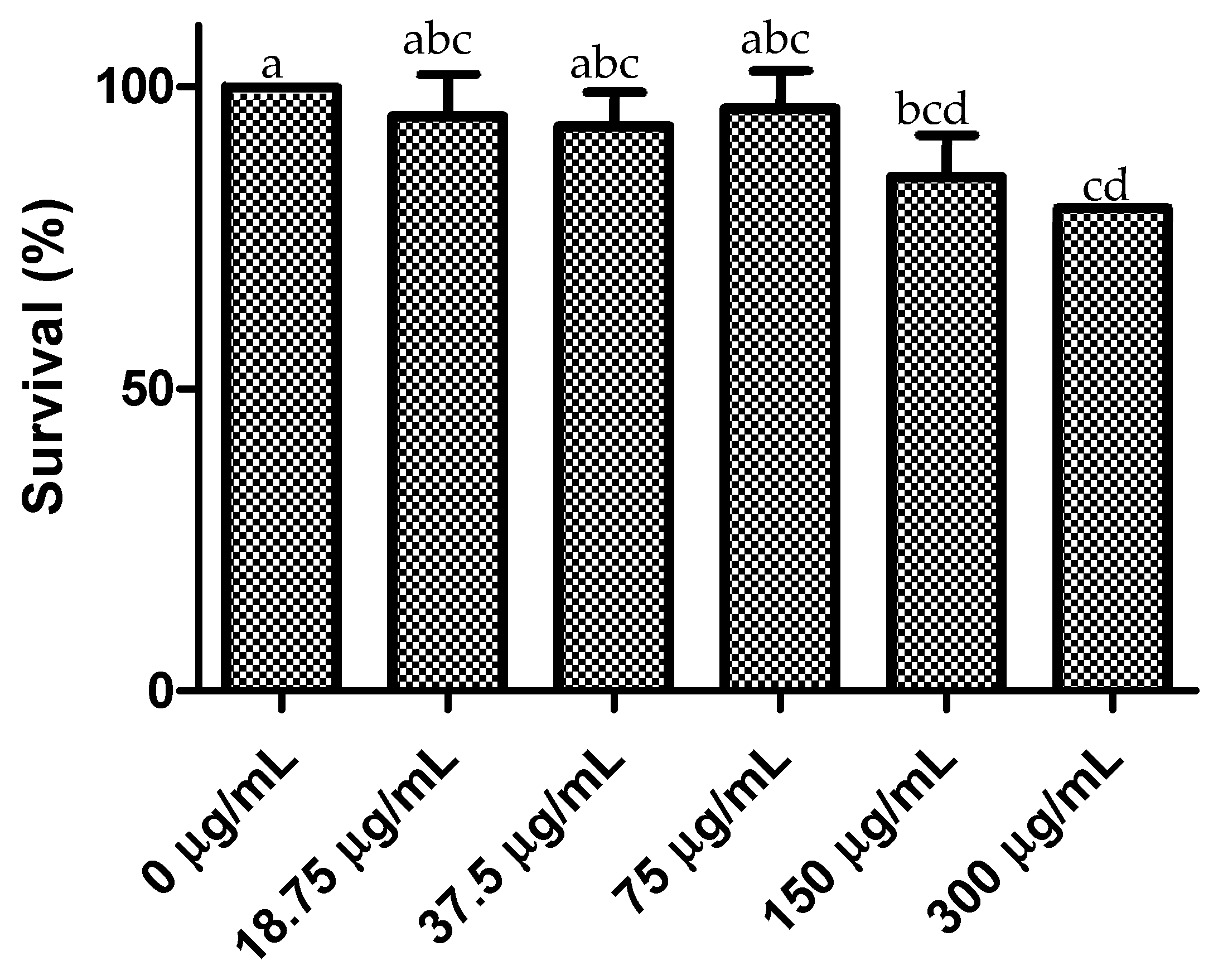
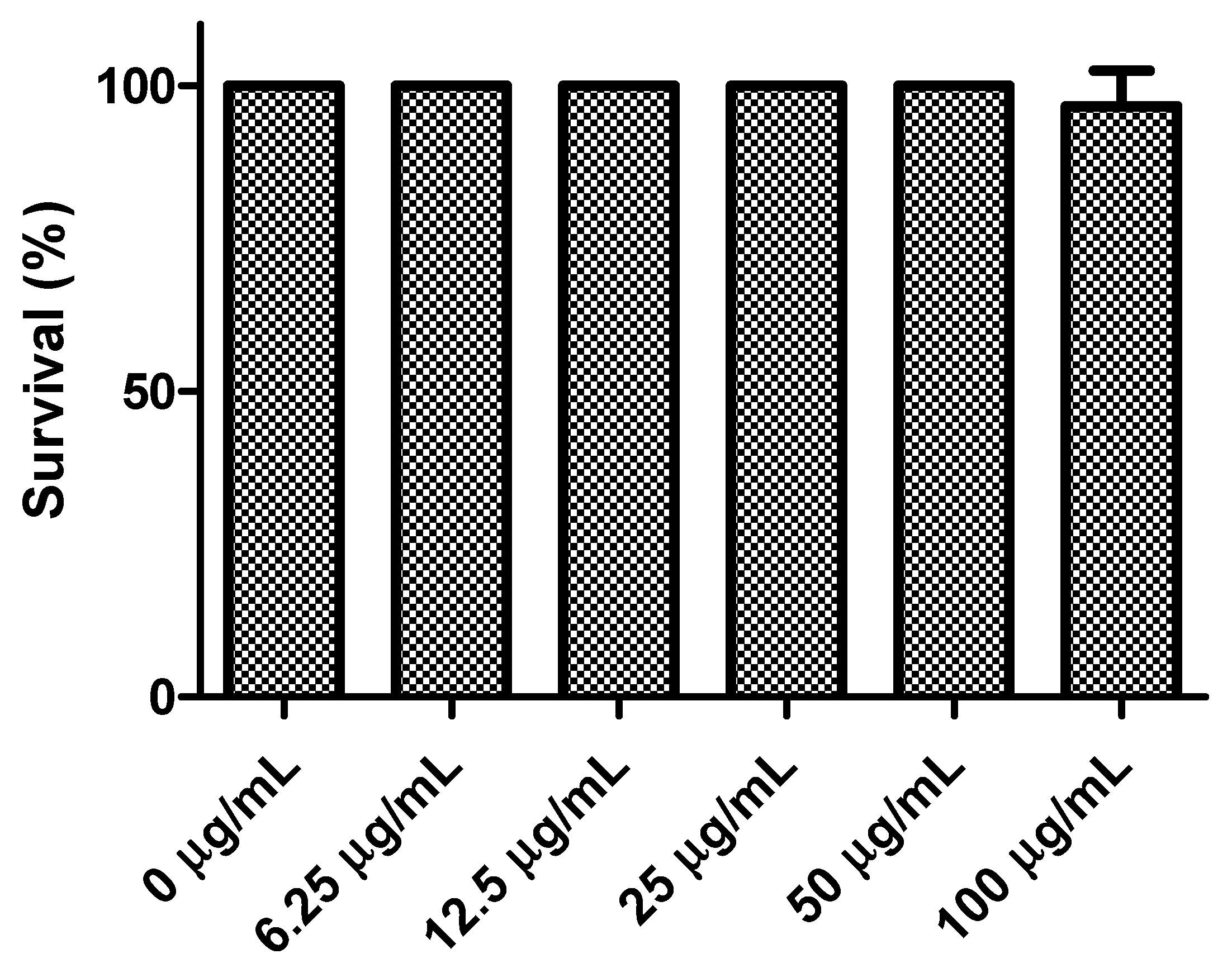
The results for acute toxicity of the leaf and stem extracts of B. hindsiana are shown in Figure 3 and Figure 4. It can be observed that the leaf extract (Figure 3) showed no toxicity at the evaluated doses (0–100 µg/mL), as a survival rate greater than 96% was observed at all doses. This indicates an LC50 value greater than 100 µg/mL. Regarding the stem extract, an LC50 value greater than the maximum evaluated dose of 300 µg/mL is expected. Similarly, the survival percentage in A. salina ranged from 100% to 80% at concentrations of 150 and 300 µg/mL, exhibiting the highest acute toxicity (p < 0.05) in A. salina, respectively.
These results report for the first time the acute toxicity of B. hindsiana extracts and indicate that the extracts obtained from this plant source so far do not present high toxicity, since the LC50 values are greater than 100 µg/mL [69].
An important aspect when exploring potential applications of medicinal plant extracts in humans or animals is ensuring their safety to prevent risks during consumption or application [10]. In addition to in vitro assays using cell lines, in vivo analyses can be used to assess toxicity. The A. salina assay is a simple and easily reproducible analysis in vivo. This assay provides an initial indication of the potential toxicity of an extract or treatment and is widely used to determine acute toxicity as A. salina is a highly sensitive system to changes in its environment that can lead to damage or death [70].
Together, the results show that the IC50 of antioxidant and antiproliferative activity are significantly lower than the LC50 values obtained in the toxicity assay. Therefore, it is possible to continue investigating this plant source to further analyze its safety and efficacy and potentially develop drugs to address the aforementioned health problems.

4. Conclusions

The results of this research provide novel and original information about the chemical composition of the plant, identifying for first time several phenolic compounds in B. hindsiana. Furthermore, this plant is observed to be a natural source with promising biological potential, particularly its high antioxidant and antiproliferative effects. In addition, B. hindsiana exhibited low cytotoxicity against healthy cells, as well as low acute toxicity. This indicates the possibility to continue studying this natural source, including the isolation of the chemical compounds responsible for its biological effects and the determination of their respective mechanisms of action. This could lead to the use of this natural source for the development of new therapeutic agents; however, product safety must be analyzed.

Author Contributions

Conceptualization, J.C.L.-R. and H.T.-M.; methodology, J.C.L.-R., H.T.-M., J.L.M.-M., M.P.-J., M.A.V.-O., N.J.S.-L. and G.A.G.A.; software, J.C.L.-R., H.T.-M., M.A.V.-O., N.J.S.-L. and G.A.G.A.; validation, J.C.L.-R. and H.T.-M.; formal analysis, J.C.L.-R., H.T.-M., M.A.V.-O. and N.J.S.-L.; investigation, J.C.L.-R. and H.T.-M.; resources, J.C.L.-R. and H.T.-M.; writing—original draft preparation, J.C.L.-R. and H.T.-M.; writing—review and editing, J.C.L.-R., H.T.-M., J.L.M.-M., M.P.-J., M.A.V.-O., N.J.S.-L. and G.A.G.A.; supervision, J.C.L.-R. and H.T.-M.; project administration, J.C.L.-R. All authors have read and agreed to the published version of the manuscript.

Funding

This research received no external funding.

Data Availability Statement

The original contributions presented in this study are included in the article. Further inquiries can be directed to the corresponding authors.

Conflicts of Interest

The authors declare no conflicts of interest.

Abbreviations

The following abbreviations are used in this manuscript:
GAEGallic acid equivalent.
TETrolox equivalent.
DMEMDulbecco’s Modified Eagle Medium.
DMSODimethyl sulfoxide.
IC50Half maximal inhibitory concentration.

References

  1. Biblioteca Digital de la Medicina Tradicional Mexicana. Copal Blanco. 2025. Available online: http://www.medicinatradicionalmexicana.unam.mx/apmtm/termino.php?l=3&t=bursera-hindsiana (accessed on 21 January 2026).
  2. Rzedowski, J.; Lemos, R.M.; de Rzedowski, G.C. Inventario del conocimiento taxonómico, así como de la diversidad y del endemismo regionales de las especies mexicanas de Bursera (Burseraceae). Acta Bot. Mex. 2005, 70, 85–111. [Google Scholar] [CrossRef]
  3. Pooja, G.; Shweta, S.; Patel, P. Oxidative stress and free radicals in disease pathogenesis: A review. Discov. Med. 2025, 2, 104. [Google Scholar] [CrossRef]
  4. Dai, X.; Huang, Z.; Lyu, R. Free Radicals in Health and Disease. MedComm 2025, 6, e70396. [Google Scholar] [CrossRef] [PubMed]
  5. Agnihotri, A.; Aruoma, O.I. Alzheimer’s disease and Parkinson’s disease: A nutritional toxicology perspective of the impact of oxidative stress, mitochondrial dysfunction, nutrigenomics and environmental chemicals. J. Am. Coll. Nutr. 2020, 39, 16–27. [Google Scholar] [CrossRef] [PubMed]
  6. Jomova, K.; Alomar, S.Y.; Valko, R.; Fresser, L.; Nepovimova, E.; Kuca, K.; Valko, M. Interplay of oxidative stress and antioxidant mechanisms in cancer development and progression. Arch. Toxicol. 2025, 100, 27–73. [Google Scholar] [CrossRef] [PubMed]
  7. Wagle, N.S.; Nogueira, L.; Devasia, T.P.; Mariotto, A.B.; Yabroff, R.; Islami, F.; Jemal, A.; Alteri, R.; Ganz, P.A.; Siegel, R.L. Cancer treatment and survivorship statistics, 2025. CA Cancer J. Clin. 2025, 75, 308–340, Correction in CA Cancer J. 2025, 75, 683. [Google Scholar] [CrossRef] [PubMed]
  8. Luo, Q.; Smith, D.P. Global cancer burden: Progress, projections, and challenges. Lancet 2025, 406, 1536–1537. [Google Scholar] [CrossRef] [PubMed]
  9. Freihat, O.; Sipos, D.; Kovacs, A. Global burden and projections of breast cancer incidence and mortality to 2050: A comprehensive analysis of GLOBOCAN data. Front. Public Health 2025, 13, 1622954. [Google Scholar] [CrossRef] [PubMed]
  10. Li, M.; Zhang, H.; Li, J.; Chen, Y.; Ren, S.; Sun, W.; Zhang, Q.; Zengin, G. Reshaping the future of cancer therapy: Taming toxicity and side effects. Biomed. Eng. Commun. 2026, 5, 11. [Google Scholar] [CrossRef]
  11. Manisha, D.; Babu, R.; Begam, A.M.; Chahal, K.S.; Harale, A.A. Medicinal Plants and Traditional Uses and Modern Applications. J. Neonatal Surg. 2025, 14, 162–175. [Google Scholar] [CrossRef]
  12. Latif, R.; Nawaz, T. Medicinal plants and human health: A comprehensive review of bioactive compounds, therapeutic effects, and applications. Phytochem. Rev. 2025, 25, 2299–2342. [Google Scholar] [CrossRef]
  13. Brower, V. Back to Nature: Extinction of Medicinal Plants Threatens Drug Discovery; Oxford University Press: Oxford, UK, 2008. [Google Scholar] [CrossRef] [PubMed]
  14. Velazquez, C.; Navarro, M.; Acosta, A.; Angulo, A.; Dominguez, Z.; Robles, R.; Robles-Zepeda, R.; Lugo, E.; Goycolea, F.M.; Velazquez, E.F.; et al. Antibacterial and free-radical scavenging activities of Sonoran propolis. J. Appl. Microbiol. 2007, 103, 1747–1756. [Google Scholar] [CrossRef] [PubMed]
  15. Velderrain-Rodríguez, G.R.; Torres-Moreno, H.; Villegas-Ochoa, M.A.; Ayala-Zavala, J.F.; Robles-Zepeda, R.E.; Wall-Medrano, A.; Gonzalez-Aguilar, G.A. Gallic acid content and an antioxidant mechanism are responsible for the antiproliferative activity of ‘Ataulfo’mango peel on LS180 cells. Molecules 2018, 23, 695. [Google Scholar] [CrossRef] [PubMed]
  16. Usia, T.; Banskota, A.H.; Tezuka, Y.; Midorikawa, K.; Matsushige, K.; Kadota, S. Constituents of Chinese propolis and their antiproliferative activities. J. Nat. Prod. 2002, 65, 673–676. [Google Scholar] [CrossRef] [PubMed]
  17. Re, R.; Pellegrini, N.; Proteggente, A.; Pnnala, A.; Yang, M.; Rice-Evans, C. Antioxidant activity applying an improved ABTS radical cation decolorization assay. Free Radic. Biol. Med. 1999, 26, 1231–1237. [Google Scholar] [CrossRef] [PubMed]
  18. Ou, B.; Hampsch-Woodill, M.; Prior, R.L. Development and validation of an improved oxygen radical absorbance capacity assay using fluorescein as the fluorescent probe. J. Agric. Food Chem. 2001, 49, 4619–4626. [Google Scholar] [CrossRef] [PubMed]
  19. Benzie, I.F.; Strain, J.J. The ferric reducing ability of plasma (FRAP) as a measure of “antioxidant power”: The FRAP assay. Anal. Biochem. 1996, 239, 70–76. [Google Scholar] [CrossRef] [PubMed]
  20. Torres-Moreno, H.; Velazquez, C.A.; Garivay-Escobar, A.; Curini, M.; Marcotullio, M.C. Antiproliferative and apoptosis induction of cucurbitacin-type triterpenes from Ibervillea sonorae. Ind. Crops Prod. 2015, 77, 895–900. [Google Scholar] [CrossRef]
  21. López-Romero, J.C.; González-Ríos, H.; Peña-Ramos, A.; Velazquez, C.; Navarro, M.; Robles-Zepeda, R.E.; Hernández, J. Seasonal effect on the biological activities of Litsea glaucescens Kunth extracts. Evid.-Based Complement. Altern. Med. 2018, 2018, 2738489. [Google Scholar] [CrossRef] [PubMed]
  22. Chávez-Magdaleno, M.; Luque-Alcaraz, A.G.; Gutiérrez-Martínez, P.; Cortez-Rocha, M.O.; Burgos-Hernández, A.; Lizardi-Mendoza, J.; Plascencia-Jatomea, M. Effect of chitosan-pepper tree (Schinus molle) essential oil biocomposites on the growth kinetics, viability and membrane integrity of Colletotrichum gloeosporioides. Rev. Mex. Ing. Quím. 2018, 17, 29–45. [Google Scholar] [CrossRef]
  23. Zhang, Y.; Cai, P.; Cheng, G.; Zhang, Y. A brief review of phenolic compounds identified from plants: Their extraction, analysis, and biological activity. Nat. Prod. Commun. 2022, 17, 1–14. [Google Scholar] [CrossRef]
  24. Emus-Medina, A.; Contreras-Angulo, L.A.; Ambriz-Perez, D.L.; Vazquez-Olivo, G.; Heredia, J.B. UV Light Stress Induces Phenolic Compounds in Plants, in Plant Phenolics in Abiotic Stress Management; Springer: Berlin/Heidelberg, Germany, 2023; pp. 415–440. [Google Scholar] [CrossRef]
  25. Beevi, S.S.; Narasu, M.L.; Gowda, B.B. Polyphenolics profile, antioxidant and radical scavenging activity of leaves and stem of Raphanus sativus L. Plant Foods Hum. Nutr. 2010, 65, 8–17. [Google Scholar] [CrossRef] [PubMed]
  26. Łepecka, A.; Kołożyn-Krajewska, D. Antioxidant Properties of Food-Derived Lactic Acid Bacteria: A Review. Microb. Biotechnol. 2025, 18, e70229. [Google Scholar] [CrossRef] [PubMed]
  27. Knez, E.; Kadac-Czapska, K.; Grembecka, M. Evaluation of spectrophotometric methods for assessing antioxidant potential in plant food samples—A critical approach. Appl. Sci. 2025, 15, 5925. [Google Scholar] [CrossRef]
  28. Wong, C.C.; Hua-Bin, L.; Ka-Wing, C.; Chen, F. A systematic survey of antioxidant activity of 30 Chinese medicinal plants using the ferric reducing antioxidant power assay. Food Chem. 2006, 97, 705–711. [Google Scholar] [CrossRef]
  29. Fidrianny, I.; Aristya, T.; Hartati, R. Antioxidant capacities of various leaves extracts from three species of legumes and correlation with total flavonoid, phenolic, carotenoid content. Int. J. Pharmacogn. Phytochem. Res. 2015, 7, 628–634. [Google Scholar]
  30. Wang, X.; Dong, B.; Gan, Q.; Li, J.; Wu, P.; Guan, Y.; Wang, J. Unraveling the vicious cycle: Oxidative stress and neurotoxicity in neurodegenerative diseases. FASEB BioAdv. 2025, 7, e70041. [Google Scholar] [CrossRef] [PubMed]
  31. Jomova, K.; Alomar, S.Y.; Valko, R.; Liska, J.; Nepovimova, E.; Kuca, K.; Valko, M. Flavonoids and their role in oxidative stress, inflammation, and human diseases. Chem. Biol. Interact. 2025, 413, 111489. [Google Scholar] [CrossRef] [PubMed]
  32. López-Romero, J.C.; Torres-Moreno, H.; Rodríguez-Martínez, K.L.; Ramírez-Audelo, V.; Vidal-Gutiérrez, M.; Hernández, J.; González-Aguilar, G.A. Fouquieria splendens: A source of phenolic compounds with antioxidant and antiproliferative potential. Eur. J. Integr. Med. 2022, 49, 102084. [Google Scholar] [CrossRef]
  33. Balaji, R.; Prakash, G.; Suganya, P.D.; Aravinthan, K.M. Antioxidant activity of methanol extract of Ocimum tenuiflorum (dried leaf and stem). Int. J. Pharm. Sci. Rev. Res. 2011, 3, 20–27. [Google Scholar]
  34. Mohan, B. Evaluation of phenolic, flavonoid contents and antioxidant activity in leaves, stem and roots of Solanum indicum Linn. Environ. Conserv. J. 2020, 21, 167–172. [Google Scholar] [CrossRef]
  35. Xiao, B.; Sun, X.; Sun, R. Chemical, structural, and thermal characterizations of alkali-soluble lignins and hemicelluloses, and cellulose from maize stems, rye straw, and rice straw. Polym. Degrad. Stab. 2001, 74, 307–319. [Google Scholar] [CrossRef]
  36. Shahidi, F.; Hossain, A. Importance of insoluble-bound phenolics to the antioxidant potential is dictated by source material. Antioxidants 2023, 12, 203. [Google Scholar] [CrossRef] [PubMed]
  37. Cartea, M.E.; Francisco, M.; Soengas, P.; Velasco, P. Phenolic compounds in Brassica vegetables. Molecules 2010, 16, 251–280. [Google Scholar] [CrossRef] [PubMed]
  38. Panat, N.A.; Amrute, B.K.; Bhattu, S.; Haram, S.K.; Sharma, G.K.; Ghaskadbi, S. Antioxidant Profiling of C3 Quercetin Glycosides: Quercitrin, Quercetin 3-β-D-glucoside and Quercetin 3-O-(6”-O-malonyl)-β-Dglucoside in cell free environment. Free Radic. Antioxid. 2015, 5, 90–100. [Google Scholar] [CrossRef]
  39. Rusmana, D.; Wahyudianingsih, R.; Elisabeth, M.; Balqis, B.; Maesaroh, M.; Widowat, W. Antioxidant activity of Phyllanthus niruri extract, rutin and quercetin. Indones. Biomed. J. 2017, 9, 84–90. [Google Scholar] [CrossRef]
  40. Qi, W.; Qi, W.; Xiong, D.; Long, M. Quercetin: Its antioxidant mechanism, antibacterial properties and potential application in prevention and control of toxipathy. Molecules 2022, 27, 6545. [Google Scholar] [CrossRef] [PubMed]
  41. Yang, J.; Guo, J.; Yuan, J. In vitro antioxidant properties of rutin. LWT-Food Sci. Technol. 2008, 41, 1060–1066. [Google Scholar] [CrossRef]
  42. Rösch, D.; Bergmann, M.; Knorr, D.; Kroh, L.W. Structure− antioxidant efficiency relationships of phenolic compounds and their contribution to the antioxidant activity of sea buckthorn juice. J. Agric. Food Chem. 2003, 51, 4233–4239. [Google Scholar] [CrossRef] [PubMed]
  43. Salido, A.A.G.; Assanga, S.B.I.; Luján, L.M.L.; Ángulo, D.F.; Espinoza, C.L.L.; Silva, A.L.A.; Rubio, J.L. Composition of secondary metabolites in mexican plant extracts and their antiproliferative activity towards cancer cell lines. Int. J. Sci. 2016, 5, 63–77. [Google Scholar] [CrossRef][Green Version]
  44. Vidal Gutiérrez, M.; Torres Moreno, H.; Velázquez Contreras, C.A.; Rascón Valenzuela, L.A.; Robles Zepeda, R.E. Actividad antioxidante y antiproliferativa de seis plantas medicinales del noroeste de México. Biotecnia 2020, 22, 40–45. [Google Scholar] [CrossRef]
  45. Jan, R.; Asaf, S.; Numan, M.; Lubna; Kim, K.M. Plant secondary metabolite biosynthesis and transcriptional regulation in response to biotic and abiotic stress conditions. Agronomy 2021, 11, 968. [Google Scholar] [CrossRef]
  46. Biesiada, A.; Tomczak, A. Biotic and abiotic factors affecting the content of the chosen antioxidant compounds in vegetables. Veg. Crops Res. Bull. 2012, 76, 55. [Google Scholar] [CrossRef]
  47. Abu-Dahab, R.; Afifi, F.; Kasabri, V.; Majdalawi, L.; Naffa, R. Comparison of the antiproliferative activity of crude ethanol extracts of nine salvia species grown in Jordan against breast cancer cell line models. Pharmacogn. Mag. 2012, 8, 319. [Google Scholar] [CrossRef] [PubMed]
  48. OMS. Crece la Carga Mundial de Cáncer en Medio de una Creciente Necesidad de Servicios. 2024. Available online: https://www.who.int/es/news/item/01-02-2024-global-cancer-burden-growing--amidst-mounting-need-for-services (accessed on 10 December 2025).
  49. OMS. Lung Cancer. 2023. Available online: https://www.who.int/news-room/fact-sheets/detail/lung-cancer (accessed on 10 December 2025).
  50. OMS. Cervical Cancer. 2025. Available online: https://www.who.int/news-room/fact-sheets/detail/cervical-cancer (accessed on 11 December 2025).
  51. McPhail, S.; Johnson, S.; Greenberg, D.; Peake, M.; Rous, B. Stage at diagnosis and early mortality from cancer in England. Br. J. Cancer 2015, 112, S108–S115. [Google Scholar] [CrossRef] [PubMed]
  52. Duggan, C.; Trapani, D.; Ilbawi, A.M.; Fidarova, E.; Laversanne, M.; Curigliano, G.; Anderson, B.O. National health system characteristics, breast cancer stage at diagnosis, and breast cancer mortality: A population-based analysis. Lancet Oncol. 2021, 22, 1632–1642. [Google Scholar] [CrossRef] [PubMed]
  53. Guneidy, R.A. Molecular basis of cancer chemoresistance: Biochemical insights. Drug Metab. Rev. 2026, 58, 94–120. [Google Scholar] [CrossRef] [PubMed]
  54. Wang, Z.; Zhang, J. Genetic and epigenetic bases of long-term adverse effects of childhood cancer therapy. Nat. Rev. Cancer 2025, 25, 129–144. [Google Scholar] [CrossRef] [PubMed]
  55. Sakai, Y.; Katsura, K.; Kotake, M.; Toyama, A. A cross-sectional survey of oral adverse events and oral management needs in outpatients receiving cancer drug therapy. Cancers 2025, 17, 641. [Google Scholar] [CrossRef] [PubMed]
  56. Hazafa, A.; Rehman, K.U.; Jahan, N.; Jabeen, Z. The role of polyphenol (flavonoids) compounds in the treatment of cancer cells. Nutr. Cancer 2020, 72, 386–397. [Google Scholar] [CrossRef] [PubMed]
  57. Ahmed, Z.S.O.; Khan, E.; Elias, N.; Elshebiny, A.; Dou, Q. Updated review on natural polyphenols: Molecular mechanisms, biological effects, and clinical applications for cancer management. Biomolecules 2025, 15, 629. [Google Scholar] [CrossRef] [PubMed]
  58. Ceylan, F.D.; Günal-Köroğlu, D.; Saricaoglu, B.; Ozkan, G.; Capanoglu, E.; Calina, D.; Sharifi-Rad, J. Anticancer potential of hydroxycinnamic acids: Mechanisms, bioavailability, and therapeutic applications. Naunyn-Schmiedeberg’s Arch. Pharmacol. 2025, 398, 469–495. [Google Scholar] [CrossRef] [PubMed]
  59. Pyo, Y.; Kwon, K.H.; Jung, Y.J. Anticancer potential of flavonoids: Their role in cancer prevention and health benefits. Foods 2024, 13, 2253. [Google Scholar] [CrossRef] [PubMed]
  60. Priyadarsini, R.V.; Murugan, R.S.; Maitreyi, S.; Ramalingam, K.; Karunagaran, D.; Nagini, S. The flavonoid quercetin induces cell cycle arrest and mitochondria-mediated apoptosis in human cervical cancer (HeLa) cells through p53 induction and NF-κB inhibition. Eur. J. Pharmacol. 2010, 649, 84–91. [Google Scholar]
  61. Nile, A.; Nile, S.H.; Shin, J.; Park, G.; Oh, J.W. Quercetin-3-glucoside extracted from apple pomace induces cell cycle arrest and apoptosis by increasing intracellular ROS levels. Int. J. Mol. Sci. 2021, 22, 10749. [Google Scholar] [CrossRef] [PubMed]
  62. Pandey, P.; Khan, F.; Farhan, M.; Jafri, A. Elucidation of rutin’s role in inducing caspase-dependent apoptosis via HPV-E6 and E7 down-regulation in cervical cancer HeLa cells. Biosci. Rep. 2021, 41, BSR20210670. [Google Scholar] [CrossRef] [PubMed]
  63. Abou Baker, D.H. Achillea millefolium L. ethyl acetate fraction induces apoptosis and cell cycle arrest in human cervical cancer (HeLa) cells. Ann. Agric. Sci. 2020, 65, 42–48. [Google Scholar] [CrossRef]
  64. Kaur, S.; Kumar, A.; Pandit, K.; Kaur, S. Modulation of mutagenicity in Salmonella typhimurium and antioxidant properties and antiproliferative effects of fractions from Cassia fistula L. on human cervical HeLa and breast MCF-7 cancer cells. Environ. Sci. Pollut. Res. 2021, 28, 6619–6634. [Google Scholar] [CrossRef] [PubMed]
  65. ben Sghaier, M.; Pagano, A.; Mousslim, M.; Ammari, Y.; Kovacic, H.; Luis, J. Rutin inhibits proliferation, attenuates superoxide production and decreases adhesion and migration of human cancerous cells. Biomed. Pharmacother. 2016, 84, 1972–1978. [Google Scholar] [CrossRef] [PubMed]
  66. Farrag, I.M.; Belal, A.; Al Badawi, M.H.; Abdelhady, A.A.; Abou Galala, F.M.; El-Sharkawy, A.; Mehany, A.B. Antiproliferative, apoptotic effects and suppression of oxidative stress of quercetin against induced toxicity in lung cancer cells of rats: In vitro and in vivo study. J. Cancer 2021, 12, 5249. [Google Scholar] [CrossRef] [PubMed]
  67. Pourabdollah-Kaleybar, V.; Pourabdollah-Kaleybar, P.; Eskandani, M.; Nazemiyeh, H. Toxicity of bioactive compounds from Halocnemum strobilaceum against A549 lung cancer cells. Toxicon 2025, 253, 108186. [Google Scholar] [CrossRef] [PubMed]
  68. Wang, L.; Du, H.; Chen, P. Chlorogenic acid inhibits the proliferation of human lung cancer A549 cell lines by targeting annexin A2 in vitro and in vivo. Biomed. Pharmacother. 2020, 131, 110673. [Google Scholar] [CrossRef] [PubMed]
  69. Clarkson, C.; Maharaj, V.J.; Crouch, N.R.; Grace, O.M.; Pillay, P.; Matsabisa, M.G.; Folb, P.I. In vitro antiplasmodial activity of medicinal plants native to or naturalised in South Africa. J. Ethnopharmacol. 2004, 92, 177–191. [Google Scholar] [CrossRef] [PubMed]
  70. Rajabi, S.; Ramazani, A.; Hamidi, M.; Naji, T. Artemia salina as a model organism in toxicity assessment of nanoparticles. DARU J. Pharm. Sci. 2015, 23, 20. [Google Scholar] [CrossRef] [PubMed]
Figure 1. Chromatogram obtained from B. hindsiana leaf extract recorded at 280 and 360 nm. 1. Protocatechuic acid, 2. catechin, 3. chlorogenic acid, 6. quercetin-3-β-glucodise, 7. rutin. 8. kaempferol-3-β-glucoside, and 9. quercetin.
Compounds 06 00040 g001
Figure 2. Chromatogram obtained from B. hindsiana stem extract recorded at 280 and 360 nm. 4. Vanillic acid, 5. p-coumaric acid, 6. quercetin-3-β-glucoside, 9. quercetin, and 10. kaempferol.
Compounds 06 00040 g002
Figure 3. Acute toxicity of Bursera hindsiana leaf extract. Data are presented as the mean ± standard deviation of three independent experiments.
Compounds 06 00040 g003
Figure 4. Acute toxicity of Bursera hindsiana stem extract. Data are presented as the mean ± standard deviation of three independent experiments. a–d Different superscripts in each bar indicate significant differences (p < 0.05).
Compounds 06 00040 g004
Table 1. Phenolic content and antioxidant activity of Bursera hindsiana extracts.
MethodExtract
LeavesStems
Total phenols (mg GAE/g)160.0 ± 13.0 b76.4 ± 7.7 a
DPPH (IC50, µg/mL)42.8 ± 2.2 a152.8 ± 8.0 b
ABTS (IC50, µg/mL)12.2 ± 1.9 a44.9 ± 3.8 b
ORAC (µm TE/g)3055.17 ± 120.07 b966.46 ± 28.85 a
FRAP (µm Fe(II)/g)1492.7 ± 70.9 b574.3 ± 46.0 a
Data are presented as the mean ± standard deviation of three independent experiments. a,b Different superscripts in each method indicate significant differences (p < 0.05).
Table 2. Phenolic profile of Bursera hindsiana extract.
CompoundExtract (µg/g)
LeavesStems
Protocatechuid acid396.27 ± 6.36NI
Catechin2753.91 ± 28.67NI
Chlorogenic acid9362.66 ± 20.64NI
Vanillic acidNI402.96 ± 3.21
p-Coumaric acidNI25.09 ± 0.40
Quercetin-3-β-glucoside46,434.90 ± 27.25777.42 ± 18.26
Rutin37,735.20 ± 18.26NI
Kaempferol-3-β-glucoside718.04 ± 0.48NI
Quercetin1400.61 ± 32.181584.96 ± 1.59
KaempferolNI123.57 ± 0.74
Total98,801.592914.00
NI: compound not identified.
Table 3. Antiproliferative activity of Bursera hindsiana extracts.
ExtractIC50 (µg/mL)
HeLaA549ARPE-19
Leaves 24.7 ± 1.7 a41.4 ± 5.3 a>200
Stems43.8 ± 1.8 b56.7 ± 3.8 b>200
Data are presented as the mean ± standard deviation of three independent experiments. a,b Different superscripts in each extract indicate significant differences (p < 0.05).
Disclaimer/Publisher’s Note: The statements, opinions and data contained in all publications are solely those of the individual author(s) and contributor(s) and not of MDPI and/or the editor(s). MDPI and/or the editor(s) disclaim responsibility for any injury to people or property resulting from any ideas, methods, instructions or products referred to in the content.