
Appl. Nano, Volume 6, Issue 3 (September 2025) – 9 articles
Cover Story (view full-size image):
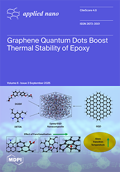
Epoxy resins are widely used in aerospace, defense, and electronics, but their relatively low glass transition temperature (Tg) limits performance under high heat. In this issue, Bamane and Keles explore a novel solution using molecular dynamics by reinforcing epoxies with graphene quantum dots (GQDs). Unlike larger nanofillers such as carbon nanotubes or graphene sheets, GQDs are zero-dimensional, nanoscale fragments with tunable surface chemistry. The study reveals that functionalized GQDs can raise epoxy’s Tg by up to ~16%, primarily by forming covalent bonds with GQD and restricting polymer chain mobility. These findings highlight the effect of GQDs as a promising nanofiller for the next generation of polymer composites with improved thermal stability. View this paper
- Issues are regarded as officially published after their release is announced to the table of contents alert mailing list.
- You may sign up for e-mail alerts to receive table of contents of newly released issues.
- PDF is the official format for papers published in both, html and pdf forms. To view the papers in pdf format, click on the "PDF Full-text" link, and use the free Adobe Reader to open them.
Previous Issue
Next Issue