Review

New Frontiers in the Digital Restoration of Hidden Texts in Manuscripts: A Review of the Technical Approaches

1
Opera del Vocabolario Italiano, National Research Council of Italy (OVI-CNR), 50141 Firenze, Italy
2
National Laboratories of Frascati, National Institute for Nuclear Physics (INFN), 00044 Frascati, Italy
3
Department of Basic and Applied Sciences for Engineering (SBAI), Sapienza University of Rome, 00161 Rome, Italy
4
Department of Classics, Sapienza University of Rome, 00185 Rome, Italy
*
Author to whom correspondence should be addressed.
Heritage 2024, 7(2), 683-696; https://doi.org/10.3390/heritage7020034
Submission received: 20 December 2023 / Revised: 22 January 2024 / Accepted: 22 January 2024 / Published: 3 February 2024
(This article belongs to the Special Issue Non-invasive Technologies Applied in Cultural Heritage)

Abstract

:
The digital restoration of historical manuscripts centers on deciphering hidden writings, made imperceptible to the naked eye due to factors such as erasure, fading, carbonization, and aging effects. Recent advancements in modern technologies have significantly improved our ability to unveil and interpret such written cultural heritage that, for centuries, had remained inaccessible to contemporary understanding. This paper aims to present a critical overview of state-of-the-art technologies, engaging in discussions about perspectives and limitations, and anticipating future applications. Serving as a practical guide, this work seeks to assist in the selection of techniques for digitally restoring ancient writings. Additionally, potential and challenges associated with integrating these techniques with advanced machine-learning approaches are also outlined.

1. Introduction

Over the centuries, the discovery of indecipherable manuscripts has been far from an uncommon occurrence. The legibility of such documents is often compromised by a range of factors, including the manuscript’s usage, storage conditions, natural disasters, the manufacture of writing support material, and the composition of ink. Technical approaches focused on physical–chemical methods aim to improve the readability of such texts and make manuscripts accessible to scholars nowadays. Since the 1990s, emphasis has been placed on non-invasive and imaging technologies, as well as on the use of portable instrumentation. These innovations resulted in an ever-increasing number of research projects of national and international prominence, most of these focused on specific categories of texts, like palimpsests, i.e., manuscripts reused in which traces of their earlier forms are barely visible. Notably, pioneering contributions include the case studies of the palimpsests of Archimedes (1999–2008) and St. Catherine’s Monastery in the Sinai (2011–2016). More recently, two Advanced Grants from the European Research Council (ERC) have brought to the forefront the challenge of restoring indecipherable manuscripts with advanced imaging techniques: the “PAGES” project (AdG 2019 n° 882588—Priscian’s Ars Grammatica in European Scriptoria) aims to read palimpsests containing erased medieval copies of the Priscian’s Ars Grammatica; on the other hand, the “GreekSchools” project (AdG 2019 n° 885222) focuses on the recovery of the philosophical texts concealed in the carbonized papyri of Herculaneum. Moreover, new projects dedicated to the study and digital acquisition of ancient manuscripts propose the development of innovative solutions for their 3D visualization (i.e., the Codex4D project [1]).
The initial step in digital restoration involves selecting the optimal spectral range in which the ink stands out against the support. The choice of the equipment is influenced by factors, such as the physical–chemical composition of both the writing support and ink, as well as issues hindering readability, like carbonization, stains, mechanical erasure, and natural fading. The groundwork for a critical review of the field has been established through prior studies. Tonazzini et al., 2019 [2], explored some of the most widely used techniques for paintings and manuscripts, while Orazi 2020 [3] delved specifically into mid-wave infrared reflectography and thermography.
The aim of this work is to provide a comprehensive overview of various technical approaches employed to enhance the readability of historical writings, discussing both the drawbacks and potential applications of well-known and lesser-known methods. In the second paragraph, we deal with the critical issue of library accessibility, a fundamental consideration in designing measurement sessions. Subsequently, the work is divided into eight paragraphs, each dedicated to a specific technique, outlining both the theory and application scope. For the lesser-known technologies, future application scenarios are envisioned. Furthermore, challenges of combining imaging techniques with modern machine-learning approaches are also outlined.

2. Accessibility of Libraries and Best Practices
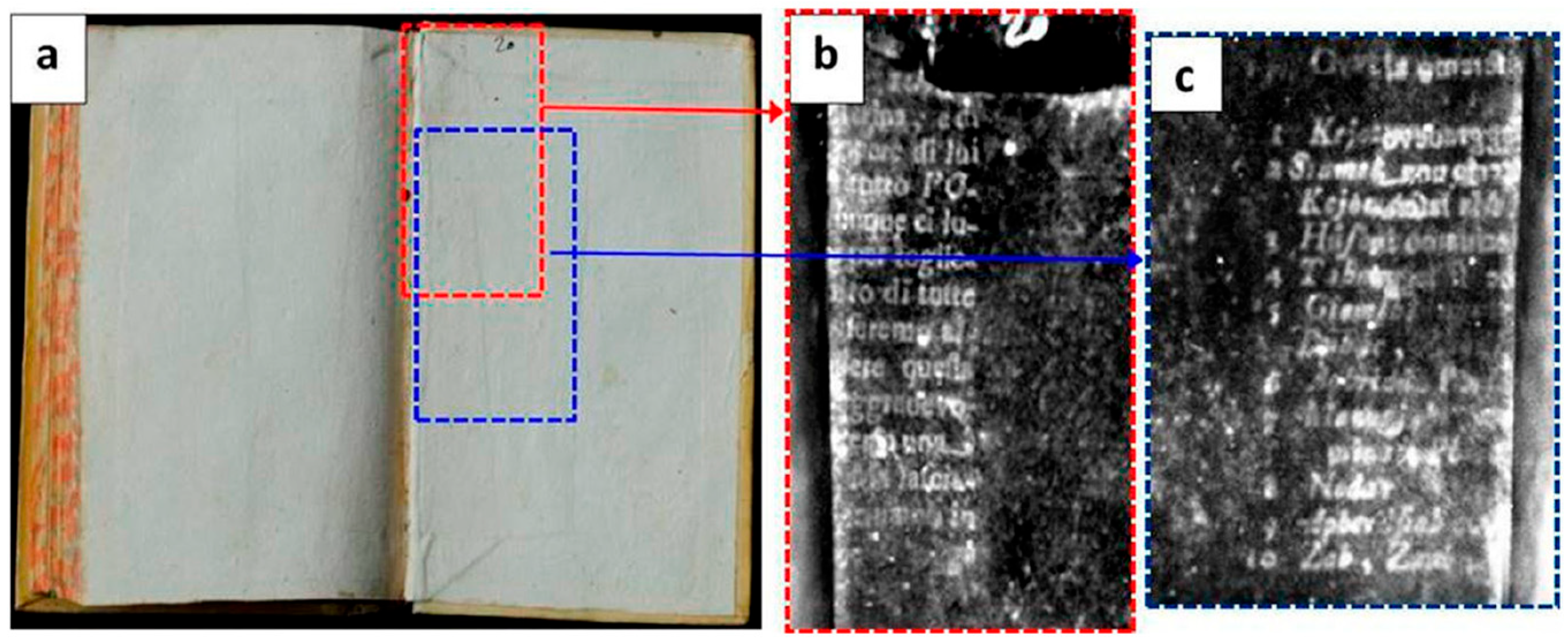
The accessibility of libraries and conservation institutes is one of the fundamental issues in the digital restoration of manuscripts. In the case of non-portable technologies, such as synchrotron radiation, institutions may send the manuscript to a specific facility. However, even though almost all technologies are portable, they need specific conditions to be used in situ; for instance, a space dedicated to performing scientific measurements, as a dark room, is necessary for the use of ultraviolet (UV) light. Usually, these requirements need to be presented in advance to the libraries through a plan, in order to reach an agreement on insurance and storage policies for both manuscripts and equipment, hours of work, types of analysis, and staff involved.
As preserving the integrity of the manuscript poses a crucial condition, several precautions must be observed to guarantee both the effectiveness of the measurement session and the safeguarding of the artefact. A single sheet of parchment can be easier to manage than a codex; in most cases, the manuscript’s binding holds tight the pages, preventing the book from opening fully. Libraries often provide scholars with tools for handling manuscripts. Alternatively, some tutorials (e.g., the instructions of Collapsible Book Cradle by Tara O’Brien) describe how to create different supports to adapt to the opening of a codex while preserving its integrity. After determining the optimal opening angle of the codex, it is essential to maintain page stability throughout the session. Various options are available for achieving this, e.g., coated lead weights or transparent polypropylene/polyester strips, which are the most widely adopted commercial alternatives. During measurements, dust or residues of various materials may accidentally cover the writing, and they can provoke light absorbance/reflections during the measurements; a fine, soft-bristled, conservator-approved brush can help remove them.

3. UV Photography

UV photography was the first technique used to recover invisible text, and has been used since the 1920s, when Wood’s lamps went on the market, nowadays substituted by LEDs.
Two techniques are the most used in UV photography: UV-induced visible fluorescence and UV-reflected imaging.
UV-induced visible fluorescence (UVIVF), or UV-fluorescence (UVF), relies on the properties of certain materials to absorb ultraviolet (UV) radiation and consequently emit visible light. The phenomenon known as fluorescence can reveal details by highlighting differences in fluorescence between various substances. Indeed, by exciting in the UV range, parchment or paper supports show intense fluorescence (with a blue/green color) whereas the imprint of the lost ink appears as darker areas. The image acquired by using a digital camera thus captures a high contrast between the two parts, allowing the reading of the lost text.
UVF was applied by Pottier et al., 2019, on 132 medieval parchment manuscripts made illegible after a fire in the Chartres library in France [4]. The advantages of UVF are the low-cost equipment, including a standard digital camera and lens, the rapidness of image acquisition, and the uncomplicated image treatment.
On the other hand, the UV-reflected (UVR) technique examines how the material reflects UV radiation by mounting a UV filter on the camera. For example, it is useful for identifying white pigments like titanium white and zinc white, which show a strong UV absorbance band [5]. Typically, UVR requires the use of specially developed lenses for UV photography and the removal of the internal UV-IR blocking filter from the digital camera. Cameras designed specifically for UVR are also employed.
According to Cosentino 2015 [5], both UVF and UVR methods are rarely conclusive; rather, they serve as the initial step toward implementing other techniques.

4. Multispectral Imaging (MSI) and Hyperspectral Imaging (HSI)

Multispectral imaging (MSI) is a widely employed technique with a rich history of application in the case of erased and partially legible texts. More precisely, in MSI, a series of images are captured in different spectral ranges with the use of bandpass filters or similar systems (Figure 1). The difference between this technique and photography with a common camera lies in the ability to select the radiation coming from the object, isolating single portions of the spectral range. For instance, in the case of dark spots covering the text, it is sometimes possible to select some spectral ranges in which they are transparent, allowing the ink to stand out [6].
Initial applications in the field involved the restoration of the illegible fragments of the Dead Sea scrolls [7,8] and the Petra Church scrolls [8,9], which had suffered from extreme degradation and a darkening of the parchment; in 1999, a similar technique was successfully applied to the reading of the carbonized Herculaneum papyri [10,11]. In all these works, the text contrast improves by selecting images in the infrared band, which are obtained through illumination using visible and infrared lamps.
Currently, two main MSI systems are utilized for text restoration. The first approach entails illuminating the manuscript with various sources, ranging from ultraviolet to infrared, while selectively filtering wavelengths using camera-mounted filters [6,12,13,14,15,16]. The second system directly employs narrow-band light-emitting diodes (LEDs) to capture images across distinct spectral bands [17,18,19,20,21,22].
While multispectral imaging typically captures 10–20 images at specific discrete wavelengths, hyperspectral imaging (HSI) collects hundreds of images across a continuous spectrum of wavelengths. Despite their higher spectral resolution, HSI scanning systems tend to have a lower spatial resolution than that of MSI [23]. At present, there are fewer applications of hyperspectral imaging for illegible manuscripts, and they include medieval palimpsests [24,25]; manuscripts corrupted with accidents such as ink bleeding, ink corrosion, and the foxing age-related process of deterioration [26]; concealed parchments recycled in bookbinding [27]; Herculaneum carbonized papyri [28].
Advanced techniques in fluorescence hyperspectral imaging include the use of lasers as exciting sources such as laser-induced fluorescence (LIF). This technique has led to significant results for hidden texts in mural painting [29] and could be potentially useful for hidden inks [30].
In the case of overlapping writings, such as palimpsests, it may not always be feasible to identify a specific spectral range where the “upper text” becomes transparent with MSI, allowing the “undertext” to become legible. Recent works have focused on the leveraging of generative adversarial networks (GANs) [31] or deep learning-based semantic segmentation to disentangle complex nodes of overlapping letters (Figure 2) [32].

5. Macro X-ray Fluorescence (XRF) Imaging

X-ray fluorescence (XRF) is one of the most used techniques for the characterization of inks in manuscripts [33,34,35]. Although the technique is not sensitive to materials with low atomic numbers (for example carbon ink), it has been revealed to be suitable for the detection of iron-based materials, i.e., iron gall ink, and pigments composed of chemical elements with medium/high atomic numbers (i.e., mercury, lead, and copper). The development of high-performing scanning systems has enabled us to carry out macro XRF (MA-XRF) imaging, discovering hidden texts on the basis of their elemental composition [36,37,38,39,40,41]. MA-XRF imaging has proven to be a successful approach when dealing with books made from recycled parchments that have suffered from darkening (Figure 3). Since iron-based inks were the most used writing materials during the Middle Ages [42], MA-XRF could map iron and other elements constituting the inks (e.g., calcium, zinc, copper, and manganese) for reconstructing the text, without any mechanical treatment of the books. This is possible because the support materials (i.e., parchment, papyri, and paper) are completely transparent to X-rays.
Another application of MA-XRF imaging involves writings covered by a pictorial layer. The recovery of the text is possible because the chemical elements of the painting are different from those used for the hidden text [18,43]. Michelin et al., 2021 [44], reveal with MA-XRF the retouched secret correspondence of French Queen Marie Antoinette. The study demonstrates that macroscopic elemental mapping combined with principal component analysis (PCA) can be a powerful tool for unraveling the superimposition of iron gall inks when they have distinct elemental compositions.
More recently, MA-XRF has revealed the presence of ruling lines lead-drawn by ancient scribes for the layout of Greek papyrus rolls. This discovery defines the experimental proof that slanted text columns were an esthetic choice of scribes [45].

6. X-ray Tomography

XRF is limited to direct application on a specific page, necessitating the book to be opened. In cases where opening the book is infeasible, X-ray computed tomography (XCT) can be utilized as an alternative method.
In XCT, the image contrast mechanism is based on differential X-ray absorption within a compound object. Micro-computed tomography (micro-CT or micro-XCT) is a variant of XCT that generates significantly higher-resolution images of objects. One of the main experiments in the field is the first “virtual unwrapping” of the En-Gedi scroll, one of the oldest Hebrew Pentateuch scrolls [46]. The ink composition, which likely contains metal, has a density that can be detected using micro-CT. This technique enables the reading of text that is concealed within the charred scroll (Figure 4).
Bukreeva et al., 2016 [47], applied X-ray phase-contrast tomography (XPCT) to reveal carbon-based ink within the Herculaneum papyrus scrolls, buried and carbonized in the eruption of Mount Vesuvius in 79 A.D. As shown in this study, standard XCT is unsuitable for distinguishing carbon fiber-based papyrus from carbon-based ink used to write on papyrus. Contrast was only achieved by capturing the phase modulation induced by the object in a coherent or partially coherent beam [47]. The study unveils, for the first time, textual portions inside the rolled-up scrolls from the Herculaneum collectionexpanding on the groundwork laid by Mocella et al., in 2015 [48]. Their pioneering work not only showcased the benefits of employing XPCT but also shed light on the challenges associated with virtually unrolling the Herculaneum papyri. In 2023, Dr. Brent Seales and his team launched the “Vesuvius Challenge”, a machine learning and computer vision competition aiming to read the X-ray scans of scrolls performed by EduceLab. The challenge in comprehending the text content arises from the intricate geometric structure of the carbonized scroll. The authors identify three key phases that lead to the reading of rolled papyri: scanning, segmentation and flattening, and ink detection. The latter occurs after the identification and flattening of the layers of papyrus, leveraging the 3D scans of the scroll (https://scrollprize.org/ (accessed on 24 January 2023).
In the last few years, the use of X-ray tomography for manuscript digitization has thus encouraged the development of algorithms to virtually unroll and unfold manuscripts that are too fragile and damaged to open [49,50,51,52,53,54]. In 2014, Ecole Polytechnique Fédérale de Lausanne (EPFL) and Ca’ Foscari University of Venice promoted the European project “Time Machine”, aimed at reading and digitizing a huge number of documents in diverse types of formats using a compact, laboratory-based radiology system [55,56].

7. Infrared Thermography (IRT)

Pulsed IRT is a type of thermography where a pulsed heat source is used for external heating; the IR radiation emitted from the sample surface is recorded as a thermogram. This technique has proven to be useful, especially for the inspection of subsurface elements [3]. In their work, Mercuri et al., 2013 [57], employed IRT to reveal fragments that had been inserted beneath the paper (Figure 5).
Pulsed IRT was also employed to reveal text beneath water stains in a thirteenth-century manuscript [58]. According to Orazi 2020 [3], the IRT technique relies on different temperature values between the inked and non-inked areas originating from thermal contrast, making the text more readable.

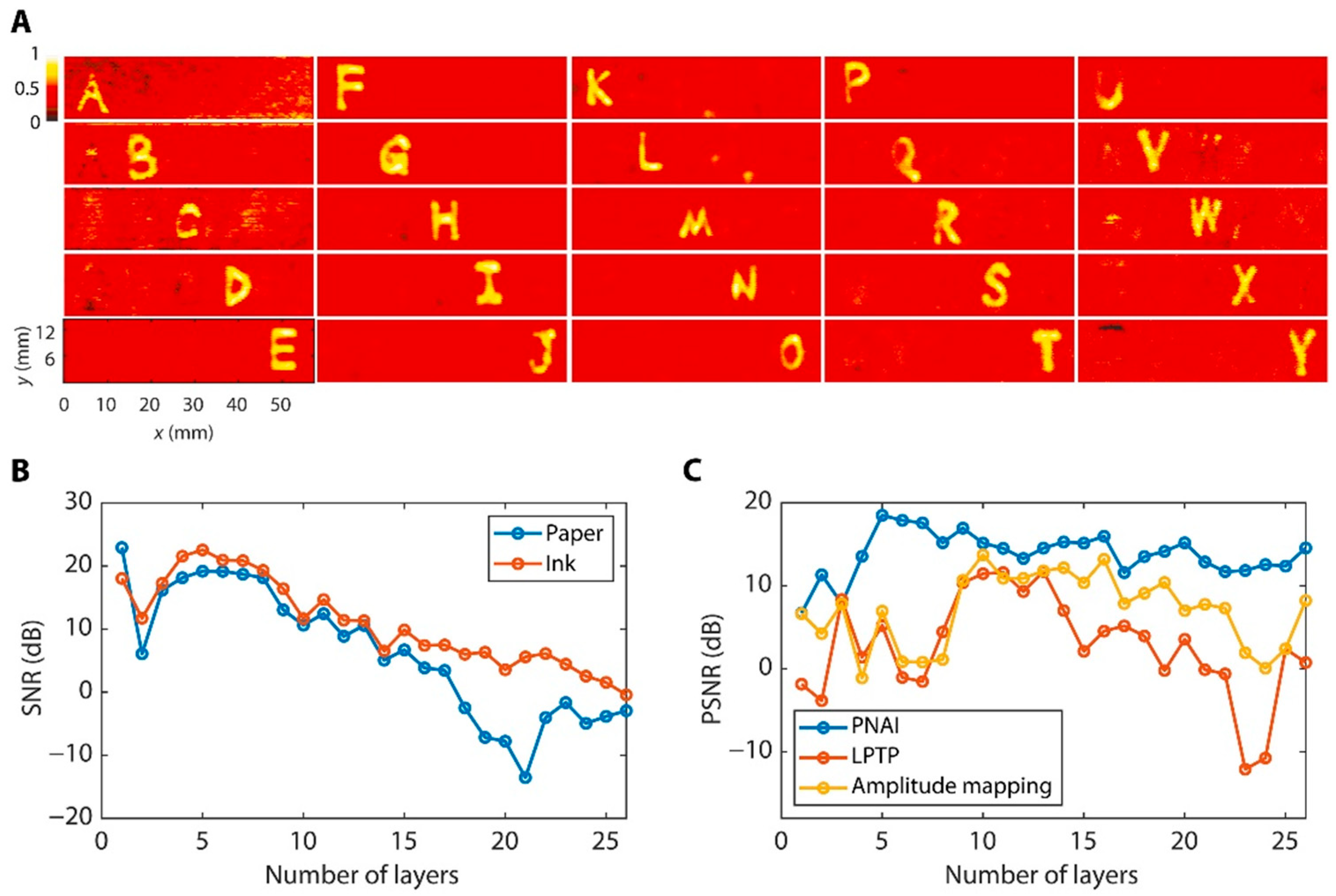
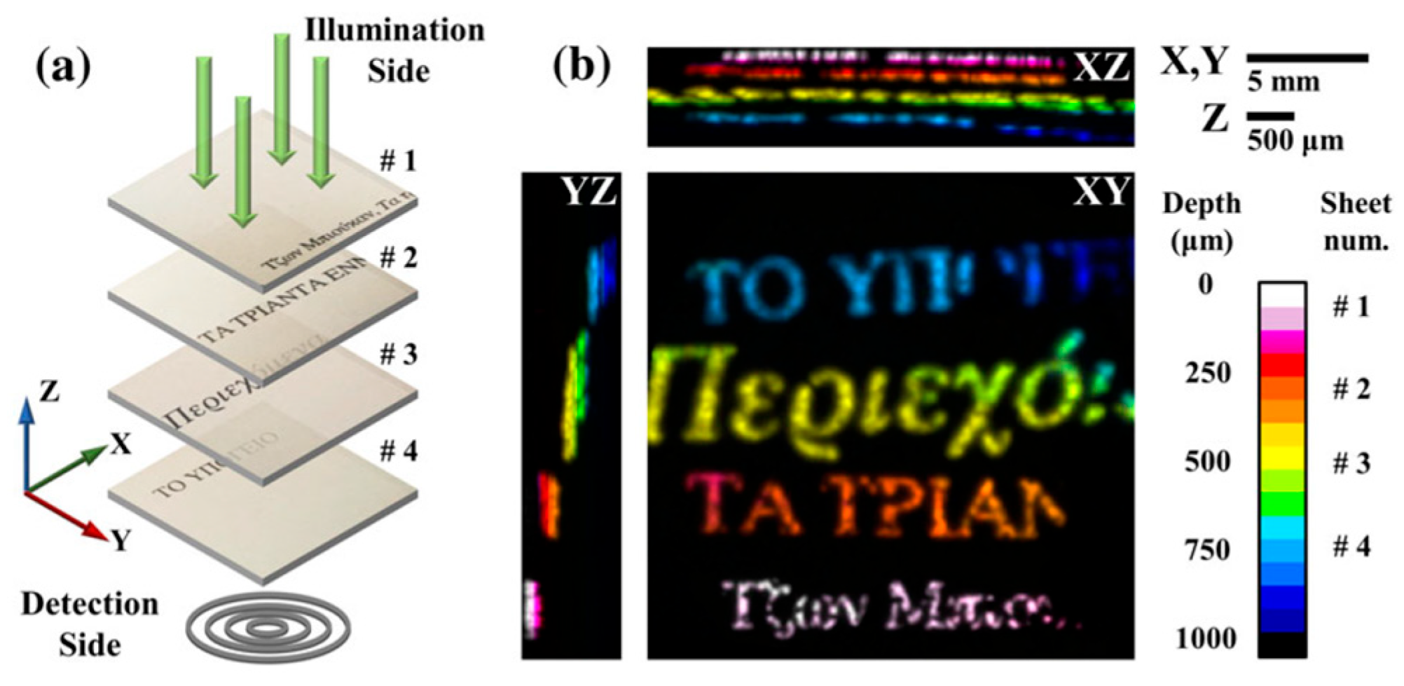
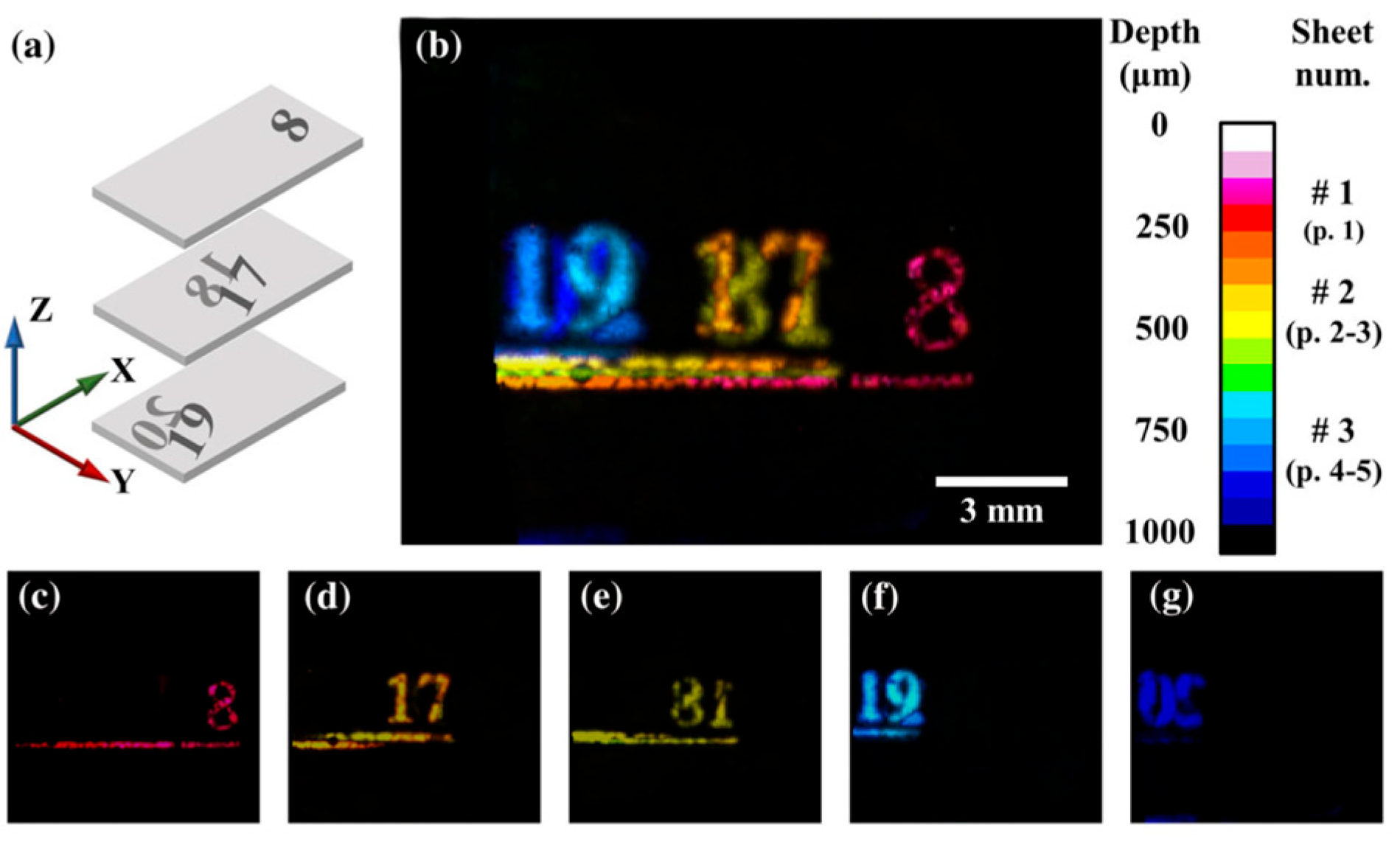
8. Terahertz (THz) Imaging

Terahertz (THz) radiation, with its deep penetration capabilities, allows for non-ionizing and non-invasive analysis. The successful acquisition of spectra has been achieved for writing materials such as iron gall inks, carbon-based inks, and traditional pigments commonly employed in manuscripts [59,60,61,62,63,64,65,66,67].
Due to the minimal attenuation provided by manuscript supports, THz radiation is advantageous for recovering hidden texts [68]. The versatility of sources and measurement geometry allows for a wide range of portable approaches.
The transmission configuration can be valuable in imaging single-sheet manuscripts and recovering underdrawings beneath ink layers [69,70] or seals [71] and detecting indentations, sieve lines, hidden watermarks or stains and alterations in aged materials [65,72,73].
THz reflection geometry is ideal for imaging layered archival documents. THz signals reveal signatures corresponding to reflections from various layers, allowing examinations of internal structures in optically opaque objects. The method is known as “time-of-flight”, and the pulse sequence provides information on the depth, position, and thickness of the inner layers [65].
Thus, it can resolve text from several layers of writing on papyrus and paper [16,60,74] and text on both sides of a single sheet [75], and reveal concealed writing under burned carbon black [65].
A THz tomosynthesis method based on a filtered back-projection algorithm allowed the recovery of graphite characters on paper up to the 50th layer [76], and the enhancement of the contrast for multiple-layered writings was successfully obtained [77,78] (Figure 6).
THz confocal microscopy has been recently applied to works of art [79] and has been able to reveal different inks used for music notes and pentagrams of a parchment manuscript [68].
The use of more powerful sources (i.e., quantum cascade lasers or Gunn diodes) combined with tomography can recover depth information overcoming possible limitations in the reflection mode [77].
THz cameras have already provided great advantages for imaging purposes in terms of discriminating between different materials, and they allow the real-time monitoring of the conservation state of artworks in a short amount of time [80,81].

9. Photoacoustic Imaging

Photoacoustic imaging applied to hidden text is a new research frontier. This technique is based on the absorption of pulsed light (i.e., laser sources) by a material, which determines the thermoelastic expansion of the surrounding environment and, consequently, a rapid pressure variation. The latter effect spreads as acoustic waves [82]. In the field of imaging applied to cultural heritage, the frequency range of the detected photoacoustic signals is in the ultrasonic interval (from a few up to several tens of MHz). As already demonstrated by the technologies that do not use visible light as a source, the main advantage of this technique is to provide high optical absorption contrast in an opaque matrix. Photoacoustic imaging takes advantage of light scattering, which enables the excitation of a broad volume, independent of the directions of the interacting photons [82]. Photoacoustic imaging has been demonstrated to be a good tool for the individuation of subsurface structures/underdrawings on pictorial tests [83] and for monitoring laser cleaning interventions [84]. Still, good results have been obtained from hidden texts of layered documents, both on single-sided printed paper sheets and in overlapping texts [85] (Figure 7 and Figure 8).
These first experimental studies demonstrate the high potential of photoacoustic signals in revealing hidden text in multi-layered documents, by performing high spatial resolution and good contrast levels. Since the application of the photoacoustic effect in cultural heritage is still under study, multi-wavelength imaging setups and further geometrical, optical, and acoustic parameter configurations could provide promising developments in this context [85].

10. Not-Conventional Sources (Synchrotron Radiation, Ion Beam Analysis (IBA) and Neutron)

To reveal texts by detecting the distribution of an element, a powerful technique is the SR-based MA-XRF, which involves synchrotron radiation (SR) as an X-ray source. The use of SR permits a higher incident flux, flux density, and collimation, resulting in short acquisition times, as well as high-contrast and high-spatial-resolution images with respect to those obtained with conventional MA-XRF [27,86,87,88,89,90,91,92,93,94]. As a synchrotron X-ray technique, we also mention synchrotron X-ray diffraction, which has demonstrated high efficiency for the detection of minerals contained in pigments or inks [95,96] and for studying the support structure (i.e., parchment, paper, and papyrus) of inked regions [97,98].
In the infrared region, synchrotron infrared microspectroscopy has been used to study inks and ink–paper material interactions in order to investigate the presence of iron gall ink in degradation processes [93,99]; these studies may also be promising as mapping techniques.
Other non-conventional sources used for the elemental analyses of manuscripts are ion beams. Among the techniques utilizing ion beam analysis (IBA), there is the particle-induced X-ray emission (PIXE) [100]. This technique shows strong potential for the analysis of manuscripts damaged by iron gall ink corrosion; indeed, PIXE mapping proves to be able to recognize heterogeneities of the elemental distributions of inks [101,102,103].
Finally, few studies have been reported about the use of neutron sources for the analysis of manuscripts and inks [104]. The interaction of neutrons with matter provides information about the elemental compositions of the inks. The advantage is that the captured and emitted particles are both highly penetrating and are used for the identification of major, minor, trace, and rare elements [105]. However, the possibility to detect hidden text is still not investigated, to the authors’ knowledge.
Although the use of non-conventional sources has many advantages for the detection of ink traces, generally, they are generated by particle accelerators, and this means that the artifact must be transported in the laboratory. However, for the PIXE technique, in recent years, a new portable particle accelerator has been developed [106], opening up new perspectives in the field.

11. Conclusions

In recent years, the digital restoration of historical manuscripts has emerged as a widely explored topic, as evident in the increasing number of related research projects and scientific publications.
This review comprehensively delves into various technologies, ranging from more conventional and well-known approaches like UV photography, multispectral and hyperspectral imaging techniques, infrared thermography, and X-ray tomography to less conventional yet promising methods such as terahertz and photoacoustic imaging, along with the utilization of synchrotron radiation, ion beams and neutron sources. A noteworthy advance among these unconventional methods is the ongoing evolution of technology, enhancing the portability of these techniques and thereby unlocking new potential applications in the digital restoration of manuscripts.
Notably, the ongoing strides in machine learning methods mark a significant advancement in addressing challenges related to the complex issue of ink detection, particularly in the case of palimpsests and carbonized papyri.

Author Contributions

Conceptualization, M.P.; methodology, M.P. and L.P.; writing—original draft preparation, M.P., L.P. and C.M.; writing—review and editing, M.P., L.P., C.M. and A.C.F.; supervision, A.C.F. and M.R. All authors have read and agreed to the published version of the manuscript.

Funding

This research received no external funding.

Data Availability Statement

Data sharing is not applicable.

Acknowledgments

We express our sincere gratitude to the ERC-PAGES project (AdG 2019 n° 882588) for its research support in the execution of this work. We heartily thank Anne-Sophie Gagnal, Pascaline Todeschini, Lise Marandet and Audrey Monet of the “Atelier de conservation-restauration du Service de Coopération Documentaire Interuniversitaire” of Université Paul Valéry- Montpellier 3 for their invaluable conservation expertise.

Conflicts of Interest

The authors declare no conflicts of interest.

Correction Statement

This article has been republished with a minor correction to the reference [32]. This change does not affect the scientific content of the article.

References

  1. Schettino, P.; Pietroni, E.; D’annibale, E. Re-Thinking Visitor Experience with Ancient Manuscripts via the Holographic Showcase: The Case of the Codex4D Project and Its First Public Results from a Mixed-Method Evaluation In Situ. Heritage 2023, 6, 6035–6065. [Google Scholar] [CrossRef]
  2. Tonazzini, A.; Salerno, E.; Abdel-Salam, Z.A.; Harith, M.A.; Marras, L.; Botto, A.; Campanella, B.; Legnaioli, S.; Pagnotta, S.; Poggialini, F.; et al. Analytical and mathematical methods for revealing hidden details in ancient manuscripts and paintings: A review. J. Adv. Res. 2019, 17, 31–42. [Google Scholar] [CrossRef] [PubMed]
  3. Orazi, N. Mid-wave Infrared Reflectography and Thermography for the Study of Ancient Books: A Review. Stud. Conserv. 2020, 65, 437–449. [Google Scholar] [CrossRef]
  4. Pottier, F.; Michelin, A.; Robinet, L. Recovering illegible writings in fire-damaged medieval manuscripts through data treatment of UV-fluorescence photography. J. Cult. Herit. 2018, 36, 183–190. [Google Scholar] [CrossRef]
  5. Cosentino, A. Practical notes on ultraviolet technical photography for art examination. Conserv. Patrim. 2015, 21, 53–62. [Google Scholar] [CrossRef]
  6. Pronti, L.; Perino, M.; Cursi, M.; Santarelli, M.L.; Felici, A.C.; Bracciale, M.P. Characterization and Digital Restauration of XIV-XV Centuries Written Parchments by Means of Nondestructive Techniques: Three Case Studies. J. Spectrosc. 2018, 2018, 2081548. [Google Scholar] [CrossRef]
  7. Bearman, G.H.; Spiro, S.I. Archaeological Applications of Advanced Imaging Techniques. Biblic. Archaeol. 1996, 59, 56–66. [Google Scholar] [CrossRef]
  8. Chabries, D.M.; Booras, S.W.; Bearman, G.H. Imaging the past: Recent applications of multispectral imaging technology to deciphering manuscripts. Antiquity 2003, 77, 359–372. [Google Scholar] [CrossRef]
  9. Chabries, D.M.; Booras, S.W.; Bikai, P. The Petra Scrolls. In Proceedings of the Image Processing, Image Quality, Image Capture Systems Conference (PICS), Montréal, QC, Canada, 22–25 April 2001. [Google Scholar]
  10. Booras, S.W.; Seely, D.R. Multispectral imaging of the Herculaneum papyri. Cronache Ercolanesi 1999, 29, 95–100. [Google Scholar]
  11. Macfarlane, R.; Del Mastro, G.; Antoni, A.; Booras, S. Update Report on the Use of the Multi-spectral images of the Herculaneum papyri. In Proceedings of the XXIV International Congress of Papyrology, Helsinki, Finland, 1–7 August 2004; The Finnish Society of Sciences and Letters: Helsinki, Finland, 2007; pp. 579–586. [Google Scholar]
  12. Knox, K.T.; Dickinson, C.; Wei, L.; Easton, R.L., Jr.; Johnston, R.H. Multispectral imaging of the Archimedes palimpsest. In Proceedings of the Image Processing, Image Quality, Image Capture, Systems Conference (PICS), Montreal, QC, Canada, 22–25 April 2001; pp. 206–210. [Google Scholar]
  13. Attas, E.M. Enhancement of Document Legibility Using Spectroscopic Imaging. Archivaria 2004, 57, 131–145. [Google Scholar]
  14. MacDonald, L.; Giacometti, A.; Campagnolo, A.; Robson, S.; Weyrich, T.; Terras, M.; Gibson, A. Multispectral Imaging of Degraded Parchment. In Computational Color Imaging; Tominaga, S., Schettini, R., Trémeau, A., Eds.; Lecture Notes in Computer Science; Springer: Berlin/Heidelberg, Germany, 2013; Volume 7786, pp. 143–157. [Google Scholar] [CrossRef]
  15. Alexopoulou, A.A.; Kaminari, A.-A.; Panagopoulos, A.; Pöhlmann, E. Multispectral documentation and image processing analysis of the papyrus of tomb II at Daphne, Greece. J. Archaeol. Sci. 2013, 40, 1242–1249. [Google Scholar] [CrossRef]
  16. Gibson, A.; Piquette, K.E.; Bergmann, U.; Christens-Barry, W.; Davis, G.; Endrizzi, M.; Fan, S.; Farsiu, S.; Fitzgerald, A.; Griffiths, J.; et al. An assessment of multimodal imaging of subsurface text in mummy cartonnage using surrogate papyrus phantoms. Herit. Sci. 2018, 6, 7. [Google Scholar] [CrossRef]
  17. Easton, R.; Knox, K.; Christens-Barry, W. Multispectral imaging of the Archimedes palimpsest. In Proceedings of the 32nd Applied Imagery Pattern Recognition Workshop, Washington, DC, USA, 15–17 October 2003; pp. 111–116. [Google Scholar]
  18. Knox, K.T. Enhancement of overwritten text in the Archimedes Palimpsest. In Electronic Imaging; Stork, D.G., Coddington, J., Eds.; SPIE: San Jose, CA, USA, 2008; p. 681004. [Google Scholar] [CrossRef]
  19. Christens-Barry, W.A.; Boydston, K.; France, F.G.; Knox, K.T.; Easton, R.L., Jr.; Toth, M.B. Camera system for multispectral imaging of documents. In IS&T/SPIE Electronic Imaging; Bodegom, E., Nguyen, V., Eds.; SPIE: San Jose, CA, USA, 2009; p. 724908. [Google Scholar]
  20. Easton, R.L., Jr.; Knox, K.T.; Christens-Barry, W.A.; Boydston, K.; Toth, M.B.; Emery, D.; Noel, W. Standardized system for multispectral imaging of palimpsests. In IS&T/SPIE Electronic Imaging; Stork, D.G., Coddington, J., Bentkowska-Kafel, A., Eds.; SPIE: San Jose, CA, USA, 2010; p. 75310D. [Google Scholar] [CrossRef]
  21. Bloechl, K.; Hamlin, H.; Easton, R.L., Jr. Text recovery from the ultraviolet-fluorescent spectrum for treatises of the Archimedes Palimpsest. In IS&T/SPIE Electronic Imaging; Stork, D.G., Coddington, J., Bentkowska-Kafel, A., Eds.; SPIE: San Jose, CA, USA, 2010; p. 753109. [Google Scholar] [CrossRef]
  22. Knox, K.T.; Easton, R.L., Jr.; Christens-Barry, W.A.; Boydston, K. Recovery of handwritten text from the diaries and papers of David Livingstone. In IS&T/SPIE Electronic Imaging; SPIE: San Francisco, CA, USA, 2011; p. 786909. [Google Scholar] [CrossRef]
  23. Jones, C.; Duffy, C.; Gibson, A.; Terras, M. Understanding multispectral imaging of cultural heritage: Determining best practice in MSI analysis of historical artefacts. J. Cult. Herit. 2020, 45, 339–350. [Google Scholar] [CrossRef]
  24. Rapantzikos, K.; Balas, C. Hyperspectral imaging: Potential in non-destructive analysis of palimpsests. In Proceedings of the IEEE International Conference on Image Processing 2005, Genova, Italy, 14 September 2005. [Google Scholar]
  25. Snijders, L.; Zaman, T.; Howell, D. Using hyperspectral imaging to reveal a hidden precolonial Mesoamerican codex. J. Archaeol. Sci. Rep. 2016, 9, 143–149. [Google Scholar] [CrossRef]
  26. Kim, S.J.; Deng, F.; Brown, M.S. Visual enhancement of old documents with hyperspectral imaging. Pattern Recognit. 2011, 44, 1461–1469. [Google Scholar] [CrossRef]
  27. Pouyet, E.; Devine, S.; Grafakos, T.; Kieckhefer, R.; Salvant, J.; Smieska, L.; Woll, A.; Katsaggelos, A.; Cossairt, O.; Walton, M. Revealing the biography of a hidden medieval manuscript using synchrotron and conventional imaging techniques. Anal. Chim. Acta 2017, 982, 20–30. [Google Scholar] [CrossRef]
  28. Tournié, A.; Fleischer, K.; Bukreeva, I.; Palermo, F.; Perino, M.; Cedola, A.; Andraud, C.; Ranocchia, G. Ancient Greek text concealed on the back of unrolled papyrus revealed through shortwave-infrared hyperspectral imaging. Sci. Adv. 2019, 5, eaav8936. [Google Scholar] [CrossRef]
  29. Almaviva, S.; Fantoni, R.; Colao, F.; Puiu, A.; Bisconti, F.; Nicolai, V.F.; Romani, M.; Cascioli, S.; Bellagamba, S. LIF/Raman/XRF non-invasive microanalysis of frescoes from St. Alexander catacombs in Rome. Spectrochim. Acta Part A Mol. Biomol. Spectrosc. 2018, 201, 207–215. [Google Scholar] [CrossRef]
  30. Nevin, A.; Spoto, G.; Anglos, D. Laser spectroscopies for elemental and molecular analysis in art and archaeology. Appl. Phys. A 2011, 106, 339–361. [Google Scholar] [CrossRef]
  31. Starynska, A.; Messinger, D.; Kong, Y. Revealing a history: Palimpsest text separation with generative networks. Int. J. Doc. Anal. Recognit. 2021, 24, 181–195. [Google Scholar] [CrossRef]
  32. Perino, M.; Ginolfi, M.; Felici, A.C.; Rosellini, M. A deep learning experiment for semantic segmentation of overlapping characters in palimpsests. In Proceedings of the IMEKO TC4 International Conference on Metrology for Archaeology and Cultural Heritage, Rome, Italy, 19–21 October 2023; pp. 825–829. [Google Scholar] [CrossRef]
  33. Ghigo, T.; Rabin, I.; Buzi, P. Black Egyptian inks in Late Antiquity: New insights on their manufacture and use. Archaeol. Anthr. Sci. 2020, 12, 70. [Google Scholar] [CrossRef]
  34. Taccetti, F.; Castelli, L.; Czelusniak, C.; Giambi, F.; Manetti, M.; Massi, M.; Mazzinghi, A.; Ruberto, C.; Arneodo, F.; Torres, R.; et al. Novel implementation of the INFN-CHNet X-ray fluorescence scanner for the study of ancient photographs, archaeological pottery, and rock art. Rend. Lincei. Sci. Fis. Nat. 2023, 34, 515–522. [Google Scholar] [CrossRef]
  35. Perino, M.; Pronti, L.; Di Forti, L.G.; Romani, M.; Taverna, C.; Massolo, L.; Manzari, F.; Cestelli-Guidi, M.; Nucara, A.; Felici, A.C. Revealing Artists’ Collaboration in a 14th Century Manuscript by Non-Invasive Analyses. Minerals 2021, 11, 771. [Google Scholar] [CrossRef]
  36. Miguel, C.; Bottura-Scardina, S.; Bottaini, C.; Valadas, S.; Candeias, A.; Bilou, F. The Power of Combining MA-XRF, Infrared Reflectography and Digital Microscopy to Unveil the Production of the 16th Century Illuminated Charter of Évora: What May Be Hidden under a Painted Surface? Heritage 2022, 5, 286–296. [Google Scholar] [CrossRef]
  37. Mazzinghi, A.; Ruberto, C.; Castelli, L.; Ricciardi, P.; Czelusniak, C.; Giuntini, L.; Mandò, P.A.; Manetti, M.; Palla, L.; Taccetti, F. The importance of being little: MA-XRF on manuscripts on a Venetian island. X-ray Spectrom. 2020, 50, 272–278. [Google Scholar] [CrossRef]
  38. Magkanas, G.; Bagán, H.; Sistach, M.; García, J. Illuminated manuscript analysis methodology using MA-XRF and NMF: Application on the Liber Feudorum Maior. Microchem. J. 2021, 165, 106112. [Google Scholar] [CrossRef]
  39. Watteeuw, L.; Van Bos, M.; Gersten, T.; Vandermeulen, B.; Hameeuw, H. An applied complementary use of macro X-ray fluorescence scanning and multi-light reflectance imaging to study Medieval Illuminated Manuscripts. The Rijmbijbel of Jacob van Maerlant. Microchem. J. 2020, 155, 104582. [Google Scholar] [CrossRef]
  40. Ricciardi, P.; Legrand, S.; Bertolotti, G.; Janssens, K. Macro X-ray fluorescence (MA-XRF) scanning of illuminated manuscript fragments: Potentialities and challenges. Microchem. J. 2016, 124, 785–791. [Google Scholar] [CrossRef]
  41. Tibúrcio, C.; Valadas, S.; Cardoso, A.; Candeias, A.; Barreira, C.; Miguel, C. On the use of EDXRF and UV–Vis FORS to unveil the production of two illuminated manuscripts from the fifteenth century portuguese royal court. Microchem. J. 2019, 153, 104455. [Google Scholar] [CrossRef]
  42. Ragib, Y.; Bat-Yehouda, M.Z. Les encres noires au Moyen Age (jusqu’a 1600). Stud. Islam. 1986, 64, 170. [Google Scholar] [CrossRef]
  43. Duivenvoorden, J.R.; Käyhkö, A.; Kwakkel, E.; Dik, J. Hidden library: Visualizing fragments of medieval manuscripts in early-modern bookbindings with mobile macro-XRF scanner. Herit. Sci. 2017, 5, 6. [Google Scholar] [CrossRef]
  44. Michelin, A.; Pottier, F.; Andraud, C. 2D macro-XRF to reveal redacted sections of French queen Marie-Antoinette secret correspondence with Swedish count Axel von Fersen. Sci. Adv. 2021, 7, eabg4266. [Google Scholar] [CrossRef]
  45. Romano, F.P.; Puglia, E.; Caliri, C.; Pavone, D.P.; Alessandrelli, M.; Busacca, A.; Fatuzzo, C.G.; Fleischer, K.J.; Pernigotti, C.; Preisler, Z.; et al. Layout of ancient Greek papyri through lead-drawn ruling lines revealed by Macro X-ray Fluorescence Imaging. Sci. Rep. 2023, 13, 6582. [Google Scholar] [CrossRef]
  46. Seales, W.B.; Parker, C.S.; Segal, M.; Tov, E.; Shor, P.; Porath, Y. From damage to discovery via virtual unwrapping: Reading the scroll from En-Gedi. Sci. Adv. 2016, 2, e1601247. [Google Scholar] [CrossRef]
  47. Bukreeva, I.; Mittone, A.; Bravin, A.; Festa, G.; Alessandrelli, M.; Coan, P.; Formoso, V.; Agostino, R.G.; Giocondo, M.; Ciuchi, F.; et al. Virtual unrolling and deciphering of Herculaneum papyri by X-ray phase-contrast tomography. Sci. Rep. 2016, 6, 27227. [Google Scholar] [CrossRef]
  48. Mocella, V.; Brun, E.; Ferrero, C.; Delattre, D. Revealing letters in rolled Herculaneum papyri by X-ray phase-contrast imaging. Nat. Commun. 2015, 6, 5895. [Google Scholar] [CrossRef]
  49. Baum, D.; Lindow, N.; Hege, H.-C.; Lepper, V.; Siopi, T.; Kutz, F.; Mahlow, K.; Mahnke, H.-E. Revealing hidden text in rolled and folded papyri. Appl. Phys. A 2017, 123, 171. [Google Scholar] [CrossRef]
  50. Liu, C.; Rosin, P.L.; Lai, Y.-K.; Hu, W. Robust Virtual Unrolling of Historical Parchment XMT Images. IEEE Trans. Image Process. 2017, 27, 1914–1926. [Google Scholar] [CrossRef]
  51. Rosin, P.L.; Lai, Y.-K.; Liu, C.; Davis, G.R.; Mills, D.; Tuson, G.; Russell, Y. Virtual Recovery of Content from X-ray Micro-Tomography Scans of Damaged Historic Scrolls. Sci. Rep. 2018, 8, 11901. [Google Scholar] [CrossRef]
  52. Stromer, D.; Christlein, V.; Martindale, C.; Zippert, P.; Haltenberger, E.; Hausotte, T.; Maier, A. Browsing through sealed historical manuscripts by using 3-D computed tomography with low-brilliance X-ray sources. Sci. Rep. 2018, 8, 15335. [Google Scholar] [CrossRef]
  53. Parker, C.S.; Parsons, S.; Bandy, J.; Chapman, C.; Coppens, F.; Seales, W.B. From invisibility to readability: Recovering the ink of Herculaneum. PLoS ONE 2019, 14, e0215775. [Google Scholar] [CrossRef]
  54. Mahnke, H.-E.; Arlt, T.; Baum, D.; Hege, H.-C.; Herter, F.; Lindow, N.; Manke, I.; Siopi, T.; Menei, E.; Etienne, M.; et al. Virtual unfolding of folded papyri. J. Cult. Herit. 2020, 41, 264–269. [Google Scholar] [CrossRef]
  55. Albertin, F.; Astolfo, A.; Stampanoni, M.; Peccenini, E.; Hwu, Y.; Kaplan, F.; Margaritondo, G. X-ray spectrometry and imaging for ancient administrative handwritten documents. X-ray Spectrom. 2015, 44, 93–98. [Google Scholar] [CrossRef]
  56. Albertin, F.; Patera, A.; Jerjen, I.; Hartmann, S.; Peccenini, E.; Kaplan, F.; Stampanoni, M.; Kaufmann, R.; Margaritondo, G. Virtual reading of a large ancient handwritten science book. Microchem. J. 2016, 125, 185–189. [Google Scholar] [CrossRef]
  57. Mercuri, F.; Gnoli, R.; Paoloni, S.; Orazi, N.; Cicero, C.; Zammit, U.; Marinelli, M.; Scudieri, F. Hidden Text Detection by Infrared Thermography/Anwendung der aktiven Infrarotthermographie (IRT) zur Erfassung von verdeckten Texten/Utilisation de la thermographie infrarouge (IRT) pour détecter des textes cachés. Restaurator 2013, 34, 195–211. [Google Scholar] [CrossRef]
  58. Mercuri, F.; Orazi, N.; Paoloni, S.; Cicero, C.; Zammit, U. Pulsed Thermography Applied to the Study of Cultural Heritage. Appl. Sci. 2017, 7, 1010. [Google Scholar] [CrossRef]
  59. Bardon, T.; May, R.K.; Taday, P.F.; Strlič, M. Systematic study of terahertz time-domain spectra of historically informed black inks. Analyst 2013, 138, 4859–4869. [Google Scholar] [CrossRef]
  60. Labaune, J.; Jackson, J.B.; Pagès-Camagna, S.; Duling, I.N.; Menu, M.; Mourou, G.A. Papyrus imaging with terahertz time domain spectroscopy. Appl. Phys. A 2010, 100, 607–612. [Google Scholar] [CrossRef]
  61. Tasseva, J.; Taschin, A.; Bartolini, P.; Striova, J.; Fontana, R.; Torre, R. Thin layered drawing media probed by THz time-domain spectroscopy. Analyst 2016, 142, 42–47. [Google Scholar] [CrossRef]
  62. Taschin, A.; Bartolini, P.; Tasseva, J.; Striova, J.; Fontana, R.; Riminesi, C.; Torre, R. Drawing materials studied by THz spectroscopy. Acta IMEKO 2017, 6, 12. [Google Scholar] [CrossRef]
  63. Kleist, E.M.; Dandolo, C.L.K.; Guillet, J.-P.; Mounaix, P.; Korter, T.M. Terahertz Spectroscopy and Quantum Mechanical Simulations of Crystalline Copper-Containing Historical Pigments. J. Phys. Chem. A 2019, 123, 1225–1232. [Google Scholar] [CrossRef]
  64. Kleist, E.M.; Korter, T.M. Quantitative Analysis of Minium and Vermilion Mixtures Using Low-Frequency Vibrational Spectroscopy. Anal. Chem. 2019, 92, 1211–1218. [Google Scholar] [CrossRef]
  65. Fukunaga, K. THz Technology Applied to Cultural Heritage in Practice. In Cultural Heritage Science; Springer: Tokyo, Japan, 2016. [Google Scholar] [CrossRef]
  66. Hong, T.; Choi, K.; Ha, T.; Park, B.C.; Sim, K.I.; Kim, J.H.; Kim, J.H.; Kwon, J.E.; Lee, S.; Kang, D.I.; et al. Terahertz time-domain and Fourier-transform infrared spectroscopy of traditional Korean pigments. J. Korean Phys. Soc. 2014, 64, 727–731. [Google Scholar] [CrossRef]
  67. Ha, T.; Lee, H.; Sim, K.I.; Kim, J.; Jo, Y.C.; Kim, J.H.; Baek, N.Y.; Kang, D.-I.; Lee, H.H. Optimal methodologies for terahertz time-domain spectroscopic analysis of traditional pigments in powder form. J. Korean Phys. Soc. 2017, 70, 866–871. [Google Scholar] [CrossRef]
  68. Flammini, M.; Bonsi, C.; Ciano, C.; Giliberti, V.; Pontecorvo, E.; Italia, P.; DelRe, E.; Ortolani, M. Confocal Terahertz Imaging of Ancient Manuscripts. J. Infrared Millim. Terahertz Waves 2016, 38, 435–442. [Google Scholar] [CrossRef]
  69. Abraham, E.; Younus, A.; El Fatimy, A.; Delagnes, J.; Nguéma, E.; Mounaix, P. Broadband terahertz imaging of documents written with lead pencils. Opt. Commun. 2009, 282, 3104–3107. [Google Scholar] [CrossRef]
  70. Mittleman, D.; Gupta, M.; Neelamani, R.; Baraniuk, R.; Rudd, J.; Koch, M. Recent advances in terahertz imaging. Appl. Phys. B Laser Opt. 1999, 68, 1085–1094. [Google Scholar] [CrossRef]
  71. Younus, A.; Delagnes, J.; Canioni, L.; Guillet, J.; Mounaix, P.; Fabre, M. Spectroscopy and terahertz imaging for sigillography applications. In Proceedings of the 2012 37th International Conference on Infrared, Millimeter, and Terahertz Waves, Wollongong, NSW, Australia, 23–28 September 2012; p. 1. [Google Scholar] [CrossRef]
  72. Bardon, T.; May, R.K.; Taday, P.F.; Strlič, M. Contrast in Terahertz Images of Archival Documents—Part II: Influence of Topographic Features. J. Infrared Millim. Terahertz Waves 2017, 38, 467–482. [Google Scholar] [CrossRef]
  73. Fukunaga, K.; Ogawa, Y.; Hayashi, S.; Hosako, I. Application of terahertz spectroscopy for character recognition in a medieval manuscript. IEICE Electron. Express 2008, 5, 223–228. [Google Scholar] [CrossRef]
  74. Redo-Sanchez, A.; Heshmat, B.; Aghasi, A.; Naqvi, S.; Zhang, M.; Romberg, J.; Raskar, R. Terahertz time-gated spectral imaging for content extraction through layered structures. Nat. Commun. 2016, 7, 12665. [Google Scholar] [CrossRef]
  75. Walker, G.C.; Labaune, J.; Bowen, J.W.; Jackson, J.-B.; Hadjiloucas, S.; Mourou, G.; Menu, M. Deconvolution: Imaging the unturned page. In Proceedings of the 2011 International Conference on Infrared, Millimeter, and Terahertz Waves, Houston, TX, USA, 2–7 October 2011; pp. 1–2. [Google Scholar] [CrossRef]
  76. Sunaguchi, N.; Sasaki, Y.; Maikusa, N.; Kawai, M.; Yuasa, T.; Otani, C. Depth-resolving THz imaging with tomosynthesis. Opt. Express 2009, 17, 9558–9570. [Google Scholar] [CrossRef]
  77. Bardon, T.; May, R.K.; Jackson, J.B.; Beentjes, G.; de Bruin, G.; Taday, P.F.; Strlič, M. Contrast in Terahertz Images of Archival Documents—Part I: Influence of the Optical Parameters from the Ink and Support. J. Infrared Millim. Terahertz Waves 2017, 38, 443–466. [Google Scholar] [CrossRef]
  78. Cui, Y.; Xu, Y.; Han, D.; Wang, X.; Shen, Z.; Hou, Y.; Liang, J.; Wang, X.; Citrin, D.S.; Zhang, L.; et al. Hidden-information extraction from layered structures through terahertz imaging down to ultralow SNR. Sci. Adv. 2023, 9, eadg8435. [Google Scholar] [CrossRef] [PubMed]
  79. Ciano, C.; Flammini, M.; Giliberti, V.; Calvani, P.; DelRe, E.; Talarico, F.; Torre, M.; Missori, M.; Ortolani, M. Confocal Imaging at 0.3 THz with Depth Resolution of a Painted Wood Artwork for the Identification of Buried Thin Metal Foils. IEEE Trans. Terahertz Sci. Technol. 2018, 8, 390–396. [Google Scholar] [CrossRef]
  80. Fukunaga, K.; Hosako, I.; Iii, I.N.D.; Picollo, M. Terahertz imaging systems: A non-invasive technique for the analysis of paintings. In SPIE Europe Optical Metrology; Pezzati, L., Salimbeni, R., Eds.; SPIE: Munich, Germany, 2009; p. 73910D. [Google Scholar] [CrossRef]
  81. Fukunaga, K.; Sekine, N.; Hosako, I.; Oda, N.; Yoneyama, H.; Sudoh, T. Real-time terahertz imaging for art conservation science. J. Eur. Opt. Soc. Publ. 2008, 3, 08027. [Google Scholar] [CrossRef]
  82. Tserevelakis, G.J.; Vrouvaki, I.; Siozos, P.; Melessanaki, K.; Hatzigiannakis, K.; Fotakis, C.; Zacharakis, G. Photoacoustic imaging reveals hidden underdrawings in paintings. Sci. Rep. 2017, 7, 747. [Google Scholar] [CrossRef]
  83. Setiawan, A.; Setiaji, F.D.; Dewantoro, G.; Wibowo, N.A. Photoacoustic Tomography System for Roughly Painted Micro Objects. J. Electromagn. Eng. Sci. 2019, 19, 197–203. [Google Scholar] [CrossRef]
  84. Tserevelakis, G.J.; Pouli, P.; Zacharakis, G. Listening to laser light interactions with objects of art: A novel photoacoustic approach for diagnosis and monitoring of laser cleaning interventions. Herit. Sci. 2020, 8, 98. [Google Scholar] [CrossRef]
  85. Tserevelakis, G.J.; Tsagkaraki, M.; Siozos, P.; Zacharakis, G. Uncovering the hidden content of layered documents by means of photoacoustic imaging. Strain 2018, 55, e12289. [Google Scholar] [CrossRef]
  86. Autran, P.-O.; Dejoie, C.; Dugand, C.; Gervason, M.; Bordet, P.; Hodeau, J.-L.; Anne, M.; Martinetto, P. Illustrating papyrus in Ancient Egypt. Sci. Rep. 2023, 13, 524. [Google Scholar] [CrossRef] [PubMed]
  87. Dhara, S.; Misra, N.; Maind, S.; Kumar, S.A.; Chattopadhyay, N.; Aggarwal, S. Forensic application of total reflection X-ray fluorescence spectrometry for elemental characterization of ink samples. Spectrochim. Acta Part B At. Spectrosc. 2010, 65, 167–170. [Google Scholar] [CrossRef]
  88. Kempson, I.M.; Paulkirkbride, K.; Skinner, W.M.; Coumbaros, J. Applications of synchrotron radiation in forensic trace evidence analysis. Talanta 2005, 67, 286–303. [Google Scholar] [CrossRef] [PubMed]
  89. Reiche, I.; Radtke, M.; Berger, A.; Görner, W.; Ketelsen, T.; Merchel, S.; Riederer, J.; Riesemeier, H.; Roth, M. Spatially resolved synchrotron-induced X-ray fluorescence analyses of metal point drawings and their mysterious inscriptions. Spectrochim. Acta Part B At. Spectrosc. 2004, 59, 1657–1662. [Google Scholar] [CrossRef]
  90. Cotte, M.; Susini, J.; Dik, J.; Janssens, K. Synchrotron-Based X-ray Absorption Spectroscopy for Art Conservation: Looking Back and Looking Forward. Acc. Chem. Res. 2010, 43, 705–714. [Google Scholar] [CrossRef] [PubMed]
  91. Cotte, M.; Pouyet, E.; Salomé, M.; Rivard, C.; De Nolf, W.; Castillo-Michel, H.; Fabris, T.; Monico, L.; Janssens, K.; Wang, T.; et al. The ID21 X-ray and infrared microscopy beamline at the ESRF: Status and recent applications to artistic materials. J. Anal. At. Spectrom. 2016, 32, 477–493. [Google Scholar] [CrossRef]
  92. Sozontov, E.A.; Demkiv, A.A.; Guryeva, P.V.; Peters, G.S.; Kolobylina, N.N.; Oukhanova, E.V.; Yatsishina, E.B. Ancient Parchments: Structural Diagnostics and Visualization of Textual Fragments of Manuscripts—A Natural-Science Approach. J. Surf. Investig. 2019, 13, 366–370. [Google Scholar] [CrossRef]
  93. Faubel, W.; Staub, S.; Simon, R.; Heissler, S.; Pataki, A.; Banik, G. Non-destructive analysis for the investigation of decomposition phenomena of historical manuscripts and prints. Spectrochim. Acta Part B At. Spectrosc. 2007, 62, 669–676. [Google Scholar] [CrossRef]
  94. Guryeva, P.V.; Demkiv, A.A.; Sozontov, E.A. Mapping a text on ancient parchment by X-ray fluorescence analysis using a synchrotron source. J. Surf. Investig. 2017, 11, 167–168. [Google Scholar] [CrossRef]
  95. Christiansen, T.; Cotte, M.; de Nolf, W.; Mouro, E.; Reyes-Herrera, J.; de Meyer, S.; Vanmeert, F.; Salvadó, N.; Gonzalez, V.; Lindelof, P.E.; et al. Insights into the composition of ancient Egyptian red and black inks on papyri achieved by synchrotron-based microanalyses. Proc. Natl. Acad. Sci. USA 2020, 117, 27825–27835. [Google Scholar] [CrossRef]
  96. Smieska, L.M.; Mullett, R.; Ferri, L.; Woll, A.R. Trace elements in natural azurite pigments found in illuminated manuscript leaves investigated by synchrotron X-ray fluorescence and diffraction mapping. Appl. Phys. A 2017, 123, 484. [Google Scholar] [CrossRef]
  97. Kennedy, C.J.; Hiller, J.C.; Lammie, D.; Drakopoulos, M.; Vest, M.; Cooper, M.; Adderley, W.P.; Wess, T.J. Microfocus X-ray Diffraction of Historical Parchment Reveals Variations in Structural Features through Parchment Cross Sections. Nano Lett. 2004, 4, 1373–1380. [Google Scholar] [CrossRef]
  98. Brun, E.; Cotte, M.; Wright, J.; Ruat, M.; Tack, P.; Vincze, L.; Ferrero, C.; Delattre, D.; Mocella, V. Revealing metallic ink in Herculaneum papyri. Proc. Natl. Acad. Sci. USA 2016, 113, 3751–3754. [Google Scholar] [CrossRef]
  99. Wilkinson, T.J.; Perry, D.L.; Martin, M.C.; McKinney, W.R.; Cantu, A.A. Use of Synchrotron Reflectance Infrared Spectromicroscopy as a Rapid, Direct, Nondestructive Method for the Study of Inks on Paper. Appl. Spectrosc. 2002, 56, 800–803. [Google Scholar] [CrossRef]
  100. Vijayan, V.; Choudhury, R.K.; Mallick, B.; Sahu, S.; Choudhury, S.K.; Lenka, H.P.; Rautray, T.R.; Nayak, P.K. External particle-induced X-ray emission. Curr. Sci. 2003, 85, 772–777. [Google Scholar]
  101. Remazeilles, C.; Quillet, V.; Calligaro, T.; Dran, J.C.; Pichon, L.; Salomon, J. PIXE elemental mapping on original manuscripts with an external microbeam. Application to manuscripts damaged by iron-gall ink corrosion. Nucl. Instrum. Methods Phys. Res. Sect. B Beam Interact. Mater. At. 2001, 181, 681–687. [Google Scholar] [CrossRef]
  102. Mathayan, V.; Sortica, M.; Primetzhofer, D. Determining the chronological sequence of inks deposited with different writing and printing tools using ion beam analysis. J. Forensic Sci. 2021, 66, 1401–1409. [Google Scholar] [CrossRef]
  103. Sottili, L.; Giuntini, L.; Mazzinghi, A.; Massi, M.; Carraresi, L.; Castelli, L.; Czelusniak, C.; Giambi, F.; Mandò, P.A.; Manetti, M.; et al. The Role of PIXE and XRF in Heritage Science: The INFN-CHNet LABEC Experience. Appl. Sci. 2022, 12, 6585. [Google Scholar] [CrossRef]
  104. Festa, G.; Christiansen, T.; Turina, V.; Borla, M.; Kelleher, J.; Arcidiacono, L.; Cartechini, L.; Ponterio, R.C.; Scatigno, C.; Senesi, R.; et al. Egyptian metallic inks on textiles from the 15th century BCE unravelled by non-invasive techniques and chemometric analysis. Sci. Rep. 2019, 9, 7310. [Google Scholar] [CrossRef]
  105. Festa, G.; Romanelli, G.; Senesi, R.; Arcidiacono, L.; Scatigno, C.; Parker, S.F.; Marques, M.P.M.; Andreani, C. Neutrons for Cultural Heritage—Techniques, Sensors, and Detection. Sensors 2020, 20, 502. [Google Scholar] [CrossRef]
  106. Mathot, S.; Anelli, G.; Atieh, S.; Bilton, A.; Bulat, B.; Callamand, T.; Calvo, S.; Favre, G.; Geisser, J.-M.; Gerardin, A.; et al. The CERN PIXE-RFQ, a transportable proton accelerator for the machina project. Nucl. Instrum. Methods Phys. Res. Sect. B Beam Interact. Mater. At. 2019, 459, 153–157. [Google Scholar] [CrossRef]
Figure 1. (a) Photography of Parch. 38 belonging to the collection of the Library of the Department of History, Anthropology, Religions, Art History, Media and Performing Arts of Sapienza University of Rome; MSI with a UV source and filters at (b) 470 nm; (c) 500 nm; (d) 532 nm; (e) 600 nm; (f) 680 nm; (g) 700 nm; and (h) 750 nm. Reproduced under the terms of Creative Commons Attribution License 4.0 from [6].
Heritage 07 00034 g001
Figure 2. Examples of the prediction of a semantic segmentation model applied on a synthetic dataset of overlapping handwritten letters. In the top panels is the segmentation of complex combinations of A + C and D + E; in the bottom panels is the segmentation of complex combinations of B + C and A + D. To enhance visualization and explore potential patterns in noise residuals, the authors employ a min–max scaling technique for the color palette representation of predicted segmentation masks. Consequently, white pixels in each panel indicate zero flux, while black pixels represent the maximum flux in the mask. To facilitate a fair comparison between different masks and identify significant signals amidst noise residuals, histograms illustrate the distribution of maximum fluxes in each panel. Reproduced under the terms of Creative Commons Attribution License 4.0 from [32].
Heritage 07 00034 g002
Figure 3. Distribution maps of calcium, iron, mercury L-lines and lead L-lines of parchment recycled into bookbinding. Reproduced under the terms of the Creative Commons Attribution 4.0 International License from [43].
Heritage 07 00034 g003
Figure 4. Complete virtual unwrapping of the En-Gedi scroll. Reproduced under the terms of Creative Commons Attribution Non-Commercial License 4.0 from [46].
Heritage 07 00034 g004
Figure 5. (a) Photograph of the paper guard of a seventeenth-century book; (b) a contrasted thermogram recorded just after the light pulse revealed a first written fragment concealed beneath the paper guard; (c) a contrasted thermogram recorded 300 ms after the light pulse revealed a second written fragment that belongs to a deeper subsurface, glued just beneath the previous one. Reproduced with permission from [3]; published by Taylor & Francis Ltd., 2020.
Heritage 07 00034 g005
Figure 6. (a) THz images from 25 layers of letters written with black gel pen on paper present excellent consistency and high contrast. The proposed methodology named pulse neighborhood average imaging (PNAI) can eliminate letter superpositions, and the quality of the resulting THz images is comparable with that of optical images of the letter on single sheets of paper. Images are normalized separately with the mean value set to 0.4. (b) The estimated signal-to-noise ratio (SNR) of THz signals for each layer and (c) the evaluation of THz image quality using the peak SNR obtained by various imaging algorithms highlighting that the PNAI method provides a substantial improvement in image contrast. Reproduced under the terms of Creative Commons Attribution Non-Commercial License 4.0 from [78].
Heritage 07 00034 g006
Figure 7. Photoacoustic imaging of a layered document with non-overlapping text; (a) representation of the sample; (b) photoacoustic images through top (XY) and side views (XZ; YZ). Reproduced with permission from [85];published by John Wiley & Sons Ltd., 2019.
Heritage 07 00034 g007
Figure 8. Photoacoustic imaging of a layered document with overlapping text; (a) representation of the sample; (b) top view of a photoacoustic image; (cg) individual reconstructions of five sequential pages. Reproduced with permission from [85]; published by John Wiley & Sons Ltd., 2019.
Heritage 07 00034 g008
Disclaimer/Publisher’s Note: The statements, opinions and data contained in all publications are solely those of the individual author(s) and contributor(s) and not of MDPI and/or the editor(s). MDPI and/or the editor(s) disclaim responsibility for any injury to people or property resulting from any ideas, methods, instructions or products referred to in the content.

