Article

Rediscovering the Painting Technique of the 15th Century Panel Painting Depicting the Coronation of the Virgin by Michele di Matteo

1
Department of Physics, University of Pavia, Via Agostino Bassi 6, 27100 Pavia, Italy
2
Arvedi Laboratory of Non-Invasive Diagnostics, CISRiC, University of Pavia, Via Bell’Aspa 3, 26100 Cremona, Italy
3
Department of Musicology and Cultural Heritage, University of Pavia, C.so Garibaldi 178, 26100 Cremona, Italy
4
Laboratory of Applied Diagnostics for Cultural Heritage, Cr.Forma Restoration School, C.so Matteotti 17, 26100 Cremona, Italy
*
Author to whom correspondence should be addressed.
Heritage 2024, 7(1), 324-337; https://doi.org/10.3390/heritage7010016
Submission received: 5 December 2023 / Revised: 30 December 2023 / Accepted: 4 January 2024 / Published: 10 January 2024
(This article belongs to the Special Issue Non-invasive Technologies Applied in Cultural Heritage)

Abstract

:
The study concerned a diagnostic spectroscopic campaign carried out on the panel painting depicting the Coronation of the Virgin (first half of the 15th century) by the late-Gothic Italian painter Michele di Matteo. The main aims were the identification of the original painting materials and the characterization of the painter’s artistic technique. A combined approach based on non- and micro-invasive techniques was employed. Visible and ultraviolet-induced fluorescence photography was used to select the areas of interest for spectroscopic analyses; X-ray radiography assessed the state of conservation of the support, while X-ray fluorescence and external reflection Fourier transform infrared spectroscopies allowed the chemical identification of pigments, binders, and varnishes. Attenuated total reflection infrared spectroscopy, optical microscopy, and scanning electron microscopy coupled with energy-dispersive X-ray spectroscopy were used to visualize and characterize the materials in the pictorial layers. The results highlighted the presence of pigments, possibly applied with an egg binder, consistent with the period of the production of the painting, as well as modern pigments used during subsequent restorations: an imprimitura with lead white and a gypsum-based ground layer. Concerning the gilding, the guazzo technique was confirmed by identifying a red bolo substrate and gold leaf.

1. Introduction

In the field of cultural heritage, the analytical scientific approach is often used to support conservation and restoration procedures, as well as to increase the information useful for the art historical study and valorization of an artwork. This type of analysis often focuses on the identification and characterization of the different materials used to produce the artwork and their distribution in the multi-layered system present in different areas of the painting. In the case of medieval panel paintings, the stratigraphic system usually includes a ground layer (e.g., gypsum and glue), an under-drawing, overlapped paint layers, a mixture of pigments and binding media (e.g., animal glue, egg, siccative oil), and an external varnish that partially covers non-original paints or restoration materials [1,2,3,4,5].
The present research focuses on the analytical campaign on the panel painting from the first half of the 15th century depicting the Coronation of the Virgin by Michele di Matteo (Bologna, 14th century—Bologna, 1469) (Figure 1). The iconography depicts Jesus Christ placing the crown on the head of his mother sitting on the same throne. The painting is attributed to Michele di Matteo, also known as Matteo de Calcina, Michele della Fornace [6,7], or Michele di Matteo Lambertini [7,8], who was a late-Gothic painter active in Bologna from 1410 to 1469 [9]. While it is known that he was an apprentice of Giovanni da Modena [6,7], his artistic identity remains ambiguous. Consequently, conducting analytical studies of the painting techniques employed by the artist could provide valuable insights into his artistic style. The panel, which is part of a private collection, is probably the only remaining portion of a dismembered polyptych [7,10] commissioned by the Corporazione dei Calzolari of Bologna around 1421–1426 [6].
The main aims of this analytical campaign were (i) the assessment of the state of preservation of the support and the pictorial layer, (ii) the characterization of both original and restoration artistic materials (e.g., pigments, lakes, binders), and (iii) the identification of the multi-layered system in order to obtain information about the painting technique (i.e., support preparation and gilding). This paper tries to provide the necessary and useful data from the first analytical campaign carried out on Michele di Matteo’s painting. The results could contribute to the understanding of his painting technique, which has never been studied from a scientific point of view, and in the future, they could be a valuable reference for works by the same painter or other artists related to him. Also, the scientific investigation intends to support future restoration procedures. To these ends, a combined multi-analytical approach based on non- and micro-invasive techniques was employed to study the painting. In particular, visible photography (VIS) and visible fluorescence induced by ultraviolet radiation (UVIF) were used to select the areas of interest for the spectroscopic analyses [4,11,12]. X-ray radiography (RX) was also necessary to assess the state of conservation of the support [12,13,14]. X-ray fluorescence (XRF) [2,14,15,16] and external reflection Fourier transform infrared (ER-FTIR) [12,13,17,18] spectroscopies allowed the chemical characterization of pigments, binders, and varnishes. Considering the historical and artistic importance of the work and the painter, a micro-invasive approach, limited to a few areas of interest, was conducted. The application of attenuated total reflection infrared (ATR-FTIR) spectroscopy, optical microscopy (OM), and scanning electron microscopy coupled with energy dispersive X-ray spectroscopy (SEM-EDS) permitted the mapping and identification of the materials in the pictorial layering, in particular the ground preparation and the gilding [1,2,17,19,20,21,22]. A limited number of fragments for micro-invasive analysis were sampled by an expert restorer, where micro-cracks or already partially detached parts were recognized. According to the literature, the above-mentioned techniques have been proven to be successful in providing information about the materials [1,3,12,14,23,24] and creating a list of possible pigments used by the artist in the different layers [25,26,27].

2. Experimental Section

Thirty-three representative measuring points for XRF analysis and thirty for ER-FTIR were selected (Figure 2b). Finally, to deepen the knowledge of the painting stratigraphy—both its morphology and composition—attenuated total reflection infrared (ATR-FTIR) spectroscopy, optical microscopy (OM) in visible and UV light, and scanning electron microscopy coupled with energy dispersive X-ray spectroscopy (SEM-EDS) were used to examine the paint cross-sections.
The VIS and UVIF images were captured with a full-frame Nikon D600 digital camera (Minato, Tokyo, Japan) mounted with a 50 mm f.1.4 Nikkor objective (Minato, Tokyo, Japan). Visible light images (f/8 as focal ratio, ISO 100) were obtained using two softbox Godox SL-60W LEDs (T = 5600 K) (Shenzhen, Guangdong, China) to provide the required illumination. The colour profile ColorChecker® Classic X-Rite (Grand Rapids, Michigan, USA) was used for colour calibration and white balancing of the images. The UVIF images (f/8 as focal ratio, ISO 400, 15 s exposure time) were acquired by exposing the panel to two Madatec CR230B-HP UV LED lamps (365 nm UV source with a power output of approx. 3 W) (Pessano con Bornago, Italy). Adobe Photoshop CS6 graphics program was used for colour calibration, white balance, and photo post-processing. The RX investigation was carried out employing Job Corporation portable X-ray equipment PORTA 100HF. The radiographic system involves the use of an X-ray photosensitive phosphors radiographic Fujifilm Imaging Plate CR UR-1 (35.4 × 43.0 cm, resolution 50 μm) that can be scanned and digitized in real time with CR35NDT Dürr NDT (Bietigheim-Bissingen, German. The exposure parameters were 44 kV voltage intensity, 50 mA current intensity, and 6 s exposure time. The software employed for acquiring, viewing, and processing images was D-Tect 4.12.1. Eleven RX images were acquired to cover the entire painting’s surface. Images were aligned, merged, and processed using Adobe Photoshop CS6 to obtain all the diagnostic information in one.
XRF analysis was carried out using ELIO, a compact, portable, and high-performance energy dispersive spectrometer produced by Bruker XGLab SRL (Milan, Italy). The X-ray source worked with an Rh anode, and the beam was collimated to a spot diameter on the sample surface of about 1.3 mm. XRF measurements were performed by fixing the tube voltage at 50 kV, the measuring time at 90 s, the tube current at 80 μA, and the acquisition channel at 2048. The data are expressed as the value of the net area counts of each element’s peak (Kα) normalized to the average net area counts of the coherent scattering peak of Rh-Kα calculated on the whole dataset. Spectra were processed using ELIO 1.6.0.29 software.
An ER-FTIR spectroscopy analysis was performed using an Alpha portable FTIR spectrometer (Bruker Optics, Germany/USA-MA) equipped with an R-Alpha external reflectance module. A Globar source produces an infrared beam that is focused with a 23° incidence angle on the material surface, at a working distance of 15 mm. The beam diameter measures 5 mm. The compact optical bench comprises a SiC globar source, a RockSolid interferometer, and an uncooled DTGS detector. The reflected beam is collected with the same angle of incidence, resulting in a near-normal reflection. Spectra were collected between 7500 and 375 cm−1, at the resolution of 4 cm−1 and with an acquisition time of 1 min. The background was acquired using a gold flat mirror. Reflection FTIR spectra were transformed to absorbance spectra by applying the Kramers-Kronig transformation (KKT). Both pseudo-absorbance and KKT spectra are exhibited in figures in the mid-IR spectral range (4000–400 cm−1). Data were processed using Bruker OPUS 7.2 software.
For the stratigraphic study, six micro-samples (Figure 2b) were detached with a scalpel from different selected areas of the painting where the surface was not in an optimal state of preservation and showed cracks, or at the interface with restored areas. However, only the fragment detached from the gilding will be further discussed in Section 3.2.6, as it resulted in the most informative micro-sample. The micro-samples were then embedded in epoxy resin (Epofix Struers and Epofix Hardener with ratio 15:2) and cut in cross-sections; the sections were then polished with fine silicon carbide sandpaper (up to 8000 mesh) and observed using an Olympus BX51TF polarized light optical microscope equipped with visible (Olympus TH4–200) and UV (Olympus U-RFL-T) lights. Images at high magnification and elemental infrmation were obtained with a scanning electron microscope (FE-SEM Tescan Mira 3XMU-series, Brno, Czech Republic), set with an accelerating voltage of 15–20 kV in a low vacuum and equipped with a Bruker Quantax 200 energy-dispersive X-ray spectrometer (Billerica, MA, USA). An ATR-FTIR analysis was performed using a Thermo Scientific NICOLET iS5 spectrometer (Waltham, MA, USA) equipped with an iD7 ATR accessory and germanium crystal. All infrared spectra were recorded within the range of 4000–500 cm−1 with 4 cm−1 resolution and 32 scans. OMNIC 7.2 software package was employed for the study of spectra.

3. Results and Discussion

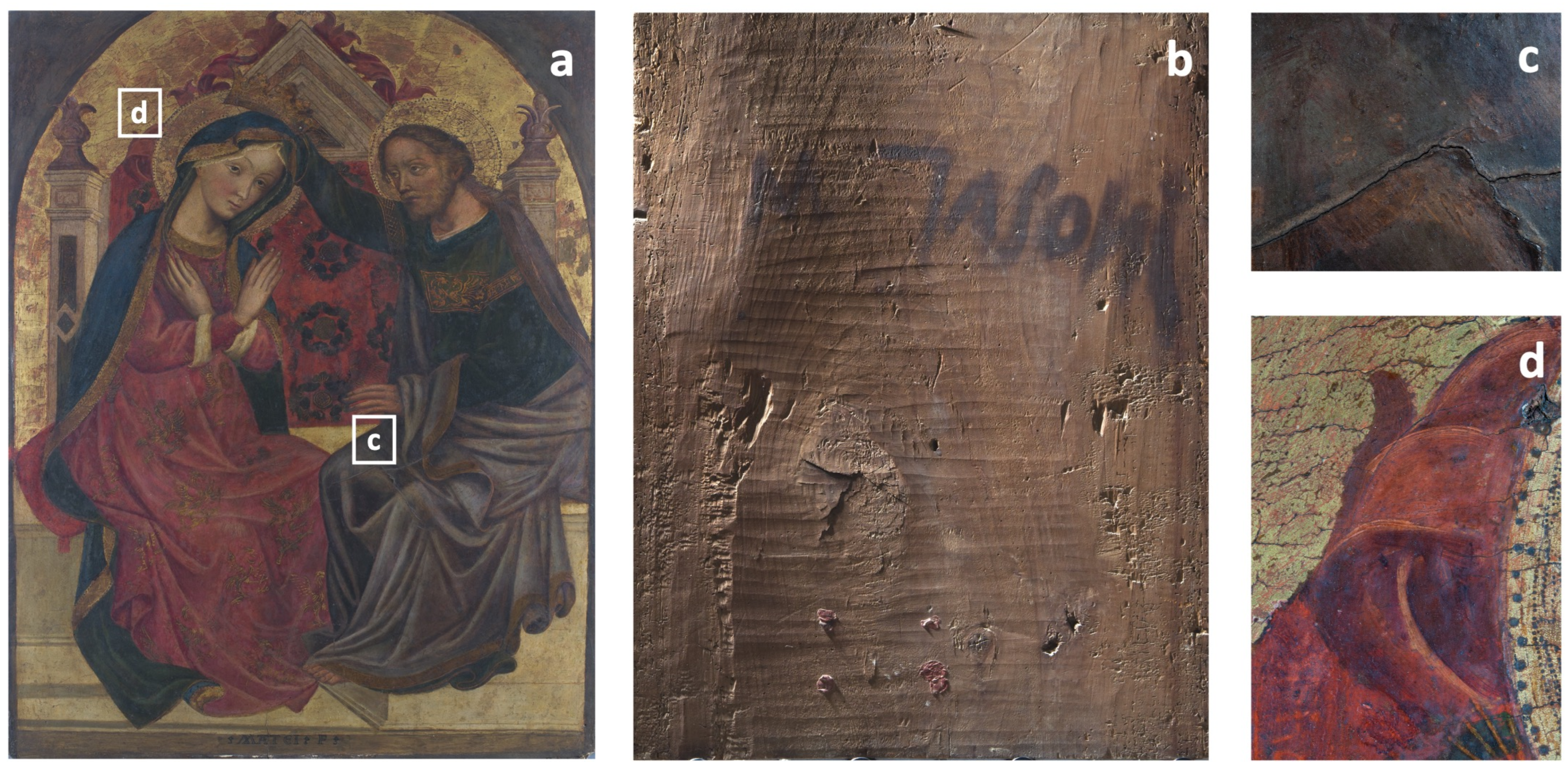
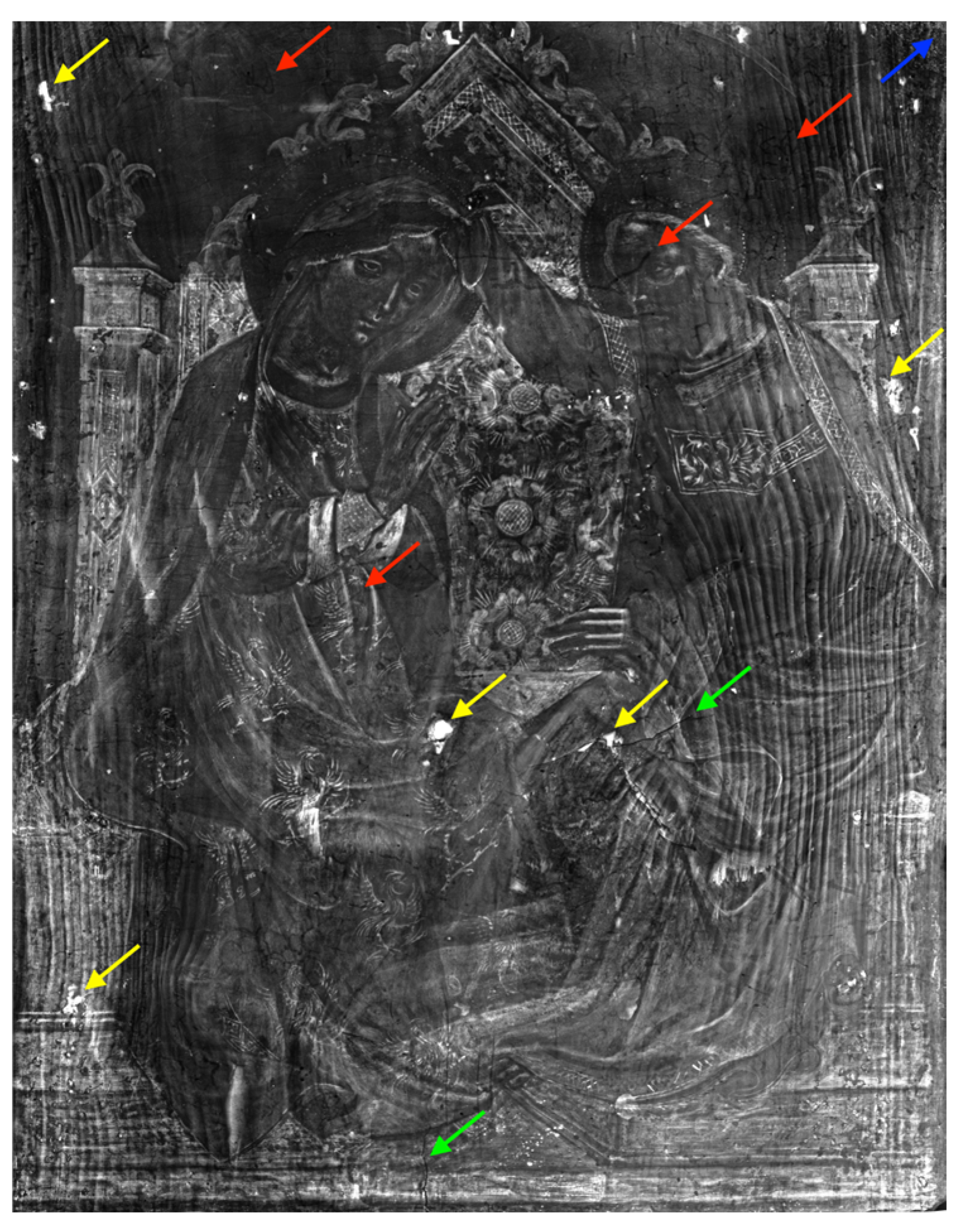
3.1. Preliminary Observations by Imaging Techniques

The visible image in Figure 1 shows the overall appearance and the state of conservation of the painting. The panel (92 × 72 × 2.5 cm) clearly shows the bending of the wooden support and cracks in the painting layers, along with some previous restoration treatments. Cracks were found in correspondence with Christ’s mantle, the golden background and halos (Figure 1c,d), and the areas in which a loss of brightness and intensity at the level of the throne structure and the figures of Christ and the Virgin were recognizable. The UVIF image (Figure 2a) confirms the presence of an even layer of varnish, characterized by a diffuse and intense light blue-green fluorescence. Other areas, which probably were the result of retouching and over-painting, are also evidenced by a different hue of fluorescence, darker than the others, observed in correspondence with Christ’s garment collar and sleeve, the Virgin’s and Christ’s mantles over their shoulders, Christ’s face, and the central decoration of the throne seatback.
High-resolution images are provided in Supplementary Information (Figures S1–S3).
The RX investigation (Figure 3) revealed the structure of the wooden support. As visible along the left and right parts of Figure 3, the panel is made of one thick wooden plank (tentatively recognized by the restorers as poplar wood) with a vertical orientation of the fibres. The presence of cracks in the central and bottom parts (marked with green arrows in Figure 3) were highlighted, and the presence of woodworm galleries proved previous xylophagous attacks, mainly spread in the areas of the background, Christ’s figure, and the Virgin’s dress (marked with red arrows in Figure 3). Moreover, the presence of stucco works in various areas over the entire surface (yellow arrows in Figure 3) was revealed due to the high radiopacity of this material, which appears clearly as white areas. On the other hand, the light grey shaded areas let us suppose the presence of radio-opaque pigments on the background, composed of elements with high atomic numbers (e.g., lead white and cinnabar [14]), which have a high absorption of X-rays. Finally, X-ray radiography proved the presence of the incamottatura, which is a commonly employed technical practice that involves raw canvas to prepare, cover, and uniform the wooden panel. It is clearly visible in the right upper part of the painting (marked with the blue arrow in Figure 3).

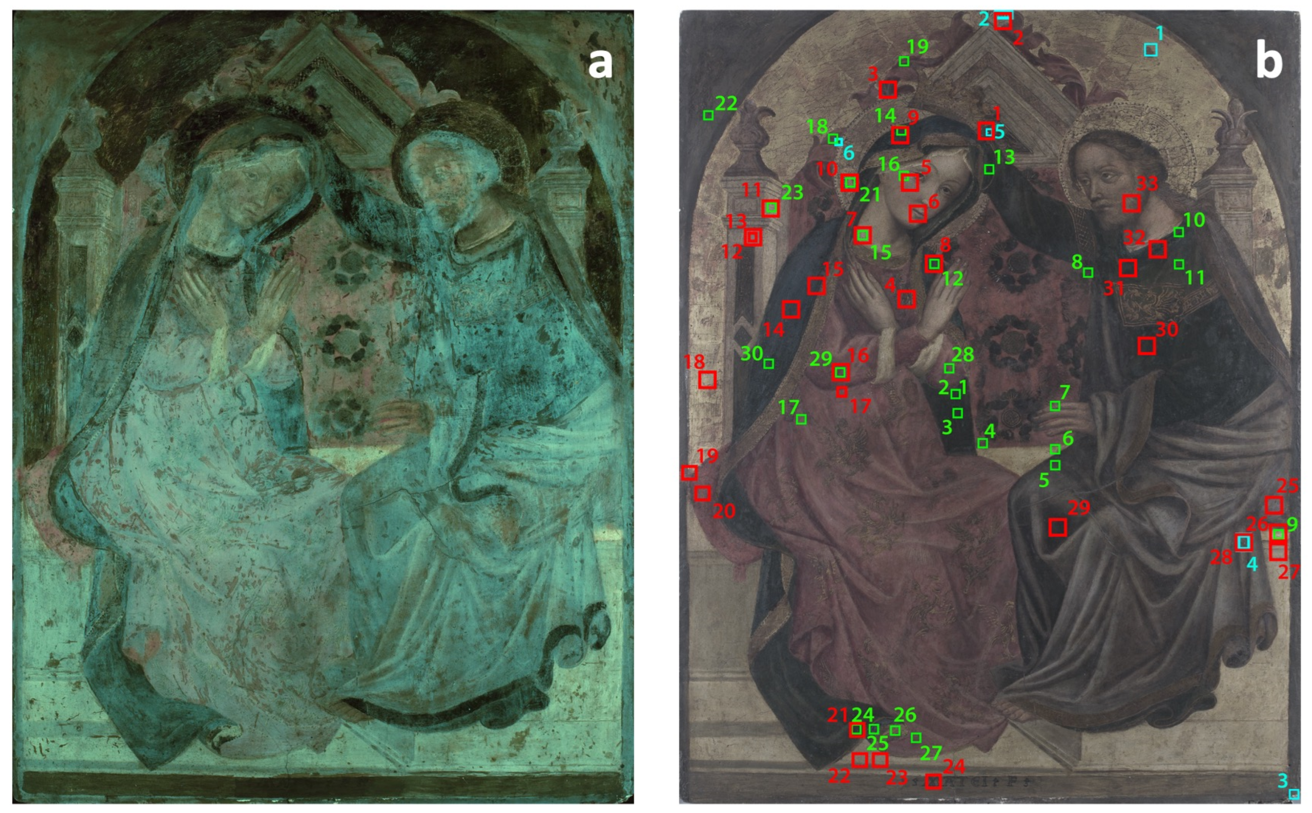
3.2. Non-Invasive Characterization of Painting Materials

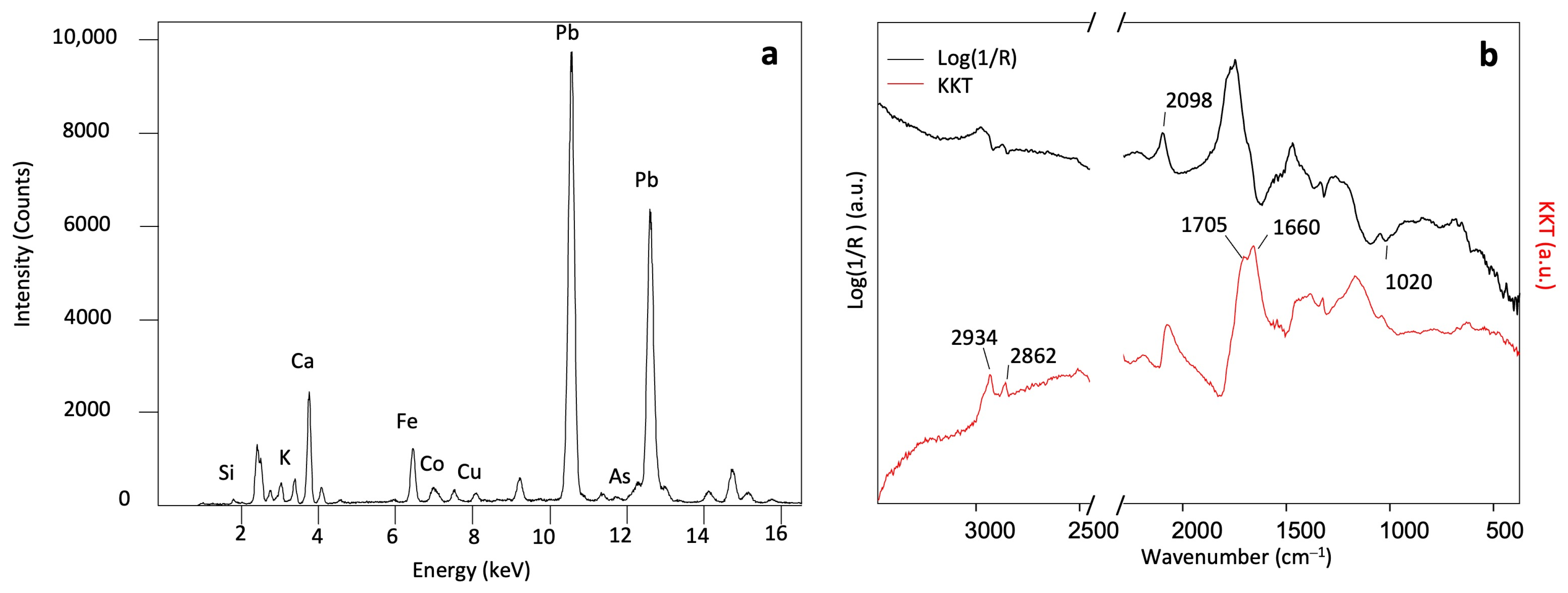
3.2.1. Blue Pigments

The XRF analysis performed on the blue areas corresponding to the Virgin’s mantle marked the presence of iron (Fe) as a major element, together with cobalt (Co), potassium (K), copper (Cu), arsenic (As), and silicon (Si) (Figure 4a). These elements suggest the presence of possibly more than one pigment: Fe could be related to Prussian blue (Fe2[Fe(CN)6]3) [14,25,27]. The presence of this pigment is also supported by the CN stretching band around 2098 cm−1 [28,29], visible in the ER-FTIR pseudo-absorbance spectrum in Figure 4b. Additionally, the presence of smalt (SiO2, K2O, Al2O3, and CoO) is suggested by Co, K, As and Si XRF peaks and by the ER-FTIR bands from silicates around 1020 cm−1 related to the Si-O-Si antisymmetric stretching mode [4,29,30]. The presence of As is considered a good marker for the smalt pigment. This additional element may be associated with some of the cobalt ores used as raw material and related to the different processes used in pigment preparation and production [31,32,33]. It is worth highlighting that the presence of Prussian blue, first synthesized in 1724, is probably due to restoration, whereas smalt is a pigment coherent with the production period of the painting [27]. Beyond the pigment signals, the ER-FTIR spectrum after the KKT showed the signal of a terpenic resin as an external varnish (CH stretching at 2934, 2862 cm−1 and C=O stretching at 1705 cm−1) [16,17,19,22,30], along with those of a proteinaceous binder (around 1660 cm−1 for amide I, while other bands overlapped with oxalates within the range of 1620–1315 cm−1) [4,16,17,19,22], possibly a tempera magra in accordance with a 15th-century painting technique [2,3,20]. The low counts of Cu detected in all the analytical spots in correspondence with the mantles of the Virgin and Christ led to the hypothesis of the use of a blue Cu-based pigment, such as azurite (2CuCO3 · Cu(OH)2) [5,25,27], as possibly the original underneath pigment used to paint this area [34], although it remained undetected by ER-FTIR. As for the areas in correspondence with Christ’s mantle, the XRF spectra highlighted the presence of Fe as the major element, without any trace of Co, suggesting the presence of Prussian blue rather than smalt.

3.2.2. Green and Blue Pigments

The dark green-bluish areas corresponding to Christ’s collared garment and the Virgin’s dress revealed high XRF counts of Cu and Fe, together with Si. Also, for greens, a mixture of different pigments cannot be ruled out: Cu can be related to malachite (CuCO3 · Cu(OH)2) or verdigris (Cu(CH3COO)2 · 2H2O) [11,25,27], the most frequent pigments used in the Middle Ages [1,11]. Fe could instead suggest a mixture with Prussian blue or green earth (hydrosilicate iron minerals) [1,14,25,27]. Unfortunately, ER-FTIR was unable to clearly detect the diagnostic features of Cu-based pigments, while the bands around 1020 cm−1 and 2098 cm−1 could reasonably support the presence of green earth and Prussian blue, respectively.

3.2.3. White Pigments
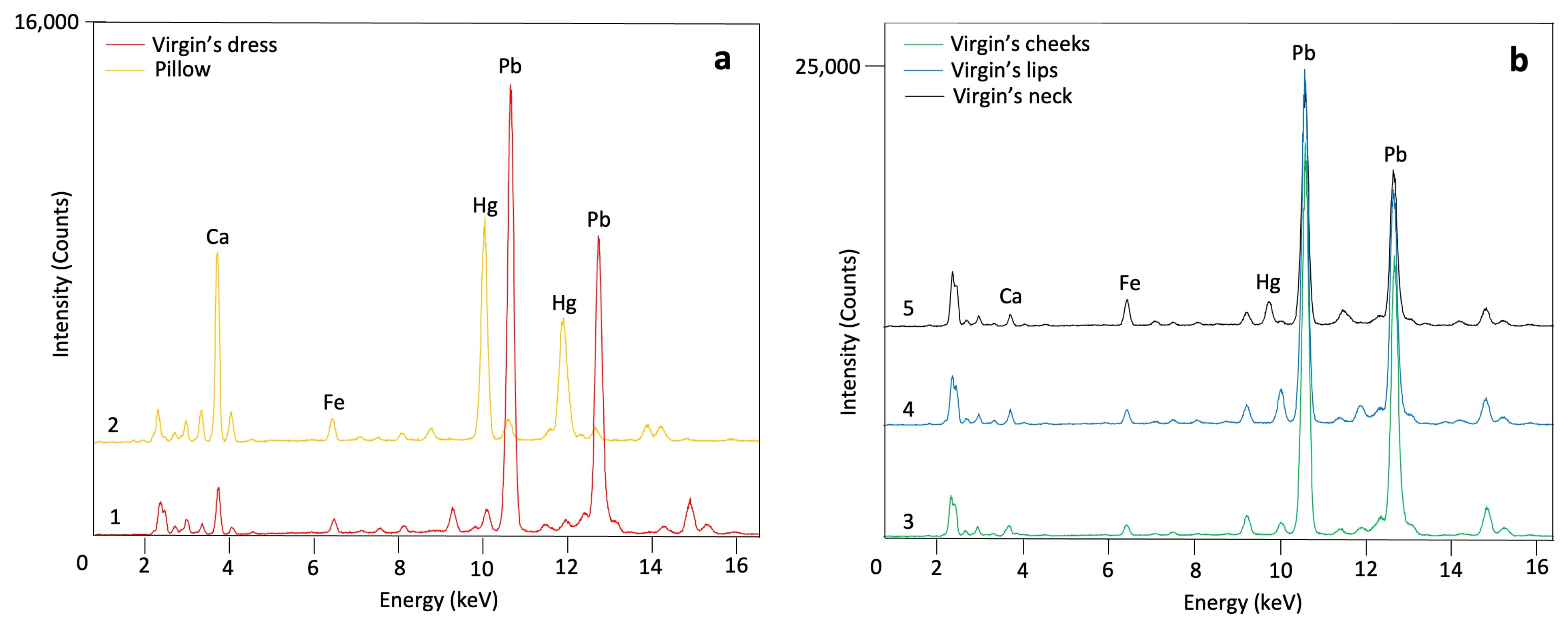
In correspondence with the whitish areas of the throne, significantly higher counts of lead (Pb), compared to the other analysis points, indicated the use of lead white ((PbCO3)2 · Pb(OH)2) [11,14,25,27]. The identification of this pigment was also confirmed by the ER-FTIR bands around 3547, 1450, and 678 cm−1 (Figure 5), related to the O-H bonds, CO32− asymmetric stretching, and in-plane bending, respectively [17,30,35]. The ubiquitous detection of Pb in any XRF spots implied the use of lead white both as imprimitura and white pigment to achieve light hues of colour in specific areas, as observed in the RX digital image (Figure 3). In addition, counts of Fe [11,14,25,27] along with ER-FTIR bands around 533 and 465 cm−1 related to the Fe-O stretching and bending bands [18,29,35] could be related to an iron-based earth pigment, possibly red ochre in low concentration.

3.2.4. Red Pigments

In the red areas of the Virgin’s dress and pillow, mercury (Hg) and Fe were detected by XRF as the principal elements, suggesting the use of cinnabar (HgS) and red ochre (Figure 6a) [11,14,25,27]. Accordingly, ER-FTIR bands around 530 and 450 cm−1 related to the Fe-O bond and those of silicates between 1020 and 1030 cm−1 supported the use of red ochre, while cinnabar remained not visible within the working range. Moreover, a difference between the investigated red backgrounds was marked by XRF measurements: the counts of Hg are significantly higher in the pillow than in the dress, implying a greater use of cinnabar.

3.2.5. Flesh Tone Pigments

The flesh tones of the Virgin’s neck, cheek, and lips have similar elemental composition but different abundance of elements. In the XRF spots 5 and 6, high counts of Hg and Pb, together with the signal of Fe, suggested the use of a mixture of cinnabar, red ochre, and lead white (Figure 6b). In the same area, the ER-FTIR spectra confirmed the presence of silicates and haematite, while in the area of the Virgin’s neck, lead carbonate was identified by the characteristic signals around 1440 and 680 cm−1 related to the O-H and C-O bonds [17,30,35]. The XRF spot 7, corresponding to the Virgin’s neck, was characterized by the same elements, although higher counts of Fe than those identified in the XRF spots 5 and 6 suggested a greater amount of red ochre.

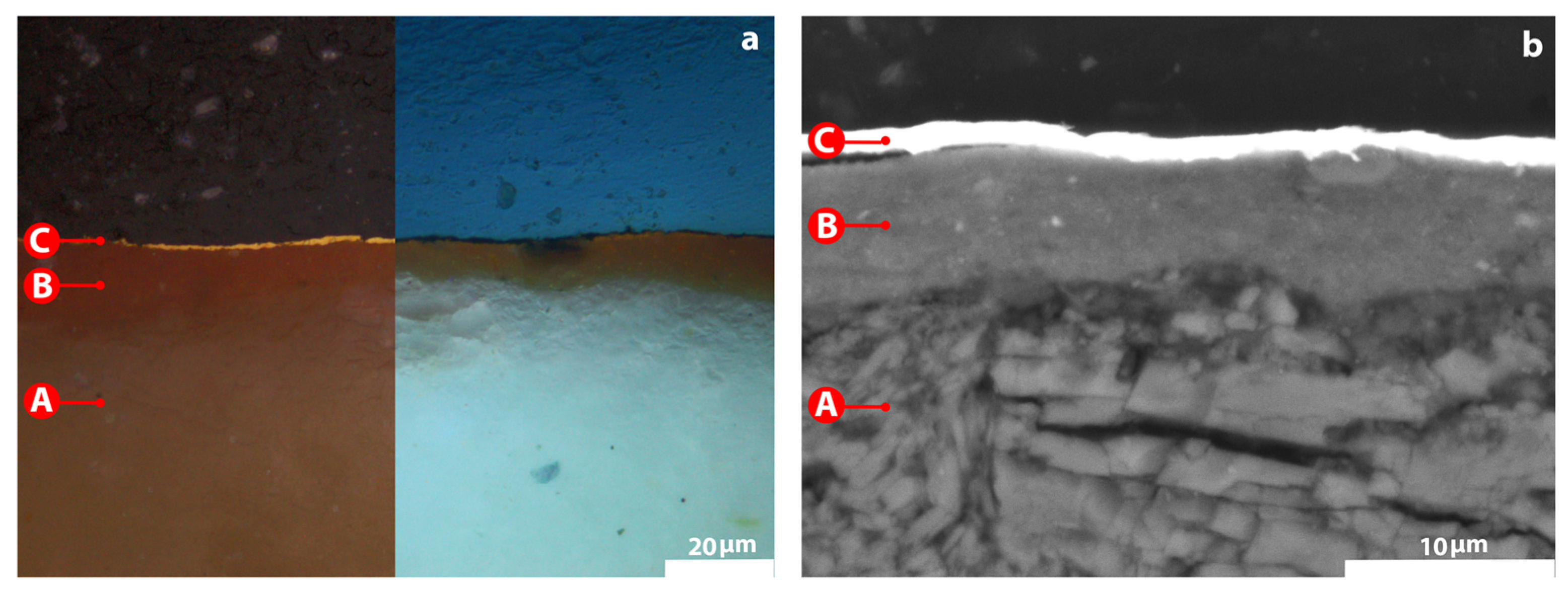
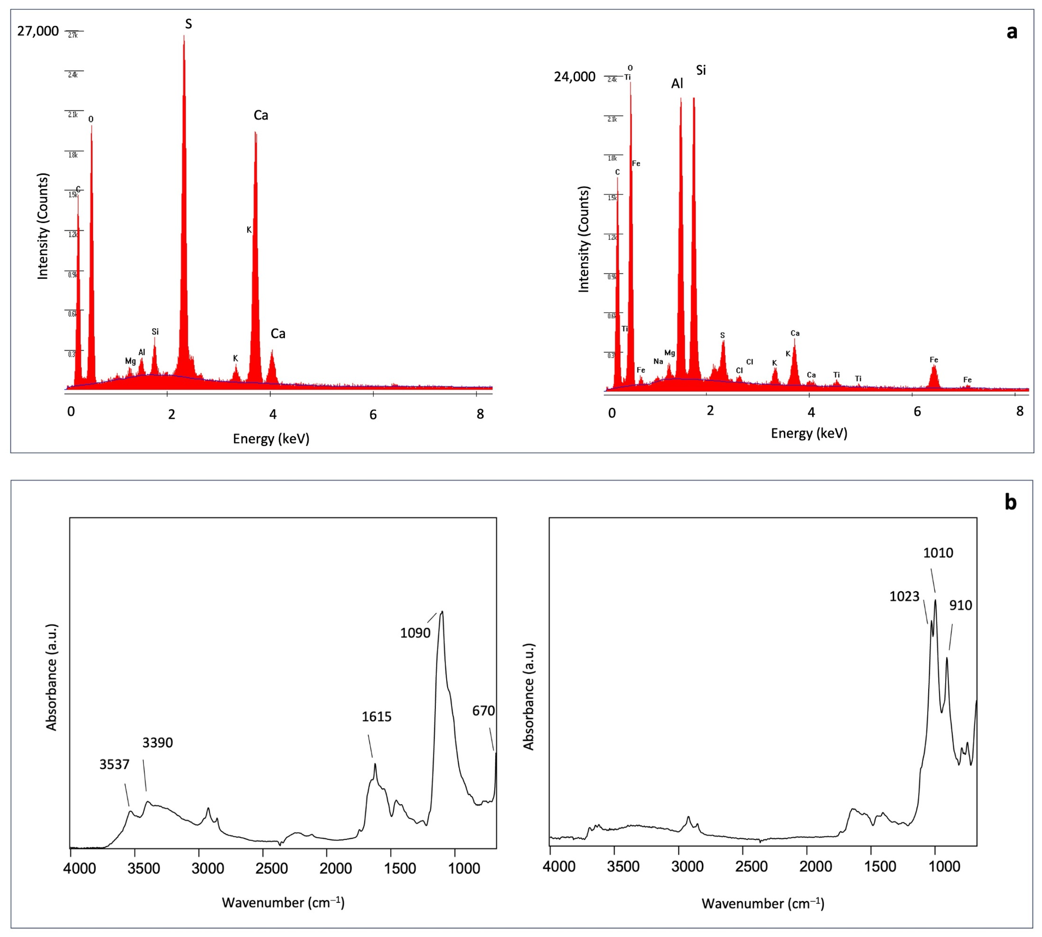
3.2.6. The Painting Technique: Ground Layer and Gilding

The execution technique of the ground layer and gilding were investigated at the microscale level thanks to the analysis with SEM-EDS and ATR-FTIR, carried out on the cross-sectioned samples from the golden background. Figure 7 highlights a structure composed of three layers: the ground layer (A), a gilding substrate or bolo (B), and gold leaf (C).
SEM-EDS analysis (Figure 8a) identified Ca and S as the main elements in the ground layer, implying the use of gypsum; while the spectrum collected in correspondence with layer B revealed high counts of aluminium (Al) and Si, which are linked to the use of clay minerals (bolo) such as kaolin [18] as preparation for the gold leaf. These results are consistent with the identification of gypsum by the ATR-FTIR bands around 3537 and 3390 cm−1, related to the stretching modes of the O-H bonds, and around 1615, 1090 and 670 cm−1, related to the stretching and bending modes of sulphate anions [4,22], along with those of proteinaceous material (commonly animal glue), appearing faintly around 1650, 1550, and 1460 cm−1, related to amide I, II, and III [16,17,19,22] (Figure 8b). A ground layer of this composition is typical of traditional Italian painting of the 15th century [10].
As for the gilding preparation (layer B), ATR-FTIR bands of kaolin were detected in the range between 3695 and 3620 cm−1 related to the O-H stretching modes, and around 1020, 1010, and 910 cm−1 related to the Si-O-Si, Si-O-Al, and Al-OH stretching modes confirming the use of bolo for the preparation [18,29]. The external layer C corresponds to the gold leaf, characterized by a high peak of gold (Au) [11,12,14] in the SEM-EDS spectrum. This type of gilding, traditionally called doratura a guazzo, was made with bolo armeno (red clay containing oxides of Fe and Al) mixed with egg. The dry bolo was burnished with an agate stone, and the gold leaf was applied by means of egg white [36].
The XRF results on the different backgrounds are summarized below (Table 1). The results are supported and completed by the ER-FTIR results.

4. Conclusions

This paper has reported and discussed the results of a diagnostic spectroscopic campaign on the painting Coronation of the Virgin by Michele di Matteo, dated around the first half of the 15th century. The multi-analytical diagnostic campaign based on non- and micro-invasive techniques allowed the assessment of the state of preservation and the characterization of the original painting materials used on the polychrome wooden panel, as well as the materials added during further interventions. The study of the multi-layered system from both the compositional and morphological point of view revealed the painting technique of the author. The painting showed some bending of the wooden support, cracks in the painting layers, and previous restoration interventions identified by the several retouches, over-paintings, modern pigments, and stucco works. The observed stratigraphy seems to be coherent with the traditional late-Gothic layered structure that generally includes a support, a ground layer, paint layers, and a varnish. The support is a thick wooden board covered in the upper part with canvas. The ground layer was made with gypsum and glue, and the presence of the imprimitura with lead white was detected by both imaging and spectroscopic techniques. For the pictorial layers, it was possible to hypothesize the presence of a selection of pigments consistent with the period of the production of the painting, as well as restoration pigments used in subsequent restorations. Cinnabar, red ochre, smalt, lead white, green earth, and green pigment based on copper were identified in red, blue, white, and green areas, possibly applied with an egg binder. Modern pigment such as Prussian blue was revealed as well. As for the gilding areas, the use of red bolo as a preparation for the gold leaf confirmed the use of the guazzo technique.
The advantages of using a multi-analytical approach proved effective in the comprehensive study of the painting. By combining and comparing the results of complementary techniques such as imaging, non-invasive spectroscopy, and micro-invasive analyses, solid and consistent outcomes were achieved. Although completing the imaging with IR reflectography could have contributed to a better localization of the pigments and a clearer distinction between retouched and original areas of the painting, the scientific investigation run so far provided necessary and useful data on the painting technique used by Michele di Matteo. These results not only contribute to the advancement of our knowledge of the painter and possibly his workshop, which have never been studied before from a scientific point of view, but can also help conservators and restorers in future restoration works and studies.

Supplementary Materials

The following supporting information can be downloaded at: https://www.mdpi.com/article/10.3390/heritage7010016/s1, Figure S1: Image of the painting in visible light; Figure S2: Image of the painting in UV light; Figure S3: X-ray radiography of the painting.

Author Contributions

Conceptualization, methodology, and data curation: C.D., M.A., F.V., T.R. and G.F.; formal analysis: all authors; investigation: all authors; writing—original draft preparation: C.D.; writing—review and editing, C.D., M.A., F.V., and G.F.; validation: F.V. and G.F. All authors have read and agreed to the published version of the manuscript.

Funding

This research received no external funding.

Data Availability Statement

The raw data supporting the conclusions of this article will be made available by the authors on request.

Acknowledgments

The authors would like to thank Mario Colella, restorer and director of the Centro Studio e Conservazione Piccolo Chiostro in Pavia, for providing the artwork and support during the analytical steps. We are grateful to Curzio Merlo and Mario Lazzari for the availability of their analytical instrumentation and support in the data interpretation. The widespread diagnostic campaign was planned during the training school Laboratori di Diagnostica per lo Studio dei Beni Culturali organized by the Arvedi Laboratory and hosted by Cr.Forma in their laboratories and classrooms. We therefore thank Cr.Forma for its technical and administrative support in organizing the school and Regione Lombardia for funding the training school.

Conflicts of Interest

The authors declare no conflicts of interest.

References

  1. Šefců, R.; Chlumská, Š.; Třeštíková, A.; Trojek, T.; Dragounová, L. Investigation of the panel painting of St Anne with the Virgin Mary and the Child Jesus using analytical and imaging methods. Appl. Radiat. Isot. 2015, 95, 8–12. [Google Scholar] [CrossRef] [PubMed]
  2. Dal Fovo, A.; Mattana, S.; Ramat, A.; Riitano, P.; Cicchi, R.; Fontana, R. Insights into the stratigraphy and palette of a painting by Pietro Lorenzetti through non-invasive methods. J. Cult. Herit. 2023, 61, 91–99. [Google Scholar] [CrossRef]
  3. Klisinska-Kopacz, A.; Obarzanowski, M.; Fraczek, P.; Moskal-del Hoyo, M.; Gargano, M.; Goslar, T.; Chmielewski, F.; Dudała, J.; del Hoyo-Meléndez, M.J. An analytical investigation of a wooden panel painting attributed to the workshop of Lucas Cranach the Elder. J. Cult. Herit. 2022, 55, 185–194. [Google Scholar] [CrossRef]
  4. Pellerito, C.; Di Marco, A.E.; Di Natale, M.C.; Pignataro, B.; Scopelliti, M.; Sebastianelli, M. Scientific studies for the restoration of a wood painting of the Galleria Interdisciplinare Regionale della Sicilia—Palazzo Mirto. Microchem. J. 2016, 124, 682–692. [Google Scholar] [CrossRef]
  5. Pięta, E.; Proniewicz, E.; Szmelter-Fausek, B.; Olszewska-Świetlik, J.; Proniewicz, L.M. Pigment characterization of important golden age panel paintings of the 17th century. Spectrochim. Acta Part A Mol. Biomol. Spectrosc. 2015, 136, 594–600. [Google Scholar] [CrossRef]
  6. cassiciaco.it. Available online: http://www.cassiciaco.it/navigazione/iconografia/pittori/quattrocento/di_matteo/di_matteo.html (accessed on 7 July 2023).
  7. Bellazzecca, E. Michele di Matteo da Bologna; Dizionario Biografico degli Italiani; Istituto dell’Enciclopedia Italiana: Roma, Italia, 2010; Volume 74. [Google Scholar]
  8. Bolognini Amorini, A. Le Vite dei Pittori ed Artefici Bolognesi, I; Tipi Governativi alla Volpe: Bologna, Italia, 1841; p. 21. [Google Scholar]
  9. Filippini, F.; Zucchini, G. Miniatori e Pittori a Bologna, II, Documenti del Secolo XV; Accademia nazionale dei Lincei: Roma, Italia, 1968; pp. 122–126. [Google Scholar]
  10. Massaccesi, F. Nuove Riflessioni Sul Percorso di M. di M.; Arte cristiana XCVII; Scuola Beato Angelico e dell’Istituto di Storia dell’Arte dell’Università Cattolica: Milano, Italia, 2009; Volume 852, pp. 171–180. [Google Scholar]
  11. Gargano, M.; Bonizzoni, L.; Grifoni, E.; Melada, J.; Guglielmi, V.; Bruni, S.; Ludwig, N. Multi-analytical investigation of panel, pigments and varnish of The Martyirdom of St. Catherine by Gaudenzio Ferrari (16th century). J. Cult. Herit. 2020, 46, 289–297. [Google Scholar] [CrossRef]
  12. Cavaleri, T.; Pelosi, C.; Giustetto, R.; Andreotti, A.; Bonaduce, I.; Calabrò, G.; Caliri, C.; Colantonio, C.; Manchinu, P.; Legnaioli, S.; et al. The northern-Italy Renaissance in a panel by Defendente Ferrari A complete study with a multi-analytical investigation. J. Archaeol. Sci. Rep. 2022, 46, 103669. [Google Scholar] [CrossRef]
  13. Galli, A.; Gargano, M.; Bonizzoni, L.; Bruni, S.; Interlenghi, M.; Longoni, M.; Passaretti, A.; Caccia, M.; Salvatore, C.; Castiglioni, I.; et al. Imaging and spectroscopic data combined to disclose the painting techniques and materials in the fifteenth century Leonardo atelier in Milan. Dyes Pigment 2021, 187, 109112. [Google Scholar] [CrossRef]
  14. Venuti, V.; Fazzari, B.; Crupi, V.; Majolino, D.; Paladini, G.; Morabito, G.; Certo, G.; Lamberto, S.; Giacobbe, L. In situ diagnostic analysis of the XVIII century Madonna della Lettera panel painting (Messina, Italy). Spectrochim. Acta Part A Mol. Biomol. Spectrosc. 2020, 228, 117822. [Google Scholar] [CrossRef]
  15. Invernizzi, C.; Daveri, A.; Rovetta, T.; Vagnini, M.; Licchelli, M.; Cacciatori, F.; Malagodi, M. A multi-analytical non-invasive approach to violin materials: The case of Antonio Stradivari “Hellier” (1679). Microchem. J. 2016, 124, 743–750. [Google Scholar] [CrossRef]
  16. Volpi, F.; Fiocco, G.; Rovetta, T.; Invernizzi, C.; Albano, M.; Licchelli, M.; Malagodi, M. New Insights on the Stradivari “Coristo” Mandolin: A Combined Non- Invasive Spectroscopic Approach. Appl. Sci. 2021, 11, 11626. [Google Scholar] [CrossRef]
  17. Pięta, E.; Olszewska-Świetlik, J.; Paluszkiewicz, C.; Zając, A.; Kwiatek, W.M. Application of ATR-FTIR mapping to identification and distribution of pigments, binders and degradation products in a 17th century painting. Vib. Spectrosc. 2019, 103, 102928. [Google Scholar] [CrossRef]
  18. Volpi, F.; Vagnini, M.; Vivani, R.; Malagodi, M.; Fiocco, G. Non-invasive identification of red and yellow oxide and sulfide pigments in wall-paintings with portable ER-FTIR spectroscopy. J. Cult. Herit. 2023, 63, 158–168. [Google Scholar] [CrossRef]
  19. Rieppi, N.; Price, B.A.; Sutherland, K.; Lins, A.P.; Newman, R.; Wang, P.; Wang, T.; Tague, T.J., Jr. Salvator Mundi: An investigation of the painting’s materials and techniques. Herit. Sci. 2020, 8, 39. [Google Scholar] [CrossRef]
  20. Stanzani, E.; Bersania, D.; Lotticia, P.; Colomban, P. Analysis of artist’s palette on a 16th century wood panel painting by portable and laboratory Raman instruments. Vib. Spectrosc. 2016, 85, 62–70. [Google Scholar] [CrossRef]
  21. Van der Werf, I.D.; Gnisci, R.; Marano, D.; De Benedetto, G.E.; Laviano, R.; Pellerano, D.; Vona, F.; Pellegrin, F.; Andriani, E.; Catalano, I.M.; et al. San Francesco d’Assisi (Apulia, South Italy) Study of a manipulated 13th century panel painting by complementary diagnostic techniques. J. Cult. Herit. 2008, 9, 162–171. [Google Scholar] [CrossRef]
  22. Lo Monaco, A.; Mattei, E.; Pelosi, C.; Santancini, M. The scientific investigation for the study and conservation of the wooden model of S. Maria della Consolazione’s church (Todi, Italy). J. Cult. Herit. 2013, 14, 537–543. [Google Scholar] [CrossRef]
  23. Fiocco, G.; Volpi, F.; Rovetta, T.; Lee, C.; Albano, M.; Weththimuni, M.; Colella, M.; Magrassi Matricardi, A.L.; Merlo, C.; Malagodi, M.; et al. Analytical investigations on polychrome artworks from the wooden ceiling of “ex-Ospedale San Matteo” in Pavia. In Proceedings of the IMEKO TC4 Conference Proceeding 2023 of the Metroarchaeo 2023 conference, Rome, Italy, 19–21 October 2023. accepted for publication. [Google Scholar]
  24. Volpi, F.; Albano, M.; Fiocco, G.; Weththimuni, M.; Malagodi, M. Unveiling Hidden Insights of Ancient Roman wall paintings in Cremona: In-Depth Knowledge Beyond the Surface with Spectroscopic Analysis. In Proceedings of the IMEKO TC4 Conference Proceeding 2023, Pordenone, Italy, 20–21 September 2023. accepted for publication. [Google Scholar]
  25. Seccaroni, C.; Moioli, P.; Fluorescenza, X. Prontuario per l’Analisi XRF Portatile Applicata a Superfici Policrome; Nardini Editore: Firenze, Italia, 2004. [Google Scholar]
  26. Eastaugh, N.; Walsh, V.; Chaplin, T.; Siddall, R. Pigment. Compendium: A Dictionary and Optical Microscopy of Historic Pigments; Elsevier: Burlington, MA, USA, 2008. [Google Scholar]
  27. Bevilacqua, N.; Borgioli, L.; Gracia, I.A. I Pigmenti Nell’Arte Dalla Preistoria Alla Rivoluzione Industriale; Il Prato: Saonara, Italia, 2010. [Google Scholar]
  28. Doménech-Carbó, A.; Doménech-Carbó, M.T.; Osete-Cortina, L.; Donnici, M.; Guasch-Ferré, N.; Gasol-Fargas, R.M.; Iglesias-Campos, M.A. Electrochemical assessment of pigments-binding medium interactions in oil paint deterioration: A case study on indigo and Prussian blue. Herit. Sci. 2020, 8, 71. [Google Scholar] [CrossRef]
  29. Silva, C.E.; Silva, L.P.; Edwards, H.; De Oliveira, L. Diffuse reflection FTIR spectral database of dyes and pigments. Anal. Bioanal. Chem. 2006, 386, 2183–2191. [Google Scholar] [CrossRef]
  30. Nodari, L.; Ricciardi, P. Non-invasive identification of paint binders in illuminated manuscripts by ER-FTIR spectroscopy: A systematic study of the influence of different pigments on the binders’ characteristic spectral features. Herit. Sci. 2019, 7, 7. [Google Scholar] [CrossRef]
  31. Van Loon, A.; Noble, P.; De Man, D.; Alfeld, M.; Callewaert, T.; Van der Snickt, G.; Janssens, K.; Dik, J. The role of smalt in complex pigment mixtures in Rembrandt’s Homer 1663: Combining MA-XRF imaging, microanalysis, paint reconstructions and OCT. Herit. Sci. 2020, 8, 90. [Google Scholar] [CrossRef]
  32. Barilaro, D.; Crupi, V.; Interdonato, S.; Majolino, D.; Venuti, V.; Barone, G.; La Russa, M.F.; Bardelli, F. Characterization of blue decorated Renaissance pottery fragments from Caltagirone (Sicily, Italy). Appl. Phys. A Mater. Sci. Process. 2008, 92, 91–96. [Google Scholar] [CrossRef]
  33. Ricciardi, P.; Dooley, K.A.; MacLennan, D.; Bertolotti, G.; Gabrieli, F.; Schmidt Patterson, C.; Delaney, J.K. Use of standard analytical tools to detect small amounts of smalt in the presence of ultramarine as observed in 15th-century Venetian illuminated manuscripts. Herit. Sci. 2022, 10, 38. [Google Scholar] [CrossRef]
  34. Galliano Lalli, C.; Innocenti, F. Appunti Sulle Caratteristiche Chimico-Fisiche Dell’Azzurrite e Del Lapislazzuli. OPD Restauro 2014, 26, 78–82. Available online: http://www.jstor.org/stable/24398163 (accessed on 3 January 2024).
  35. Giménez, P.; Linares, A.; Sessa, C.; Bagán, H.; García, J.F. Capability of Far-Infrared for the selective identification of red and black pigments in paint layers. Spectrochim. Acta Part A Mol. Biomol. Spectrosc. 2022, 266, 120411. [Google Scholar] [CrossRef]
  36. Cennini, C. Il Libro dell’arte, a Cura di F. Frezzato; Neri Pozza Editore: Vicenza, Italia, 2016. [Google Scholar]
Figure 1. Photography in VIS light of the (a) front and (b) back of the painting Coronation of the Virgin (92 × 72 × 2.5 cm) by Michele di Matteo. Enlarged details of the cracks in the painting layers in correspondence with (c) Christ’s mantle and (d) the golden background and halos.
Heritage 07 00016 g001
Figure 2. (a) UVIF image of the panel painting Coronation of the Virgin. (b) Visible image of the panel where ER-FTIR analytical spots (green squares), XRF spots (red squares) and micro-sampling areas (light blue squares) are highlighted.
Heritage 07 00016 g002
Figure 3. RX of the painting where the presence of cracks (green arrows), previous xylophagous attacks (red arrows), stucco (yellow arrows), and canvas (blue arrows) are highlighted.
Heritage 07 00016 g003
Figure 4. (a) XRF spectrum (spot 9 in Figure 2b, red square). (b) ER-FTIR pseudo-absorbance and KKT spectra (spot 14 in Figure 2b, green square) of the blue area of the Virgin’s mantle over her head.
Heritage 07 00016 g004
Figure 5. ER-FTIR pseudo-absorbance spectrum (spot 9 in Figure 2b, green square) of the white area of the throne structure.
Heritage 07 00016 g005
Figure 6. Comparison of XRF spectra collected (a) on the red areas of the (1) Virgin’s dress (spot 16 in Figure 2-b, red square) and (2) pillow (spot 20 in Figure 2b, red square), and (b) on the flesh tone areas of the Virgin’s (3) cheeks and (4) lips (spots 5 and 6 in Figure 2b, red squares) and (5) Virgin’s neck (spot 7 in Figure 2b, red square).
Heritage 07 00016 g006
Figure 7. Cross-section of sample 2 of the area of the golden background (area 2 in Figure 2b, light blue square) observed under (a) the OM in VIS light (left) and UVIF (right), and (b) the SEM-EDS. The cross-section shows the ground layer (A), bolo layer (B), and gold leaf (C).
Heritage 07 00016 g007
Figure 8. (a) SEM-EDS and (b) ATR-FTIR spectra acquired in correspondence of the ground layer A and gilding preparation layer B, left and right, respectively.
Heritage 07 00016 g008
Table 1. Summary of the analytical results identified in the selected areas.
Investigated Area (XRF Spot)ColourPigment AttributionMarker XRF ElementsMaker FTIR BandsReferences
8—Virgin’s mantle over her shoulderBluePrussian blue, Smalt, Blue Cu-based pigmentFe, Co, K, As, Cu, Si2098 cm−1, 1020 cm−1[4,5,14,16,17,19,22,25,27,28,29,30]
9—Virgin’s mantle over her headBluePrussian blue, Smalt, Blue Cu-based pigmentFe, K, Co, Cu, Si, As2098 cm−1, 1020 cm−1[4,5,14,16,17,19,22,25,27,28,29,30]
14—Virgin’s mantleBluePrussian blue, Blue Cu-based pigmentFe, Cu-[5,14,25,27]
15—Virgin’s mantleBluePrussian blue, Blue Cu-based pigmentFe, Cu-[5,14,25,27]
25—Christ’s mantleBluePrussian blue, Blue Cu-based pigmentFe, Cu-[5,14,25,27]
28—Christ’s mantleBluePrussian blue, Blue Cu-based pigmentFe, Cu-[5,14,25,27]
29—Christ’s mantleBluePrussian blue, Blue Cu-based pigmentFe, Cu-[5,14,25,27]
21—Virgin’s dressDark Green/BlueGreen pigment based on copper, Prussian blue, Green earthCu, Fe, Si1020 cm−1,
2098 cm−1
[4,11,14,25,27,28,29,30]
30—Christ’s garmentDark Green/BlueGreen pigment based on copper, Prussian blue, Green earthCu, Fe, Si-[11,14,25,27]
31—Christ’s garment collarDark Green/BluePrussian blue, Green earth, Green pigment based on copperFe, Cu, Si-[11,14,25,27]
32—Christ’s garment collarDark Green/BlueGreen pigment based on copper, Prussian blue, Green earthCu, Fe, Si-[11,14,25,27]
11—Throne structureWhiteLead white, Iron-based earthPb, Fe3547 cm−1, 1450 cm−1, 678 cm−1[11,14,17,18,25,27,29,30,35]
12—Throne structureWhiteLead white, Iron-based earthPb, Fe-[11,14,25,27]
13—Throne structureWhiteLead white, Iron-based earthPb, Fe-[11,14,25,27]
22—Throne structureWhiteLead white, Iron-based earthPb, Fe-[11,14,25,27]
23—Throne structureWhiteLead white, Iron-based earthPb, Fe-[11,14,25,27]
26—Throne structureWhiteLead white, Iron-based earthPb, Fe3547 cm−1, 1450 cm−1, 678 cm−1[11,14,17,18,25,27,29,30,35]
27—Throne structureWhiteLead white, Iron-based earthPb, Fe-[11,14,25,27]
16—Virgin’s dress sleeveRedCinnabar, Red ochreHg, Fe530 cm−1, 450 cm−1, 1020 cm1, 1030 cm−1[4,11,14,18,25,27,29,35]
17—Virgin’s dress sleeveRedCinnabar, Red ochreHg, Fe-[11,14,25,27]
19—PillowRedCinnabar, Red ochreHg, Fe-[11,14,25,27]
20—PillowRedCinnabar, Red ochreHg, Fe-[11,14,25,27]
5—Virgin’s cheekFlesh tonesCinnabar, Lead white, HaematitePb, Hg, Fe1090 cm−1, 1040 cm−1, 480 cm−1, 540 cm−1[4,11,14,18,25,27,29,35]
6—Virgin’s lipsFlesh tonesCinnabar, Lead white, Haematite/Red ochrePb, Hg, Fe-[11,14,25,27]
7—Virgin’s neckFlesh tonesRed ochre, CinnabarPb, Fe, Hg1440 cm−1, 680 cm−1[11,14,17,25,27,30,35]
Disclaimer/Publisher’s Note: The statements, opinions and data contained in all publications are solely those of the individual author(s) and contributor(s) and not of MDPI and/or the editor(s). MDPI and/or the editor(s) disclaim responsibility for any injury to people or property resulting from any ideas, methods, instructions or products referred to in the content.

