Article

The Potential Drug Target (Glycolysis Pathway) of the Spore Stage of the Pathogen Enterocytozoon hepatopenaei in Shrimp Farming


1
School of Marine Sciences, Ningbo University, Ningbo 315211, China
2
Key Laboratory of South China Sea Fishery Resources Exploitation & Utilization, Ministry of Agriculture and Rural Affairs, Guangzhou 510300, China
*
Author to whom correspondence should be addressed.
Fishes 2026, 11(4), 229; https://doi.org/10.3390/fishes11040229
Submission received: 20 March 2026 / Revised: 9 April 2026 / Accepted: 11 April 2026 / Published: 15 April 2026
(This article belongs to the Special Issue Advances in the Immunology of Aquatic Animals)

Abstract

Enterocytozoon hepatopenaei (EHP) is a specialized parasitic microsporidian that causes significant economic losses to the shrimp farming industry. The glycolysis pathway plays an important role in the survival of EHP spores in vitro. In this study, key enzyme genes involved in the glycolysis pathway of EHP were analyzed, and purified spores were treated with KOH and a low temperature (−20 °C) to promote or inhibit germination. Quantitative analysis and enzyme activity of the initiating key gene hexokinase (HK) and the core link gene glyceraldehyde 3-phosphate dehydrogenase (GAPDH) were conducted to explore the energy response characteristics of germinating spores. The results showed that HK and GAPDH genes had significant differences from the host, based on phylogenetic analysis. The expression of HK gene and enzyme activity increased after promoting germination treatment. The expression of the GAPDH gene was stable, but the activity of the GAPDH enzyme increased significantly after germination promotion. These findings indicate that the inhibition of the HK gene expression level and GAPDH protein level can block spore germination and invasion in vitro, which could be used as potential control targets for EHP.
Key Contribution: The in vitro survival of EHP; a parasite of shrimp; the role of specific glycolytic enzymes during spore germination; a potential drug target.

1. Introduction

Pacific white shrimp, Litopenaeus vannamei, is one of the most widely farmed shrimp species globally. Enterocytozoon hepatopenaei (EHP) is the predominant parasite in shrimp aquaculture, and has caused great economic losses in the L. vannamei industry [1,2,3,4]. EHP mainly parasitizes the hepatopancreatic cell basement membrane, causing the epithelial cells to detach from the membrane in shrimp. EHP does not cause acute death in infected L. vannamei, and there are no obvious clinical symptoms of infection, but significant growth retardation occurs after a few months [5]. This leads to a large amount of feed wastage, which is also an important cause of major economic losses.
During microsporidia evolution, microsporidia have achieved efficient parasitism through gene compaction and degradation [6]. Many core genes have been lost, including some related to the energy generation pathway [7,8]. Most microsporidia have lost mitochondrial genes that allow oxidative phosphorylation, and their energy supply is highly dependent upon their hosts [9]. In its nonparasitic cycle, the spore stage of microsporidium is highly dependent on its glycolytic pathway. Microsporidium lacks a functional glycolysis pathway, and different microsporidia retain different glycolysis pathway enzymes. When mature spores are present in the environment, their main energy source derives from the metabolism of glucose via their own glycolysis pathway in the absence of oxidative phosphorylation [10]. EHP is like most microsporidians, and its genes are extremely compact. High-throughput sequencing showed that only the genes encoding hexokinase (HK) and 3-phosphoglyceraldehyde dehydrogenase (GAPDH) are retained in the glycolytic pathway of EHP [11]. Therefore, the HK and GAPDH of EHP may be related to the still unclear energy source in the mature spore germination process.
In this study, we performed bioinformatics analysis on key gene sequences involved in the glycolytic pathway of EHP. Then, purified spores were treated to promote or inhibit germination, and the effectiveness of the various treatments was determined with light microscopic and scanning electron microscopic analyses. The expression of key enzymes in the glycolytic pathway was quantified and the enzyme activities in the spores were determined after different treatments to investigate the response characteristics of the glycolytic pathway in EHP spores. This study aims to explore energy changes during the germination process of EHP, identify suitable germination blocking sites, and provide appropriate target information for the prevention and treatment of EHP and drug screening.

2. Materials and Methods

2.1. Animal Samples

EHP-infected L. vannamei were obtained from commercial shrimp ponds in Zhejiang Province, China, and were maintained at room temperature (25 °C) in 150 L plastic tanks with circulating seawater containing 25 ppt salinity and proper aeration. The EHP infection status of the animals was confirmed using nested PCR [12]. Meanwhile, transmission electron microscopy (TEM) observation experiments were performed on shrimp hepatopancreatic tissues. The use of experimental animals was reviewed and approved by the Ethics Committee of Ningbo University.

2.2. Purification of EHP Spores

The hepatopancreatic tissues from shrimp with EHP infection were homogenized with Tris–EDTA (TE) buffer (pH 7.8) (Solarbio, Beijing, China) to a final concentration of 10% (v/v) in a 50 mL centrifuge tube. A quarter volume of ether was added to the hepatopancreas homogenate, which was thoroughly mixed with a vortex oscillator (Golden Biotechnology Corp, Beijing, China) for 5 min, and centrifuged at 2500 rpm at 4 °C for 5 min. The top layer was removed and the process was repeated three times. After low-speed centrifugation at 5000 rpm at 4 °C for 20 min, the supernatant was discarded, and the precipitate was redissolved in TE. A crude extract of EHP spores was thus obtained. The EHP spore extract was purified with discontinuous density Percoll gradient separation (60%, 50%, 40%, and 30%), with horizontal centrifugation at 5000 rpm for 20 min. The white band of spores was carefully collected with a 1 mL syringe and the purified spores were washed with TE buffer at 10,000 rpm for 2 min.
After washing five times repeatedly, the precipitate was the purified EHP. Meanwhile, the hepatopancreatic tissues from shrimp without EHP infection were used as a control.

2.3. Treatment and Observation of Purified Spores

As previously reported [13], 200 μL of 0.1 mol/L KOH was added to the spores in group A (germination promotion group), which were then stored at room temperature for 12 h; in group B (germination inhibition group), 200 μL of TE buffer was added to the spores, which were stored at −20 °C for 12 h; in group C (control group), 200 μL of TE buffer was added to the spores, which were stored at room temperature for 12 h. A drop (10 μL) of 2% (w/v) phloxin B (Beyotime Biotechnology, Shanghai, China) in sterilized distilled water was placed on a microscope slide and 5 μL of purified treated spores was added to it [14].

2.4. Scanning Electron Microscopy (SEM) Analysis

The EHP spores were fixed with 2.5% glutaraldehyde (Solarbio, Beijing, China) at 4 °C for 12 h. The fixation solution was decanted, and the samples were rinsed three times with 0.1 M phosphate buffer (pH 7.0) (Solarbio, Beijing, China) for 15 min each time. The samples were fixed with 1% osmic acid solution (Solarbio, Beijing, China) for 2 h. The osmic acid waste solution was carefully removed, and the samples were rinsed three times with 0.1 M phosphate buffer (pH 7.0) for 15 min each time. The samples were dehydrated with a gradient of ethanol solution (Sinopharm, Shanghai, China) (30%, 50%, 70%, 80%, 90%, and 95%) for 15 min at each concentration and then treated twice with 100% ethanol for 20 min each time. The samples were treated with an equivolumetric mixture of ethanol and isoamyl acetate (Sinopharm, Shanghai, China) for 30 min, and then with pure isoamyl acetate for 1 h. After critical point drying, the film was coated and observed with SEM (Hitachi, Tokyo, Japan).

2.5. Phylogenetic Analysis of HK and GAPDH Genes

HK and GAPDH are respectively the initiating core gene and the core link gene in glycolysis. Phylogenetic trees of the HK and GAPDH genes of EHP, other microsporidians, commercially cultured decapods, and other cultured aquatic species were constructed. The phylogenetic analysis was performed using MEGA v.7 with the neighbor-joining method. The data were resampled with 1000 bootstrap replicates to determine the confidence indices.

2.6. Absolute Quantification of Key Enzyme Genes Involved in EHP Spore Glycolysis

To investigate the expression changes in key genes in glycolysis of EHP spores in germination promotion and germination inhibition treatment, HK and GAPDH absolute quantitative methods of EHP were established. Primer Premier 5.0 was used to design primers (Table 1) that were specific for the HK and GAPDH genes of EHP, based on a genome sequence (No. MNPJ01000002.1). The optimal annealing temperature was determined to be 55 °C.
Using the optimized real-time quantitative PCR reaction system, the quantitative standard samples were diluted 10 times in a gradient manner and amplified. The quantitative amplification curves of the standard plasmids reflected the exponential growth and plateau phase of PCR within the range of 7.9 × 10⁶~7.9 × 10² and 2.43 × 10⁹~2.43 × 10⁴ copies/μL. Based on the experimental results, standard curves corresponding to the relationship between the threshold cycle number (Ct) of the amplification products and the logarithm of the initial amount of the standard template [log(SQ)] were established. The standard curve of the threshold cycle number of the amplification products and the logarithm of the initial amount of the template was calculated as Ct (HK) = −3.5057 log (SQ) + 32.552, with a correlation coefficient R2 of 0.9994. Ct (GAPDH) = −4.6854 log (SQ) + 58.02, with a correlation coefficient R2 of 0.999.
After different treatments, the total RNA was extracted from the purified spores with the RNeasy Mini Kit (Qiagen, Dusseldorf, Germany), according to the manufacturer’s instructions. Reverse transcription reagent (Omega Biotek, Norcross, GA, USA) was used to reverse transcribe the RNA into cDNA, and the designed primers were used for qPCR detection under optimal qPCR conditions. The constructed standard curves were used to estimate the gene expression of the key glycolytic enzymes in each treatment group.

2.7. Activities of Key Enzymes in the Glycolysis Pathway

In order to explore the changes in key enzyme activities in the glycolysis pathway of EHP spores after different treatments, HK and GAPDH enzyme activities were determined, respectively. Hexokinase activity was measured according to the previous reports [14], with some modifications. The reaction solution (1 mL) contained 30 mmol/L HEPES–NaOH (pH 7.5), 0.6 mmol/L EDTA, 1 mmol/L MgCl2, 1 mmol/L NADP, 9 mmol/L KCl, 1 mmol/L ATP, 2 U of glucose-6-phosphate dehydrogenase, and 2 mmol/L glucose, and was reacted at room temperature for 4 min. The corresponding chemical reagents were purchased from Sinopharm, Shanghai, China.The absorbance at a wavelength of 340 nm (A340) was measured to determine the increase in absorbance caused by the reduction in NADP. The enzyme solution (20 μL) was added to 230 μL of reaction solution. An increase in A340 of 1 unit per 1 mL of reaction solution was deemed to indicate a change in the enzyme activity of 1 U/mL.
The principle underlying the measurement of GAPDH activity is that GAPDH catalyzes the oxidation of glyceraldehyde 3-phosphate to 1,3-phosphate glyceraldehyde and NAD+, and A340 increases as the NAD+ in the reaction mixture decreases. The reaction solution consisted of 30 mmol/L HEPES–NaOH (pH 7.5), 15 mmol/L sodium pyrophosphate, 30 mmol/L sodium phosphate, 2 mmol/L NAD+, 10 mmol/L dithiothreitol, and 0.5 mmol/L GAPDH. An aliquot (20 μL) of enzyme solution was added to 230 μL of the reaction solution [15]. An increase in A340 of 1 unit per 1 mL of reaction solution was deemed to indicate an increase in the enzyme activity of 1 U/mL.

2.8. Statistical Analysis

The experimental data was collated by Microsoft Excel, and the experimental results were statistically analyzed by SPSS 2023 Statistics software (One-Way ANOVA), and the Duncan method was used for multiple comparisons between groups. When p < 0.05, the difference was statistically significant.

3. Results

3.1. Symptoms and TEM of EHP Infected L. vannamei

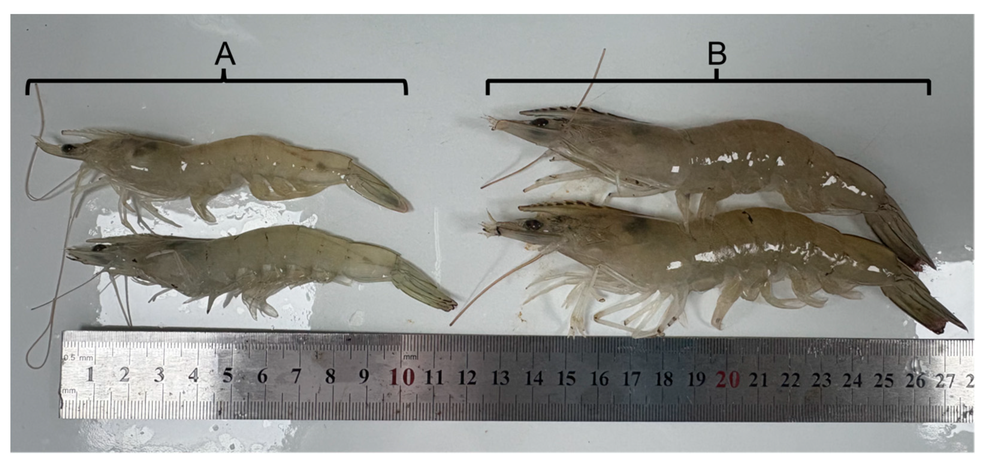
The growth of L. vannamei with EHP infection is significantly delayed (Figure 1). The body lengths of shrimp with EHP was 10.6 ± 0.3 cm, while the healthy shrimp was 15.3 ± 0.5 cm in the same breeding cycle (p < 0.05, n = 10).
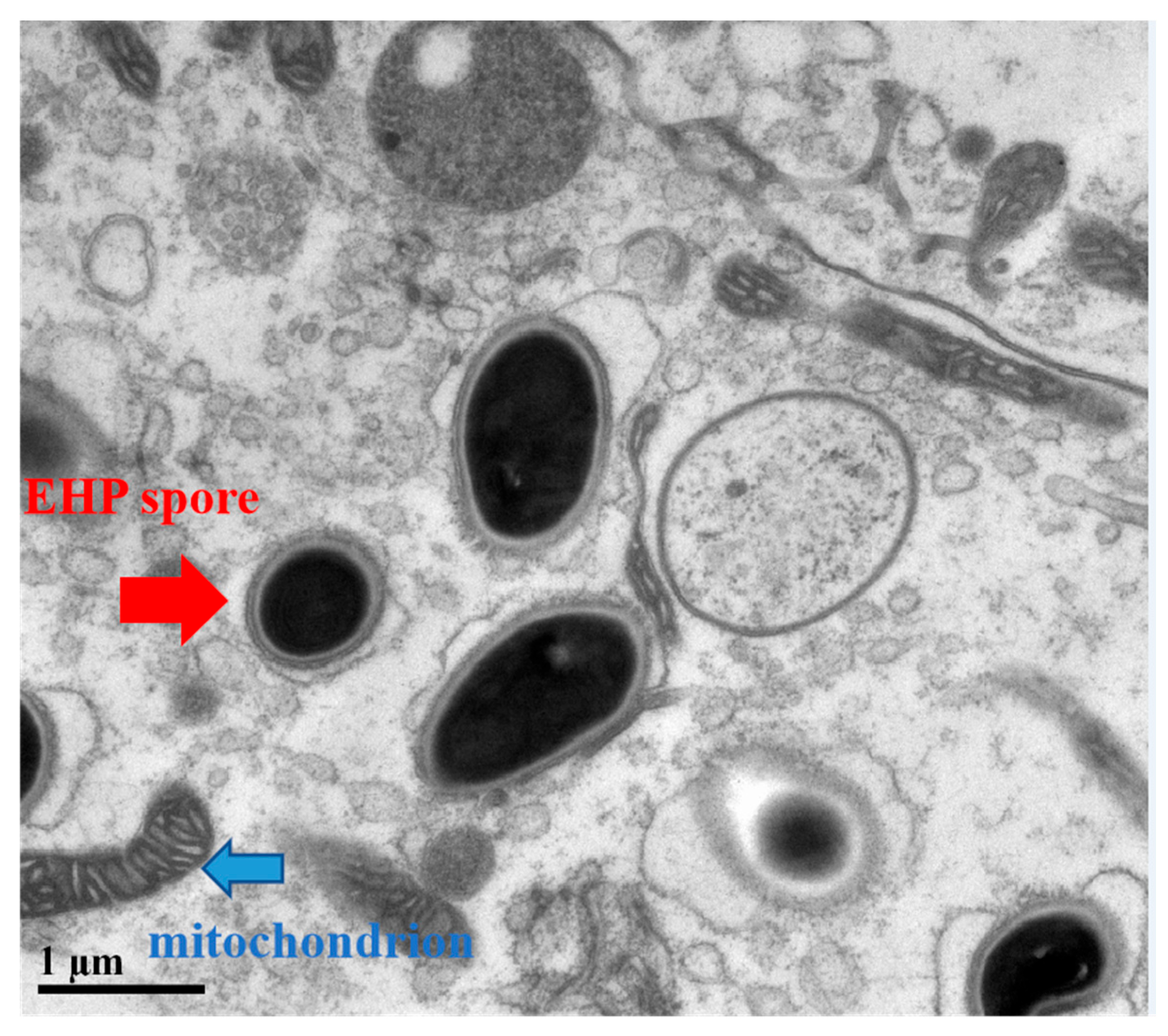
The results of the TEM of hepatopancreas of L. vannamei showed that EHP exists in the cytoplasm, surrounded by mitochondria, vacuoles, and other organelles. Mature spores have a characteristic thick spore wall and are about 1 um in size (Figure 2).

3.2. EHP Spores

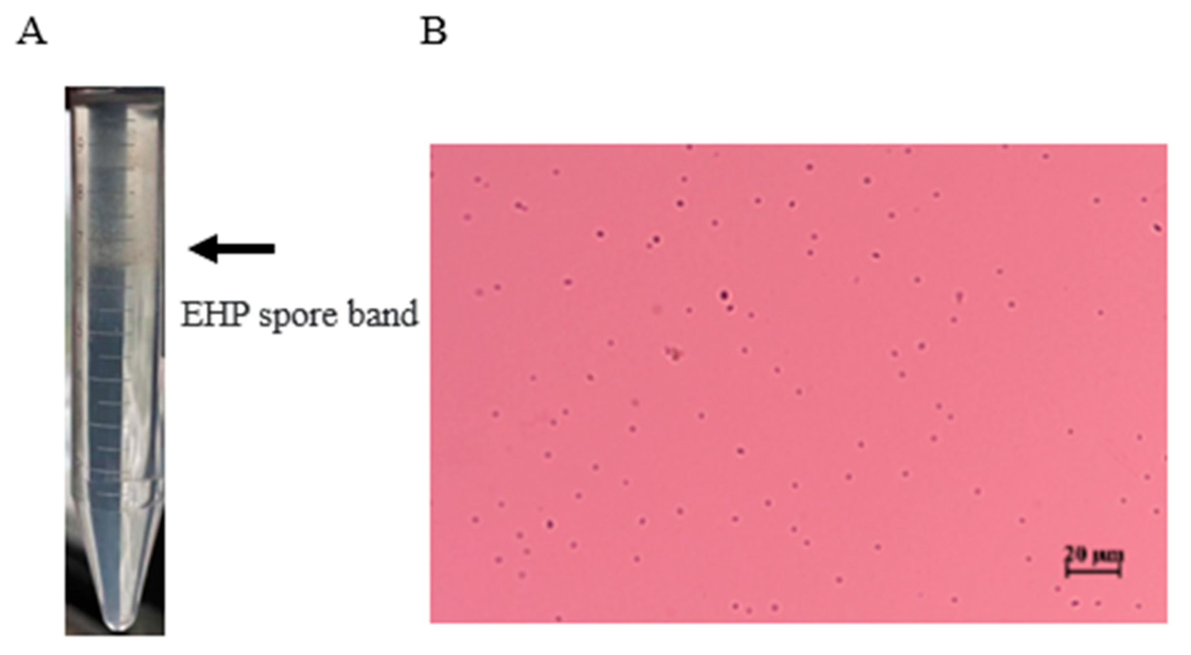
In this study, the hepatopancreases of EHP-positive L. vannamei were used to prepare a crude extract of EHP spores, which were then purified with Percoll density gradient centrifugation. The EHP spores from the EHP-infected L. vannamei were then isolated from the visible white band in the gradient (Figure 3A) with a 1 mL syringe. Phloxin B staining showed that the band contained spores and almost no impurities (Figure 3B).

3.3. Measuring the Germination Rate of Treated Spores

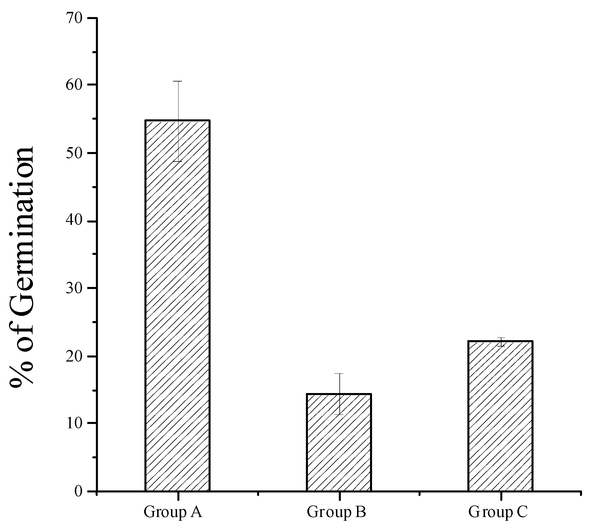
Germinated spores have a poor refractive index and stain only slightly with phloxin B, whereas ungerminated mature spores have a strong refractive index. The spores in the different treatment groups were stained with phloxin B, which showed that the germination rate of group A was significantly higher than that of groups B and C (p < 0.01), and the germination rate of group C was slightly higher than that of group B (Figure 4). These results confirm that KOH promoted the germination of the EHP spores, whereas a low temperature inhibited their germination.

3.4. SEM Analysis

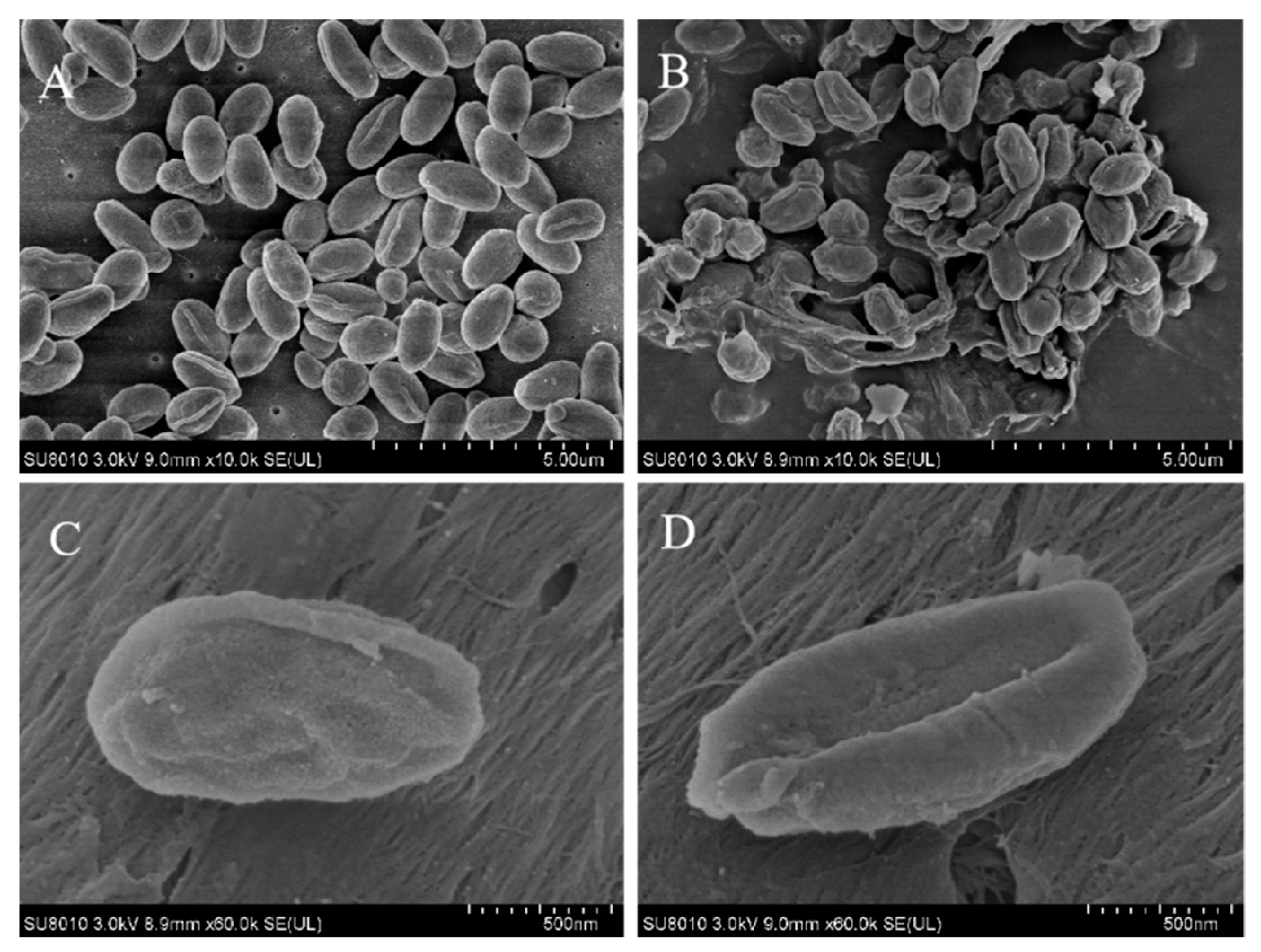
SEM showed that the size of the EHP spores was about 1 μm. Most spores in the control group (treated with TE buffer) were intact mature spores (Figure 5A), whereas a large number of empty cysts were observed in group A (Figure 5B). These results indicate that the addition of 0.1 M KOH to relatively clean spores of EHP induced spore germination.

3.5. Phylogenetic Analysis

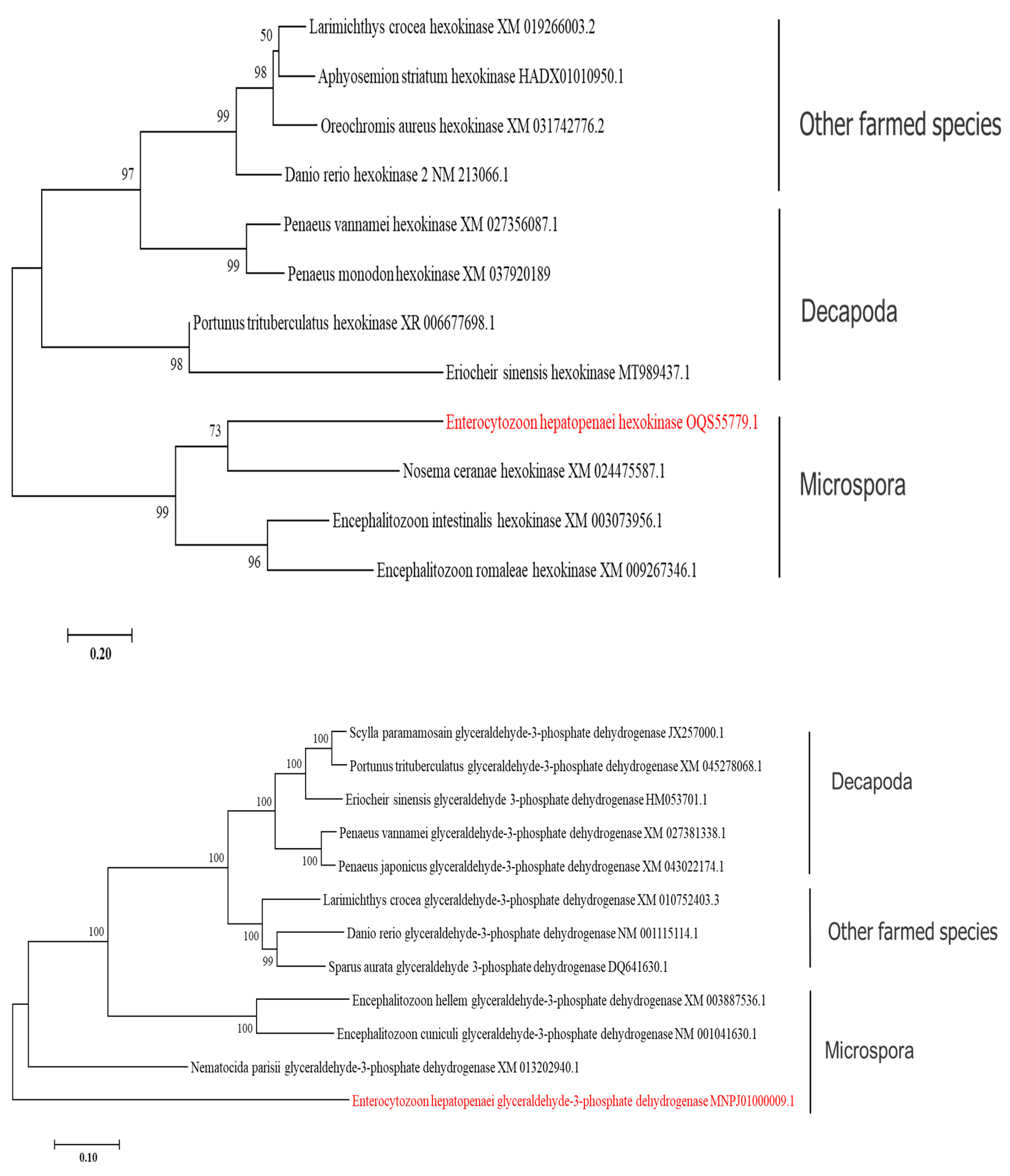
Phylogenetic trees were constructed from the HK and GAPDH gene sequences of microsporidia, commercially cultured decapods, and other cultured aquatic species. The phylogenetic tree (Figure 6) based on the HK and GAPDH genes showed that EHP is genetically very distant from its host. On the HK tree, the microsporidia clustered in one clade, and the commercially farmed shrimp, crabs, and fish clustered in another clade. On the GAPDH tree, EHP was located on a separate branch from the shrimp, crabs, and fish.

3.6. Quantitative Results for HK and GAPDH Genes

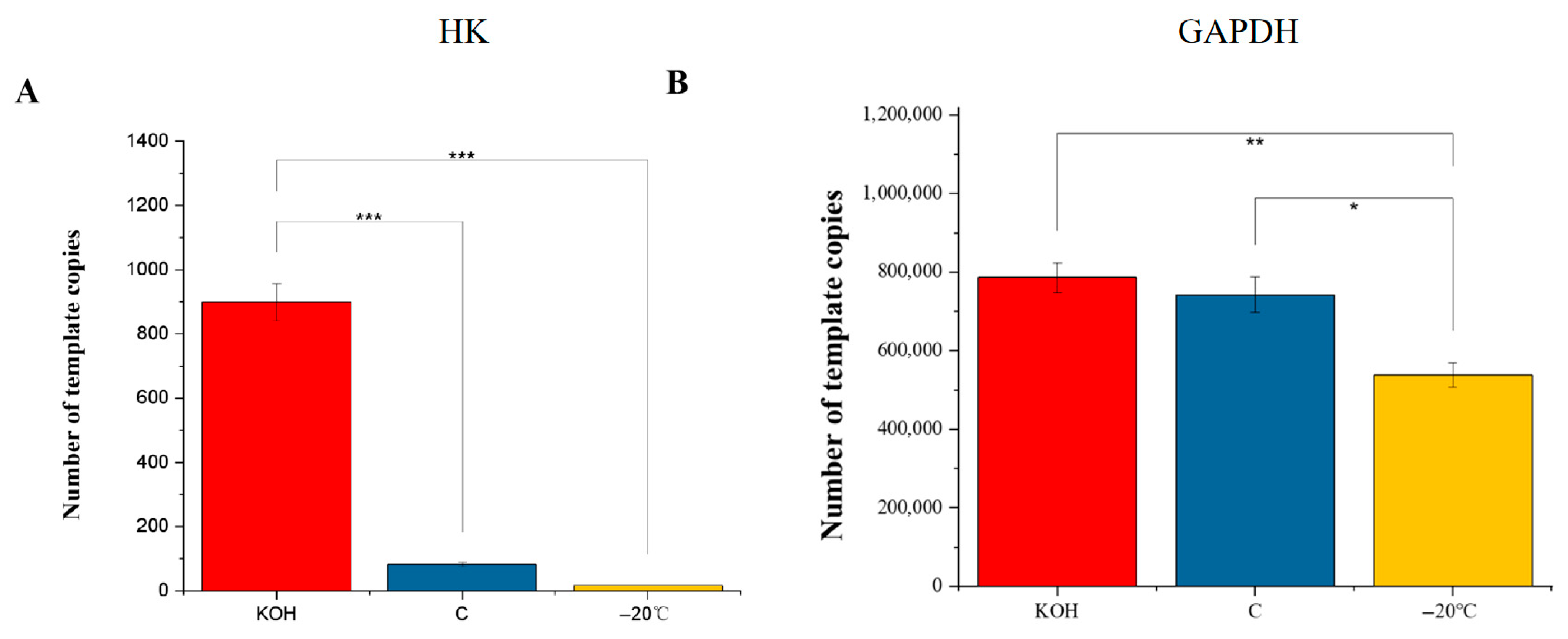
The established fluorescence quantitative detection method confirmed that the specificity, sensitivity, and repeatability of the method were all good. After the germination-promoting treatment, the expression of the HK gene was significantly higher than in the control group (p < 0.01), whereas the expression of the GAPDH gene was basically unchanged. After germination inhibition, the expression of the HK gene was downregulated, but the amplitude was small. However, the expression of the GAPDH gene was decreased at a low temperature (Figure 7).

3.7. Enzyme Activities of HK and GAPDH

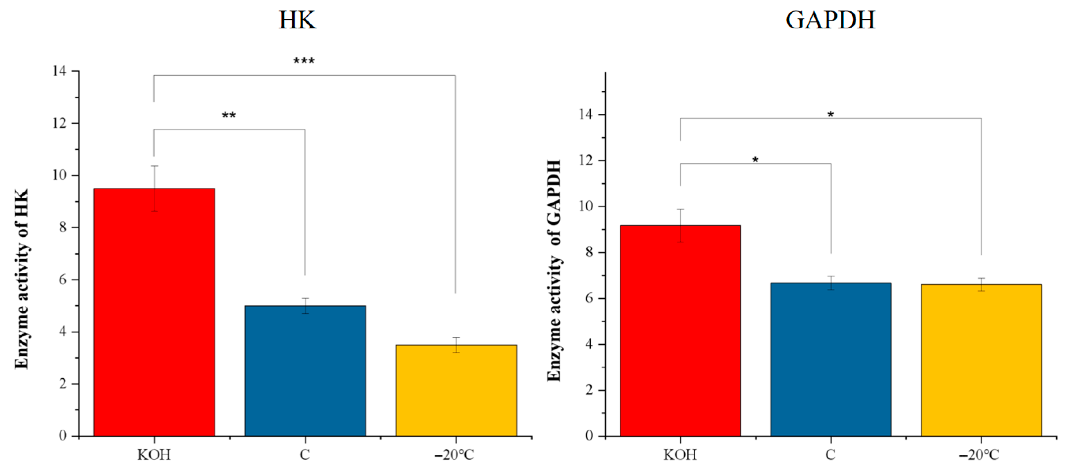
The activities of HK and GAPDH were measured in the purified spores after different treatments. After the germination-promoting treatment, the activities of HK and GAPDH increased, but the change in GAPDH was relatively small. After the germination-inhibiting treatment, the GAPDH activity remained unchanged, whereas the activity of HK decreased (Figure 8). Compared with the control group, the GAPDH activity changed little and increased only slightly after the promotion of germination.

4. Discussion

Enterocytozoon hepatopenaei is an obligate intracellular parasite in the hepatopancreas of L. vannamei, for which effective control drugs are lacking, leading to significant loss. When shrimp are infected with EHP, the parasite competes for the host’s energy. The feed conversion rate of the host then decreases during the feeding process, resulting in growth retardation and serious reductions in the economic benefits of aquaculture. Most microsporidians have lost their mitochondrial genomes and are unable to follow the complete oxidative phosphorylation pathway. Glycolysis is a basic energy-producing pathway and is considered to be the most primitive method of energy acquisition. Using high-throughput sequencing, the complete genomic sequence of EHP was determined, and it was found that it encodes only some genes of a consequently incomplete glycolysis pathway [11]. However, the survival of EHP is largely dependent upon the glycolysis pathway in vitro. In the process of invasion, the spore germinates under external stimulation, releases a polar filament that combines with the host cell, and transfers its sporoplasm into the host cell through the polar tube. Spores enter the in vivo stage from the in vitro stage to develop and complete their life cycles within the host. In the in vitro phase, microsporidia mainly rely on their own glycolysis pathway to maintain their survival. Investigating the response characteristics of two enzymes in the glycolysis pathway of spores in different states in vitro can provide new reference information for its control with drugs.
Phloxin B can be used to trigger and visualize the activity in microsporidians of any species, and can therefore be used to analyze EHP spores in their active stage [10]. Reports of the germination of microsporidia showed that 0.1 KOH promoted the germination of EHP spores, and the survival rate of the microsporidium was significantly improved when the temperature increased from 4 °C to 15 °C. However, the purified spores did not germinate or extrude their polar tubes at 4 °C. Several studies have shown that EHP spores were 100% inactivated after incubation at −20 °C for 2 h [10,16,17]. Based on the literature, we selected KOH to induce spore germination (group A), maintained spores at room temperature as the control group (group C), and inhibited the germination of purified spores at −20 °C (group B). Meanwhile, 12 h was selected to ensure the majority of the inhibition. The germination rate was calculated with light microscopy, and confirmed that KOH triggered the germination of microsporidium and that the germination rate of EHP was inhibited at a low temperature. An SEM analysis showed that the spores were about 1 μm in length. Broken sprouting spores and empty cysts were clearly visible in the KOH treatment group, whereas most spores were intact in the control group. The spore size and the effect of different treatments were consistent with previous reports [10].
HK and GAPDH were the initiating key gene and the core link gene in the glycolysis pathway, respectively. The phylogenetic analysis of the HK and GAPDH genes, which are expressed in the glycolysis pathway of EHP, showed that the HK gene of EHP clustered with those of other microsporidia, but differed greatly from those of its host and other aquatic animals. The EHP GAPDH gene clustered separately from those of other microsporidia. These results indicate that the HK and GAPDH primers used in this study can also be used as specific primers for the detection of EHP. It also can be used as an auxiliary primer to evaluate the control effect of EHP. They could also serve as auxiliary primers to monitor the effectiveness of EHP control measures.
After confirming that the spores were effectively treated, we examined the expression of HK and GAPDH genes. The results showed that the gene expression of HK increased and the activity of hexokinase increased significantly after spore germination promotion treatment. The gene expression level of GAPDH was stable after germination promotion, but the activity of the GAPDH enzyme was significantly increased. These results suggested that the glycolysis pathway of EHP may be activated during germination, and the glucose metabolism capacity may be increased. The main energy source during spore germination may be the truncated glycolysis pathway involving HK and GAPDH.
Hexokinase is a key enzyme in the glucose metabolic pathway and exists in almost all organisms. It is the first enzyme in the glycolytic pathway and one of the three rate-limiting enzymes of glycolysis [18]. It catalyzes hexose phosphorylation to glucose 6-phosphate or fructose 6-phosphate, and plays an important role in microsporidial metabolism and the synthesis of amylose [19]. The results of this study show that during the germination of EHP spores, the expression of HK increased significantly at the transcriptome level, and the apparent enzyme activity reflected the increased activities of the spore enzymes. The activity of the HK, one of the main rate-limiting enzymes of glycolysis, was enhanced, indicating an increase in glycolysis during the germination of EHP. In the extracellular environment, mature spores extrude a polar tube, releasing the cell precursor into the host cell through the lumen of the polar filament. It follows that accelerated carbohydrate metabolism promotes the completion of the invasion process because germination and the transfer of the cell precursor require large amounts of energy. It has been reported that the growth and development of Toxoplasma gondii is almost inhibited when its HK expression is knocked out by RNA interference, confirming the importance of HK in the glycolysis pathway [20]. Some scholars have reported that increasing the gene expression and activity of HK in tumor cells under hypoxic conditions provides tumor cells with sufficient energy for their growth and proliferation [21]. At present, HK has become an important target for the development of novel antiparasitic drugs. For EHP, which is extremely dependent on the glycolysis pathway during the spore stage, inhibition of HK expression or enzyme activity can interfere with EHP’s energy metabolism and germination infection in vitro, thus inhibiting EHP’s survival and transmission in vitro.
GAPDH is another key enzyme in the glycolytic pathway, and is thus involved in the energy metabolism of cells. GAPDH is expressed at high levels in almost all tissues [22]. In the same tissues or cells, the expression of the GAPDH protein is generally constant and remains constant, regardless of the influence of inducing substances. Therefore, it is known as a ‘housekeeping protein’ [23]. GAPDH is a multifunctional protein that not only participates in glycolysis, but is also involved in membrane fusion, vascular bundle formation, nuclear RNA transfer, DNA repair, apoptosis, and cell adhesion in humans and fungi [24,25,26]. GAPDH plays an important role in the mutual recognition of pathogen and host, and in the infection process involving both. There was no significant difference in GAPDH gene expression between the germination-promoting group and the control group, indicating that the expression of glyceraldehyde 3-phosphate dehydrogenase was still stable during germination, and the related physiological activities regulated by this enzyme could be normally expressed. Meanwhile, the observed increase in GAPDH enzyme activity during spore germination suggests that potential post-translational modification regulation may exist in spores, and targeting GAPDH activity may inhibit spore germination and subsequent host invasion. Of course, in addition to the analysis based on the differences in evolutionary trees, it is also necessary to further deepen and refine the research to determine whether substances targeting this potential target will also affect the related sites of the host.
In conclusion, it is speculated that the energy source of EHP spore survival and germination invasion in vitro may come from the special glycolysis pathway. HK and GAPDH, the key genes of the glycolytic pathway for EHP spore survival in vitro, are significantly different from the main host gene sequences and play a key role in spore germination. Inhibition of the HK gene expression level and GAPDH protein levels may block spore germination and invasion in vitro and can be used as a potential control target for EHP.

5. Conclusions

HK and GAPDH, the key genes of the glycolytic pathway for EHP spore survival in vitro, are significantly different from the main host gene sequences and play a key role in spore germination. Inhibition of the HK gene expression level and GAPDH protein levels can block spore germination and invasion in vitro. The energy source of EHP spore survival and germination invasion in vitro may come from the special glycolysis pathway. HK and GAPDH can be used as a potential control target for EHP.

Author Contributions

Conceptualization, P.Z. and C.M.; methodology, B.Z. and J.F.; formal analysis, B.Z.; investigation, D.Q.; resources, J.C.; writing—review and editing, B.Z. and R.M.; project administration, R.M.; funding acquisition, R.M. All authors have read and agreed to the published version of the manuscript.

Funding

This study was supported by the National Natural Science Foundation of China (42306128); the Fund of the Key Laboratory of South China Sea Fishery Resources Exploitation & Utilization, Ministry of Agriculture and Rural Affairs, P.R. China (FREU2021-02); the Key Scientific and Technological Grant of Zhejiang for Breeding New Agricultural Varieties (No. 2021C02069-5-2); and the Key Project of Ningbo Science and Technology Bureau (No. 2023S003).

Institutional Review Board Statement

The use of experimental animals was reviewed and approved by the Ethics Committee of Ningbo University (Approval code: SYXK20190005 and approval date: 23 June 2024).

Data Availability Statement

The data presented in this study are available upon request from the corresponding author.

Conflicts of Interest

The authors declare that they have no conflicts of interest concerning this article.

References

  1. Tangprasittipap, A.; Srisala, J.; Chouwdee, S.; Somboon, M.; Chuchird, N.; Limsuwan, C.; Sritunyalucksana, K. The microsporidian Enterocytozoon hepatopenaei is not the cause of white feces syndrome in whiteleg shrimp Penaeus (Litopenaeus) vannamei. BMC Vet. Res. 2013, 9, 139. [Google Scholar] [CrossRef] [Scilit]
  2. Rajendran, K.V.; Shivam, S.; Praveena, P.E.; Rajan, J.J.S.; Kumar, T.S.; Avunje, S.; Vijayan, K.K. Emergence of Enterocytozoon hepatopenaei (EHP) in farmed Penaeus (Litopenaeus) vannamei in India. Aquaculture 2016, 454, 272–280. [Google Scholar] [CrossRef] [Scilit]
  3. Tang, K.F.J.; Han, J.E.; Aranguren, L.F.; White-Noble, B.; Schmidt, M.M.; Piamsomboon, P.; Hanggono, B. Dense populations of the microsporidian Enterocytozoon hepatopenaei (EHP) in feces of Penaeus vannamei exhibiting white feces syndrome and pathways of their transmission to healthy shrimp. J. Invertebr. Pathol. 2016, 140, 1–7. [Google Scholar] [CrossRef] [Scilit]
  4. Salachan, P.V.; Jaroenlak, P.; Thitamadee, S.; Itsathitphaisarn, O.; Sritunyalucksana, K. Laboratory cohabitation challenge model for shrimp hepatopancreatic microsporidiosis (HPM) caused by Enterocytozoon hepatopenaei (EHP). BMC Vet. Res. 2017, 13, 302. [Google Scholar] [CrossRef] [Scilit]
  5. Santhoshkumar, S.; Sivakumar, S.; Vimal, S.; Majeed, S.A.; Taju, G.; Haribabu, P.; Hameed, A.S.S. Biochemical changes and tissue distribution of Enterocytozoon hepatopenaei (EHP) in naturally and experimentally EHP-infected whiteleg shrimp, Litopenaeus vannamei (Boone, 1931), in India. J. Fish Dis. 2017, 40, 529–539. [Google Scholar] [CrossRef] [Scilit] [PubMed]
  6. Nakjang, S.; Williams, T.A.; Heinz, E.; Watson, A.K.; Foster, P.G.; Sendra, K.M.; Embley, T. Reduction and Expansion in Microsporidian Genome Evolution: New Insights from Comparative Genomics. Genome Biol. Evol. 2013, 5, 2285–2303. [Google Scholar] [CrossRef] [Scilit] [PubMed]
  7. Haag, K.L.; James, T.Y.; Pombert, J.F.; Larsson, R.; Schaer, T.M.M.; Refardt, D.; Ebert, D. Evolution of a morphological novelty occurred before genome compaction in a lineage of extreme parasites. Proc. Natl. Acad. Sci. USA 2014, 111, 15480–15485. [Google Scholar] [CrossRef] [Scilit]
  8. Hacker, C.; Howell, M.; Bhella, D.; Lucocq, J. Strategies for maximizing ATP supply in the microsporidian Encephalitozoon cuniculi: Direct binding of mitochondria to the parasitophorous vacuole and clustering of the mitochondrial porin VDAC. Cell. Microbiol. 2014, 16, 565–579. [Google Scholar] [CrossRef] [Scilit] [PubMed]
  9. Heinz, E.; Williams, T.A.; Nakjang, S.; Noel, C.J.; Swan, D.C.; Goldberg, A.V.; Embley, T.M. The Genome of the Obligate Intracellular Parasite Trachipleistophora hominis: New Insights into Microsporidian Genome Dynamics and Reductive Evolution. PLoS Pathog. 2012, 8, e1002974. [Google Scholar] [CrossRef] [Scilit]
  10. Aldama-Cano, D.J.; Sanguanrut, P.; Munkongwongsiri, N.; Ibarra-Gamez, J.C.; Itsathitphaisarn, O.; Vanichviriyakit, R.; Thitamadee, S. Bioassay for spore polar tube extrusion of shrimp Enterocytozoon hepatopenaei (EHP). Aquaculture 2018, 490, 156–161. [Google Scholar] [CrossRef] [Scilit]
  11. Boakye, D.W.; Jaroenlak, P.; Prachumwat, A.; Williams, T.A.; Bateman, K.S.; Itsathitphaisarn, O.; Williams, B.A.P. Decay of the glycolytic pathway and adaptation to intranuclear parasitism within Enterocytozoonidae microsporidia. Environ. Microbiol. 2017, 19, 2077–2089. [Google Scholar] [CrossRef] [Scilit]
  12. Jaroenlak, P.; Sanguanrut, P.; Williams, B.A.; Stentiford, G.D.; Flegel, T.W.; Sritunyalucksana, K.; Itsathitphaisarn, O. A Nested PCR Assay to Avoid False Positive Detection of the Microsporidian Enterocytozoon hepatopenaei (EHP) in Environmental Samples in Shrimp Farms. PLoS ONE 2016, 11, e0166320. [Google Scholar] [CrossRef] [Scilit] [PubMed]
  13. Kurtti, T.J.; Brooks, M.A. The rate of development of a microsporidan in moth cell culture. J. Invertebr. Pathol. 1977, 29, 126–132. [Google Scholar] [CrossRef] [Scilit] [PubMed]
  14. Martinez-Barajas, E.; Randall, D.D. Purification and characterization of a glucokinase from young tomato (Lycopersicon esculentum L. Mill.) fruit. Planta 1998, 205, 567–573. [Google Scholar] [CrossRef] [Scilit] [PubMed]
  15. Lee, Y.-P.; Lardy, H.A. Influence of Thyroid Hormones on l-α-Glycerophosphate Dehydrogenases and Other Dehydrogenases in Various Organs of the Rat. J. Biol. Chem. 1965, 240, 1427–1436. [Google Scholar] [CrossRef] [Scilit]
  16. Thitamadee, S.; Prachumwat, A.; Srisala, J.; Jaroenlak, P.; Salachan, P.V.; Sritunyalucksana, K.; Itsathitphaisarn, O. Review of current disease threats for cultivated penaeid shrimp in Asia. Aquaculture 2016, 452, 69–87. [Google Scholar] [CrossRef] [Scilit]
  17. Chaijarasphong, T.; Munkongwongsiri, N.; Stentiford, G.D.; Aldama-Cano, D.J.; Thansa, K.; Flegel, T.W.; Itsathitphaisarn, O. The shrimp microsporidian Enterocytozoon hepatopenaei (EHP): Biology, pathology, diagnostics and control. J. Invertebr. Pathol. 2021, 186, 107458. [Google Scholar] [CrossRef] [Scilit]
  18. Yi, J.W.; Ge, H.T.; Abbas, F.; Zhao, J.T.; Huang, X.M.; Hu, G.B.; Wang, H.C. Function of a nonenzymatic hexokinase LcHXK1 as glucose sensor in regulating litchi fruit abscission. Tree Physiol. 2022, 42, 2412–2426. [Google Scholar]
  19. Ciscato, F.; Ferrone, L.; Masgras, I.; Laquatra, C.; Rasola, A. Hexokinase 2 in Cancer: A Prima Donna Playing Multiple Characters. Int. J. Mol. Sci. 2021, 22, 11112. [Google Scholar] [CrossRef] [Scilit]
  20. Ananvoranich, S.; Al Rayes, M.; Al Riyahi, A.; Wang, X. RNA silencing of glycolysis pathway in Toxoplasma gondii. J. Eukaryot. Microbiol. 2006, 53, S162–S163. [Google Scholar] [CrossRef] [Scilit]
  21. Wolf, A.; Agnihotri, S.; Micallef, J.; Mukherjee, J.; Sabha, N.; Cairns, R.; Guha, A. Hexokinase 2 is a key mediator of aerobic glycolysis and promotes tumor growth in human glioblastoma multiforme. J. Exp. Med. 2011, 208, 313–326. [Google Scholar] [CrossRef] [Scilit]
  22. Bolton, S.G.; Pluth, M.D. Efficient inhibition of glyceraldehyde-3-phosphate dehydrogenase (GAPDH) by sulfuration with solubilized elemental sulfur. Free Radic. Biol. Med. 2022, 185, 46–51. [Google Scholar] [CrossRef] [Scilit]
  23. Barber, R.D.; Harmer, D.W.; Coleman, R.A.; Clark, B.J. GAPDH as a housekeeping gene: Analysis of GAPDH mRNA expression in a panel of 72 human tissues. Physiol. Genom. 2005, 21, 389–395. [Google Scholar] [CrossRef] [Scilit] [PubMed]
  24. Lazarev, V.F.; Guzhova, I.V.; Margulis, B.A. Glyceraldehyde-3-phosphate Dehydrogenase is a Multifaceted Therapeutic Target. Pharmaceutics 2020, 12, 1103. [Google Scholar] [CrossRef] [Scilit] [PubMed]
  25. Hannaert, V.; Opperdoes, F.R.; Michels, P.A. Comparison and evolutionary analysis of the glycosomal glyceraldehyde-3-phosphate dehydrogenase from different Kinetoplastida. J. Mol. Evol. 1998, 47, 728–738. [Google Scholar] [CrossRef] [Scilit]
  26. Viscogliosi, E.; Muller, M. Phylogenetic relationships of the glycolytic enzyme, glyceraldehyde-3-phosphate dehydrogenase, from parabasalid flagellates. J. Mol. Evol. 1998, 47, 190–199. [Google Scholar] [CrossRef] [Scilit] [PubMed]
Figure 1. Body lengths of EHP positive and healthy L. vannamei. (A) L. vannamei infected with EHP. (B) The healthy L. vannamei.
Fishes 11 00229 g001
Figure 2. TEM observation of hepatopancreas of L. vannamei with EHP infection. Note: The red arrow shows the EHP mature spore and the blue arrow shows the mitochondria.
Fishes 11 00229 g002
Figure 3. Spore purification and Phloxin B staining observation. Notes: (A) Percoll gradient purification of EHP spores. The black arrow indicates the band formed by the spores after ultracentrifugation. (B) Purified EHP spores were visualized after phloxin B staining.
Fishes 11 00229 g003
Figure 4. Germination rate of EHP spores. Notes: Group A was KOH treatment; group B was low-temperature treatment; and group C was the control group. The values of the bars and whiskers indicate the means ± standard errors, respectively (n = 3).
Fishes 11 00229 g004
Figure 5. Scanning electron microscopic analysis of purified EHP spores. Notes: (A) Purified EHP spores were treated with 0.1 M KOH to promote germination. (B) Purified EHP spores were treated with TE buffer. (C) Intact mature spores. (D) Broken spores after germination.
Fishes 11 00229 g005
Figure 6. Phylogenetic tree based on the HK and GAPDH genes of various species, including EHP.
Fishes 11 00229 g006
Figure 7. Number of template copies of HK and GAPDH in different groups. (A) is the expression of HK gene in different groups, (B) is the expression of GAPDH gene in different groups. Notes: The values of the bars and whiskers indicate the means ± standard errors, respectively (n = 3); * p ≤ 0.05, ** p ≤ 0.01, and *** p ≤ 0.001.
Fishes 11 00229 g007
Figure 8. HK and GAPDH enzyme activities. Notes: The values of the bars and whiskers indicate the means ± standard errors, respectively (n = 3); * p ≤ 0.05, ** p ≤ 0.01, and *** p ≤ 0.001.
Fishes 11 00229 g008
Table 1. Quantitative real-time PCR (qPCR) primer sequences for EHP.
PrimerSequence (5′-3′)PCR Length
EHP-HK-FTGACTGTGTGGCAATGATGT131 bp
EHP-HK-RACTGTCCTGCCTCAGTAACAC
EHP-GAPDH-FAATCAGCACGAAATAGACTT128 bp
EHP-GAPDH-RCAACTTCTTCCCAAACGA
Disclaimer/Publisher’s Note: The statements, opinions and data contained in all publications are solely those of the individual author(s) and contributor(s) and not of MDPI and/or the editor(s). MDPI and/or the editor(s) disclaim responsibility for any injury to people or property resulting from any ideas, methods, instructions or products referred to in the content.