


Abstract
Macrophage migration inhibitory factor (MIF) is broadly acknowledged as a central pro-inflammatory regulator, owing to its multifaceted functions including immune cell recruitment, initiation and amplification of pro-inflammatory cytokine cascades, enhancement of macrophage viability, facilitation of macrophage polarization toward a pro-inflammatory state, and attenuation of glucocorticoid-mediated immunosuppression. However, functional investigations of MIF in Mytilus coruscus remain limited. In this study, we identified the MIF gene in M. coruscus, and bioinformatic analyses revealed that the gene encodes a 115-amino-acid polypeptide that exhibits close phylogenetic affinity with MIF homologs from other mollusks. McMIF was predominantly expressed in immune-related tissues, with notably high expression levels in the digestive gland. Upon Vibrio alginolyticus infection, both the mRNA and protein levels of McMIF were significantly upregulated, suggesting that McMIF is involved in the antibacterial immune response of M. coruscus. Meanwhile, the m6A modification level of McMIF was markedly reduced following infection, suggesting a potential relationship between m6A modification and the antibacterial immune function of MIF. Furthermore, knockdown of McMIF followed by LPS stimulation led to an increased level of apoptosis in digestive gland cells, suggesting that McMIF is involved in the inhibition of apoptosis induced by immune stimulation. Collectively, these findings provide insights into the immunological characteristics of McMIF in M. coruscus.
Key Contribution:
This study identifies McMIF in M. coruscus for the first time and reveals that McMIF m6A modification and gene expression are altered following V. alginolyticus infection. Suppression of McMIF expression enhanced LPS-induced apoptosis, indicating that McMIF may be linked to the innate immune response in M. coruscus.
1. Introduction
Macrophage migration inhibitory factor (MIF) is a broadly acknowledged modulator of innate immunity and a multifunctional cytokine. It enhances the expression and secretion of pro-inflammatory molecules, including interleukin-1β and interleukin-6, while suppressing macrophage motility [1,2], and contributes to the pathological progression of a wide range of immune-related and autoimmune disorders [3], including spondyloarthritis [4], myocarditis [5], and neuroinflammation [6]. In recent years, MIF genes have been detected in both invertebrates [7] and vertebrates [8]. MIF homologs in different vertebrates typically participate in both innate and adaptive immune responses [9]. In invertebrates, which lack a developed adaptive immune system (such as mollusks and arthropods), MIF homologs may play a role in innate immunity [10]. Nevertheless, MIF homologs have been characterized in only a limited number of mollusk species to date, encompassing the abalone (Haliotis diversicolor supertexta), the snail (Biomphalaria glabrata), the pearl oyster (Pinctada fucata), the Mediterranean mussel (Mytilus galloprovincialis), the clam (Meretrix meretrix), and the scallop (Chlamys farreri) [11]. Research by Roger T [12] showed that MIF facilitates the recognition of Gram-negative bacteria and stimulates pro-inflammatory cytokine generation through the enhancement of Toll-like receptor 4 (TLR4) expression, which is crucial for the rapid response of mollusks to bacterial pathogens. Li [13] observed that in scallops, MIF accelerates the migration of fibroblasts to scrape wounds, promoting wound healing. This suggests that MIF may assist in the migratory phase of wound healing in scallops.
To date, studies have demonstrated that MIF regulates its biological activity through covalent and structural modifications, including oxidation [14], haptenation [15], methylation [16], and glycosylation [17]. Meanwhile, over 160 distinct chemical modifications have been cataloged in RNA molecules, distributed across various biological domains, species, and molecular types [18]. As early as the 1970s, changes in m6A (N6-methyladenosine) in eukaryotic mRNA and lncRNA were detected, indicating that RNA methylation is a post-transcriptional regulatory mechanism [19]. When methylation occurs at the nitrogen atom at position 6 of RNA adenosine, m6A is formed [20], which significantly affects all stages of the RNA lifecycle [21]. However, no studies have yet revealed the relationship between MIF methylation modifications and inflammatory responses. The present study seeks to investigate and validate the biological significance of MIF methylation modifications in immune regulation.
Mytilus coruscus (M. coruscus), an economically important marine bivalve species, is primarily distributed in the Yellow Sea and East China Sea [22]. Owing to its exceptional nutritional profile and considerable commercial value, this species has emerged as a vital component of the human food supply [23]. Nonetheless, with environmental changes and human activities, the risk of pathogenic bacterial infections in these bivalves is gradually increasing [24]. Because bivalves lack antibody-mediated mechanisms in their adaptive immune system, their immune defense primarily relies on the innate immune system [25].
Although MIF plays a crucial regulatory role in the innate immune defense of invertebrates, functional studies of MIF in M. coruscus remain lacking, and its molecular mechanisms in immune responses in this species are still unclear. Therefore, in this study, we cloned and identified the MIF homologous gene from the bivalve M. coruscus, analyzed its expression patterns, and further investigated its immune function.
2. Materials and Methods
2.1. Animals
All 150 M. coruscus needed for the experiment were purchased from Donghe Market in Zhoushan City. They were acclimated for one week under experimental conditions of 25‰ salinity and water temperature of 25 ± 1 °C. During the acclimation period, Spirulina was supplied daily and the water was changed regularly to ensure that the daily mortality rate of M. coruscus was below 5%. The day before the experiment began, feeding was suspended, and individuals exhibiting healthy growth status and comparable body size (shell length: 9 ± 0.5 cm; body weight: 80 ± 2.5 g) were chosen for subsequent experimentation.
2.2. Bioinformatic Analysis
The open reading frame (ORF) of McMIF was predicted using DNAMAN 9.0 software, and the deduced amino acid sequence was obtained. Multiple sequence alignment of McMIF with reported MIF homologs from other species was performed, and sequence conservation was visualized using ESPript 3.0 [26] and WebLogo 3.0 [27]. On this basis, the domain architecture of the protein was identified using SMART (v3.0) [28]. The three-dimensional structure of McMIF was predicted using the SWISS-MODEL [29] online server based on homology modeling. The optimal template was automatically selected by the server according to sequence similarity and model quality parameters. For phylogenetic analysis, McMIF amino acid sequences from different species were retrieved from the NCBI [30] database. Multiple sequence alignment was performed using Clustal Omega [31], and a phylogenetic tree was constructed using MEGA 11 [32] software based on the Neighbor-Joining method. The reliability of the phylogenetic tree was evaluated using 5000 bootstrap replicates.
2.3. Bacterial Infection and Sample Collection
The experimental strain of V. alginolyticus was isolated from naturally infected mussels. Bacterial cultures were established by streaking onto 2216E agar plates (Hopebio, Qingdao, China) and maintained in an inverted position at 28 °C for 24 h. Individual colonies were subsequently selected and propagated in TSB liquid medium (Hopebio, Qingdao, China) under continuous agitation (200 rpm) at 37 °C for 12 h. The resulting culture was centrifuged at 12,000 rpm for 3 min, and the obtained pellet was washed and resuspended in sterile PBS to prepare the bacterial inoculum. Thirty healthy adult M. coruscus were randomly allocated into three experimental groups (A, B, and C), with 10 individuals in each group. Each individual received an intramuscular injection of 100 µL V. alginolyticus suspension (1 × 10⁷ CFU/mL) [33] into the adductor muscle. Subsequently, at six designated time points (0, 6, 12, 24, 48, and 72 h post-challenge), one mussel was randomly selected from each group for dissection and collection of digestive gland tissues. Samples within each experimental group were pooled to minimize individual biological variation. In parallel, digestive gland, hemolymph, gonads, mantle, and gill tissues were also harvested from unchallenged healthy mussels, with three biological replicates prepared per tissue type. All collected samples were immediately snap-frozen and stored at −80 °C until subsequent RNA and protein extraction.
2.4. Quantitative Real-Time PCR Analysis
Total RNA was reverse-transcribed into cDNA using the PrimeScript™ RT reagent Kit (TaKaRa, Osaka, Japan) with gDNA Eraser according to the manufacturer’s instructions. Briefly, genomic DNA was removed in a 10 μL reaction at 42 °C for 2 min, and reverse transcription was subsequently performed in a final volume of 20 μL at 37 °C for 15 min, followed by enzyme inactivation at 85 °C for 5 s. The synthesized cDNA was used for RT-PCR. Based on the McMIF mRNA sequence, the primer pair McMIF-F and McMIF-R was designed to amplify a 134 bp fragment spanning nucleotides 4–137 of the transcript. The primers were synthesized by Sangon Biotech (Shanghai, China). Quantitative assessment of gene expression levels in vivo was conducted on an Applied Biosystems 7500 Real-Time PCR System. Each RT-PCR reaction was prepared in a final volume of 10 µL, comprising 5 µL of TB Green premix, 0.4 µL of forward and reverse primers (10 mM each), 1 µL of cDNA template, and 3.2 µL of nuclease-free ddH2O. The primer sequences employed in this study were designed using Primer-BLAST in NCBI and are summarized in Table 1, with Mcβ-actin utilized as the endogenous reference gene. The thermal cycling protocol consisted of an initial denaturation at 95 °C for 30 s, followed by 40 amplification cycles of 95 °C for 5 s and 55 °C for 34 s. Relative transcript abundance was calculated using the 2^(−ΔΔCt) method [34]. All experiments were performed with three biological replicates (n = 3).
Table 1.
Primers used in this study.
2.5. RNA Interference
McMIF small interfering RNA (siRNA) and control siRNA (siNC) were designed and synthesized by GenePharma (Shanghai, China) as shown in Table 1. Primers used in this study. The siRNA powder was reconstituted in DEPC-treated water to achieve a working concentration of 20 µM. For in vivo assays, six healthy adult M. coruscus were randomly selected and infected according to the procedure described in Section 2.3. Each individual received an intramuscular injection of 100 µL of V. alginolyticus suspension (1 × 10⁷ CFU/mL) into the adductor muscle. At 24 h post-infection, the infected mussels were randomly assigned to either the siNC control group or the siRNA treatment group, with three mussels in each group representing three independent biological replicates. Each individual was administered 100 µL of the siRNA solution via injection into the adductor muscle. Twenty-four hours post-injection, three mussels from each group were selected for digestive gland tissue collection, and mRNA expression levels were analyzed with β-actin as the internal control to assess knockdown efficiency. For in vitro studies, digestive gland–derived cells were isolated and maintained in culture. A mixture consisting of 200 µL DMEM medium supplemented with 12.5 µL siRNA and 12.5 µL TransIntro® EL Transfection Reagent (TransGen Biotech, Beijing, China) was added to 2 mL of primary cells. After 8 h of transfection, the medium was replaced with fresh medium containing LPS, and the cells were further incubated for another 24 h to induce an immune response. The cells were then collected for apoptosis analysis. All experiments were performed with three biological replicates (n = 3).
2.6. m6A Methylation Analysis
Total RNA was isolated from normal digestive gland tissues and digestive gland tissues collected at 24 h after V. alginolyticus infection using Trizol reagent (Solarbio, Beijing, China), and RNA quality was assessed using a NanoDrop 2000 spectrophotometer and agarose gel electrophoresis. m6A methylation enrichment was carried out according to the m6A MeRIP Kit (CloudSeq Biotechnology, Shanghai, China). Total RNA was adjusted to 1 µg/µL with RNase-free water, and RNA fragmentation was performed using 10× Fragmentation Buffer and Stop Buffer. m6A antibody-coated PGM magnetic beads and IgG-coated PGM magnetic beads were incubated with the fragmented RNA for 1 h at 4 °C. After washing, methylated RNA fragments were obtained, and RNA methylation fragments were purified using MS magnetic beads. Eluted RNA was collected, reverse-transcribed into cDNA, and methylation levels were detected using RT-PCR. For MeRIP-qPCR analysis, methylation levels were evaluated based on the enrichment of the target transcript in the m6A-immunoprecipitated fraction rather than by direct detection of individual methylated nucleotides. Ct values from the IP, Input, and IgG samples were used to calculate the relative enrichment of McMIF transcripts. The percentage of input was calculated for both the m6A-IP and IgG groups, and fold enrichment was obtained by normalizing the % input of the IP group to that of the IgG group. Relative methylation levels between experimental groups were then compared based on the fold enrichment values. Each sample was analyzed in triplicate.
2.7. RNA Dot Blot
Aliquots of m6A-enriched RNA samples (200 or 400 ng) were serially diluted and applied to a polyvinylidene fluoride (PVDF) membrane (pore size: 0.25 µm; Merck KGaA, Darmatadt, Germany) pre-activated with methanol. The RNA samples were denatured at 95 °C for 3 min to eliminate secondary structures, followed by immediate cooling on ice to prevent refolding. The membrane was then incubated at 37 °C for 1 h to allow RNA immobilization. Afterward, the membrane was washed with TBS containing 0.02% Tween-20 for 10 min under gentle agitation to remove unbound RNA. Blocking was performed at room temperature for 1 h using TBS supplemented with 5% skim milk and 0.02% Tween-20. The membrane was subsequently incubated overnight at 4 °C with an m6A-specific primary antibody (1:2000 dilution; HuaBio, Hangzhou, China). Following three washes (10 min each), the membrane was incubated with HRP-conjugated goat anti-rabbit IgG secondary antibody (1:200,000 dilution; Sangon, Shanghai, China) for 1.5 h at room temperature. After additional washing steps, signals were visualized using an enhanced chemiluminescence (ECL) substrate and captured with a chemiluminescence imaging system.
2.8. Western Blot
Proteins were extracted from digestive gland tissues using cell lysis buffer (Biotime, Shanghai, China), and protein concentrations were determined using the BCA Protein Assay Kit (Solebao, Beijing, China). Protein samples (35 µg per well) were separated on 10% SDS-PAGE precast gels (GenScript, Nanjing, China) and transferred to a PVDF membrane (Merck KGaA, Darmatadt, Germany) by wet transfer. The membrane was blocked at room temperature for 1 h in 5% skim milk and then incubated overnight at 4 °C with a polyclonal antibody specific to McMIF. The McMIF antibody (1:8000 dilution, Ata Genix, Wuhan, China) was diluted in 1× TBST (1× TBS, 0.05% Tween-20). Following incubation, the membrane was washed three times with 1× TBST and then incubated at room temperature for 1.5 h with the corresponding HRP Conjugated Goat anti-Rabbit IgG polyclonal Antibody (1:150,000 dilution, HuaBio, Hangzhou, China, HA1001). After three washes with TBST (10 min each), the membrane was incubated with Western Lightning ECL Substrate (Solarbio, Beijing, China) for 30 s and imaged using a chemiluminescence imaging system. Mcβ-actin was used as the internal reference protein. The same procedure described above was followed, using the β-actin antibody (1:10,000 dilution, ABclonal, Wuhan, China, AC006). Western blot experiments were performed in at least three independent biological replicates, and representative images are shown. Band intensities were quantified using ImageJ 1.54 software. The relative protein expression levels were calculated by normalizing the intensity of McMIF bands to that of Mcβ-actin.
2.9. Analysis of Cell Apoptosis
Primary digestive gland cells were assigned to two groups: siNC (negative control) and siMIF (target gene knockdown). Upon reaching approximately 60% confluency, the transfection mixture was introduced into the cultured cells, and cellular proliferation was assessed at pre- and post-transfection time points. Following an 8 h siRNA transfection period, the cells were subsequently exposed to LPS stimulation for 24 h, with siNC-transfected cells used as the negative control. The treated digestive gland cells were then harvested for downstream analysis. Apoptotic rates were evaluated using an Annexin-V FITC Apoptosis Detection Kit (Elabscience, Wuhan, China) in accordance with the provided protocol. Briefly, collected cells were resuspended in 190 μL of Annexin-V FITC binding buffer, and then stained with 5 μL of PI and 5 μL of Annexin-V FITC simultaneously. After a 20 min incubation at 25 °C in the dark, apoptosis was quantified on a CytoFLEX flow cytometer (Beckman Coulter, Shanghai, China). All experiments were performed with three biological replicates (n = 3).
3. Results
3.1. McMIF Molecular Characterization
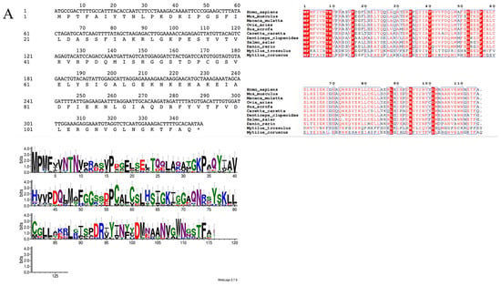
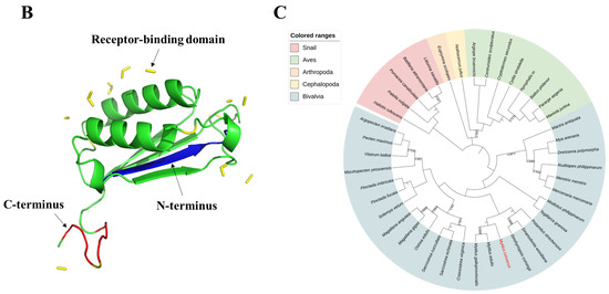
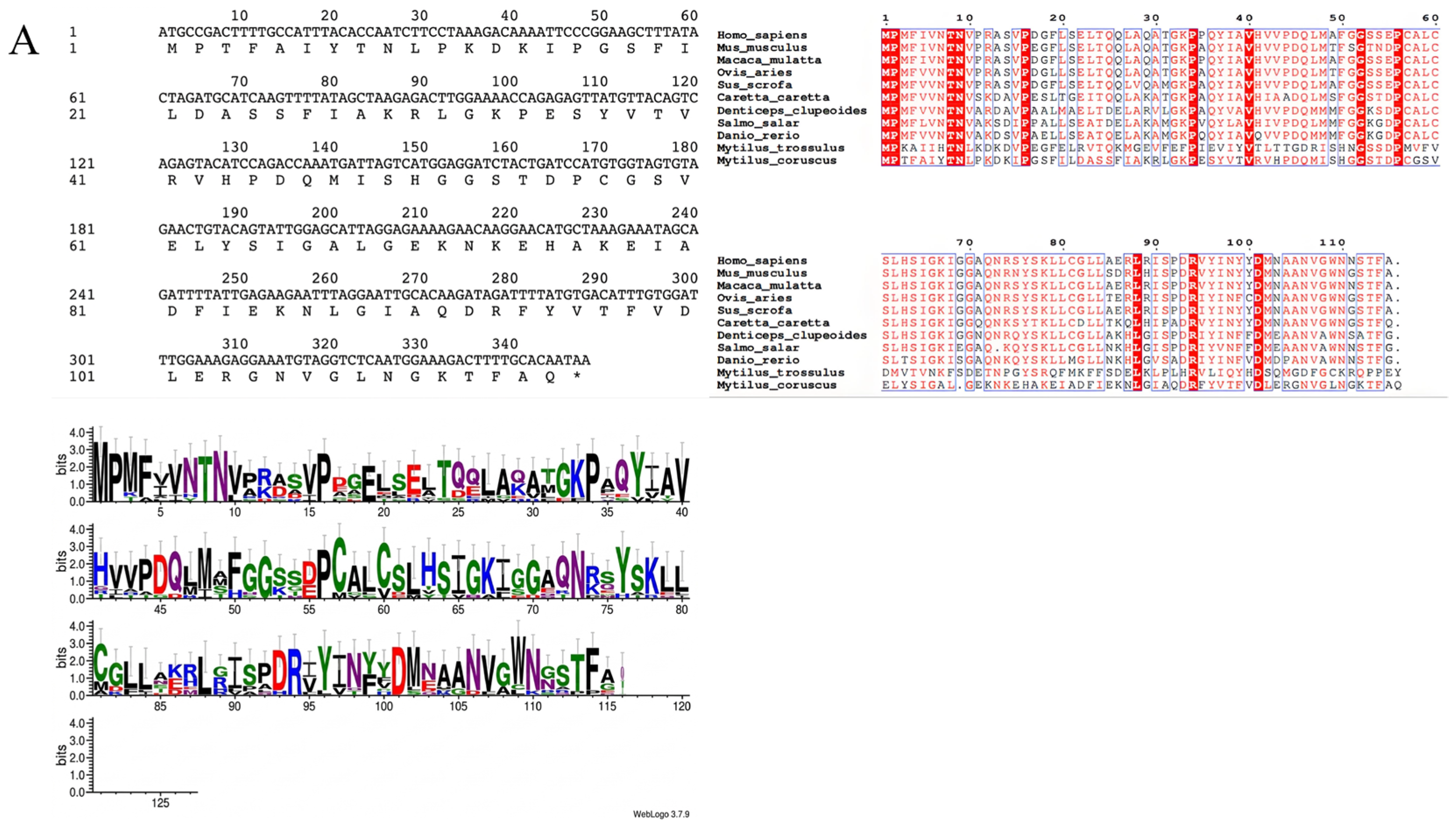
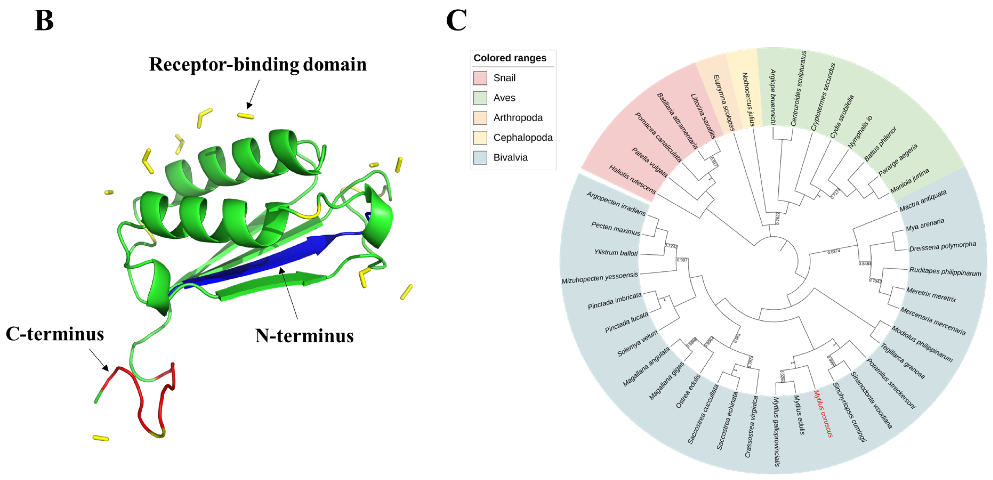
The full-length sequence of the McMIF gene was imported into DNAMAN for analysis, revealing an open reading frame (ORF) that encodes 115 amino acids. A comparison of the McMIF sequence with homologous MIF sequences from other species demonstrated a marked polarization in its conservation. Specifically, the conservation of key functional sites was exceptionally high, while the conservation of variable regions, particularly across different taxonomic groups, was notably reduced (Figure 1A). Structural prediction showed that McMIF consists of an N-terminal region and a C-terminal region, which together form a compact α/β mixed fold. Further structural analysis revealed that several surface residues in both regions were spatially aligned with the receptor-binding regions previously reported in other MIF proteins. Comparative analysis with canonical MIF structures further suggested that some amino acid residues located in the N-terminal and C-terminal regions may serve as potential interaction sites for the CD74 receptor (Figure 1B). Phylogenetic analysis revealed that McMIF clusters most closely with MIF from Mactra antiquata, Mya arenaria, and Ruditapes philippinarum. This group, in turn, forms a clade with MIFs from Modiolus philippinarum, Tegillarca granosa, Solemya velum, and Ylistrum balloti, all of which constitute a distinct branch within the Bivalvia class (Figure 1C).
Figure 1.
Molecular characteristics of McMIF. (A) The open reading frame (ORF) and deduced amino acid sequence of McMIF. Schematic representation of McMIF homologous sequence conservation generated using ESPript 3.0 and Weblogo 3.0. (B) Predicted three-dimensional structure of McMIF generated by SWISS-MODEL. (C) Phylogenetic analysis of the selected McMIF sequences performed using MEGA 11 software, with 5000 bootstrap replicates using the Neighbor-Joining method.
3.2. Expression Profile and Immune Induction Analysis
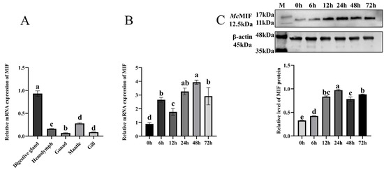
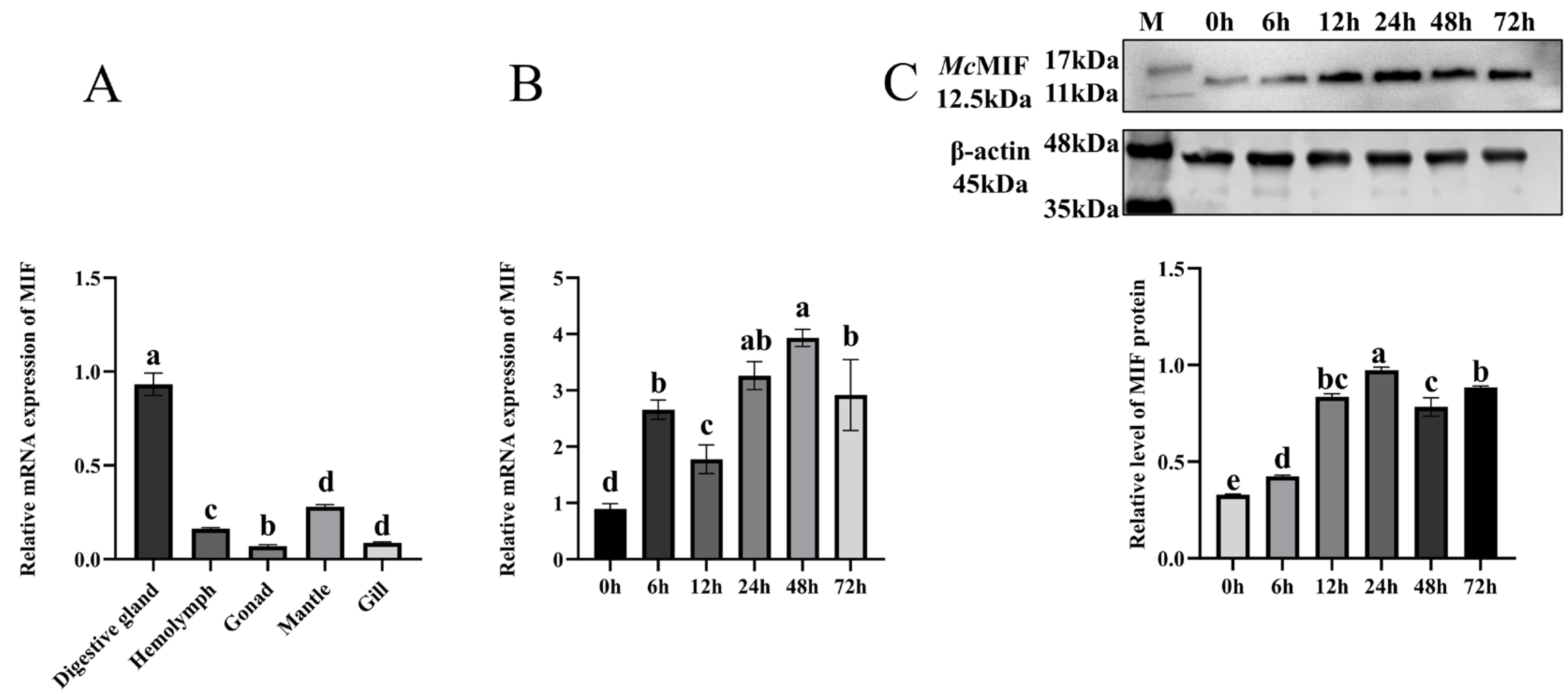
The expression levels of McMIF in the digestive gland, hemolymph, gonads, mantle, and gills were measured using RT-PCR. The results revealed that McMIF expression was highest in the digestive gland (Figure 2A). Changes in McMIF mRNA expression in the digestive gland of M. coruscus were also assessed following infection with V. alginolyticus. Relative to the unchallenged control, McMIF transcript abundance in the digestive gland was markedly elevated following infection, with a peak expression level observed at 48 h, showing a three-fold increase (Figure 2B). Western blot analysis revealed a tendency toward increased McMIF protein levels in the digestive gland following V. alginolyticus infection, with higher expression observed at 24 and 72 h (Figure 2C).
Figure 2.
Expression of McMIF. (A) Tissue-specific distribution of McMIF transcript levels across different tissues, as determined by RT-PCR. (B) Time-course analysis of McMIF transcript abundance in the digestive gland at multiple intervals (0, 6, 12, 24, 48, and 72 h) after V. alginolyticus challenge, as quantified by RT-PCR. (C) Immunoblot detection of McMIF protein levels in the digestive gland at designated intervals (0, 6, 12, 24, 48, and 72 h) after V. alginolyticus challenge. Quantification of relative McMIF protein levels is presented. M: Marker. Values are presented as mean ± SD (n = 3), with error bars representing the standard deviation. Groups labeled with different letters indicate statistically significant differences (p < 0.05), as determined by one-way ANOVA followed by Tukey’s multiple comparisons test.
3.3. Analysis of m6A Modification Levels of McMIF Post-Infection
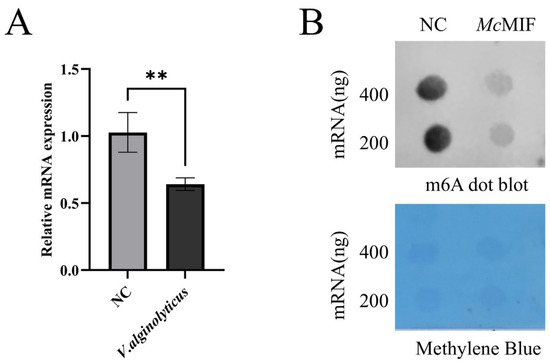
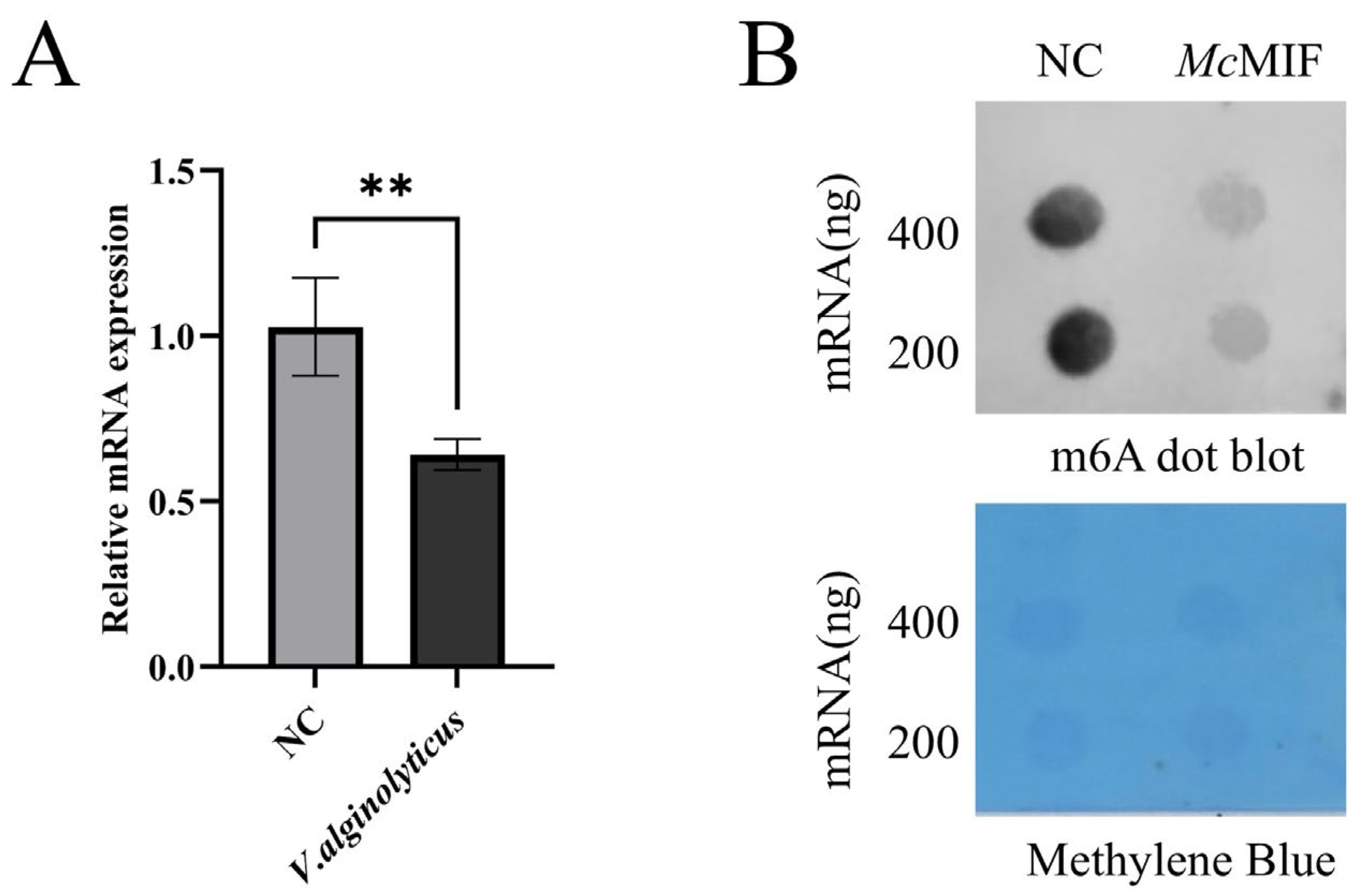
MeRIP-qPCR results demonstrated that the m6A methylation level of McMIF mRNA was markedly reduced at 24 h post-V. alginolyticus challenge relative to the unchallenged control group (Figure 3A). Subsequently, Dot blot analysis was employed to assess changes in the m6A modification levels of McMIF. The analysis indicated an attenuated staining signal on the PVDF membrane (Figure 3B).
Figure 3.
m6A Methylation Dynamics in Response to V. alginolyticus Challenge. (A) Relative m6A methylation levels of McMIF transcripts in the digestive gland of the non-infected control group (NC) and the group at 24 h after V. alginolyticus infection were determined by RT-PCR. (B) McMIF m6A levels in the digestive gland following V. alginolyticus infection, as detected by RNA Dot blot analysis. Methylene blue staining was used as a loading control. Values are presented as mean ± SD (n = 3), with error bars representing the standard deviation. Statistical differences between groups were evaluated using an unpaired Student’s t-test. ** indicates statistically significant differences at p < 0.005.
3.4. Impact of McMIF Gene Silencing on Cellular Apoptosis
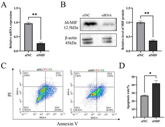
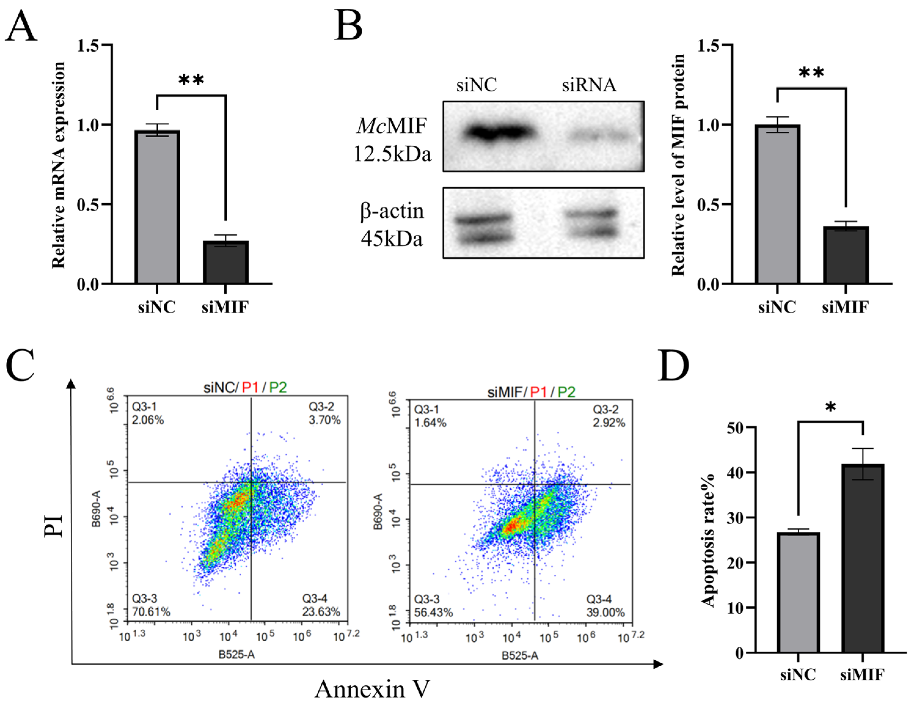
siRNA was used to silence McMIF expression in infected individuals of M. coruscus. After 24 h of transfection with McMIF-specific siRNA, McMIF expression was reduced by 70.33% compared to the siNC group (Figure 4A). Western blot analysis further confirmed the successful silencing of the McMIF gene by siRNA (Figure 4B). Furthermore, flow cytometric analysis was utilized to evaluate apoptotic levels in digestive gland cells following siMIF treatment. The findings demonstrated that, at 24 h post-transfection, the apoptotic rate in the siMIF group (39.00 ± 2.92%) was markedly elevated compared to that of the siNC group (23.63 ± 3.70%) (Figure 4C,D).
Figure 4.
Apoptosis analysis following McMIF knockdown (A) McMIF expression is significantly downregulated by siMIF. (B) Immunoblot assessment of McMIF protein abundance across experimental groups (siNC, siMIF) upon V. alginolyticus challenge. The relative expression level of McMIF protein is shown. (C) Representative flow cytometry dot plots illustrating apoptotic rates in digestive gland cells following 24 h transfection with siNC or siMIF under LPS stimulation. (D) Quantitative comparison of apoptosis levels across experimental groups. Values are presented as mean ± SD (n = 3), with error bars representing the standard deviation. Statistical differences between groups were evaluated using an unpaired Student’s t-test. * indicates statistically significant differences at p < 0.05; ** indicates statistically significant differences at p < 0.005.
4. Discussion
MIF, as a crucial immune regulatory factor, regulates the activation of inflammatory signaling pathways and the inhibition of cell migration, thereby significantly influencing the balance between innate and adaptive immunity and playing a critical role in the immune homeostasis of various organisms [35]. Based on previously obtained direct RNA sequencing data, McMIF-related sequence information was identified and retrieved [36]. Bioinformatic analysis revealed that McMIF encodes a polypeptide consisting of 115 amino acids, and its overall structural framework is highly similar to that of vertebrate MIF proteins. Several conserved key residues and predicted functional regions associated with the MIF family were retained in McMIF, suggesting that it may preserve the fundamental structural features required for mediating immune-related activities. Similar structural conservation has also been reported in other molluscan MIF homologs, such as those from M. galloprovincialis [37], Pinctada fucata [38], and Ruditapes philippinarum [11], indicating that the MIF family has remained highly conserved during molluscan evolution. The online SMART analysis predicted the presence of a potential CD74-binding region in McMIF. In vertebrates, extracellular MIF can bind to CD74 and activate downstream signaling pathways associated with inflammation and immune responses [39,40]. However, to date, no typical CD74 homolog has been identified in M. coruscus. Therefore, whether M. coruscus possesses a mechanism similar to that in vertebrates, in which MIF binds to CD74 and activates related signaling pathways to participate in immune regulation, remains to be further validated by functional studies. Phylogenetic analysis showed that McMIF clusters with other molluscan MIF proteins into an independent clade, suggesting a high degree of conservation within mollusks, along with a certain level of lineage-specific divergence.
To clarify the biological function of McMIF in modulating innate immune defense mechanisms, we employed RT-PCR to study the expression dynamics of McMIF following infection with V. alginolyticus. Tissue distribution analysis demonstrated that McMIF is highly expressed in the digestive gland, mantle, and hemolymph, which is consistent with the important roles of these tissues in mussel immune defense. The digestive gland, as a major metabolic and immune organ, is enriched with hemolymph and participates in pathogen recognition and clearance. In Ruditapes philippinarum, the MIF homolog (RpMIF) is highly expressed in the digestive gland and hemolymph, and recombinant RpMIF protein has been shown to significantly enhance hemocyte phagocytic activity and regulate immune-related gene expression, indicating its important role in innate immunity in invertebrates [11]. The mantle, being directly exposed to the external environment, is involved in early immune surveillance. In Chlamys farreri, the MIF homolog (CfMIF) exhibits relatively high expression in the mantle and is significantly upregulated following stimulation with lipopolysaccharide, peptidoglycan, and β-glucan, suggesting its involvement in pathogen recognition and immune responses [13]. Following V. alginolyticus infection, McMIF exhibited a typical inducible expression pattern, characterized by early upregulation, reaching a peak at the intermediate stage, and subsequently declining. Western blot experiments further confirmed this observation. Such temporal variation suggests that McMIF may primarily function during the early to middle stages of antibacterial defense. Studies suggest that MIF induces excessive inflammatory responses either directly or by stimulating the secretion of pro-inflammatory cytokines [41]. In bivalve innate immunity, the expression of immune-related genes often displays stimulus-dependent and time-specific dynamics. For example, MIF homologs in Chlamys farreri are also significantly upregulated only at specific time points following immune stimulation [13]. Therefore, the lack of significant changes at some time points in this study may reflect temporal specificity of immune responses, tissue-specific differences, or the gradual restoration of homeostasis following the initial immune activation. This phenomenon is consistent with the adaptive strategy of bivalves to finely regulate immune responses in order to minimize the physiological burden caused by prolonged activation [42]. It should be noted that pooled samples were used in this study to minimize individual variability. Future studies with larger biological sample sizes (n > 3) are required to further validate and strengthen the reliability and robustness of the present findings.
The present study revealed that McMIF m6A modification levels were significantly decreased following V. alginolyticus infection. As a reversible RNA modification, m6A can influence RNA fate by regulating transcript stability, translation efficiency, splicing, or intracellular localization. In vertebrates, m6A regulatory mechanisms have been shown to participate in innate immune activation and the expression of inflammation-related genes [43,44]. Moreover, m6A reader proteins, such as YTHDF2, can promote the degradation of m6A-modified transcripts, whereas demethylation may enhance transcript stability under certain conditions [45,46]. Therefore, the decreased m6A modification level observed in McMIF may have potential implications for its post-transcriptional regulation. In recent years, the role of m6A modification in immune regulation in invertebrates has attracted increasing attention. Studies have shown that in marine invertebrates, m6A modification levels dynamically change under pathogen infection and participate in regulating innate immune responses. For instance, transcriptome-wide analyses in shrimp (Marsupenaeus japonicus) revealed that virus-induced changes in m6A methylation are closely associated with differential expression of metabolism-related genes, indicating that m6A-mediated regulation contributes to metabolic reprogramming and antiviral immune responses [47].
During infection, apoptosis serves as a key defense mechanism to limit pathogen spread [48]. Therefore, we performed flow cytometry analysis, which revealed that the apoptotic rate in the McMIF knockdown group was significantly higher compared to the control group. This suggests that MIF induces cell proliferation during physiological growth [49]. This is consistent with the well-established function of MIF as a survival factor in mammals. Schindler et al. [50] demonstrated using MIF knockout models that the absence of MIF significantly increases cellular sensitivity to apoptotic stimuli and reduces cell survival capacity. Similar observations have also been reported in various invertebrates. In other invertebrate species, dsRNA-mediated knockdown of MIF significantly reduces its transcription level and increases mortality, indicating its crucial role in immune regulation and maintenance of cell survival [51]. Therefore, the increased apoptosis observed after McMIF knockdown suggests that it may play a regulatory role in balancing immune defense and cell survival.
Despite the scarcity of dedicated studies on marine invertebrates, comparable investigations in higher organisms and cell culture systems offer a solid theoretical basis for the current work. For invertebrates such as bivalves, which lack an adaptive immune system, rapid and reversible post-transcriptional regulatory mechanisms are particularly important for early responses to environmental pathogens [52]. Further studies are needed to determine whether and how m6A regulatory factors influence McMIF mRNA stability or translation efficiency, and whether McMIF participates in immune regulation through receptor-mediated pathways analogous to those in vertebrates.
5. Conclusions
The present study demonstrates that McMIF is closely associated with the antibacterial immune response in M. coruscus and may be associated with the regulation of cell survival under infection conditions. More importantly, this study provides the first experimental evidence in marine invertebrates that the expression dynamics of MIF genes in response to pathogenic stimulation are accompanied by changes in RNA m6A modification. These findings not only expand the current understanding of the immunological functions of MIF in mollusks, but also suggest that epitranscriptomic regulation may play a crucial role in the innate immune responses of bivalves. These observations present promising opportunities for devising innovative disease control approaches in aquaculture practices and for facilitating the selective cultivation of bivalve varieties with enhanced tolerance to both environmental stressors and pathogenic threats.
Author Contributions
Conceptualization, B.S. and P.Q.; methodology, X.W. and X.S.; software, validation, formal analysis, X.W. and C.Y.; data curation, writing—original draft preparation, writing—review and editing, visualization, X.W.; supervision, B.S.; funding acquisition, P.Q. All authors have read and agreed to the published version of the manuscript.
Funding
This research was funded by the Technology Collaboration Projects of Zhejiang Provincial Department of Agriculture and Rural Affairs (Grant No. 2024SNJF057), and the Special Grant of Zhoushan for Breeding Aquatic Animals (Grant No. 2025Y001-2).
Institutional Review Board Statement
The animal study protocol was approved by the animal experimental ethical inspection, institutional animals care and use committee of Zhejiang Ocean University (protocol code 2026082 and date of approval 5 December 2025).
Data Availability Statement
The original contributions presented in this study are included in the article. Further inquiries can be directed to the corresponding author.
Acknowledgments
The authors would like to express their gratitude to Zhejiang Ocean University.
Conflicts of Interest
The authors declare no conflicts of interest.
References
- Kang, I.; Bucala, R. The immunobiology of MIF: Function, genetics and prospects for precision medicine. Nat. Rev. Rheumatol. 2019, 15, 427–437. [Google Scholar] [CrossRef] [Scilit]
- Zhu, W.-Y.; Jin, X.; Ma, Y.-C.; Liu, Z.-B. Correlations of MIF polymorphism and serum levels of MIF with glucocorticoid sensitivity of sudden sensorineural hearing loss. J. Int. Med. Res. 2020, 48, 300060519893870. [Google Scholar] [CrossRef] [Scilit]
- Kong, Y.-Z.; Chen, Q.; Lan, H.-Y. Macrophage migration inhibitory factor (MIF) as a stress molecule in renal inflammation. Int. J. Mol. Sci. 2022, 23, 4908. [Google Scholar] [CrossRef] [Scilit]
- Wu, B.; Nakamura, A. Deep insight into the role of MIF in spondyloarthritis. Curr. Rheumatol. Rep. 2022, 24, 269–278. [Google Scholar] [CrossRef] [Scilit] [PubMed]
- Pressley, K.R.; Naseem, Y.; Nalawade, S.; Forsthuber, T.G. The distinct functions of MIF in inflammatory cardiomyopathy. Front. Immunol. 2025, 16, 1544484. [Google Scholar] [CrossRef] [Scilit]
- Nasiri, E.; Sankowski, R.; Dietrich, H.; Oikonomidi, A.; Huerta, P.T.; Popp, J.; Al-Abed, Y.; Bacher, M. Key role of MIF-related neuroinflammation in neurodegeneration and cognitive impairment in alzheimer’s disease. Mol. Med. 2020, 26, 34. [Google Scholar] [CrossRef] [Scilit]
- Baeza Garcia, A.; Pierce, R.J.; Gourbal, B.; Werkmeister, E.; Colinet, D.; Reichhart, J.-M.; Dissous, C.; Coustau, C. Involvement of the cytokine MIF in the snail host immune response to the parasite schistosoma mansoni. PLoS Pathog. 2010, 6, e1001115. [Google Scholar] [CrossRef] [Scilit] [PubMed]
- Jeong, H.; Lee, S.-Y.; Seo, H.; Kim, B.-J. Recombinant Mycobacterium smegmatis delivering a fusion protein of human macrophage migration inhibitory factor (MIF) and IL-7 exerts an anticancer effect by inducing an immune response against MIF in a tumor-bearing mouse model. J. Immunother. Cancer 2021, 9, e003180. [Google Scholar] [CrossRef] [Scilit] [PubMed]
- Bozza, M.T.; Martins, Y.C.; Carneiro, L.A.M.; Paiva, C.N. Macrophage migration inhibitory factor in protozoan infections. J. Parasitol. Res. 2012, 2012, 413052. [Google Scholar] [CrossRef] [Scilit]
- Huang, W.-S.; Duan, L.-P.; Huang, B.; Wang, K.-J.; Zhang, C.-L.; Jia, Q.; Nie, P.; Wang, T. Macrophage migration inhibitory factor (MIF) family in arthropods: Cloning and expression analysis of two MIF and one D-dopachrome tautomerase (DDT) homologues in mud crabs, scylla paramamosain. Fish Shellfish Immunol. 2016, 50, 142–149. [Google Scholar] [CrossRef] [Scilit]
- Wang, D.; Yang, D.; Wang, Q.; Zhao, Y.; Li, C.; Wei, Q.; Han, Y.; Zhao, J. Two macrophage migration inhibitory factors (MIFs) from the clam ruditapes philippinarum: Molecular characterization, localization and enzymatic activities. Fish Shellfish Immunol. 2018, 78, 158–168. [Google Scholar] [CrossRef] [Scilit] [PubMed]
- Roger, T.; David, J.; Glauser, M.P.; Calandra, T. MIF regulates innate immune responses through modulation of toll-like receptor 4. Nature 2001, 414, 920–924. [Google Scholar] [CrossRef] [Scilit]
- Li, F.; Huang, S.; Wang, L.; Yang, J.; Zhang, H.; Qiu, L.; Li, L.; Song, L. A macrophage migration inhibitory factor like gene from scallop chlamys farreri: Involvement in immune response and wound healing. Dev. Comp. Immunol. 2011, 35, 62–71. [Google Scholar] [CrossRef] [Scilit] [PubMed]
- Dickerhof, N.; Schindler, L.; Bernhagen, J.; Kettle, A.J.; Hampton, M.B. Macrophage migration inhibitory factor (MIF) is rendered enzymatically inactive by myeloperoxidase-derived oxidants but retains its immunomodulatory function. Free Radic. Biol. Med. 2015, 89, 498–511. [Google Scholar] [CrossRef] [Scilit]
- Karlsson, I.; Samuelsson, K.; Simonsson, C.; Stenfeldt, A.-L.; Nilsson, U.; Ilag, L.L.; Jonsson, C.; Karlberg, A.-T. The fate of a hapten—From the skin to modification of macrophage migration inhibitory factor (MIF) in lymph nodes. Sci. Rep. 2018, 8, 2895. [Google Scholar] [CrossRef] [Scilit]
- Mao, Y.; Jiang, F.; Xu, X.-J.; Zhou, L.-B.; Jin, R.; Zhuang, L.-L.; Juan, C.-X.; Zhou, G.-P. Inhibition of IGF2BP1 attenuates renal injury and inflammation by alleviating m6A modifications and E2F1/MIF pathway. Int. J. Biol. Sci. 2023, 19, 593–609. [Google Scholar] [CrossRef] [Scilit]
- Kassaar, O.; Pereira Morais, M.; Xu, S.; Adam, E.L.; Chamberlain, R.C.; Jenkins, B.; James, T.D.; Francis, P.T.; Ward, S.; Williams, R.J.; et al. Macrophage migration inhibitory factor is subjected to glucose modification and oxidation in alzheimer’s disease. Sci. Rep. 2017, 7, 42874. [Google Scholar] [CrossRef] [Scilit]
- Mathlin, J.; Le Pera, L.; Colombo, T. A census and categorization method of epitranscriptomic marks. Int. J. Mol. Sci. 2020, 21, 4684. [Google Scholar] [CrossRef] [Scilit]
- Desrosiers, R.; Friderici, K.; Rottman, F. Identification of methylated nucleosides in messenger RNA from novikoff hepatoma cells. Proc. Natl. Acad. Sci. USA 1974, 71, 3971–3975. [Google Scholar] [CrossRef] [Scilit]
- Zhou, Y.; Kong, Y.; Fan, W.; Tao, T.; Xiao, Q.; Li, N.; Zhu, X. Principles of RNA methylation and their implications for biology and medicine. Biomed. Pharmacother. 2020, 131, 110731. [Google Scholar] [CrossRef] [Scilit] [PubMed]
- Roundtree, I.A.; Evans, M.E.; Pan, T.; He, C. Dynamic RNA modifications in gene expression regulation. Cell 2017, 169, 1187–1200. [Google Scholar] [CrossRef] [Scilit] [PubMed]
- Wang, X.; Huang, W.; Wei, S.; Shang, Y.; Gu, H.; Wu, F.; Lan, Z.; Hu, M.; Shi, H.; Wang, Y. Microplastics impair digestive performance but show little effects on antioxidant activity in mussels under low pH conditions. Environ. Pollut. 2020, 258, 113691. [Google Scholar] [CrossRef] [Scilit] [PubMed]
- Bei, Y.; Si, X.; Ma, W.; Qi, P.; Ye, Y. The role of NFAT5 in immune response and antioxidant defense in the thick-shelled mussel (Mytilus coruscus). Animals 2025, 15, 726. [Google Scholar] [CrossRef] [Scilit]
- Zannella, C.; Mosca, F.; Mariani, F.; Franci, G.; Folliero, V.; Galdiero, M.; Tiscar, P.G.; Galdiero, M. Microbial diseases of bivalve mollusks: Infections, immunology and antimicrobial defense. Mar. Drugs 2017, 15, 182. [Google Scholar] [CrossRef] [Scilit] [PubMed]
- Zhao, J.; Dong, Z.; Zhu, L.; Song, W.; Qi, P. An interleukin-17 isoform from thick shell mussel mytilus coruscus serves as a mediator of inflammatory response. Molecules 2023, 28, 1806. [Google Scholar] [CrossRef] [Scilit]
- Robert, X.; Gouet, P. Deciphering key features in protein structures with the new ENDscript server. Nucleic Acids Res. 2014, 42, W320–W324. [Google Scholar] [CrossRef] [Scilit]
- Crooks, G.E.; Hon, G.; Chandonia, J.-M.; Brenner, S.E. WebLogo: A sequence logo generator: Figure 1. Genome Res. 2004, 14, 1188–1190. [Google Scholar] [CrossRef] [Scilit]
- Letunic, I.; Doerks, T.; Bork, P. SMART: Recent updates, new developments and status in 2015. Nucleic Acids Res. 2015, 43, D257–D260. [Google Scholar] [CrossRef] [Scilit]
- Waterhouse, A.; Bertoni, M.; Bienert, S.; Studer, G.; Tauriello, G.; Gumienny, R.; Heer, F.T.; de Beer, T.A.P.; Rempfer, C.; Bordoli, L.; et al. SWISS-MODEL: Homology modelling of protein structures and complexes. Nucleic Acids Res. 2018, 46, W296–W303. [Google Scholar] [CrossRef] [Scilit]
- Sayers, E.W.; Beck, J.; Bolton, E.E.; Brister, J.R.; Chan, J.; Comeau, D.C.; Connor, R.; DiCuccio, M.; Farrell, C.M.; Feldgarden, M.; et al. Database resources of the national center for biotechnology information. Nucleic Acids Res. 2024, 52, D33–D43. [Google Scholar] [CrossRef] [Scilit]
- Sievers, F.; Higgins, D.G. Clustal omega for making accurate alignments of many protein sequences. Protein Sci. 2018, 27, 135–145. [Google Scholar] [CrossRef] [Scilit] [PubMed]
- Tamura, K.; Stecher, G.; Kumar, S. MEGA11: Molecular evolutionary genetics analysis version 11. Mol. Biol. Evol. 2021, 38, 3022–3027. [Google Scholar] [CrossRef] [Scilit]
- Li, D.; Guo, M.; Lv, Z.; Shao, Y.; Liang, W.; Li, C. METTL3 activates PERK-eIF2α dependent coelomocyte apoptosis by targeting the endoplasmic reticulum degradation-related protein SEL1L in echinoderms. Biochim. Biophys. Acta BBA Gene Regul. Mech. 2023, 1866, 194927. [Google Scholar] [CrossRef] [Scilit]
- Livak, K.J.; Schmittgen, T.D. Analysis of relative gene expression data using real-time quantitative PCR and the 2−ΔΔCT method. Methods 2001, 25, 402–408. [Google Scholar] [CrossRef] [Scilit] [PubMed]
- Bernhagen, J.; Calandra, T.; Mitchell, R.A.; Martin, S.B.; Tracey, K.J.; Voelter, W.; Manogue, K.R.; Cerami, A.; Bucala, R. MIF is a pituitary-derived cytokine that potentiates lethal endotoxaemia. Nature 1993, 365, 756–759. [Google Scholar] [CrossRef] [Scilit] [PubMed]
- Li, H.; Fu, P.; Guo, B.; Liao, Z.; Yan, X.; Qi, P. Nanopore direct RNA sequencing deciphering the m6A epitranscriptome and its role in the innate immune response of the mussel mytilus coruscus: A prospective study of RNA modification in marine molluscs. Aquaculture 2025, 598, 742040. [Google Scholar] [CrossRef] [Scilit]
- Parisi, M.-G.; Toubiana, M.; Mangano, V.; Parrinello, N.; Cammarata, M.; Roch, P. MIF from mussel: Coding sequence, phylogeny, polymorphism, 3D model and regulation of expression. Dev. Comp. Immunol. 2012, 36, 688–696. [Google Scholar] [CrossRef] [Scilit]
- Cui, S.; Zhang, D.; Jiang, S.; Pu, H.; Hu, Y.; Guo, H.; Chen, M.; Su, T.; Zhu, C. A macrophage migration inhibitory factor like oxidoreductase from pearl oyster pinctada fucata involved in innate immune responses. Fish Shellfish Immunol. 2011, 31, 173–181. [Google Scholar] [CrossRef] [Scilit]
- Leng, L.; Metz, C.N.; Fang, Y.; Xu, J.; Donnelly, S.; Baugh, J.; Delohery, T.; Chen, Y.; Mitchell, R.A.; Bucala, R. MIF signal transduction initiated by binding to CD74. J. Exp. Med. 2003, 197, 1467–1476. [Google Scholar] [CrossRef] [Scilit]
- Osipyan, A.; Chen, D.; Dekker, F.J. Epigenetic regulation in macrophage migration inhibitory factor (MIF)-mediated signaling in cancer and inflammation. Drug Discov. Today 2021, 26, 1728–1734. [Google Scholar] [CrossRef] [Scilit]
- Tahara, T.; Shibata, T.; Nakamura, M.; Yamashita, H.; Yoshioka, D.; Okubo, M.; Yonemura, J.; Maeda, Y.; Maruyama, N.; Kamano, T.; et al. Effect of polymorphisms of IL-17A, -17F and MIF genes on CpG island hyper-methylation (CIHM) in the human gastric mucosa. Int. J. Mol. Med. 2009, 24, 563–569. [Google Scholar] [CrossRef] [Scilit]
- Liu, Z.; Li, M. The Neuroendocrine-Immune Regulation in Response to Environmental Stress in Marine Bivalves. Front. Physiol. 2018, 9, 01456. [Google Scholar] [CrossRef] [Scilit]
- Winkler, R.; Gillis, E.; Lasman, L.; Safra, M.; Geula, S.; Soyris, C.; Nachshon, A.; Tai-Schmiedel, J.; Friedman, N.; Le-Trilling, V.T.K.; et al. m6A modification controls the innate immune response to infection by targeting type I interferons. Nat. Immunol. 2019, 20, 173–182. [Google Scholar] [CrossRef] [Scilit]
- Wang, H.; Hu, X.; Huang, M.; Liu, J.; Gu, Y.; Ma, L.; Zhou, Q.; Cao, X. Mettl3-mediated mRNA m6A methylation promotes dendritic cell activation. Nat. Commun. 2019, 10, 1898. [Google Scholar] [CrossRef] [Scilit]
- Wang, L.; Weichselbaum, R.R.; He, C. N6 -methyladenosine reader YTHDF2 in cell state transition and antitumor immunity. RNA 2025, 31, 395–401. [Google Scholar] [CrossRef] [Scilit]
- Gao, Z.; Zha, X.; Li, M.; Xia, X.; Wang, S. Insights into the m6A demethylases FTO and ALKBH5: Structural, biological function, and inhibitor development. Cell Biosci. 2024, 14, 108–127. [Google Scholar] [CrossRef] [Scilit] [PubMed]
- Sun, X.; Chen, Y.-L.; Xin, F.; Zhang, S. Transcriptome-wide identification and analysis reveals m6A regulation of metabolic reprogramming in shrimp (Marsupenaeus japonicus) under virus infection. BMC Genom. 2024, 25, 1103. [Google Scholar] [CrossRef] [Scilit] [PubMed]
- Vu, A.; Glassman, I.; Campbell, G.; Yeganyan, S.; Nguyen, J.; Shin, A.; Venketaraman, V. Host cell death and modulation of immune response against mycobacterium tuberculosis infection. Int. J. Mol. Sci. 2024, 25, 6255. [Google Scholar] [CrossRef] [Scilit] [PubMed]
- Mitchell, R.A.; Metz, C.N.; Peng, T.; Bucala, R. Sustained mitogen-activated protein kinase (MAPK) and cytoplasmic phospholipase A2 activation by macrophage migration inhibitory factor (MIF): Regulatory role in cell proliferation and glucocorticoid action. J. Biol. Chem. 1999, 274, 18100–18106. [Google Scholar] [CrossRef] [Scilit]
- Schindler, L.; Zwissler, L.; Krammer, C.; Hendgen-Cotta, U.; Rassaf, T.; Hampton, M.B.; Dickerhof, N.; Bernhagen, J. Macrophage migration inhibitory factor inhibits neutrophil apoptosis by inducing cytokine release from mononuclear cells. J. Leukoc. Biol. 2021, 110, 893–905. [Google Scholar] [CrossRef] [Scilit]
- Liu, S.; Ladera-Carmona, M.J.; Poranen, M.M.; Van Bel, A.J.E.; Kogel, K.-H.; Imani, J. Evaluation of dsRNA delivery methods for targeting macrophage migration inhibitory factor MIF in RNAi-based aphid control. J. Plant Dis. Prot. 2021, 128, 1201–1212. [Google Scholar] [CrossRef] [Scilit]
- Ge, Y.; Chen, R.; Ling, T.; Liu, B.; Huang, J.; Cheng, Y.; Lin, Y.; Chen, H.; Xie, X.; Xia, G.; et al. Elevated WTAP promotes hyperinflammation by increasing m6A modification in inflammatory disease models. J. Clin. Invest. 2024, 134, e177932. [Google Scholar] [CrossRef] [Scilit]

Disclaimer/Publisher’s Note: The statements, opinions and data contained in all publications are solely those of the individual author(s) and contributor(s) and not of MDPI and/or the editor(s). MDPI and/or the editor(s) disclaim responsibility for any injury to people or property resulting from any ideas, methods, instructions or products referred to in the content. |
© 2026 by the authors. Licensee MDPI, Basel, Switzerland. This article is an open access article distributed under the terms and conditions of the Creative Commons Attribution (CC BY) license.