You are currently viewing a new version of our website. To view the old version click .

Journal of Imaging, Volume 5, Issue 7

July 2019 - 4 articles

Cover Story: 
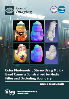
This seven-band color photometric stereo estimates the shape from a single-shot image. The target object is illuminated with seven light sources with wavelengths of 750nm, 632nm, 610nm, 550nm, 520nm, 470nm, and 430nm. The illuminated object is captured by a seven-band multispectral camera. Conventional three-band color photometric stereo suffers from the problem that the target object should be white, though this method overcomes the problem and estimates the surface normal of multicolored objects. The first column of this image shows the target object, the second column is the pseudocolor representation of the estimatedsurface normal, and the third column is the shape integrated from thesurface normal. View this paper
  • Issues are regarded as officially published after their release is announced to the table of contents alert mailing list .
  • You may sign up for email alerts to receive table of contents of newly released issues.
  • PDF is the official format for papers published in both, html and pdf forms. To view the papers in pdf format, click on the "PDF Full-text" link, and use the free Adobe Reader to open them.

Articles (4)

  • Article
  • Open Access
13 Citations
5,646 Views
19 Pages

Relative humidity (RH) changes are a natural environmental effect that forces organic materials to a constant cycle of achieving equilibrium. The present work is part of an ongoing research based on the hypothesis that the inevitable deleterious effe...

  • Article
  • Open Access
9 Citations
7,481 Views
29 Pages

One of the main problems faced by the photometric stereo method is that several measurements are required, as this method needs illumination from light sources from different directions. A solution to this problem is the color photometric stereo meth...

  • Article
  • Open Access
8 Citations
6,851 Views
12 Pages

Total internal reflection fluorescence microscopy with polarized excitation (P-TIRF) can be used to image nanoscale curvature phenomena in live cells. We used P-TIRF to visualize rat basophilic leukemia cells (RBL-2H3 cells) primed with fluorescent a...

  • Editorial
  • Open Access
4,439 Views
4 Pages

In recent years, following the tremendous growth of the Web, extremely large amounts of digital multimedia content are being produced every day and are shared online mainly through several newly emerged channels, such as social networks [...]

Get Alerted

Add your email address to receive forthcoming issues of this journal.

XFacebookLinkedIn
J. Imaging - ISSN 2313-433X