1. Introduction
Amylases are enzymes secreted by animals, plants, and microorganisms that catalyze the hydrolysis of starch [
1]. They are divided into three different groups as α, β, and γ [
2]. Among these, α-amylase is the most extensively used form in the detergent, paper, and food industries. Amylase is used especially in clear-type fruit juice production processes in the food industry. Juice clarity represents a key quality parameter, particularly for fruits such as apples [
3].
In order to meet the demand for amylase enzyme used in industry, studies on recombinant production are being carried out using expression systems such as
E. coli,
Bacillus lichenifromis,
Saccharomyces cerevisiae, and
P. pastoris [
4,
5,
6,
7,
8,
9,
10]. Microorganisms, particularly bacteria, fungi, and yeasts, constitute the primary biological sources of α-amylase.
A. niger is frequently selected as a gene source for recombinant amylase production due to its ability to naturally produce high levels of stable and industrially relevant amylases. Enzymes derived from this organism are typically well-suited for industrial conditions, including a broad range of pH and temperature stability [
11]. Furthermore, the genetic sequences of amylase genes from
A. niger are well characterized, facilitating their cloning and heterologous expression in different host systems. In addition, the commercially available enzyme (Amylase AG 300 L, Novozymes, Bagsværd, Denmark) is produced from
A. niger; therefore, in this study,
A. niger was selected as the genetic source for amylase enzyme production.
A. niger α-amylase consists of a total of 567 amino acids. Its approximate molecular weight is around 58 kDa. Numerous studies have demonstrated the suitability of
A. niger as a genetic source for recombinant α-amylase production, particularly in yeast-based expression systems. In
P. pastoris, the methanol-inducible alcohol oxidase I (
AOX1) promoter is commonly used for recombinant amylase production [
8,
12].
P. pastoris is a methylotrophic yeast that can grow rapidly in relatively inexpensive media, similar to bacterial systems. However, as a eukaryotic host, it offers additional advantages by enabling post-translational modifications, including polypeptide folding, disulfide bond formation, and glycosylation, which are often required for functional expression of eukaryotic proteins. This makes the
Pichia expression system advantageous over bacteria for recombinant protein production. In addition, the availability of vectors harbouring strong promoters such as
AOX1 and the constitutive glyceraldehyde-3-phosphate dehydrogenase (
GAP) promoters provides great flexibility and convenience in host-vector design for recombinant protein production.
AOX1 and
GAP promoters of
P. pastoris are frequently used promoters. Their unique strengths and regulation mechanisms provide the host organism with the ability to express foreign proteins. Although the main goal in recombinant protein production is to obtain maximum protein in minimum time, sometimes weaker promoters are preferred to ensure the correct folding of proteins. Therefore, the discovery of alternative promoters and the study of their regulation mechanisms remain ongoing. The
P. pastoris alcohol dehydrogenase (ADH2) gene, which shares 74% sequence homology to the
S. cerevisiae ADH gene, has been characterized, and its promoter region has been proposed as an alternative to the
AOX1 and
GAP promoters [
13,
14]. In a 5 L bioreactor study, the native
ADH2 promoter achieved an enzyme activity of 3725 U/mL, whereas
AOX1 and
GAP promoters resulted in 2095 U/mL and 580 U/mL, respectively. The authors demonstrate that the
ADH2 promoter can drive effective protein expression under 30 °C, pH 5, and 30% dissolved oxygen [
13]. Subsequent studies have focused on modifying this promoter to enhance its performance. In particular, the regulatory regions of the
ADH2 promoter were identified, and five synthetic ADH2 promoter variants were generated by selective deletion or insertion of these elements [
15]. These synthetic promoters were tested for ethanol-induced production of extracellular xylanase at the shake flask level and were shown to increase xylanase production at rates ranging from 165% to 200% of the native
ADH2 promoter. Among these variants, the synthetic
ADH2 promoter 5 (SNT5) exhibited the highest activity and was further validated at the fermentor scale, demonstrating its suitability for industrial processes [
15].
Based on these previous studies, the synthetic
ADH2 promoter SNT5 represents a promising alternative to traditional promoters [
13,
15]. It provides advantages such as methanol-free expression and a high-yield product. This makes them particularly valuable for optimizing recombinant protein production in different industrial and experimental settings. Therefore, synthetic promoters can be considered as alternatives to conventional promoters in
P. pastoris.
Building on these findings, the present study aimed to develop an expression system for the extracellular production of A. niger α-amylase under the control of the ethanol-inducible SNT5 promoter in P. pastoris. The recombinant expression vector was introduced into the MK155-PDI strain, which overexpresses protein disulfide isomerase (PDI) to support correct protein folding and secretion. Recombinant α-amylase production was subsequently evaluated at the 5 L bioreactor scale, and the biochemical properties and potential application of the produced enzyme were investigated. To the best of our knowledge, this is the first report of A. niger α-amylase expression in P. pastoris driven by an ethanol-inducible synthetic promoter.
2. Materials and Methods
2.1. Strains and Culture Media
P. pastoris MK115-PDI strain [
14] and
E. coli NEB5-α strains were used for recombinant protein expression and cloning studies, respectively. For routine cultivation and strain development of
P. pastoris, YPD media (1% yeast extract, 2% peptone, 2% glucose); BMGY (2% peptone, 1% yeast extract, 1% glycerol, 1.34% YNB, 4 × 10
−5% biotin and 100 mM phosphate buffer pH 7.0); BMEY (10 g/L yeast extract, 20 g/L soytone, 13.4 g/L YNB, 4 × 10
−5% biotin, 10% pure ethanol and 0.1 M potassium phosphate buffer pH 6.0) were used. For the cultivation of
E. coli cells, LB Miller (0.5% yeast extract, 1% peptone, and 1% NaCl) and/or LB Lennox medium (1% peptone, 0.5% yeast extract, and 0.5% NaCl) were employed. Appropriate antibiotics were added to both liquid and solid media according to the resistance gene on the plasmid used.
2.2. Gene Source and Expression Vector Construction
A. niger was used as a gene source, and a codon-optimized DNA region for
P. pastoris was provided in the pUC57 cloning vector (GenScript, Piscataway, NJ, USA). The pUC57-AnAMY plasmid and pSNT5α vector [
15] were digested with
XhoI-
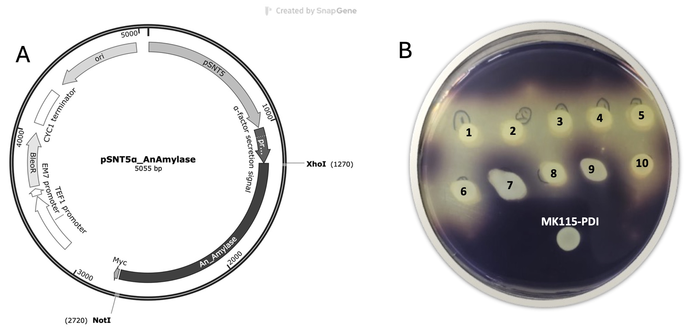
NotI cloning enzymes and then extracted from the agarose gel. Ligation was performed to create the pSNT5α-AnAMY expression vector, and the transformant cells were selected by plating on LB-Lenox agar plates containing zeocin containing. Plasmid DNA isolation was performed from the selected transformant cells, and the plasmid was verified by digesting with restriction enzymes. The confirmed plasmid was linearized with the
BsiWI enzyme and transformed into the competent
P. pastoris MK115-PDI strain by electroporation (Eppendorf SE, Hamburg, Germany). YPD agar plates containing 100 μg/mL and 500 μg/mL zeocin were used to select transformant cells.
2.3. Genomic DNA Isolation
The selected clones were inoculated in YPD broth and incubated overnight for genomic DNA isolation. 2 mL of culture was taken, and the yeast cells were precipitated. 200 μL of yeast cell lysis buffer (2% Triton X-100, 1% SDS, 0.1 M NaCl, 10 mM Tris HCl (pH 8), 1 mM EDTA (pH 8)) was added to dissolve the cells, and the mixture was then frozen at −80 °C for at least 30 min. The frozen mixture was incubated at 95 °C for 2 min and vortexed. After this freeze–thaw process was repeated three times, 200 μL of chloroform was added to the mixture and vortexed again for 2 min. Phase separation was then achieved by centrifugation at 20,000× g for 5 min. Following centrifugation, the upper phase was transferred to a new tube containing 400 μL of cold ethanol and kept at room temperature for 5 min to precipitate the gDNA. The samples were centrifuged again, and the mixture was washed with 500 μL of 70% ethanol. The remaining ethanol was removed using an evaporator (Eppendorf Concantrator Plus-5301, Hamburg, Germany). The obtained gDNAs were dissolved in 20 μL elution buffer (TE buffer: 10 mM Tris, pH 8.5), and 1 μL RNAse enzyme (RNase A, DNase, and protease-free, Thermo Scientific™, Waltham, MA, USA) was added to remove RNAs and kept at 37 °C for 1 h.
2.4. Determination of Gene Copy Number
Gene copy number of the transformant cells was determined by qPCR. To amplify the SNT5 promoter region and actin gene in the PCR reaction, primers ADH2prom-Frt: CCACCCCTCCCCAATCTC and ADH2prom-Rrt: AGCTAGTAGCTGATGGAAGAAGG, ActinrtF: GCTTTGTTCCACCCATCTGT, and ActinrtR: TGCATACGCTCAGCAATACC were used, respectively. The reaction mixture was prepared according to the Maxima SYBR Green/ROX Qpcr master mix (Thermo Fisher Scientific, Waltham, MA, USA) protocol and after the initial denaturation step at 95 °C for 10 min, the qPCR reaction was completed by repeating the steps of denaturation at 95 °C for 15 s, annealing at 60 °C for 30 s and extension at 72 °C for 30 s 40 times. The gene copy number was determined by relative quantification according to the 2
−ΔΔCt method as described before [
16].
2.5. Agar Plate Assay
In order to screen α-amylase enzyme activity in the transformant yeast strains, a Lugol iodine assay was employed. Firstly, the clones were developed in YPD broth until they reached 12–13 OD600, and then transferred to BMGY pH 6.0 medium. The developed culture was plated on ME agar (0.34% yeast nitrogen base, 4 × 10⁻⁵% biotin, 1% ethanol, 1.5% agar) plates containing 0.4% starch and incubated at 28 °C for 2 days. Since the SNT5 promoter used in protein production is an ethanol-inducible promoter, 100 μL of ethanol was added to the lids of the plates at 24 and 48 h. At the end of the second day, 10 mL of 5% Lugol solution was added to the developing colonies, and a clear zone formation was observed around the colony to confirm the amylase activity.
2.6. Expression of Recombinant Amylase in a Shake Flask
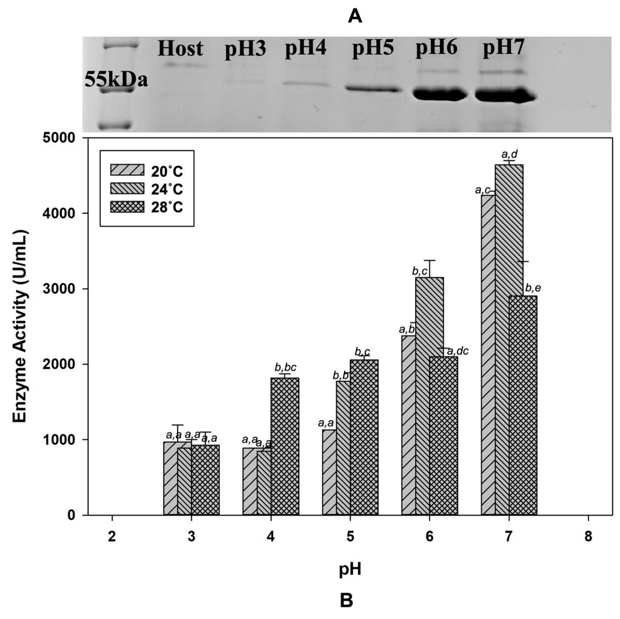
Clones containing a single copy expression cassette were inoculated into 3 mL YPD broth medium and incubated at 225 rpm and at 28 °C until the OD600 reached 6–8. Then, these cultures were inoculated into 30 mL of BMGY (pH 6) medium in 250 mL shake flasks with an initial OD600 of 0.1. The cells with approximately 10–13 OD600 in BMGY medium were centrifuged at +4 °C, 3000× g for 15 min. Supernatants were discarded, and the cell pellet obtained was suspended in 30 mL of BMEY medium with different pHs (pH 3.0, 4.0, 5.0, 6.0, 7.0) and incubation was continued for 96 h at different temperatures (20 °C, 24 °C, 28 °C) with a constant agitation rate. Since protein production was carried out under the control of the ethanol-inducible SNT5 promoter, ethanol was added every 12 h throughout protein expression to a final concentration of 1% in BMEY medium. Samples were taken during 120 h of production. Supernatant samples were passed through a 0.45 PES filter and stored at −80 °C to be used in total protein, enzyme activity, and SDS-PAGE analyses.
2.7. Expression of Recombinant Amylase in a 5 L Bioreactor
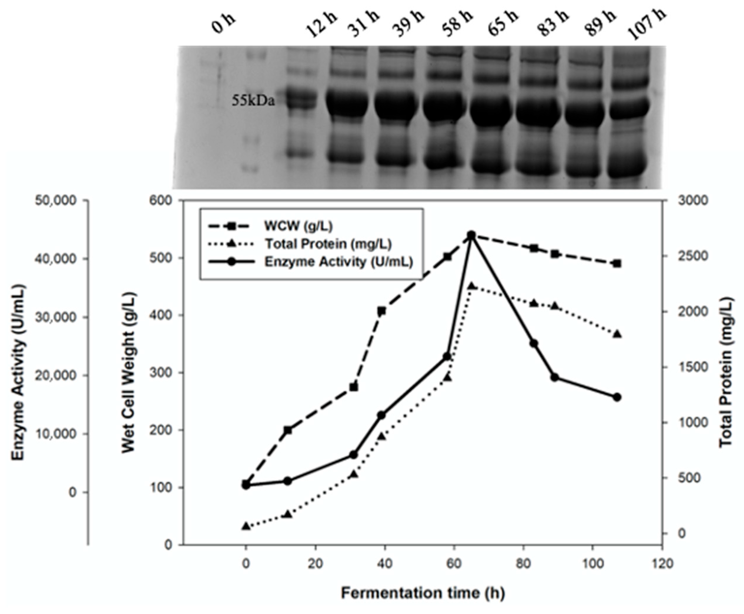
To produce recombinant α-amylase enzyme on a large scale, a 5 L bioreactor (Sartorius Stedim BIOSTAT B, Göttingen, Germany) was used. The frozen culture was transferred to 100 mL of BMGY (pH 6.0) medium and incubated for approximately 20 h at 28 °C at 225 rpm (OD600 nm approximately 10). This culture was used for inoculation into 2 L of FM22 medium (42.2 g/L KH2PO4, 5 g/L (NH4)2SO4, 0.79 g/L CaSO4, 14.9 g/L K2SO4, 4.13 g/L MgSO4·7H2O, 40 g/L glycerol, pH 6.0). After FM22 medium was autoclaved, its pH was adjusted to 6.0 using 25% (v/v) NH4OH and 1 mL of PTM4 salt (2 g/L CuSO4·5H2O, 0.08 g/L NaI, 3 g/L MnSO4·H2O, 0.2 g/L Na2MoO4·2H2O, 0.02 g/L H3BO3, 0.5 g/L CaSO4·2H2O, 0.5 g/L CoCl2, 7 g/L ZnCl2, 22 g/L FeSO4·7H2O, 0.2 g/L biotin, 1 mL H2SO4) was added for 1 L of medium. The batch phase continued for approximately 20 h and was completed with the peak of DO. After this carbon source depletion signal, the fed-batch phase was started with the solution containing 50% glucose, 10% ethanol, and 0.4% PTM4. During the fed-batch phase, the feed rate was kept constant at 7.3 mL/L/h during the first 2 h of fermentation, and the feed rate was gradually increased to 31.2 mL/L/h and kept constant at 31.2 mL/L/h until the end of fermentation. In the batch phase, temperature and pH were kept constant at 28 °C and 6.0, respectively, and at the beginning of the fed-batch phase, temperature and pH were adjusted to optimal protein production conditions (24 °C and pH 6.0). Fermentation pH was controlled with 25% (v/v) NH4OH. The DO level was controlled at 20% saturation by stirring, adding air (1.5 vvm), and pure oxygen. pH and oxygen concentration were determined with appropriate probes (Hamilton, Reno, NV, USA). Supernatants were collected at different times during fermentation, and these samples were analyzed for wet cell weight (WCW g/L), SDS-PAGE, total protein (g/L), and enzyme activity (U/L). All analyses were performed in triplicate.
2.8. Amylase Activity Measurement
The dinitrosalicylic acid (DNSA) colorimetric method was used to measure amylase activity. For the measurement of amylase enzyme activity, 200 µL of supernatant sample was incubated with 200 µL 1% soluble starch solution at 50 °C for 5 min. Then, 200 µL of the DNA solution was added and incubated at 95 °C for 15 min, and the samples were kept on ice for 3 min. After the addition of 1800 µL of pure water, the absorbance of the samples was measured at 540 nm. Different dilutions of the 0.3% (w/v) pure maltose solution were used as a standard. One enzyme unit for α-amylase was defined as the amount of enzyme required to release 1.0 mg of maltose in 1 min at 50 °C and pH 6.9.
2.9. Total Protein Determination
Total protein concentration in supernatant samples was measured according to the Coomassie Plus—The Better Bradford Assay Kit (Thermo Fisher Scientific, Waltham, MA, USA) protocol. Bovine serum albumin was used as a standard, and protein concentrations were calculated by measuring absorbance at 595 nm.
2.10. SDS-PAGE Analysis
The supernatant samples were mixed with 4X SDS gel loading buffer (200 mM Tris-Cl, pH 6.8, 8% SDS, 0.4% Bromphenol Blue, 40% glycerol, 100 mM DTT) at a ratio of 1:3 and incubated at 70 °C for 10 min. 20 μL of the prepared samples were loaded into the 10% polyacrylamide gel. Protein samples were separated by electrophoresis at 120 V for 60 min. The gel was stained with Coomassie brilliant blue (10% Acetic acid, 50% Methanol, 0.1% Coomassie blue staining) for 1 h and washed with wash buffer (10% Acetic acid, 10% Methanol). The acrylamide gel was imaged with the ChemiDoc MP imaging system (Bio-Rad, Hercules, CA, USA).
2.11. Ultrafiltration Process
At the end of protein production in the bioreactor, the culture was harvested, and the protein sample was concentrated by the ultrafiltration technique. For this purpose, a 30 kDa cut-off Sartocon Slice Cassette (Sartorius AG, Goettingen, Germany) was used, and the process was completed by applying 2 bar pressure. The retentate part was collected, and total protein concentration and enzyme activity were subsequently determined.
2.12. Endo-Hf Treatment
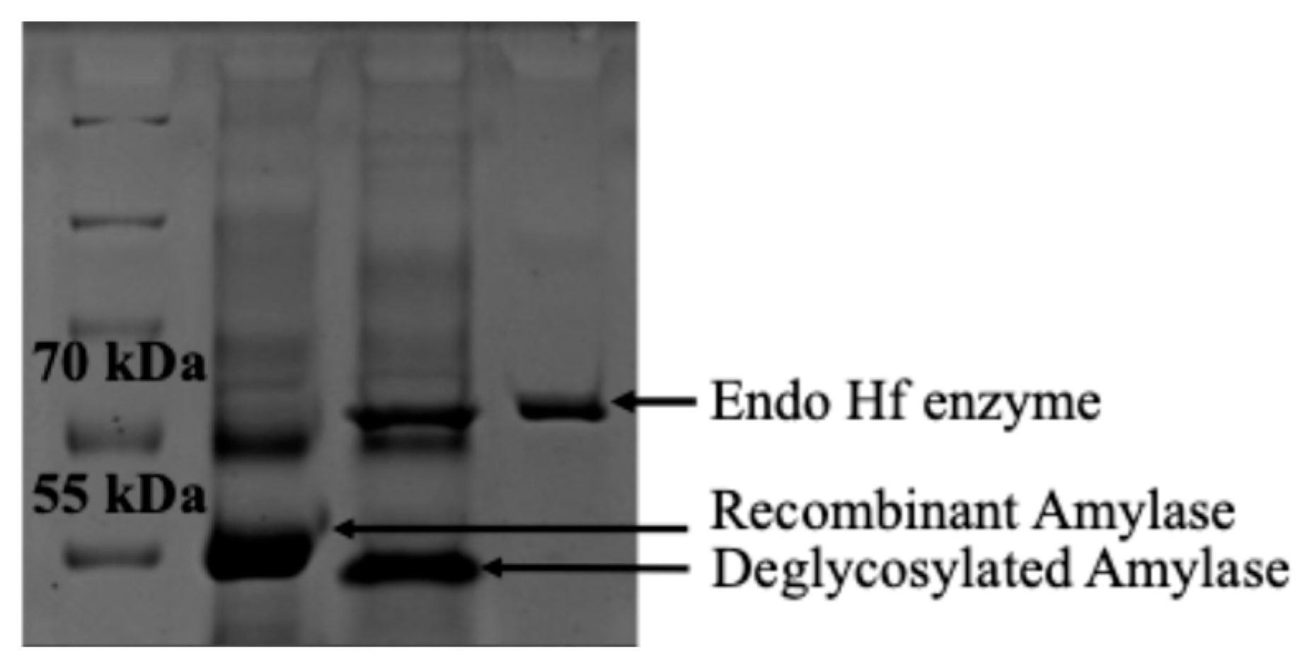
In order to see whether the produced recombinant α-amylase enzyme undergoes glycosylation, a reaction was performed with EndoHf enzyme. Approximately 3 μg of protein sample was denatured with denaturation buffer by incubating at 100 °C for 10 min and then kept on ice for 1 min. 0.6 μL of EndoHf enzyme was added, and the final volume was completed to 40 μL and then incubated at 37 °C for 3 h. After incubation, samples were analyzed by SDS-PAGE.
2.13. Determination of Biochemical Properties of Recombinant Amylase
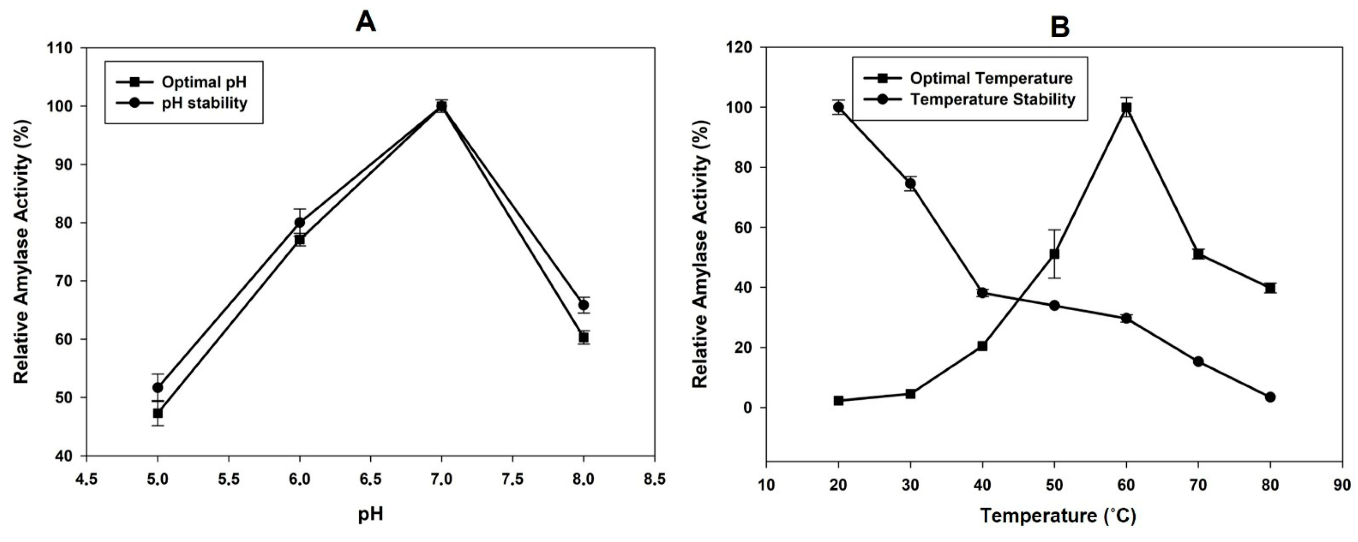
The optimal temperature of α-amylase activity was tested over a range of 30–80 °C by using 1% (w/v) starch solution at pH 6.9. For the thermal stability of α-amylase enzyme, supernatants were incubated at temperatures between 30 and 80 °C for 1 h, and then the enzyme activity was measured immediately under standard conditions.
In order to determine the optimal pH of α-amylase enzyme activity, a substrate solution containing 1% (w/v) starch was prepared using various buffers in the pH range of 5.0–9.0 and tested. To investigate the effect of pH on enzyme stability, supernatant samples were incubated with 20 mM sodium phosphate buffer containing 6.7 mM NaCl adjusted to pH 5.0–9.0 at room temperature for 1 h, and enzyme activity was measured by the standard method after adding 1% soluble starch solution to each buffer. Relative enzyme activities were calculated by assuming the activity of the enzyme as 100% at the temperature and pH specified in the standard method.
2.14. Effect of Metal Ions on Amylase Enzyme Activity
The effect of FeCl2, MgCl2, CuCl2, ZnCl2, and CaCl2 on the amylase enzyme activity was determined at final concentrations of 2 mM, 5 mM, and 10 mM of the metal ions. Relative activities were calculated by assuming the control activity as 100%.
2.15. Application of Recombinant Amylase Enzyme on Apple Juice
The effectiveness of various units of the produced α-amylase enzyme (1400 U–690,000 U) was investigated in raw apple juice. Initially, to gelatinize the starch potentially present in the juice, the samples were heated at 90 °C for 5 min. This process rendered the starch more susceptible to enzymatic hydrolysis.
Following this, the juice samples were cooled to 50 °C, and the enzyme treatments at the specified dosages were applied. The samples were then incubated in a water bath for 2 h. To inactivate the enzyme after treatment, the samples were heated again at 90 °C for 5 min. After cooling to room temperature, the samples were centrifuged at 8000 rpm for 10 min at 4 °C and filtered through a coarse filter before analysis.
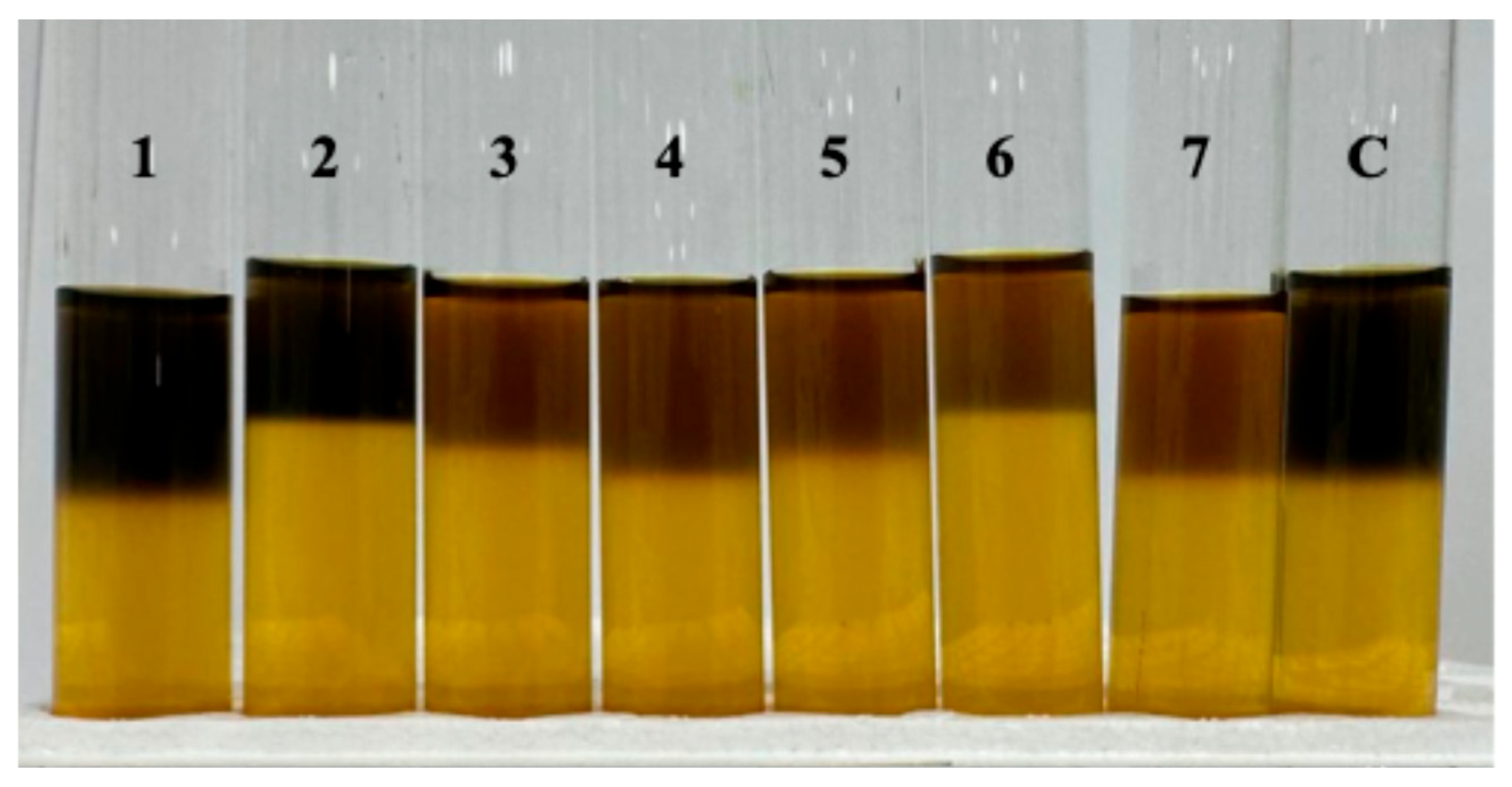
To detect the presence of residual starch in the apple juice samples, iodine tests were conducted. The iodine solution (10%) was prepared by dissolving 0.1 g iodine in 10 mL of ethanol, adding 2 g of potassium iodide, and diluting to 100 mL with distilled water. For the test, 10 mL of apple juice was placed in a test tube, which was held at a slight angle. A few drops of iodine solution were carefully added along the inner wall of the tube to reach the surface of the liquid. The colour change at the surface was observed to assess the degree of starch hydrolysis.
2.16. Statistical Analyses
Descriptive statistics were expressed as the mean, standard deviation, and minimum–maximum values. The normality of the data was assessed with the Shapiro–Wilk test, while homogeneity of variances was evaluated using Levene’s test. A two-way ANOVA was conducted to examine the effects of pH, temperature, and their interaction on the activity. For significant findings, pairwise comparisons for pH and temperature were performed using Tukey’s post hoc test, and Bonferroni correction was applied for comparisons of temperature levels within each pH group. All statistical analyses were carried out with SPSS software (version 23.0), and a p-value < 0.05 was considered statistically significant.
4. Discussion
In this study, the use of a codon-optimized gene sequence and a PDI-overexpressing host strain was intended to support efficient secretion and correct folding of the recombinant enzyme, which is particularly relevant for fungal α-amylases expressed in yeast systems.
From examination of past studies using
A. niger as a genetic source for recombinant production of amylase enzyme in
P. pastoris, it was seen that the methanol-inducible
AOX1 promoter is generally used. In these studies, maximum amylase activities reported at shake flask level production were found to be 31.33 U/mL and 2838 U/mL [
8,
12]. In another study,
Bacillus subtilis PY22 was used as a gene source for amylase enzyme, and maximum enzyme activity was reported as 44 U/mL [
7]. It has been reported that 603.4 U/mL enzyme activity was obtained from the recombinant production of
Thermomyces dupontii α-amylase enzyme in
P. pastoris [
9]. The codon-optimized
Bacillus licheniformis α-amylase gene was expressed in
P. pastoris under the control of the
AOX1 promoter, and the maximum enzyme activity reached 420 U/mL [
20]. Compared to these reports, the enzyme activity obtained in the present study indicated that the synthetic SNT5 promoter supports enhanced α-amylase production at the shake flask level, suggesting that SNT5 constitutes an effective alternative to
AOX1 for small-scale expression.
In the literature, there are limited studies about the recombinant production of
A. niger amylase enzyme in
P. pastoris, and the scale-up production of amylase enzyme was not reported in these studies [
8,
12]. However, in another study,
B. licheniformis amylase enzyme was produced in a 50 L bioreactor, and 11,000 U/mL enzyme activity was obtained [
20].
Rhizopus oryzae amylase enzyme was produced in
P. pastoris at the bioreactor level and reached 448.6 U/mL enzyme activity at the end of 120 h of fermentation [
21]. Furthermore, using
Bacillus acidicola as a gene source, it was reported that 750 U/mL enzyme activity was achieved in a bioreactor with eight copies of the expression cassette in a
Pichia clone [
22]. Comparing the results obtained in this study with those in the literature reveals that we have achieved the highest enzyme activity reported to date. Therefore, it was concluded that the synthetic
ADH2 promoter (SNT5) used for the first time in this study is an efficient promoter to produce recombinant
A. niger amylase enzyme in the
P. pastoris expression system.
The molecular weight of the
A. niger amylase protein was determined as 58 kDa, and studies using different gene sources showed that the molecular weight of the amylase protein generally ranges between 50 and 70 kDa [
7,
8,
9,
21,
23,
24,
25]. Within this study, the produced enzyme was determined to have a molecular weight of 55 kDa and to undergo glycosylation. The observed N-linked glycosylation is consistent with previous reports on yeast-expressed fungal α-amylases and may contribute to secretion efficiency and stability of the recombinant enzyme [
26].
The temperature profile observed in this study is consistent with previously reported characteristics of fungal α-amylases. Eight α-amylase enzymes belonging to the
A. niger amylase family have been characterized and reported optimal temperatures of 30–40 °C for seven enzymes, while one enzyme exhibited an optimal temperature of 60 °C [
8], in agreement with the findings of the present study. In another study, the optimal temperature of the
A. niger amylase enzyme was determined as 70 °C [
12]. In addition, the other studies using different gene sources such as
B. subtilis PY22,
B. licheniformis, and
T. dupontii reported that the optimal temperature of amylase enzyme was 60 °C, 90 °C, and 60 °C, respectively. These findings indicate that the temperature optimum of the recombinant enzyme produced in this study falls within the expected range for industrially relevant α-amylases. In the literature, amylase enzymes show the optimal activity in the pH range 2.5–4.5 and 5.0–6.5 according to their acidic or neutral character [
27,
28]. Based on its pH optimum and stability profile, the recombinant α-amylase produced in this study can be classified as a neutral amylase enzyme.
The stimulatory effect of Ca
2+ is consistent with the well-documented Ca
2+ dependency of α-amylases, while inhibitory effects observed at higher concentrations of other metal ions agree with previous biochemical studies [
29]. Calcium ions are known to stabilize the enzyme structure and enhance catalytic efficiency. In previous studies, 1 mM Ca
+2 was reported to activate α-amylase activity, whereas Mg
+2, Zn
+2, Cu
+2, and Fe
+2 exhibited inhibitory effects on α-amylases derived from
T. dupontii and
A. amylolytica [
9,
24]. In another study expressing the
A. oryzae amylase enzyme in
P. pastoris, the effect of metal ions on enzyme activity was evaluated by testing 4 mM Ca
+2, Mg
+2, Fe
+2, and Cu
+2 [
30]. They reported that Ca
+2, Mg
+2, and Fe
+2 had an activating effect on enzyme activity, while Cu
+2 had an inhibitory effect. The concentration-dependent effects observed in the present study, particularly the differential behaviour of Ca
2+ and Zn
2+ at higher concentrations, are therefore in agreement with previous biochemical reports and highlight the metal sensitivity of α-amylase activity.
When the recombinant enzyme was applied to fruit juice, it was observed that it degraded starch in a dose-dependent manner. The requirement for higher enzyme concentrations is likely related to differences in formulation, purification level, and stabilization rather than intrinsic catalytic limitations. Similarly, it has been reported that crude amylase extracts require purification and concentration optimization to achieve comparable hydrolytic performance [
31]. At low concentrations, the recombinant α-amylase enzyme displayed a dark blue coloration, suggesting incomplete starch degradation. The effectiveness of enzymatic starch hydrolysis is influenced by multiple parameters, including enzyme concentration, substrate availability, pH, temperature, and incubation time [
3].



 ,
,