Article

Probiotic Potential of Lactic Acid Bacteria Strains Isolated from Artisanal Cheeses: Impact on Listeria monocytogenes Infection

Centro de Referencia para Lactobacilos (CERELA, CONICET-FML-FECIC), Chacabuco 145, Tucumán T4000ILC, Argentina
*
Authors to whom correspondence should be addressed.
These authors contributed equally to this work.
Fermentation 2025, 11(6), 343; https://doi.org/10.3390/fermentation11060343
Submission received: 7 May 2025 / Revised: 28 May 2025 / Accepted: 5 June 2025 / Published: 12 June 2025

Abstract

Listeriosis is a disease associated with the consumption of food contaminated with Listeria monocytogenes. Probiotic lactic acid bacteria (LAB) or their postbiotics have been of interest for their anti-listerial effect. This study focused on isolating LAB from artisanal cheeses and characterizing their potential as probiotics. Twelve LAB isolates exhibiting typical LAB traits were evaluated for their ability to survive in simulated gastric juice, hydrolyze bile salts, auto-aggregate, hydrophobicity, and antagonistic activity against L. monocytogenes. The four most promising LAB strains demonstrated anti-listerial probiotic potential and were identified as Latilactobacillus (Lat.) curvatus SC076 and Lactiplantibacillus (Lact.) paraplantarum SC291, SC093, and SC425. The antimicrobial activity of these strains was mainly attributed to bacteriocin-like substances and organic acids. While three Lact. paraplantarum strains were resistant to ampicillin, Lat. curvatus was sensitive to all tested antibiotics. All selected strains exhibited no hemolytic, gelatinase, and lecithinase activity. Exposure to LAB supernatants resulted in a significant reduction in the adhesion and intracellular count of L. monocytogenes in Caco-2 cells, with Lat. curvatus SC076 showing the most significant effect. Based on its probiotic characteristics, Lat. curvatus SC076 is a promising candidate for functional foods, pending further in vivo studies to assess its potential in the food industry.

Graphical Abstract

1. Introduction

Listeria monocytogenes is the causative agent of animal and human listeriosis, a disease resulting from the consumption of contaminated food such as processed meat, dairy products, pre-packaged sandwiches, prepared vegetables, salads, and fruits [1,2]. Many listeriosis cases are classified as sporadic, but foodborne outbreaks are frequently observed [3]. Immunocompromised individuals, the elderly (65 and older), pregnant women, and infants are at the highest risk of infection with a high fatality rate (20−30%). L. monocytogenes can spread from intestinal tissue to the central nervous system, causing septicemia or meningitis [4]. Controlling this pathogen is challenging due to its complex regulatory mechanisms, which allow it to adapt to adverse environmental conditions and transition from saprophytism to virulence [5].
On the other hand, several strains of lactobacilli and bifidobacteria, commonly used as probiotics, have been reported to inhibit intestinal pathogens [6,7]. Probiotics are defined as live microorganisms that, when administered in adequate amounts, confer a health benefit to the host [8]. Their mechanism of action is not completely elucidated. However, it is known that they enhance microbial balance, compete with pathogens for adhesion sites, restore epithelial barrier function, and stimulate the epithelial immune response [9]. In addition, probiotics must be able to (i) resist gastrointestinal conditions, (ii) hydrolyze bile salts, (iii) adhere to intestinal epithelial cells, (iv) inhibit the adhesion of pathogens, and (v) have antimicrobial activity toward pathogenic bacteria [10].
In particular, lactic acid bacteria (LAB) produce various antimicrobial compounds such as hydrogen peroxide, organic acids, biosurfactants, functional peptides, as well as bacteriocins [11]. The latter are ribosomally synthesized small inhibitory peptides or complex proteins that selectively target phylogenetically related bacteria [12]. Notably, many bacteriocins exhibit activity against L. monocytogenes [13]. In this context, although a considerable number of well-characterized commercial probiotic strains are available around the world, the discovery of new strains is still of industrial interest [14]. The aim of this study was to isolate, identify, and evaluate the probiotic potential of LAB strains from artisanal cheeses with a focus on the control of L. monocytogenes by decreasing bacterial adhesion and invasion of eukaryotic cells.

2. Materials and Methods

2.1. Sampling and LAB Isolation

The isolates were collected from artisanal cheeses. Ten grams of food were placed in sterile stomacher bags; 90 mL of 0.85% NaCl was added and mixed for 2 min in a stomacher machine (Stomacher Lab-Blender 400, A.J. Seward Lab. London, England). Tenfold serial dilutions were plated on Man, Rogosa, and Sharpe (MRS) agar and incubated for 48 h at 30 °C. All putative LAB colonies were subjected to microscopic observation to determine cell shape, Gram staining, oxidase, and catalase activity tests. Selected colonies were kept at −80 °C in MRS broth containing 15% glycerol. Unless otherwise stated, all the used media were supplied by Britania (Buenos Aires, Argentina).

2.2. Strains and Culture Conditions

L. monocytogenes FBUNT (Facultad de Bioquímica, Química y Farmacia, Universidad Nacional de Tucumán, Argentina) and the bacteriocin producer Lat. curvatus CRL705 were grown in Tryptone Soya Broth (TSB; BBL, Cockeysville, MD, USA) supplemented with 0.5% yeast extract (YE) and MRS broth at 30 °C, respectively. Lat. curvatus CRL705 was used as a positive control, producing a two-component bacteriocin (lactocin 705) and an anti-listerial bacteriocin (lactocin AL705) [15].

2.3. Bacterial Resistance to the Simulated Gastrointestinal (SGI) Tract and Bile Salt Hydrolase (BSH) Activity

2.3.1. Survival of LAB to SGI Conditions

Bacterial viability after simulated gastric and intestinal exposure was evaluated according to Marchesi et al. [16] with modifications. Briefly, the artificial gastric fluid consisted of 3 g L−1 pepsin (Sigma-Aldrich, St. Louis, MO, USA), pH 3 adjusted with 1N HCl. The composition of the intestinal juice was 3 g L−1 Oxgall (dehydrated fresh bile; Sigma-Aldrich), 15 g L−1 pancreatin (Sigma-Aldrich), 1 mg mL−1 chymotrypsin (Sigma-Aldrich), 1 mg mL−1 trypsin (Sigma-Aldrich), pH 7.5 adjusted with 1N NaOH. The bacterial suspension (~1 × 10⁹ CFU mL−1) was inoculated into the simulated gastric juice (5 mL) and incubated at 37 °C in a shaker (MaxQ 4450, Thermo Fisher, Waltham, MA, USA) for 2 h. Then, the cells were harvested by centrifugation (6500× g, 5 min), washed twice with sterilized saline solution (9 g L−1 NaCl), and transferred to simulated intestinal juice for 2 h with agitation at 37 °C. Viable bacteria after gastrointestinal exposition were enumerated by serial dilution and plating on MRS agar, followed by incubation at 30 °C for 48 h. The colony counts were expressed as the log10 of CFU mL−1.

2.3.2. Bile Salt Tolerance

The overnight culture of each selected strain was spotted (10 μL) on MRS agar plates supplemented with 5 g L−1 chenodeoxycholic acid (Sigma-Aldrich, St. Louis, MO, USA) and 0.37 g L−1 CaCl2 (Sigma-Aldrich, St. Louis, MO, USA) and incubated aerobically for 48 h at 37 °C. A positive result was evidenced by a precipitation zone around the spots [17].

2.4. Antimicrobial Activity Against L. Monocytogenes

The LAB strains resistant to the SGI conditions were cultured in an MRS medium, and cell-free supernatants (CFS) were tested for their antimicrobial activity using the spot-on-lawn assay. CFS was neutralized (pH 7.0) with 4 N NaOH and subsequently treated with 500 U mL−1 catalase (Sigma-Aldrich, St. Louis, MO, USA) or 1 mg mL−1 proteinase K (Invitrogen, Carlsbad, CA, USA) for 1 h at 37 °C. Then, 5 μL of each CFS was spotted in semisolid TSBYE plates, overlayed inoculated with L. monocytogenes FBUNT (~106 UFC mL−1). After 48 h of incubation at 30 °C, the presence or absence of growth around the spots was interpreted as (+) or (−), respectively.

2.5. Hydrophobicity and Auto-Aggregation Assay

Microbial adhesion to xylene (nonpolar solvent) and toluene (acidic solvent) was performed according to Perez Ibarreche et al. [18]. Briefly, antimicrobial-producing LAB was grown for 16-18 h in an MRS medium at 30 °C. Each culture was centrifuged (7000× g for 10 min), washed twice with 0.85% NaCl, and resuspended in the same solution (A600:0.3 to 0.7; A0). Then, mixtures of each cell suspension (3 mL) with the solvents (0.5 mL) using a vortex for 60 s were made. The A600 (A1) was measured in the presence of 2-phase formation. The LAB adhesion to solvent expressed as a percentage was calculated as follows: [(A0 − A1)/A0] × 100. Then, the level of bacterial auto-aggregation was determined. LAB cultures grown separately in MRS during 16 h at 30 °C were allowed to settle at room temperature for 2 h. Initial DO600 (ODinitial) readings and after sedimentation (OD2h) were performed. Finally, the auto-aggregation percentage was calculated as [ODinitial − (OD2h)]/ODinitial × 100 [19]. The different scores by hydrophobicity and auto-aggregation were defined as high (61 to 80%), medium (31 to 60%), and low (0 to 30%).

2.6. Genetic Identification of the Selected Strains

2.6.1. Sequencing the 16S rRNA Gene

The putative LAB isolates with anti-listerial activity were identified by ribosomal 16S-DNA sequencing. Total genomic DNA was extracted according to Pospiech and Neumann [20]. Universal primers 27F (5′-GTGCTGCAGAGAGTTTGATCCTGGCTCAG-3′) and 1492R (5′-CACGGATCCTACGGGTACCTTGTTACGACTT-3′) were used for the sequencing reaction [21]. The PCR reaction mixture contained: dNTPs (0.2 mM), DNA template (50 ng), forward and reverse primers (10 pmol each), Taq polymerase (0.5 U), and 1x PCR buffer containing MgCl2. The amplification conditions were as follows: an initial denaturation step at 94 °C for 5 min, followed by 35 cycles of denaturation at 94 °C for 30 s and 52 °C for 30 s, an extension at 72 °C for 1 min and 30 s, and a final extension step at 72 °C for 10 min. PCR reactions were carried out in an XP Cycler thermal cycler (Bioer, Hangzhou, China), and PCR products were separated by electrophoresis on a 1.2% (w/v) agarose at 80 V. A 100 pb DNA ladder (Embiotec, Buenos Aires, Argentina) was used as a molecular size marker. Gels were stained with GelRed (Biotium Inc., Fremont, CA, USA) and visualized under an ultraviolet light transilluminator (320 nm). Purified amplicons were sequenced in an automatic DNA sequencer (Applied Biosystems model 3130, Foster, CA, USA) at the CERELA-CONICET sequencing service. The sequences were aligned using MEGA software version 11 and then compared with those in the GenBank using the BLASTN tool (NCBI). Identification at the species level was achieved for sequences with 99–100% homology. The sequences were deposited in the GeneBank NCBI database with the accession numbers PV533605 (Lat. curvatus SC076), PV533606 (Lact. paraplantarum SC291), PV533608 (Lact. paraplantarum SC293), and PV533611 (Lact. paraplantarum SC425).

2.6.2. Multiplex PCR Assay

The PCR reaction mixture (25 μL) was performed using the recA gene-based primers paraF (5′-GTCACAGGCATTACGAAAAC-3′), pentF (5′-CAGTGGCGCGGTTGATATC-3′), planF (5′-CCGTTTATGCGGAACACCTA-3′), and pREV (5′-TCGGGATTACCAAACATCAC-3′), following the protocol described by Torriani et al. [22].

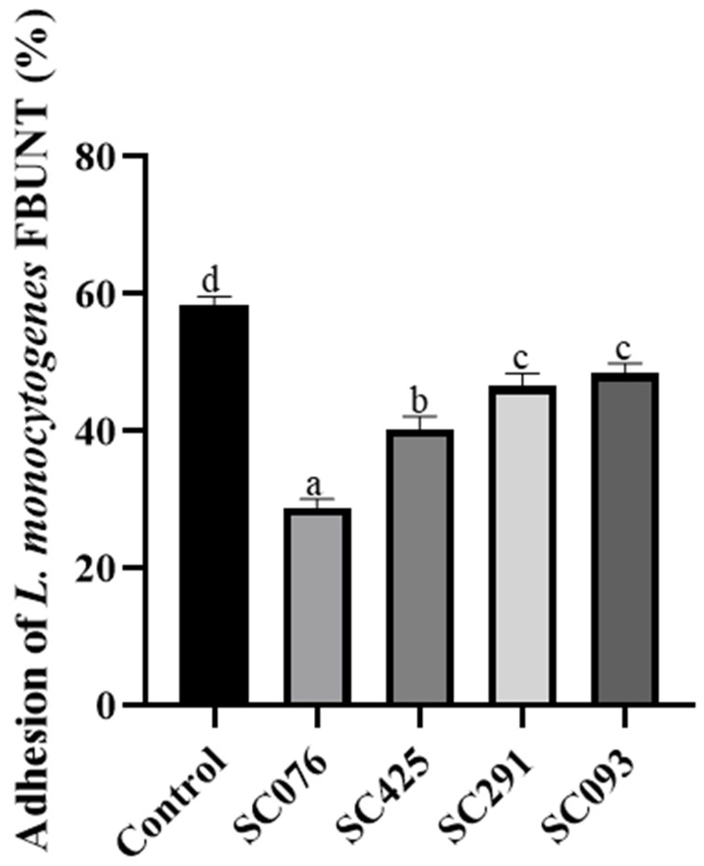
2.7. Adhesion Assays

The adhesion of LAB strains and L. monocytogenes to Caco-2 cells (ATCC HBT37) was evaluated separately, combining the pathogen with each CFS. Caco-2 cells were obtained from the American Type Culture Collection (Rockville, MD, USA). This cell line was grown as monolayers in 24-well plates with Dulbecco’s Modified Eagle Medium (DMEM, Sigma-Aldrich) under 5% CO2 at 37 °C [23]. Overnight cultures of the selected LAB strains and L. monocytogenes FBUNT were harvested by centrifugation (8500× g for 10 min) and washed twice with sterile phosphate-buffered saline (PBS). The LAB and L. monocytogenes cultures were added separately to the monolayers, reaching final concentrations of 108 and 107 CFU mL−1, respectively. After incubating for 30 min, the Caco-2 cell monolayers were washed with PBS, removing non-adhering bacteria. Finally, the LAB strains and the pathogen were detached from the cell monolayers with trypsin by centrifugation (1000× g for 10 min) and enumerated in MRS and TSBYE agar plates, respectively. In order to evaluate the effect of CFS on L. monocytogenes adhesion, the cell monolayers were inoculated with the pathogen combined with each LAB CFS and processed as described above. Adhesion capacity was expressed as an adhesion index, calculated as the number of adherent bacterial cells divided by the initial inoculum size, multiplied by 100.

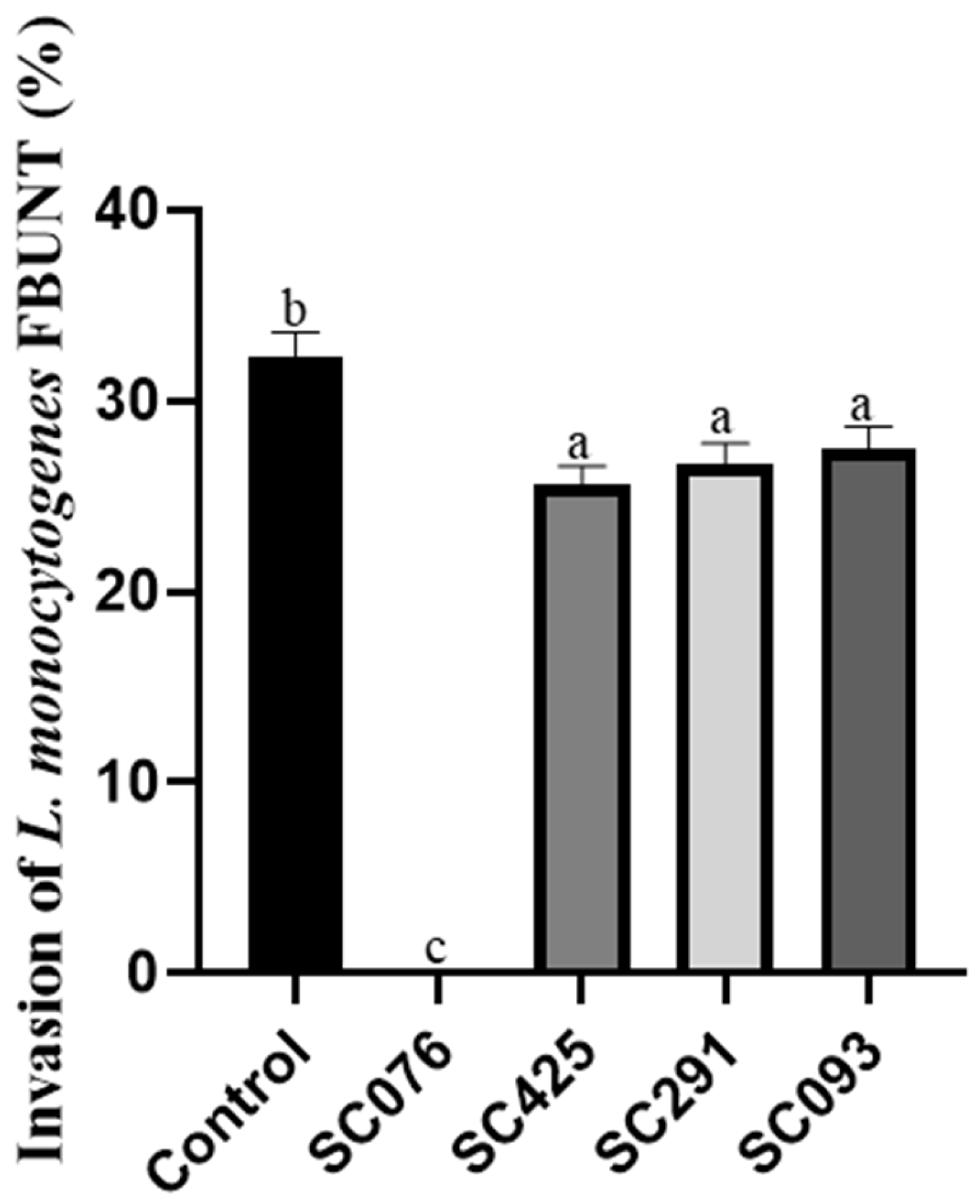
2.8. Invasion Assays

In order to evaluate the effect of LAB CFS on L. monocytogenes invasion of epithelial cell monolayers, the pathogen (~107 CFU mL−1) was inoculated in the presence or absence of CFS during 60 min at 37 °C in a 5% CO2 atmosphere. Then, 250 mg mL−1 of gentamicin (60 min at 37 °C) was added to eliminate non-internalized bacteria. Finally, Caco-2 cells were lysed with 0.1% (v/v) Triton X-100 (Sigma-Aldrich) and internalized L. monocytogenes cells were enumerated in TSBYE agar from the dilutions of the lysates. After each addition of reagents, the Caco-2 cells were washed twice with PBS.

2.9. Safety Evaluation

2.9.1. Antibiotic Sensitivity

The antimicrobial resistance of the selected LAB strains was evaluated using the microplate dilution method, following ISO 10932:2010/IDF 223:2010 standard [24]. Seven clinically relevant antibiotics were tested: ampicillin (AMP), gentamicin (GEN), kanamycin (KAN), erythromycin (ERY), tetracycline (TET), chloramphenicol (CHL), and streptomycin (STR). Minimum inhibitory concentrations (MIC, μg mL−1) were determined for each antibiotic and compared with the EFSA- FEEDAP [25] cut-off values. All antibiotics were purchased from Sigma-Aldrich (MO, USA).

2.9.2. Hemolytic, Gelatinase, and Lecithinase Activity

The pathogenic potential of selected LAB strains was evaluated based on their hemolytic, gelatinase, and lecithinase activities. The phenotypic assays in agarized media with specific substrates for each enzyme were performed. Hemolytic activity was assessed on Columbia Blood Agar (Oxoid, Basingstoke, UK) supplemented with 5% defibrinized horse blood and incubated aerobically for 48 h at 37 °C. Hemolytic reactions were evidenced by the presence of a clear zone (β-haemolysis), green zone (α-haemolysis), or the absence of a zone (γ-haemolysis) around the colonies [26]. The evaluation of gelatinase and lecithinase enzymes was performed in selected LAB according to Miranda et al. [27]. The gelatinase production was detected by inoculating LAB on peptone–yeast extract agar containing gelatin (30 g L−1, Difco). Plates were incubated for 18 h at 37 °C and subsequently cooled at room temperature for 2 h. A turbid halo around the colonies indicated positive gelatinase activity. The ability of the LAB selected to express lecithinase activity was evaluated in egg yolk agar medium prepared in accordance with Miranda et al. [27]. Plates were incubated at 37 °C for 48 h. A positive lecithinase reaction was evidenced by the formation of an opaque zone around the colonies.

2.10. Statistical Analysis

All experiments were conducted in triplicate using three independent bacterial cultures to ensure reproducibility. Mean values and standard deviations were calculated for each experiment. Significant differences (p ≤ 0.05) were determined using ANOVA followed by Tukey’s test, performed with the InfoStat software version 2020 (InfoStat Group, Córdoba, Argentina).

3. Results and Discussion

Over the years, human and animal intestinal microbiota, as well as fermented foods, have been identified as potential reservoirs of probiotics [28]. Herein, LAB was isolated from artisanal cheeses and evaluated for their probiotic properties. Thirty isolates presumptively identified as LAB based on Gram staining, morphology, and catalase activity were divided into Gram-positive, catalase-negative cocci (18 isolates) and Gram-positive, catalase-negative rods (12 isolates). Bacteria with cocci morphology were not selected because they could belong to the Enterococcus genus, which not only exhibits intrinsic resistance to antibiotics but also has the capacity to acquire new resistance mechanisms to antimicrobial agents [29]. The 12 rod-shaped isolates were chosen for subsequent screening, considering their resistance to SGI conditions and their ability to produce antimicrobial substances.
The passage through the GI tract constitutes the first defense mechanism that all ingested microorganisms, including probiotics, must overcome [30]. Therefore, it is essential that probiotics administered orally resist the conditions of the stomach and small intestine. Out of the twelve isolates, five putative LAB strains (SC347, SC358, SC379, SC162, and SC274) showed significant decreases (greater than two log CFU mL−1) after treatment with SGI juice (Figure 1). High tolerance was recorded in seven isolates, with survival percentages ranging from 91 to 95% after 4 h of incubation. Most strains showed survival rates exceeding 90% in SGI juice after 4 h of incubation, suggesting that they could reach the intestinal tract in large numbers as viable cells and potentially confer health benefits.
Similarly, commercial protective cultures such as Lact. plantarum LP and LPP were resistant to GI conditions [31]. Conversely, bacteriocinogenic LAB strains only enhanced their survival during SGI exposure when encapsulated in alginate beads [23]. In addition to the ability to tolerate the acidic environment of SGI juice, probiotics are also expected to hydrolyze bile salts. Bacteria with this capacity can increase their intestinal survival and persistence, thus enhancing their overall probiotic effect. Out of seven LAB strains evaluated, SC076, SC291, SC093, and SC425 were able to hydrolyze bile salts, as evidenced by visual observation of precipitation zones around the inoculation spot (Table 1). This property is associated with the presence of conjugated bile salt hydrolase [23].
According to the FAO/WHO guidelines [10], another important requirement for selecting probiotic bacteria is the production of antimicrobial compounds. The four selected strains were evaluated for their antimicrobial activity against L. monocytogenes FBUNT. All isolates (SC076, SC291, SC093, and SC425) were able to inhibit pathogen growth (Table 1). The SC076 strain through a bacteriocin-like substance, as evidenced by treatment with proteases, which completely abolished its antimicrobial action. The CFS from SC291, SC093, and SC425 showed inhibitory activity at a pH close to 3.5, while a pH of 7.0 allowed L. monocytogenes to grow. This finding indicated that acid production played a major role in the inhibitory effect of these LAB.
Then, isolates producing antimicrobial compounds (SC076, SC291, SC093, and SC425) were identified by sequencing the 16S rRNA gene as Lat. curvatus SC076, Lact. plantarum SC291, SC093 and SC425. Additionally, since the isolates SC291, SC093, and SC425 belong to Lact. plantarum group, recA gene amplification was performed to differentiate them at the species level. Based on the size of the specific amplicons, these strains were identified as Lact. paraplantarum (Supplementary Figure S1). Recent studies have shown the key role of Lat. curvatus and Lact. plantarum among LAB species in promoting host health benefits. Lat. curvatus SMFM2016-NK-fermented milk has been shown to mitigate periodontal and intestinal inflammation by changing oral and gut microbiota [32]. In addition, Lat. curvatus DN317 and Wikim38, which exhibit immunomodulatory effects, have been proposed as potential therapeutic agents for various inflammatory diseases [33,34]. Furthermore, strains from the Lact. plantarum species have demonstrated probiotic potential, either alone [31,35] or in combination with Lat. plantarum strains [36,37].
On the other hand, adherence to the host’s intestinal cells is a relevant characteristic required for probiotic strains to colonize the gut and exert beneficial effects [38]. The four Lactobacillus strains demonstrated adhesion capacity to Caco-2 cells ranging from 43 to 48% (Table 2).
These results are in agreement with those reported by other authors regarding Lactobacillus strains’ adhesion to Caco-2 and HT-29 cell lines [39,40]. In this study, the inhibition of L. monocytogenes FBUNT adhesion to Caco-2 cells by CFS from selected LAB is shown in Figure 2. All CFSs significantly reduced the adhesion of the pathogen to Caco-2 cells. However, the CFS from Lat. curvatus SC076 produced the highest inhibition of pathogen adhesion (30%) compared to the control (58%). Similarly, the adhesion of L. monocytogenes to HT-29 cells was counteracted by the bacteriocinogenic strain L. sakei CTC494 in competition experiments [41].
Regarding LAB hydrophobicity and auto-aggregation, these properties were evaluated due to their relevance for adhesion to epithelial surfaces [42]. The four isolates exhibited moderate cell surface hydrophobicity, ranging from 46 to 59% with xylene and 45 to 55% with toluene (Table 2). Based on their sedimentation characteristics, auto-aggregation at 2 h showed moderate aggregative values for the selected LAB strains (Table 2). Our results highlight the potential for effective colonization of Lat. curvatus SC076, Lact. paraplantarum SC291, SC093, and SC425, aligning with previous reports [35,43]. Unlike other authors, who found that adhesion to Caco-2 cells did not significantly correlate with auto-aggregation and hydrophobicity levels [44]. This difference was attributed to the fact that LAB adhesion is a multifactorial phenomenon, beginning with the initial contact with the host cell, followed by diverse surface interactions.
In this study, the co-inoculation of each CFS with L. monocytogenes played a protective role against its invasion of Caco-2 cells (Figure 3). The CFS from Lat. curvatus SC076 was able to completely inhibit the invasion of L. monocytogenes into Caco-2 cells. A similar result was obtained when applying CFS from bacteriocin-producing Lat. curvatus CRL705 and CRL1532 as an anti-invasive strategy [23]. The CFS from Lact. paraplantarum SC291, SC425, and SC093 slightly reduced the invasive capacity of the pathogen by 18, 19, and 15%, respectively. This could be due to the lower effectiveness of the organic acids in this process or to the lack of bacteriocin production. Regarding the control samples, L. monocytogenes FBUNT, in the absence of antimicrobial treatments, showed a 37% ± 1.90 invasion into Caco-2 cells. Similar results were reported by Gomes et al. [45] in the invasion of L. monocytogenes ATCC19115 using Caco-2 cells as an in vitro model.
It is worth noting that only Lat. curvatus SCB076, a strain producing a bacteriocin-like substance, completely abolished the invasion of L. monocytogenes into the cell line. Based on these results, we could hypothesize that this antimicrobial compound may act on the expression of key genes involved in the invasion of the pathogen. In agreement with this, Ye et al. [46] reported that antimicrobial metabolites from Enterococcus faecium in co-cultures with L. monocytogenes reduced the expression of virulence genes in the pathogen, attenuating its pathogenicity. Furthermore, functional metabolites such as bacteriocins have been shown to modify the virulence gene expression profile of L. monocytogenes [47,48].
The assessment of the antimicrobial susceptibility profile is a relevant criterion for potential probiotic selection. Notably, LAB used as probiotics must be safe for the host and must not have transferable or acquired antimicrobial resistance [33]. In accordance with the guidelines proposed by the European Food Safety Authority [25], a comprehensive antibiotic susceptibility test was conducted on the four chosen LAB isolates (Table 3). Multiple antibiotics were employed to evaluate their susceptibility to different drug classes. All Lact. paraplantarum strains demonstrated resistance to ampicillin. Lact. paraplantarum SC093 also displayed resistance to kanamycin and tetracycline, while SC425 and SC291 were resistant to kanamycin and tetracycline, respectively. Lat. curvatus SC076 was the only strain susceptible to all the antibiotics assayed (Table 3).
It is worth mentioning that Lat. curvatus was listed as a Qualified Presumption of Safety (QPS)-recommended biological agent for food or feed by the European Food Safety Authority [49]. The antibiotic profile of Lat. curvatus SC076 partially coincides with the antibiotic resistance/sensitivity reported for LAB. These bacteria are generally considered sensitive to broad-spectrum antibiotics (e.g., tetracycline and chloramphenicol), glycopeptides (vancomycin), and β-lactam antibiotics (e.g., ampicillin and penicillin) but resistant to aminoglycosides (e.g., streptomycin and gentamicin) [50,51]. Contrary to our findings, Lat. curvatus Z12 displayed resistance to ampicillin, tetracycline, and chloramphenicol but was sensitive to penicillin G [52]. Factors inherent to cell wall impermeability, defects in the autolysis system, and multidrug transporters could explain the differences in resistance/sensitivity to antibiotics detected in LAB [53].
On the other hand, the selection of strains devoid of hemolytic, gelatinase, and lecithinase activity as probiotics demonstrates their nonvirulent nature. The hemolytic behavior of the selected LAB strains was evaluated. The absence of red blood cell rupture or destruction on the agar plate was interpreted as γ-hemolytic activity (no hemolysis). All isolates were non-hemolytic, which supports the non-pathogenic status of the probiotic. Similar results, indicating that numerous LAB strains exhibit a non-hemolytic profile, were reported [54,55]. Furthermore, the assessment of the hemolytic activity of 73 Lactobacillus strains detected only four α-hemolytic strains, indicating that this profile is not common in this genus [51]. Like most LAB strains [27,35,56], the four selected strains did not show lecithinase and gelatinase activity in the specific media and thus are considered safe for use.

4. Conclusions

In conclusion, four LAB strains isolated from artisanal cheeses were identified by 16S rRNA sequencing as Lat. curvatus SC076 and Lact. paraplantarum SC291, SC093 and SC425. These LAB strains met the requirements for probiotics, as evaluated by their anti-listerial activity, survivability in SGI conditions, and good cell surface properties. The bacteriocin-like substance-producing Lat. curvatus SC076 caused a significant reduction in the adhesion of L. monocytogenes FBUNT and completely inhibited its invasion in Caco-2 cells. Furthermore, this strain was found to be safe and incapable of producing virulence-associated enzymes. The suitable properties exhibited by Lat. curvatus SC076 make it a promising candidate for functional foods. Further in vivo assays are necessary for its application in the food industry.

Supplementary Materials

The following supporting information can be downloaded at https://www.mdpi.com/article/10.3390/fermentation11060343/s1, Figure S1: Amplification products of the recA gene obtained from multiplex PCR.

Author Contributions

Conceptualization, C.B., C.M. and P.C.; methodology, C.B. and C.M.; formal analysis, C.M. and L.M.M.; investigation, C.B., C.M. and L.M.M.; resources, L.M.M. and P.C.; writing—original draft preparation, P.C.; writing—review and editing, P.C., S.S. and L.M.M.; funding acquisition, P.C., S.S. and L.M.M. All authors have read and agreed to the published version of the manuscript.

Funding

This work was financed by the Agencia Nacional de Promoción Científica y Tecnológica (PICT-2020-00377 and PICT-2021-0278), Consejo Nacional de Investigaciones Científicas y Técnicas (CONICET; PIP-2021-1858).

Institutional Review Board Statement

Not applicable. This study does not involve humans or animals.

Informed Consent Statement

Not applicable.

Data Availability Statement

The original data presented in the study are openly available in the GeneBank NCBI database with the accession numbers PV533605 (Lat. curvatus SC076), PV533606 (Lact. paraplantarum SC291), PV533608 (Lact.paraplantarum SC293), and PV533611 (Lact. paraplantarum SC425).

Conflicts of Interest

The authors declare no conflicts of interest.

References

  1. Hernandez-Milian, A.; Payeras-Cifre, A. What is new in listeriosis? BioMed Res. Int. 2014, 2014, 358051. [Google Scholar] [CrossRef] [PubMed]
  2. Ferreira, V.; Wiedmann, M.; Teixeira, P.; Stasiewicz, M.J. Listeria monocytogenes persistence in food-associated environments: Epidemiology, strain characteristics, and implications for public health. J. Food. Prot. 2014, 77, 150–170. [Google Scholar] [CrossRef] [PubMed]
  3. Koopmans, M.M.; Brouwer, M.C.; Vázquez-Boland, J.A.; van de Beek, D. Human Listeriosis. Clin. Microbiol. Rev. 2023, 36, e0006019. [Google Scholar] [CrossRef] [PubMed]
  4. Lecuit, M. Listeria monocytogenes, a model in infection biology. Cell. Microbiol. 2020, 22, e13186. [Google Scholar] [CrossRef]
  5. Radoshevich, L.; Cossart, P. Listeria monocytogenes: Towards a complete picture of its physiology and pathogenesis. Nat. Rev. Microbiol. 2018, 16, 32–46. [Google Scholar] [CrossRef]
  6. Matera, M. Bifidobacteria, Lactobacilli… when, how and why to use them. Glob. Pediatr. 2024, 8, 100139. [Google Scholar] [CrossRef]
  7. Sarita, B.; Samadhan, D.; Hassan, M.Z.; Kovaleva, E.G. A comprehensive review of probiotics and human health-current prospective and applications. Front. Microbiol. 2025, 15, 1487641. [Google Scholar] [CrossRef]
  8. Hill, C.; Guarner, F.; Reid, G.; Gibson, G.R.; Merenstein, D.J.; Pot, B.; Morelli, L.; Canani, R.B.; Flint, H.J.; Salminen, S.; et al. Expert consensus document. The International Scientific Association for Probiotics and Prebiotics consensus statement on the scope and appropriate use of the term probiotic. Nat. Rev. Gastroenterol. Hepatol. 2014, 11, 506–514. [Google Scholar] [CrossRef]
  9. Yan, F.; Polk, D.B. Probiotics and probiotic-derived functional factors-mechanistic insights into applications for intestinal homeostasis. Front. Immunol. 2020, 11, 1428. [Google Scholar] [CrossRef]
  10. Food and Agriculture Organization of the United Nations/World Health Organization (FAO/WHO). Guidelines for the Evaluation of Probiotics in Food. Report of a Joint FAO/WHO working group in London Ontario, Canada, April 2002. Available online: https://www.mhlw.go.jp/file/05-Shingikai-11121000-Iyakushokuhinkyoku-Soumuka/0000197343.pdf (accessed on 20 December 2024).
  11. Mani-López, E.; Arrioja-Bretón, D.; López-Malo, A. The impacts of antimicrobial and antifungal activity of cell-free supernatants from lactic acid bacteria in vitro and foods. Compr. Rev. Food Sci. Food Saf. 2022, 21, 604–641. [Google Scholar] [CrossRef]
  12. Darbandi, A.; Asadi, A.; Mahdizade Ari, M.; Ohadi, E.; Talebi, M.; Halaj Zadeh, M.; Darb Emamie, A.; Ghanavati, R.; Kakanj, M. Bacteriocins: Properties and potential use as antimicrobials. J. Clin. Lab. Anal. 2022, 36, e24093. [Google Scholar] [CrossRef] [PubMed]
  13. Fontana, C.; Cocconcelli, P.; Vignolo, G.; Saavedra, L. Occurrence of antilisterial structural bacteriocins genes in meat borne lactic acid bacteria. Food Control 2015, 47, 53–59. [Google Scholar] [CrossRef]
  14. Chua, J.C.L.; Hale, J.D.F.; Silcock, P.; Bremer, P.J. Bacterial survival and adhesion for formulating new oral probiotic foods. Crit. Rev. Food Sci. Nutr. 2020, 60, 2926–2937. [Google Scholar] [CrossRef]
  15. Castellano, P.; Vignolo, G. Inhibition of Listeria innocua and Brochothrix thermosphacta in vacuum-packaged meat by addition of bacteriocinogenic Lactobacillus curvatus CRL705 and its bacteriocins. Lett. Appl. Microbiol. 2006, 43, 194–199. [Google Scholar] [CrossRef]
  16. Marchesi, A.; Silva, J.A.; Wiese, B.; Nader-Macías, M.E.F. Survival of beneficial vaginal Lactobacilli (BVL) to different gastrointestinal tract conditions. Curr. Pharm. Des. 2020, 26, 3608–3618. [Google Scholar] [CrossRef]
  17. Franz, C.M.; Specht, I.; Haberer, P.; Holzapfel, W.H. Bile salt hydrolase activity of Enterococci isolated from food: Screening and quantitative determination. J. Food Prot. 2001, 64, 725–729. [Google Scholar] [CrossRef]
  18. Pérez Ibarreche, M.; Castellano, P.; Vignolo, G. Evaluation of anti-Listeria meat borne Lactobacillus for biofilm formation on selected abiotic surfaces. Meat Sci. 2014, 96, 295–303. [Google Scholar] [CrossRef]
  19. Maldonado, N.C.; de Ruiz, C.S.; Otero, M.C.; Sesma, F.; Nader-Macías, M.E. Lactic acid bacteria isolated from young calves-characterization and potential as probiotics. Res. Vet. Sci. 2012, 92, 342–349. [Google Scholar] [CrossRef]
  20. Pospiech, A.; Neumann, B. A versatile quick-prep of genomic DNA from gram-positive bacteria. Trends Genet. 1995, 11, 217–218. [Google Scholar] [CrossRef]
  21. Lane, D.J. 16S/23S rRNA sequencing. In Nucleic Acid Techniques in Bacterial Systematics; Stackebrandt, E., Goodfellow, M., Eds.; John Wiley and Sons: New York, NY, USA, 1991; Volume 4, pp. 115–175. [Google Scholar]
  22. Torriani, S.; Felis, G.E.; Dellaglio, F. Differentiation of Lactobacillus plantarum, L. pentosus, and L. paraplantarum by recA gene sequence analysis and multiplex PCR assay with recA gene-derived primers. Appl. Environ. Microbiol. 2001, 67, 3450–3454. [Google Scholar] [CrossRef]
  23. Castellano, P.; Pérez Ibarreche, M.; Longo Borges, L.; Niño Arias, F.C.; Ross, G.R.; De Martinis, E.C.P. Lactobacillus spp. impair the ability of Listeria monocytogenes FBUNT to adhere to and invade Caco-2 cells. Biotechnol. Lett. 2018, 40, 1237–1244. [Google Scholar] [CrossRef] [PubMed]
  24. ISO 10932:2010/IDF 223:2010 standard; Milk and Milk Products—Determination of the Minimal Inhibitory Concentration (Mic) of Antibiotics Applicable to Bifidobacteria and Non-Enterococcal Lactic Acid Bacteria (LAB). International Organization for Standardization: Geneva, Switzerland, 2010.
  25. EFSA-FEEDAP. Guidance on the assessment of bacterial susceptibility to antimicrobials of human and veterinary importance. EFSA J. 2012, 10, 2740–2749. [Google Scholar] [CrossRef]
  26. De Vuyst, L.; Foulquie Moreno, M.R.; Revets, H. Screening for enterocins and detection of hemolysin and vancomycin resistance in enterococci of different origins. Int. J. Food Microbiol. 2003, 84, 299–318. [Google Scholar] [CrossRef] [PubMed]
  27. Miranda, M.H.; Aristimuño Ficoseco, C.; Nader-Macías, M.E.F. Safety, environmental and technological characterization of beneficial autochthonous lactic bacteria, and their vaginal administration to pregnant cows for the design of homologous multi-strain probiotic formulas. Braz. J. Microbiol. 2021, 52, 2455–2473. [Google Scholar] [CrossRef] [PubMed]
  28. Zhang, Z.; Lv, J.; Pan, L.; Zhang, Y. Roles and applications of probiotic Lactobacillus strains. Appl. Microbiol. Biotechnol. 2018, 102, 8135–8143. [Google Scholar] [CrossRef]
  29. Torres, C.; Alonso, C.A.; Ruiz-Ripa, L.; León-Sampedro, R.; Del Campo, R.; Coque, T.M. Antimicrobial Resistance in Enterococcus spp. of animal origin. Microbiol. Spectr. 2018, 6, 4. [Google Scholar] [CrossRef]
  30. de Melo Pereira, G.V.; de Oliveira Coelho, B.; Magalhães Júnior, A.I.; Thomaz-Soccol, V.; Soccol, C.R. How to select a probiotic? A review and update of methods and criteria. Biotechnol. Adv. 2018, 36, 2060–2076. [Google Scholar] [CrossRef]
  31. Aljasir, S.F.; D’Amico, D.J. Probiotic potential of commercial dairy-associated protective cultures: In vitro and in vivo protection against Listeria monocytogenes infection. Food Res. Int. 2021, 149, 110699. [Google Scholar] [CrossRef]
  32. Choi, Y.; Park, E.; Kim, S.; Ha, J.; Oh, H.; Kim, Y.; Lee, Y.; Seo, Y.; Kang, J.; Lee, S.; et al. Fermented milk with Lactobacillus curvatus SMFM2016-NK alleviates periodontal and gut inflammation, and alters oral and gut microbiota. J. Dairy Sci. 2021, 104, 5197–5207. [Google Scholar] [CrossRef]
  33. Zommiti, M.; Connil, N.; Hamida, J.B.; Ferchichi, M. Probiotic characteristics of Lactobacillus curvatus DN317, a strain isolated from chicken ceca. Probiotics Antimicrob. Proteins 2017, 9, 415–424. [Google Scholar] [CrossRef]
  34. Jo, S.G.; Noh, E.J.; Lee, J.Y.; Kim, G.; Choi, J.H.; Lee, M.E.; Song, J.H.; Chang, J.Y.; Park, J.H. Lactobacillus curvatus WiKim38 isolated from kimchi induces IL-10 production in dendritic cells and alleviates DSS-induced colitis in mice. J. Microbiol. 2016, 54, 503–509. [Google Scholar] [CrossRef] [PubMed]
  35. Reuben, R.C.; Roy, P.C.; Sarkar, S.L.; Rubayet Ul Alam, A.S.M.; Jahid, I.K. Characterization and evaluation of lactic acid bacteria from indigenous raw milk for potential probiotic properties. J. Dairy Sci. 2020, 103, 1223–1237. [Google Scholar] [CrossRef] [PubMed]
  36. Choi, I.D.; Kim, S.H.; Jeong, J.W.; Lee, D.E.; Huh, C.S.; Hong, S.S.; Sim, J.H.; Ahn, Y.T. Triglyceride-lowering effects of two probiotics, Lactobacillus plantarum KY1032 and Lactobacillus curvatus HY7601, in a rat model of high-fat diet-induced hypertriglyceridemia. J. Microbiol. Biotechnol. 2016, 26, 483–487. [Google Scholar] [CrossRef]
  37. Jeung, W.H.; Shim, J.-J.; Woo, S.-W.; Sim, J.-H.; Lee, J.-L. Lactobacillus curvatus HY7601 and Lactobacillus plantarum KY1032 cell extracts inhibit adipogenesis in 3T3-L1 and HepG2 cells. J. Med. Food 2018, 21, 876–886. [Google Scholar] [CrossRef]
  38. Monteagudo-Mera, A.; Rastall, R.A.; Gibson, G.R.; Charalampopoulos, D.; Chatzifragkou, A. Adhesion mechanisms mediated by probiotics and prebiotics and their potential impact on human health. Appl. Microbiol. Biotechnol. 2019, 103, 6463–6472. [Google Scholar] [CrossRef]
  39. Aazami, N.; Salehi Jouzani, G.; Khodaei, Z.; Meimandipour, A.; Safari, M.; Goudarzvand, M. Characterization of some potentially probiotic Lactobacillus strains isolated from Iranian native chickens. J. Gen. Appl. Microbiol. 2014, 60, 215–221. [Google Scholar] [CrossRef]
  40. Mukherjee, S.; Ramesh, A. Dual-label flow cytometry-based host cell adhesion assay to ascertain the prospect of probiotic Lactobacillus plantarum in niche-specific antibacterial therapy. Microbiology 2017, 163, 1822–1834. [Google Scholar] [CrossRef]
  41. Garriga, M.; Rubio, R.; Aymerich, T.; Ruas-Madiedo, P. Potentially probiotic and bioprotective lactic acid bacteria starter cultures antagonise the Listeria monocytogenes adhesion to HT29 colonocyte-like cells. Benef. Microbes 2015, 6, 337–343. [Google Scholar] [CrossRef]
  42. Kotzamanidis, C.; Kourelis, A.; Litopoulou-Tzanetaki, E.; Tzanetakis, N.; Yiangou, M. Evaluation of adhesion capacity, cell surface traits and immunomodulatory activity of presumptive probiotic Lactobacillus strains. Int. J. Food Microbiol. 2010, 140, 154–163. [Google Scholar] [CrossRef]
  43. Nandha, M.C.; Shukla, R.M. Exploration of probiotic attributes in lactic acid bacteria isolated from fermented Theobroma cacao L. fruit using in vitro techniques. Front. Microbiol. 2023, 14, 1274636. [Google Scholar] [CrossRef]
  44. Liu, C.; Xue, W.J.; Ding, H.; An, C.; Ma, S.J.; Liu, Y. Probiotic potential of Lactobacillus strains isolated from fermented vegetables in shaanxi, China. Front. Microbiol. 2022, 12, 774903. [Google Scholar] [CrossRef] [PubMed]
  45. Gomes, B.C.; Rodrigues, M.R.; Winkelströter, L.K.; Nomizo, A.; de Martinis, E.C. In vitro evaluation of the probiotic potential of bacteriocin producer Lactobacillus sakei 1. J. Food Prot. 2012, 75, 1083–1089. [Google Scholar] [CrossRef] [PubMed]
  46. Ye, K.; Zhang, X.; Huang, Y.; Liu, J.; Liu, M.; Zhou, G. Bacteriocinogenic Enterococcus faecium inhibits the virulence property of Listeria monocytogenes. LWT-Food Sci. Technol. 2018, 89, 87–92. [Google Scholar] [CrossRef]
  47. Montiel, R.; Quesille-Villalobos, A.; Alessandria, V.; Medina, M.; Cocolin, L.S.; Rantsiou, K. Antilisterial effect and influence on Listeria monocytogenes gene expression of enterocin or Enterococcus faecalis in sliced dry-cured ham stored at 7 °C. J. Food Prot. 2019, 82, 1598–1606. [Google Scholar] [CrossRef]
  48. Melian, C.; Bentencourt, E.; Castellano, P.; Ploper, D.; Vignolo, G.; Mendoza, L.M. Biofilm genes expression of Listeria monocytogenes exposed to Latilactobacillus curvatus bacteriocins at 10 °C. Int. J. Food Microbiol. 2022, 370, 109648. [Google Scholar] [CrossRef]
  49. EFSA Panel on Biological Hazards. Scientific opinion on the maintenance of the list of QPS biological agents intentionally added to food and feed. EFSA J. 2013, 11, 3449. [Google Scholar] [CrossRef]
  50. Argyri, A.A.; Zoumpopoulou, G.; Karatzas, K.A.; Tsakalidou, E.; Nychas, G.J.; Panagou, E.Z.; Tassou, C.C.l. Selection of potential probiotic lactic acid bacteria from fermented olives by in vitro tests. Food Microbiol. 2013, 33, 282–291. [Google Scholar] [CrossRef]
  51. Chen, Y.; Yu, L.; Qiao, N.; Xiao, Y.; Tian, F.; Zhao, J.; Zhang, H.; Chen, W.; Zhai, Q. Latilactobacillus curvatus: A candidate probiotic with excellent fermentation properties and health benefits. Foods 2020, 9, 1366. [Google Scholar] [CrossRef]
  52. Zhang, Y.; Liang, S.; Zhao, M.D.; Yang, X.; Choi, S.H.; Li, G.Y. Screening and identification of Latilactobacillus curvatus Z12 from rumen fluid of an adult female sika deer as a potential probiotic for feed additives. Front. Vet. Sci. 2021, 8, 753527. [Google Scholar] [CrossRef]
  53. Ammor, M.S.; Flórez, A.B.; Mayo, B. Antibiotic resistance in non-enterococcal lactic acid bacteria and bifidobacteria. Food Microbiol. 2007, 24, 559–570. [Google Scholar] [CrossRef]
  54. Olufemi, F.O.; Chijioke, O.M.; Popoola, S.T.; Oluwafemi, O.F. In vitro study of potential probiotic lactic acid bacteria isolated from the gut of chickens in Abeokuta, Nigeria. Alex. J. Vet. Sci. 2018, 58, 73–84. [Google Scholar]
  55. Jawan, R.; Abbasiliasi, S.; Mustafa, S.; Kapri, M.R.; Halim, M.; Ariff, A.B. In vitro evaluation of potential probiotic strain Lactococcus lactis Gh1 and its bacteriocin-like inhibitory substances for potential use in the food industry. Probiotics Antimicrob. Proteins 2021, 13, 422–440. [Google Scholar] [CrossRef] [PubMed]
  56. Silva, J.A.; Marchesi, A.; Aristimuño Ficosecco, M.C.; Nader-Macías, M.E.F. Functional and safety characterization of beneficial vaginal lactic acid bacteria for the design of vaginal hygiene products. J. Appl. Microbiol. 2022, 133, 3041–3058. [Google Scholar] [CrossRef] [PubMed]
Figure 1. Survival of LAB strains isolated from artisanal cheeses in SGI juice. LAB growth (Log UFC mL−1) in control samples (black bars) and after exposure to SGI conditions (gray bars). Data are means of three replicates, and error bars indicate SD. The asterisk indicates significant differences in the LAB growth between controls and treated samples (p ≤ 0.05).
Fermentation 11 00343 g001
Figure 2. Protective effect of CFSs from Lat. curvatus SC076 (SC076), Lact. paraplantarum SC291 (SC291), Lact. paraplantarum SC093 (SC093), and Lact. paraplantarum SC425 (SC425) against the adhesion of L. monocytogenes to Caco-2 cells. Data are the means of three replicates, and error bars indicate SD. Different letters indicate significant differences (p ≤ 0.05).
Fermentation 11 00343 g002
Figure 3. Protective effect of CFSs from Lat. curvatus SC076 (SC076), Lact. paraplantarum SC291 (SC291), Lact. paraplantarum SC093 (SC093), and Lact. paraplantarum SC425 (SC425) against the invasion of Caco-2 cells by L. monocytogenes. Data are the means of three replicates, and error bars indicate SD. Different letters indicate significant differences (p ≤ 0.05).
Fermentation 11 00343 g003
Table 1. Characterization of the antimicrobial compound produced by selected LAB and hydrolysis of bile salts.
StrainsAntimicrobial Activity Against L. monocytogenes FBUNTBile Salt Hydrolysis
CFS*CFS Neutralized (N)CFS N+CatalaseCFS N+Proteinase K
SC076+++++++++-+
SC425+---+
SC291+---+
SC093+---+
CRL705+++++++++-+
CFS*: cell free supernatant; + Positive activity. Score established according to the diameter of the inhibition halos: + ≤ 5 mm, and +++ ≥ 10 mm. CRL705: Lat. curvatus CRL705 used as a positive control.
Table 2. Percentage of auto-aggregation, hydrophobicity (toluene and xylene), and adhesion of lactobacilli strains to Caco-2 cells.
StrainsAuto-Aggregation (%)Hydrophobicity (%)Adhesion (%)
XileneToluene
SC07633.62a ± 1.1346.00a ± 6.6049.96a ± 5.1343.17a ± 3.52
SC42538.64a ± 2.1253.37a ± 0.7945.74a ± 5.1348.29a ± 1.17
SC29137.39a ± 2.1355.09a ± 2.4848.89a ± 12.5746.24a ± 1.90
SC09335.82a ± 0.1059.09a ± 4.9955.35a ± 5.9943.18a ±1.52
The mean values ± SD of three replicates are presented. Percentage of adhesion: log CFU mL−1 bacteria adhered/log CFU mL−1 initial cell load × 100. The mean values with de same letters do not have significant differences (p ≤ 0.05).
Table 3. MICs of lactobacilli strains isolated from artisanal cheeses.
Strains ATB
AMPGENKANERYTETCHLSTR
Cut-Off Value216641328n.r
Lact. paraplantarumSC093MIC4161280.125648
SC291MIC48640.125648
SC425MIC481280.125324
Lat. curvatusSC076Cut-off value4166418464
MIC44804216
Cut-off values proposed by the EFSA-FEDAAP [25] and MIC are expressed in µg mL−1; n.r, not required.
Disclaimer/Publisher’s Note: The statements, opinions and data contained in all publications are solely those of the individual author(s) and contributor(s) and not of MDPI and/or the editor(s). MDPI and/or the editor(s) disclaim responsibility for any injury to people or property resulting from any ideas, methods, instructions or products referred to in the content.

