Article

Characterization of a Vaginal Limosilactobacillus Strain Producing Anti-Virulence Postbiotics: A Potential Probiotic Candidate

Stephan Angeloff Institute of Microbiology, Bulgarian Academy of Sciences, Akad. G. Bonchev St. Bl. 26, 1113 Sofia, Bulgaria
*
Authors to whom correspondence should be addressed.
Fermentation 2025, 11(6), 350; https://doi.org/10.3390/fermentation11060350
Submission received: 13 May 2025 / Revised: 8 June 2025 / Accepted: 11 June 2025 / Published: 16 June 2025

Abstract

:
The search for probiotics to help limit antibiotic resistance is a major scientific challenge. The exploration of Lactobacillus postbiotics represents a promising approach to prevent pathogen invasion. With this aim, Limosilactobacillus fermentum Lf53, with a broad-spectrum of antagonistic activity, was characterized as a candidate probiotic strain with promising transit tolerance and broad spectrum of activity. A study on growth and postbiotic production in modified MRS broth with different carbohydrates and its vegan variant was carried out. This study presents a comprehensive approach to characterizing the anti-virulence properties of postbiotics derived from Lf53. The promising antibacterial, antibiofilm, and anti-quorum sensing activities of the cell-free supernatants (CFS) were assessed as part of the probiotic’s barrier mechanisms. Biofilm inhibition of P. aeruginosa revealed remarkable suppressive effects exerted by the three tested postbiotics, two of which (nCFS and aCFS) exhibited over 50% inhibition and more than 60% for lysates. The postbiotics’ influence on the production of violacein and pyocyanin pigments of Chromobacterium violaceum and Pseudomonas aeruginosa, which are markers for quorum sensing, highlighted their potential in regulating pathogenic mechanisms. The Lf53 lysates showed the most significant inhibition of violacein production across multiple assays, showing 29.8% reduction. Regarding pyocyanin suppression, the postbiotics also demonstrated strong activity. These are the first reported data on complex postbiotics (metabiotics and parabiotics) demonstrating their potential as anti-virulence agents to help combat pathogens associated with antibiotic-resistant infections.

1. Introduction

Probiotics (pro—for and bios—life), according to the current definition, are live microorganisms which, when administered in adequate amounts, provide health benefits to the host [1]. Live probiotic bacterial cultures exert beneficial effects on human health by releasing their metabolic products and interacting with immunocompetent and eukaryotic cells of the gastrointestinal tract [2]. They are first-generation agents for correcting microecological disorders in the gut microbiome, often progressing to a more serious imbalance, termed dysbiosis [3]. More than 200 medical and physiological problems or disorders can be scientifically linked to dysbiotic alterations in the microbiome. The most frequent dysbiotic agents are pathogens. New forms and options are being sought to limit the pathogens causing these unwanted changes. One such approach involves the application of bioactive compounds [4], such as the post-translational modification of primary metabolites, ex. bacteriocin-like inhibitory substances (BLIS), enzymes, peptides, and structural components of lactic acid bacteria (LAB) produced during their microbial fermentation. Among the wide range of compounds are organic acids (lactic, acetic, phenyllactic, propionic, and butyric), bacteriocins, biosurfactants, postbiotics, and probiotics capable of influencing bacterial virulence factors [5]. According to the new ISAAPP definition, a postbiotic is a “preparation of inanimate microorganisms and/or their components that confers a health benefit on the host” [6]. Effective postbiotics must contain inactivated microbial cells or cell components, with or without metabolites, that contribute to observed health benefits. The metabolites produced during the fermentation process have been proven to have probiotic potential in many studies. In order to clarify the mechanisms of their beneficial action, the term metabiotics can be used. Metabiotics are bioactive compounds derived from probiotic microorganisms that have a positive effect on human health without containing live bacteria. At the same time, dead cells or their cell fragments/lysates, termed parabiotics, have been shown to be among the active modulators of the immune response in combination with other positive effects in vivo. Like postbiotics, parabiotics are characterized by the absence of viable cells; however, they may possess biological activity that leads to health effects. They are stable—not affected by temperature, acidity, etc.—and can be used for specific targeting. Therefore, only complex research including probiotics and their metabiotics/parabiotics or postbiotics would reveal the promising therapeutic potential of LAB. The metabiotics/parabiotics may fight against pathogenic bacteria and are a promising tool to continue to develop pharmabiotics. Various studies have demonstrated the involvement of live probiotic cultures of Streptococcus thermophilus and Lactobacillus acidophilus, which exhibit the ability to protect human intestinal epithelial cells from damage caused by enteroinvasive pathogens such as Escherichia coli. Thus, they help maintain transepithelial resistance, prevent the adhesion and invasion of pathogens, and limit the occurrence of gastrointestinal infections [7,8]. Among the significant activities of probiotics are their effects related to modulating and maintaining the immune response. They also play a role in autophagy and cellular homeostasis, promoting the degradation and recycling of damaged organelles and proteins [9]. Special focus is placed on the potential of LAB to inhibit biofilm formation—a key survival strategy for many pathogens. Biofilms are heterogeneous structures with a high tolerance to antibiotics and the ability to colonize abiotic and biotic surfaces [10,11]. Of particular interest is their functionality and mode of action, which have recently been the subject of research. The focus of scientific research also includes experiments related to biofilms as causative agents of a number of infections [12,13,14,15]. Their persistent colonization often leads to chronicity, particularly those on medical implantable and non-implantable devices [16,17,18,19,20]. Inhibition and/or eradication of biofilm formation is a critical component in the implementation of any effective anti-virulence strategy, given the high resistance of biofilms to antimicrobial agents. Although still an emerging field of knowledge, it is known that some metabolic products from lactobacilli show promising activity in modulating the behavior of pathogens in the formation of biofilms [21,22,23,24]. The complex processes of biofilm formation and eradication, bioluminescence, fluorescence, pigment production, enzyme activity, etc. are under the control of the intercellular bacterial communication system quorum sensing (QS) [25]. Quorum sensing is a key regulatory mechanism in many pathogenic bacteria, coordinating gene expression involved in a number of energy-intensive processes. Interruption of the QS cascade is an important element of strategies to weaken bacterial virulence. In the context of this statement, it has been found that low molecular weight organic acids, bacteriocins, and lipid derivatives from lactic acid bacteria can block signal transduction by inhibiting some of the signaling molecular receptors, prevent biofilm formation, reduce gene expression, and thereby suppress virulence [26,27,28]. The beneficial effects are usually strain-dependent. Probiotics must be live and present in high amounts in a daily ingested dose. Each product should state the minimum daily amount needed to provide specific health benefits. They may be marketed as supplements or functional probiotic products. In this case, they may be labeled as probiotics with a health benefit claim, such as helping to reinforce the body’s natural defenses, only if there is scientific evidence to support the claim [6]. Therefore, the term “probiotic” may be used if the claim is supported by evidence. This requires the presentation of scientific data on the probiotics’ benefits. In this respect, many candidate probiotic strains have to be characterized, according to the EFSA’s criteria for in vitro selection [29]. Among the functional characteristics, besides biological activity, indicators such as transit tolerance to assess their survival in the gastrointestinal tract (GIT), vitality combined with technological relevance are mandatory. Shelf life is determined by the conditions that can guarantee probiotic vitality and activity. However, this is often limiting. Therefore, in the efforts to prevent and combat dysbiosis, serious expectations are placed on postbiotics. Their characterization and application methods are the new scientific challenge for microbiologists and biotechnologists. These are discussed with the potential of new molecules/pharmabiotics, with a wider horizon of application for both the sick population and healthy population, building on first-generation probiotics [30]. Our hypothesis is that lactobacilli with a broad spectrum of activity play an important role in maintaining homeostasis of the microbiome, actively participating in defense mechanisms. However, further determination of their mechanisms of action and functional properties is needed to confirm their promise as implementable probiotic strains. With this aim, we studied a pre-selected strain, Limosilactobacillus fermentum Lf53. It was previously characterized as an antagonist against clinical pathogens [31] and producer of metabolites with positive effects on intestinal epithelial cells [32]. Our study focuses on the innovative concept of evaluating the ability of active postbiotics produced during the fermentation of Limosilactobacillus fermentum Lf53 to inhibit QS-regulated characteristics of model pathogenic bacteria. Using a combined approach to quantifying the inhibition of violacein and pyocyanin production, assessing the antibiofilm potential of the cell-free supernatants, as well as morphological analysis of the probiotic strain analyzed by scanning electron microscopy (SEM), we present a comprehensive characterization of their potential and effectiveness. Our findings aim to support the discovery and implementation of other limiting resistance products based on microbial metabolites. In addition, transit tolerance in simulated gastric acidity conditions, catabolic potential to carbon sources in the media, and the growth were assessed. The complex characterization of functionality was conducted in accordance with guidelines for the in vitro selection of probiotics [29].

2. Materials and Methods

2.1. Microorganisms, Culture Conditions, and Antimicrobial Activity Tests

The vaginal strain Lf53 is a part of the microbial collection of the Laboratory of “Lactic acid bacteria & Probiotics” (Institute of Microbiology Stephan Angeloff, Bulgarian Academy of Sciences). It was isolated from a vaginal-swab sample on blood agar plate and was identified as Limosilactobacillus fermentum [33]. The strain was stored in lyophilized form. Prior to the assay, the strain was pre-cultured twice in De Man Rogosa Scharp (MRS) broth (HiMedia, Mumbai, India). Test pathogens Escherichia coli ATCC 25922 and HB101 and Streptococus mutans DSMZ 20523 were stored at −20 °C in Brain Heart Infusion (BHI) broth supplemented with 20% v/v glycerol. Prior to the in vitro test, they were pre-cultivated in BHI broth, and an overnight culture (0.5 McFarland) diluted 100× was used in agar diffusion and microplate antibacterial in vitro tests as described previously [34].
The bacterial strains used for biofilm formation and violacein and pyocyanin assays included Pseudomonas aeruginosa 15692 (ATCC) [35], Staphylococcus aureus 29213 (ATCC), Chromobacterium violaceum 30191 (DSMZ), and Bacillus subtilis 168 (provided by the Culture Collection of the Institute of Microbiology, Bulgarian Academy of Sciences). Strain storage and maintenance were conducted as previously described [36]. Prior to each experiment, bacterial strains were cultivated for 18 h in an appropriate growth medium specific to each strain’s nutritional requirements. Incubation was performed under static conditions at 30 °C for C. violaceum and at 37 °C for P. aeruginosa, S. aureus, and B. subtilis.

2.2. Fermentation of L. fermentum Lf53, Postbiotics/Metabiotics Production, and Transit Tolerance

L. fermentum Lf53 was cultivated in different culture media in order to assess technological important growth parameters: variants of modified Man Rogosa Scharp broth, Tween and meat extract omitted, and modified with different carbon sources (glucose MRS-Glu; lactose MRS-Lac; sucrose MRS-Suc), at 20 g/L and MRS with soya peptone (Merck, Darmstadt, Germany) instead of Trypton −10 g/L (aMRS). The control media were the commercially available MRS broth (HiMedia, Mumbai, India) and HiVeg MRS (HiMedia, Mumbai, India) as a broth with vegetarian ingredients.
The biomass was collected from Lf53 culture in 1.5 L modified MRS broth (meat extract and Tween 80 omitted) with an initial pH of 6.5. The fermentation was carried out in a Sartorius Biostat A bioreactor in 48 h with 150–200 rpm at 37 °C, without pH control. The cell-free supernatants (CFS/metabiotics) were collected after centrifugation for 10 min at 6000 rpm at 4 °C using a Hermle centrifuge (Labortechnik GmbH, Wehingen, Germany). The cells were harvested and ultra-sonicated to destroy them. The obtained postbiotics/metabiotics (named cell lysates—CLs) were stored at −20 °C prior to in vitro tests. The transit tolerance of L. fermentum Lf53 was assessed as described previously [37]. Briefly, a simulated gastric fluid with a low pH of 2.5, 0.8% NaCl (w/v), and 3 mg/mL pepsin from porcine gastric mucosa (Sigma-Aldrich, Saint Louis, MO, USA) was used and washed cells from an exponential Lf53 culture were incubated 3 h at 37 °C. Subsequently, the growth in all variants of MRS broth (at 37 °C, 24 h, of the treated and the control cells in PBS) was monitored (OD 600 nm).
The cell-free supernatants from L. fermentum LF53, cultivated in modified MRS broth (meat extract and Tween 80 omitted), with glucose as carbon source (20 g/L), initial pH 6.5 at 37 °C in a Incubator Nuve EN400 (Ankara, Turkey) were collected by centrifugation for 10 min at 6000 rpm at 4 °C using a Hermle centrifuge (Labortechnik GmbH, Wehingen, Germany). They were then collected and were divided into two equal volumes. The first half was not treated and presented acid CFS (aCFS), while for the other half of the volume, the pH was corrected with a sodium base (5 N) to values of pH 6.0–6.5 and labeled as neutralized cell-free supernatants (nCFS). This adjustment was performed to assess the presence of other produced metabolites with inhibitory activity. The cell-free supernatants (CFS) of Lf53—acidic (aCFS) and neutralized nCFS (to pH 5.5 with 5N NaOH)—and cell-derived lysates (CLs) were assessed in vitro. They were stored at −20 °C.
The biomass was collected from the Lf53 culture (48 h) in 1.5 L modified MRS broth (meat extract and Tween 80 omitted), supplemented with glucose (20 g/L), initial pH 6.5. The fermentation was carried out in a Sartorius Biostat A bioreactor for 48 h at 150–200 rpm at 37 °C, without pH control. The cell-free supernatants (CFS/metabiotics) were collected after centrifugation for 10 min at 6000 rpm at 4 °C (Hermle centrifuge, Wehingen, Germany). The cells were harvested, washed with a sterile PBS (50 mM of K2HPO4, 50 mM of KH2PO4, pH 6.5), and ultra-sonicated to destroy them. An ultrasonic homogenizer—a Bandelin Sonoplus 2070 (BANDELIN electronic GmbH & Co., KG, Berlin, Germany)—was used, set at 20 kHz, 5 × 10 s pulses, followed by 5 × 30 s in an ice bath. After sonication, the obtained cell lysates were centrifugated for 20 min on 10,000× g at 4 °C and the supernatant was collected and immediately frozen at −20 °C.
Verification for cell lysis was performed via microscopic control with a fresh covering microscopic preparation (800× magnitude, Boeco, Hamburg, Germany) of the cell culture at the end of the sonication procedure (5 × 10 s at 20% power pulse in ice). The obtained parabiotics (named cell lysates–CL) were stored at −20 °C prior to the in vitro tests.

2.3. Biofilm Formation Assays

Postbiotics 10% (v/v) were diluted in M63 medium to a final volume of 1 mL, followed by the addition of 10 µL of bacterial culture at a final concentration of 1 × 10⁶ CFU/mL. Aliquots of 150 µL were dropped into each well (in six replicates per sample) of 96-well U-bottomed polystyrene microtiter plates (Corning, Corning, NY, USA). Control groups were prepared using a bacterial inoculum diluted in M63 without the implementation of postbiotics. Plates were then incubated for 24 h at 30 °C for C. violaceum and 37 °C for the other strains, under static conditions. Following incubation, planktonic cells were removed by washing and biofilms were stained with 0.1% crystal violet for 15 min. Wells were subsequently rinsed thoroughly with PBS. The retained dye was solubilized using 70% ethanol for Gram-negative strains and a 95% ethanol–acetone solution (4:1) for Gram-positive strains. Absorbance was measured at 595 nm using an ELISA plate reader (LTEK INNO, Gyeonggi-do, Republic of Korea).

2.4. Screening for Quorum Sensing Inhibition by CFS

2.4.1. Anti-Quorum Sensing Assay—Qualitative Activity by Agar Well Diffusion Method

The anti-quorum sensing activity of the tested postbiotics was assessed using the agar well diffusion method against the bioreporter strain C. violaceum [37]. The test culture was grown for 24 h at 30 °C in Luria Bertran (LB) broth (HiMedia, Mumbai, India). The starting inoculum was adjusted to a final concentration of 1 × 10⁵ CFU/mL using densitometric calibration according to the McFarland standard and then was spread onto LA Petri dishes. Postbiotics were pipetted into wells and allowed to diffuse at 4 °C, followed by incubation at 30 °C for 24 h. Quorum sensing inhibition was evaluated by measuring the diameter of the pigment-free inhibition zone around each well. Gentamicin was used as a positive control and LB broth as a negative control. Each CFS was tested in triplicate on separate plates.

2.4.2. Anti-Quorum Sensing Assay—Quantitative Inhibition of Violacein Production

An in vitro assay was performed to quantitatively assess the inhibition of violacein production by postbiotics, following a modified method [36]. A 24-hour culture of C. violaceum was suspended in LB broth and supplemented with the tested postbiotics. A control sample without postbiotics was included. The amount of synthesized violacein pigment was measured by absorbance at 585 nm using an ELISA plate reader (LTEK INNO, Gyeonggi-do, Republic of Korea).

2.4.3. Anti-Quorum Sensing Assay—Quantitative Inhibition of Pyocyanin Production

The quantification of pyocyanin released after co-incubation with postbiotics was performed according to a previously described method with modifications [38]. An 18-hour culture of P. aeruginosa densitometrically measured by MacFarland units, to a final cell concentration of 1 × 10⁸ CFU/mL, was incubated at 37 °C for 24 h without shaking with the addition of 10% postbiotics. The next step involved centrifugation of 10 mL of cultures at 10,000 rpm for 20 min, decanting the supernatant, and extracting the released pigment with 5 mL of chloroform. The samples were centrifuged again at 10,000 rpm for 20 min and the precipitate at the bottom of the tube (2 mL) was transferred to new tubes. To the resulting pigment-containing suspension, 1.5 mL of 0.2 M HCl was added. After intensively vortexing, the extracted pigment in the upper layer was transferred to a 96-well U-bottomed plate and measured at 520 nm using an ELISA plate reader (LTEK INNO, Gyeonggi-do, Republic of Korea).

2.5. Scanning Electron Microscopy (SEM)

Changes in the morphology of bacterial cells during their passage through a simulated GIT passage were assessed by SEM. For this purpose, suspensions of overnight L. fermentum Lf53 were centrifuged, cells were pelleted, and half of them were subjected to acid shock and prepared for observation under scanning electron microscopy. The same preparation was made with the other half of the same exponential culture, which served as a control. Thus, the gastric juice (pH 2.0) for the control, was replaced with sterile saline. Treated cells with simulated gastric juice and the control were washed with cacodylate buffer (pH 7.2) and fixed in 4% glutaraldehyde in 0.1 M sodium cacodylate buffer for 2 h at 4 °C. A post-fixation procedure was performed in 1% osmium tetroxide at 4 °C, followed by dehydration through a graded ethanol series. The dehydrated samples were mounted onto specimen holders and sputter-coated with gold using a vacuum evaporator (Edwards, Irvine, CA, USA). Imaging was conducted with a Lyra/Tescan scanning electron microscope (TESCAN GROUP a. s., Brno, Czech Republic) at an accelerating voltage of 20 kV.

2.6. Statistical Analysis

The data for biofilm, violacein, and pyocyanin inhibition were analyzed using the Shapiro–Wilk test to assess normality. Based on the results, one-way ANOVA was conducted, followed by Tukey’s post hoc tests to evaluate statistically significant differences between all tested samples and the control. Results are presented as mean values ± standard deviation (SD), analyzed with OriginPro 6.1 software.

3. Results

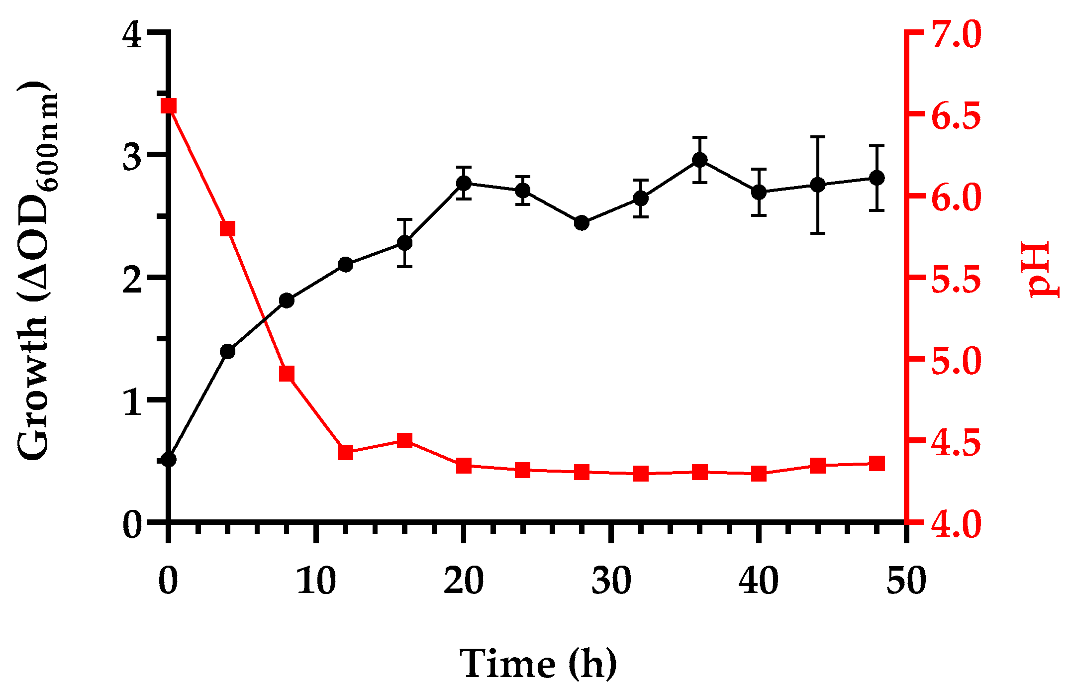
3.1. Growth and Postbiotics Production

The in vitro selection of a candidate probiotic strain involves a wide range of experiments, allowing the evaluation of functional and technologically relevant characteristics, in line with the definition of probiotics. With the understanding that these depend on fermentation conditions and are strain-specific, as an initial step the ability of L. fermentum LF53 to grow on variants of the elective medium for lactic acid bacteria (LAB), De Man Rogosa Sharp commercial media and laboratory-made variants with different carbon sources were evaluated (Figure 1). L. fermentum Lf53 grew equally well on MRS medium (HiMedia, Mumbai, India) and on the vegan variant HiVeg (Hi Media, Mumbai, India) (Figure 1a,b). The growth of the modified MRS variants (without meat extract and Tween 80) with glucose (MRS-Glu), sucrose (MRS-Suc), and lactose (MRS-Lac) as a sole carbon source showed similar parameters and dynamics, with a single main log phase after a short adaptation period in lactose growth (Figure 1e,f). Growth depends on the efficiency of lactose and sucrose degradation and the tolerance to acidity. In parallel, a laboratory modification of MRS broth (noted as veg MRS) was developed in which the casein peptone was replaced by soy peptone (Merck, Darmstadt, Germany) with no meat extract and Tween 80 added (Figure 1c). All media variants had an initial pH of 6.5. In cases of probiotic production, the rapid growth and accumulation of biomass are very important. Therefore, an additional fermentation in a Sartorius Biostat A bioreactor in MRS broth—1.5 L (Tween and meat extract omitted), at 37 °C, without pH correction—was carried out (Figure 2). The growth and dynamics in pH were monitored. Cell-free spent cultures (CFS) in MRS broth (Figure 2) were collected at 24 h and the harvested cells were used to obtain cell lysates.
During the fermentation in MRS broth with glucose in the bioreactor, with slow agitation, L. fermentum acidified rapidly to pH 4.3 in the first 10 h.

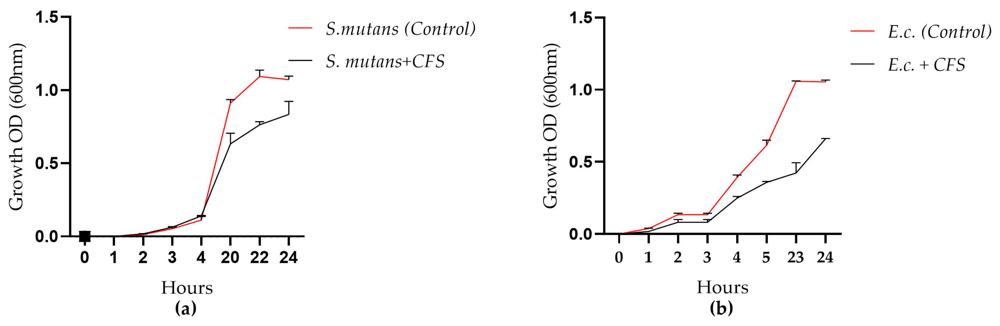
3.2. Antimicrobial Activity Assessment Against Gram (+) and Gram (−) Microorganisms

A preliminary agar diffusion assay confirmed a broad spectrum of antimicrobial activity against E. coli HB 101 (25 mm inhibition zone), E. coli ATCC 25922 (23 mm zone), and Bacillus subtilis 168 (11 mm). The acid CFS possess inhibitory activity with an inhibition zone of 20 mm against Pseudomonas aeruginosa—a clinical outpatient strain—by the agar well diffusion method. Activity against Streptococcus mutans DSMZ 20523 and E. coli ATCC 25922 was confirmed in a microplate model system in the presence of metabolites produced during the fermentation of L. fermentum Lf53 in MRS broth (Figure 3).

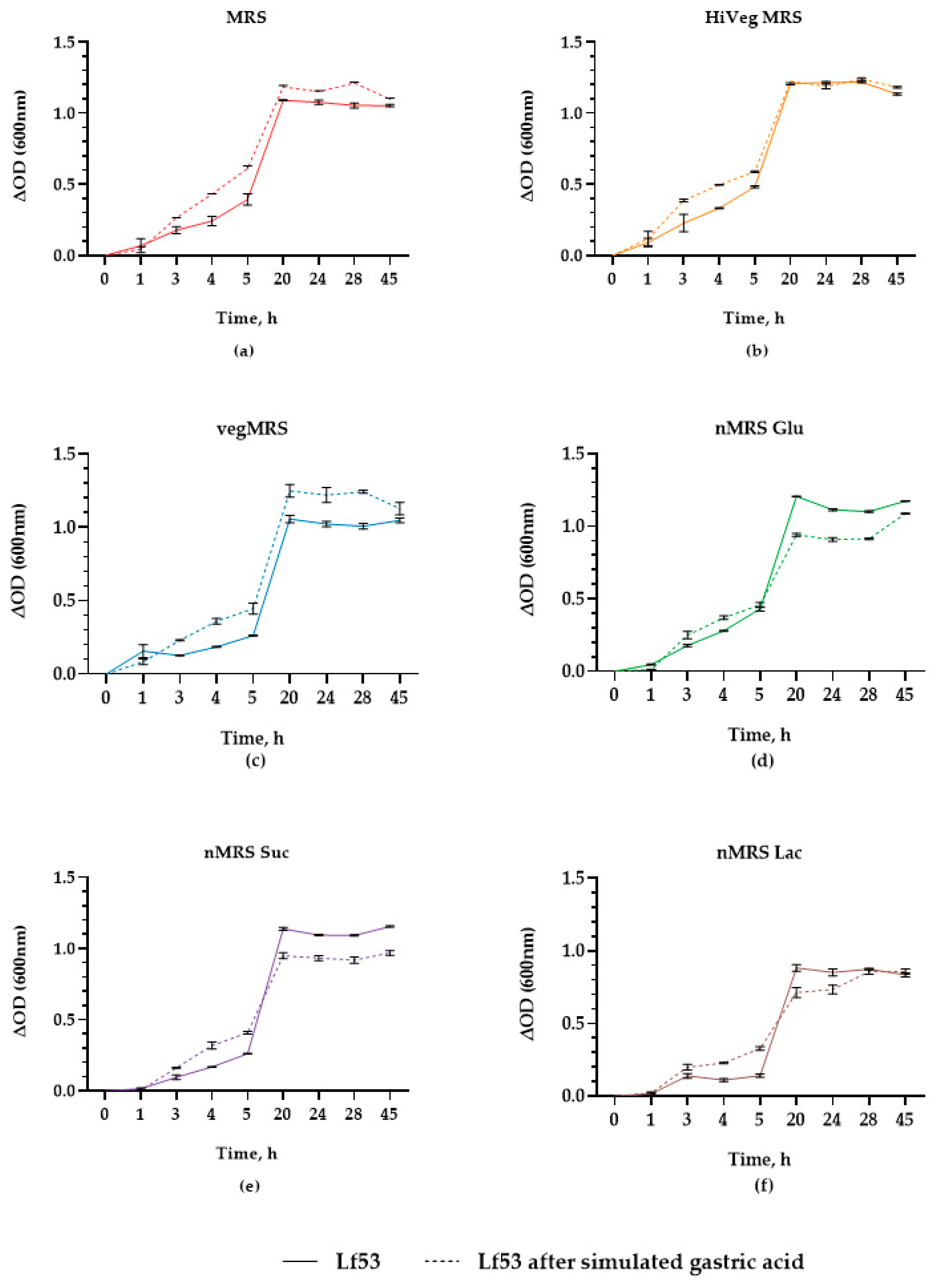
3.3. In Vitro Assessment Transit Tolerance in Simulated Gastric Conditions

The strain Lf53 was tested in vitro for transit tolerance. After 3 h of treatment in simulated gastric juice, the growth capability in the same variant of cultural MRS media was spectrophotometrically monitored (OD 600 nm) for 45 h in a microplate (Figure 1a–e, dotted lines). All variants confirmed a high viability in experimental conditions simulating passage in the GIT, which is an important probiotic characteristic.
Additional scanning electron microscopy analysis of the treated Lf53 cells confirmed their high transit tolerance. The morphological changes, in addition to growth monitoring of Lf53 (Figure 1) after gastric acid shock, were visualized (Figure 4). Experimental conditions were set to simulate the gastrointestinal tract environment, and its impact on the morphology of bacterial cells was assessed. Both the control (Figure 4a) and the sample (Figure 4b) were incubated 2 h at 37 °C in physiological saline and were monitored for possible morphological changes or injury.
The SEM micrographs primarily show the presence of multicellular populations consisting of both long and short rod-shaped cells, with a dense and multilayered pattern resembling a “honeycomb” (white star) typical of biofilm communities (Figure 4a,b). In the sample of treated cells with simulated gastric juice, numerous short and long cells were observed, including cells in the division phase with bacterial septa present (white arrow). The distribution of dividing cells in the acid sample even exceeds that in the control. Moreover, the cell membranes remained intact with smooth surfaces and no signs of altered permeability. The size of the cells was normal for the species, with no aberrantly shaped cells or visible deformations observed (Figure 4).

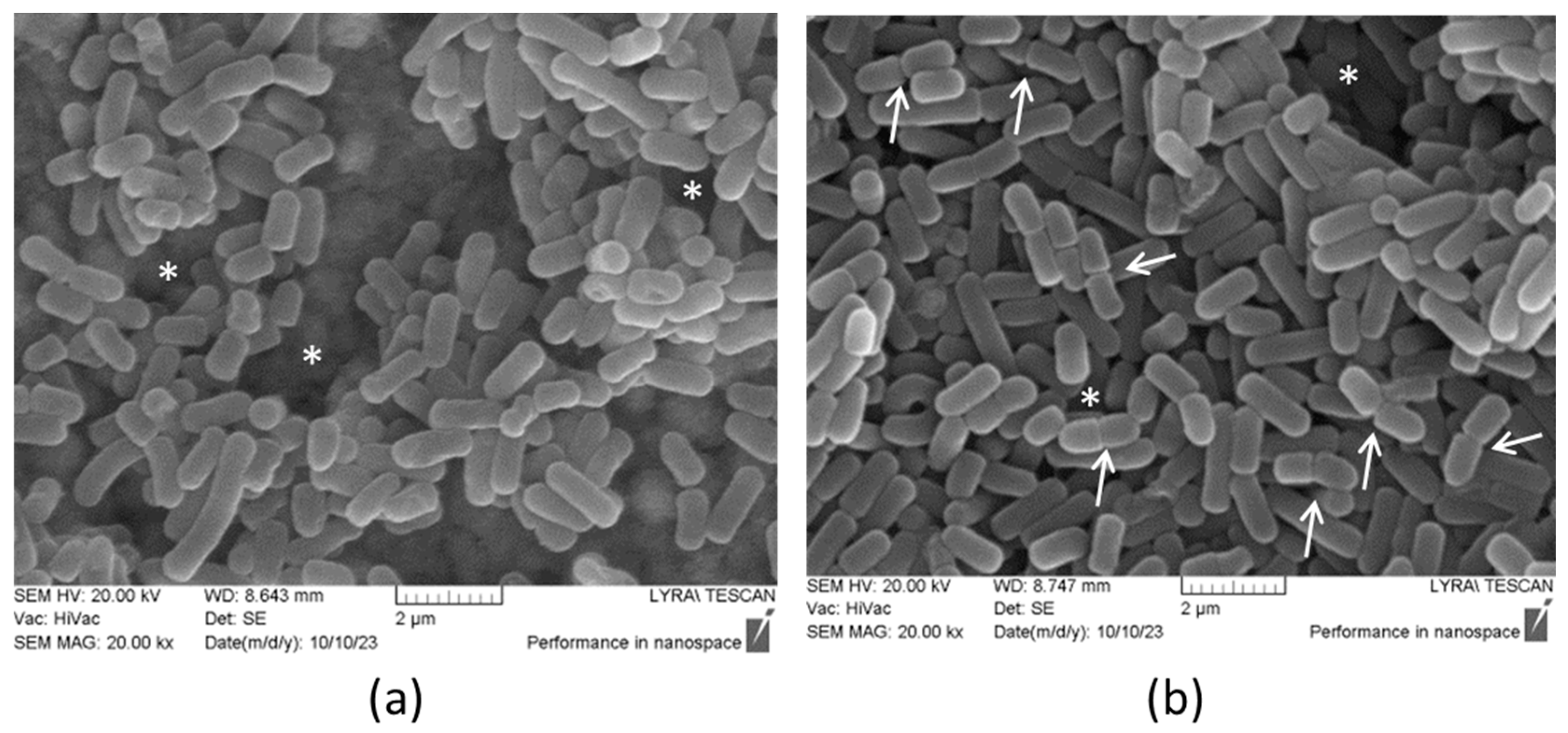
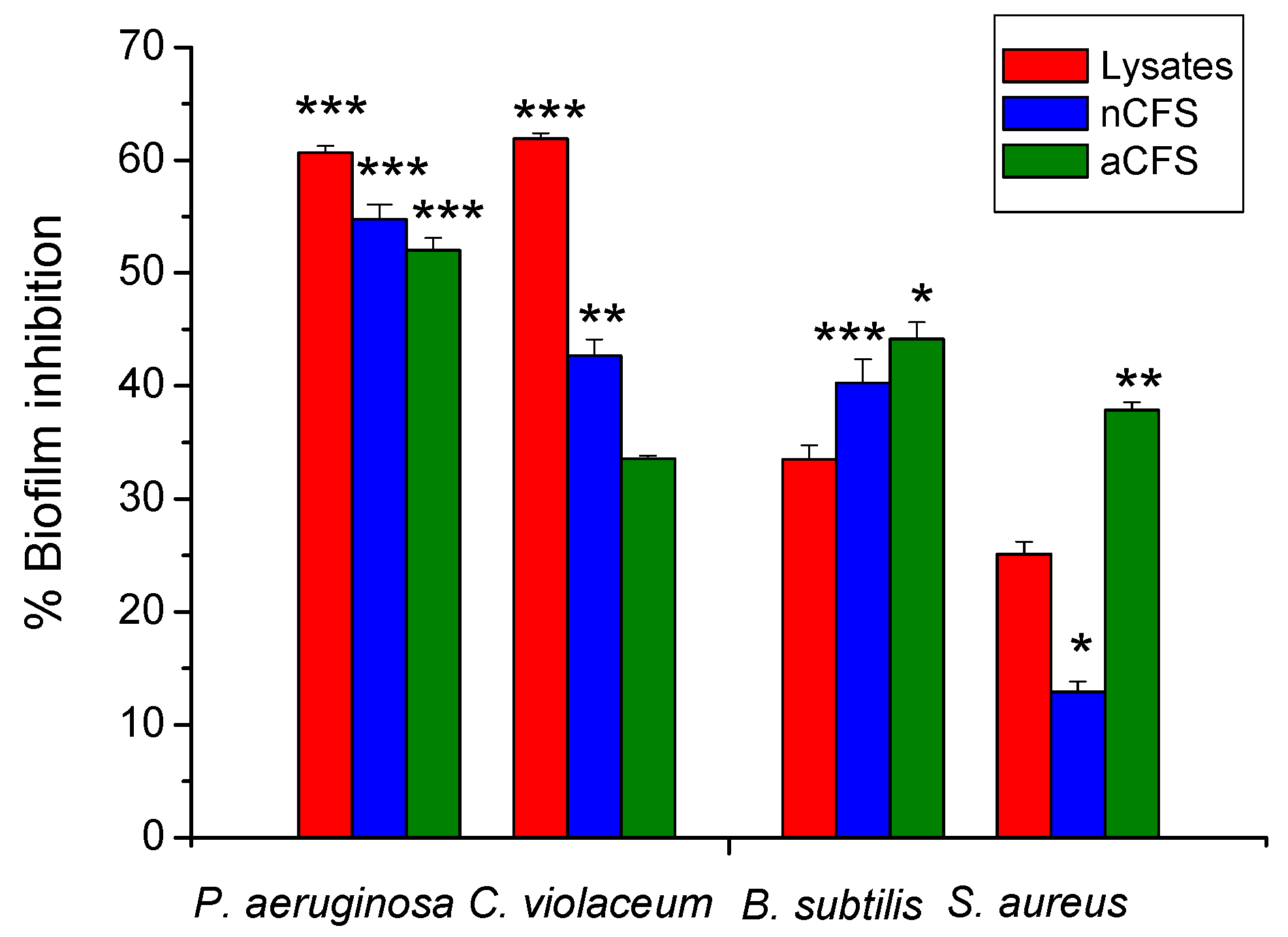
3.4. Inhibitory Activity of Produced Postbiotics on Biofilm Formation

To evaluate the inhibitory potential of the tested postbiotics on biofilm formation, a comparative analysis was conducted on four bacterial strains (see Section 2). Some of the bacterial strains used in the present study are considered potential causative agents of biofilm-associated infections and infections related to implantable and non-implantable medical devices [11].
The crystal violet staining method was applied to analyze biofilm inhibition following a 24-hour co-incubation with the postbiotics (Figure 5). The resulting data were quantified by comparison with untreated control samples. Based on the comparative analysis, both Gram-negative strains showed a consistent trend of high inhibition levels—over 50% with all three tested postbiotics types. For P. aeruginosa, the most significant biofilm inhibition was observed with the lysates (60.67%), followed by the nCFS (54.77%) and the aCFS (52.02%). A similar inhibitory pattern was noted for C. violaceum, where the lysates again showed the highest statistically significant effect, achieving 61.88% inhibition.
In contrast, the two Gram-positive strains demonstrated a different trend, with the aCFS showing greater efficacy. Biofilm formation in B. subtilis was suppressed by more than 44% following treatment with the aCFS. The lowest inhibition was recorded for S. aureus with the nCFS, showing only a 12.98% reduction in biofilm development. The analysis highlights the excellent biofilm-modulating potential of the tested postbiotics.

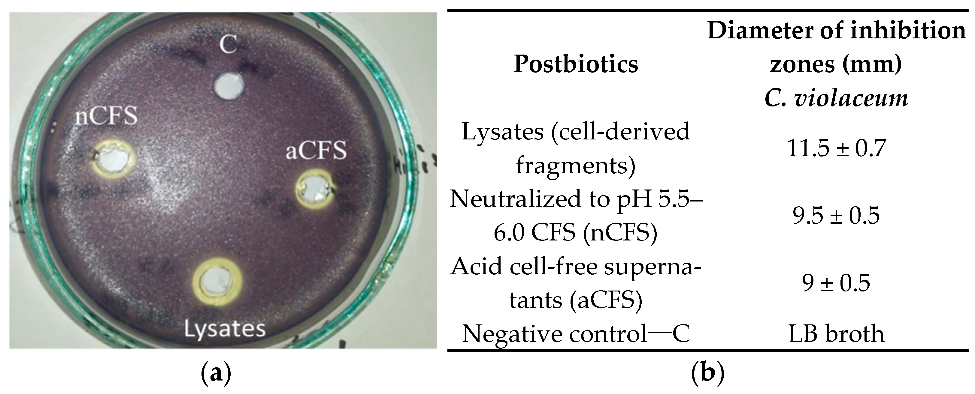
3.5. Effects of Postbiotics on Violacein Production—Qualitative Assay

To evaluate the anti-quorum sensing potential of the tested postbiotics, an agar diffusion assay was performed using the model biosensor strain C. violaceum. In contrast to the negative control (Figure 6a), which exhibited no inhibition zone, treatment with the postbiotics resulted in the appearance of inhibition zones of various diameters (Figure 6a), indicative of suppressed violacein pigment production (Figure 6b). The anti-quorum sensing activity of postbiotics ranged from 9 to 11.5 mm, with the largest inhibition zone observed for the lysed supernatant (Figure 6a,b).

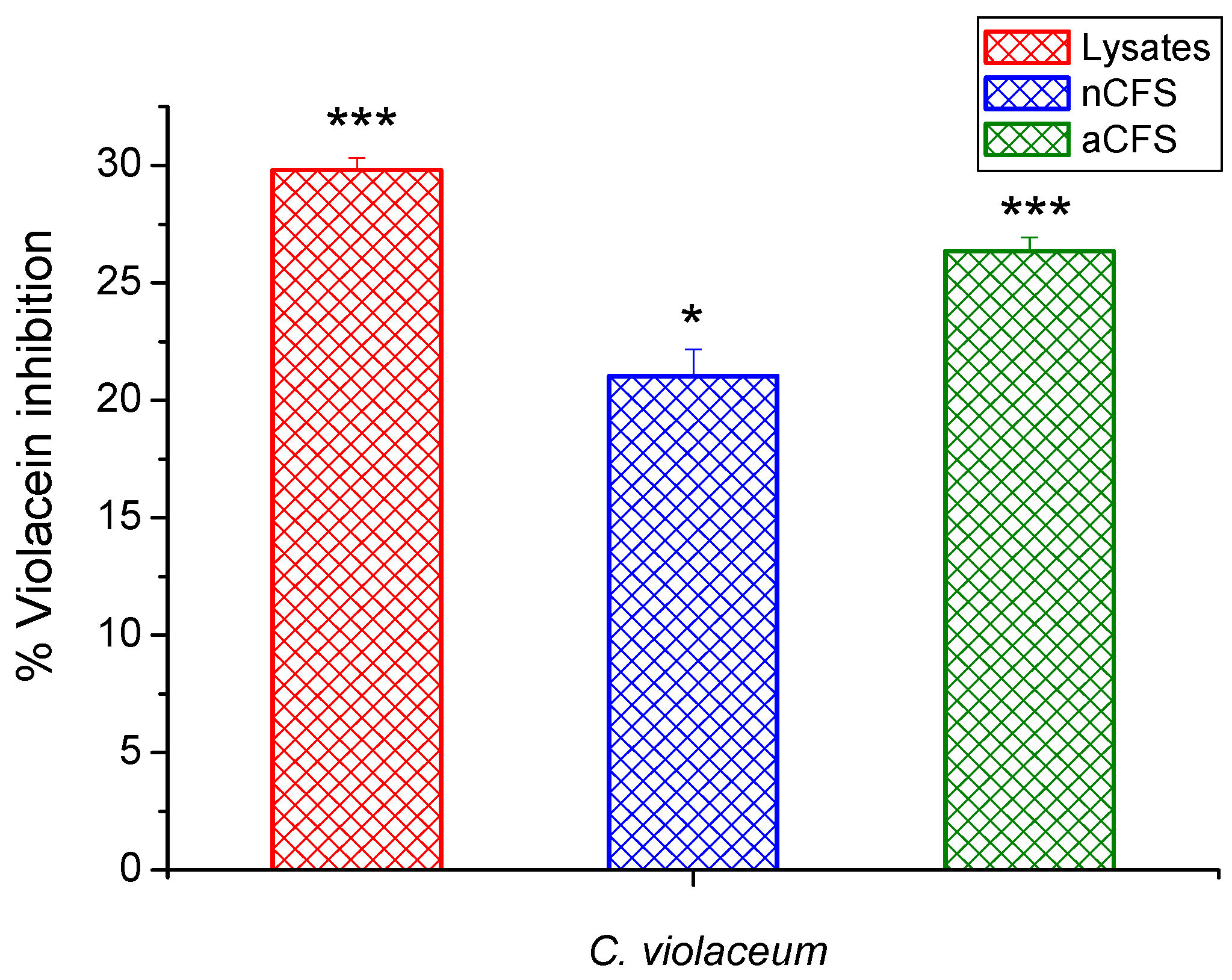
3.6. Effects of Postbiotics on Violacein Production—Quantitative Assay

To confirm the hypothesis that violacein pigment synthesis is suppressed by the tested postbiotics, we conducted a quantitative extraction of the pigment following co-cultivation with each postbiotic. The results demonstrated the highest level of pigment inhibition in the presence of the lysates (29.8%), followed by the aCFS (26.3%), with the lowest effect observed for the nCFS (21.03%) (Figure 7). These quantitative findings are consistent with the suppression pattern observed in the agar diffusion assay. Moreover, this analysis correlates with the biofilm inhibition results for C. violaceum, where the lysed sample exhibited significant biofilm-modulating activity, exceeding 60%. Our findings contribute to novel insights into the potential application of postbiotics as quorum sensing inhibitors, with specificity toward suppressing key mechanisms such as bacterial virulence and pathogenicity, an area warranting further investigation.

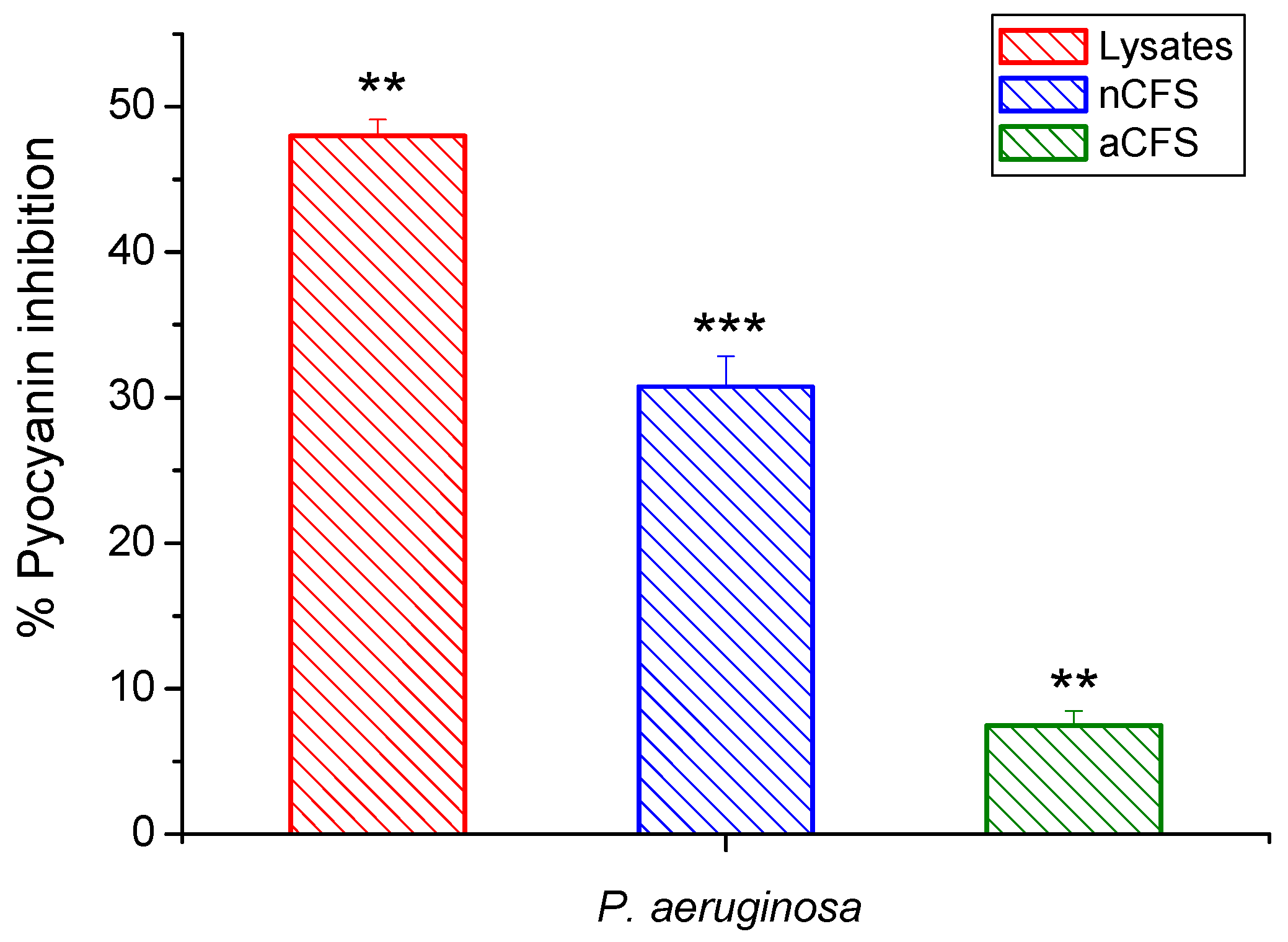
3.7. Effects of Postbiotics on Pyocyanin Production—Quantitative Assay

The conducted analysis demonstrated the ability of the tested postbiotic to inhibit the synthesis of an important virulence factor, pyocyanin. An extraction was performed to quantify the pigment levels. The highest percentage of pigment inhibition, compared to the untreated control, was observed with the lysates at 49% (Figure 8). In comparison, the inhibition percentages for the other two supernatants were 30% for the nCFS form and 7% for the aCFS. We hypothesize that certain intracellular components (such as bacteriocins) are released in the lysed form, which may suppress pyocianyn production. The nCFS showed lower, yet measurable, inhibition, likely due to a reduction in its activity after neutralization.

4. Discussion

Lactobacilli are ubiquitous and proven probiotics. They are essential microorganisms involved in various fermentation processes, producing a huge variety of metabolic products that add significant value in vivo to the health of the gut microbiome. The postbiotics/metabiotics produced are involved in barrier mechanisms against the invasion of pathogens by disease-causing microorganisms, limiting their biofilms and virulence factors. These activities are often species- and/or strain-specific and depend on many factors and bacterial growth conditions. Therefore, an initial step in the evaluation of candidate-probiotic strain Lf53 involved growth in culture medium based on commercial MRS (De Mann Rogosa Sharp) ingredients in modifications (Figure 1). The tested carbohydrates promoted appropriate growth of Lf53 (Figure 1) and likely enhanced the synthesis of desirable metabolic products. A very short lag phase was observed in cultures grown in both commercial media—MRS (HIMedia, Mumbai, India) and HiVeg MRS (HiMedia, Mumbai, India). Our results confirmed those of a study on L. plantarum strains from traditional dairy products [39]. The growth of Lf53 strain in the media with vegan composition is of practical importance, in view of the growing demand for vegan products and diet (Figure 1b). A weaker growth was shown when glucose (Merck, Germany) was replaced by disaccharides, and specifically in the case of lactose (Figure 1e,f). L. fermentum, as a heterofermentative species, is able to use several carbohydrates: maltose, mannose, melibiose, raffinose, ribose, arabinose, cellobiose, galactose, sucrose, trehalose, and xylose [40]. Some of them, however, may be strain-dependently fermented. In the variant with soya peptone in the culture medium, the same kinetics, with a very short lag phase and rapid exponential growth (Figure 1c), were seen. Garrote A. et al. (2025) reported highest cell density and rapid growth for Pediococcus acidilactici and Weissella cibaria in a medium supplemented with soya peptone [40]. The obtained results with variations with only one growth-promoting ingredient in the media as a carbon source (Figure 1 and Figure 2) could be used in the early stages of medium formulation [41].
A fast-acidifying capability of L. fermentum Lf53 (reaching pH 4.3 within the first 10 h) was observed during the fermentation of glucose in MRS broth in a bioreactor (Figure 2). Biologically, this is explainable, as the pH decrease values obtained correspond to the physiological parameters of the ecological niche from which the strain was isolated. A low pH is among the proven protective mechanisms of the urovaginal tract. This acidity has a microbicidal effect on a number of pathogens. Our preliminary studies show that values below 4.5 are reported in at least 108 CFU/mL [42]. Therefore, the selected fermentation conditions will ensure a high cell density in a short fermentation time in cases of further production of cell-derived fragments (postbiotics/parabiotics). The high acidifying ability could probably explain the observed resistance to gastric acidity (Figure 1a–f, dotted lines on graphs). Moreover, in MRS, cells treated with simulated gastric juice grew faster than untreated ones (Figure 1a). Similar growth kinetics were seen in L. fermentum 664 after 4 h exposure to a low pH of 2.0, with exceptional survival rates, reaching up to 107 ± 5% [43]. Barman et al. (2025) [44] reported that vaginal LAB strains could survive at least 2 h in acidic conditions (pH 2.5). Among them, L. fermentum 3 displayed the highest survival rate of 84% compared to the control, the widely accepted probiotic Lacticaseibacillus rhamnosus GG, at 75%. L. fermentum Lf53 fulfills the key criterion for in vitro selection to survive transport to the active site where beneficial effects are expected. Any probiotic capable of promoting eubiosis in the intestinal tract must survive passage through the acidic environment of the stomach for the time in which food normally persists. The strain-specific high transit tolerance to gastric acidity and pepsin has been a proven pre-selection criterion. These promising results are the basis for a follow-up panel to evaluate the probiotic potential of L. fermentum Lf53. Following, we adopted a modern and promising strategy aimed at evaluating the antibiofilm and anti-quorum sensing potential of metabolic products derived from LAB. This approach reflects a growing scientific interest in alternatives to conventional antimicrobials, particularly in the context of increasing resistance among pathogenic microorganisms.
The pathogenesis of biofilm-associated infections—regardless of their linkage to implantable medical devices—is frequently marked by a protracted clinical course and limited therapeutic efficacy. Biofilms’ persistent nature and inherent resistance to conventional antimicrobial treatments render their inhibition and eradication a significant clinical challenge. Compounding this issue is the global rise in antimicrobial resistance, particularly among biofilm-forming pathogens, which further accentuates the urgent need for innovative and effective antibiofilm strategies. The antibiotic resistance of biofilms is, on one hand, attributed to the structure of the exopolysaccharide matrix and on the heterogeneity of bacterial phenotypes distributed within the biofilm [11]. Due to these characteristics, pathogenic bacteria in biofilms exhibit much higher resistance to antibiotics and disinfectants compared to planktonic cells [45]. The World Health Organization classifies certain strains in the so-called ESKAPE group, known as critically important, and some of those included in this study belong to this group [46].
The anti-virulence potential of three CFS was evaluated. The results obtained from the inhibition of biofilm formation under the influence of the tested CFS metabiotics and lysates showed higher effectiveness against P. aeruginosa and C. violaceum, with a similar pattern of suppression. The highest percentage of inhibition was observed with lysates, followed by the nCFS and aCFS. Notably, data from P. aeruginosa showed that biofilm inhibition exceeded 50% for all three tested postbiotics. Furthermore, when measuring the lysate activity in C. violaceum, the values calculated against the untreated control sample revealed more than 60% biofilm-inhibiting capacity. The obtained results demonstrate and propose new, currently undescribed biofilm inhibitors isolated from a vaginal-swab sample. The antimicrobial and anti-virulence activity of Lactiplantibacillus metabolites have been documented by other authors. In a study by Ali et al. 2023, it was found that a biosurfactant produced by Lactiplantibacillus plantarum inhibits quorum sensing-regulated virulence factors and biofilm formation in P. aeruginosa and C. violaceum [18]. At sub-MIC concentrations (2.5 mg/mL), there was a reduction in pyocyanin production by 66.63%, total protease activity by 60.95%, LasA and LasB elastase activities by 51.33% and 56.62%, a decrease in exopolysaccharide production by 53.11%, and inhibition of biofilm formation by 59.80% in P. aeruginosa and 68.12% in C. violaceum. The authors confirmed that Lactobacillus metabolites effectively suppress QS and related virulence factors in these strains. Studies on the mechanism of 3-phenyllactic acid reported the inhibition of adhesion and biofilm formation in P. aeruginosa, as well as the expression of virulence factors such as pyocyanin, protease, and rhamnolipids [47]. Considering that during their growth and fermentation processes, lactobacilli produce a wide range of organic acids, it is well known that these compounds exert inhibitory effects on the growth of various bacteria, yeasts, and fungi [48,49,50,51]. Most scientific studies indicate that the observed pH reduction is largely due to lactic acid production, while other acids are synthesized to a significantly lesser extent. In this regard, we hypothesize that acidification may also contribute to the high biofilm-inhibiting activity of the supernatants. We obtained similar data for the two tested Gram-positive microorganisms—Bacillus subtilis and Staphylococcus aureus. In our previous studies, we reported the antimicrobial and biofilm-inhibiting activity of postbiotics from lactobacilli isolated from katak (a traditional fermented dairy product). We investigated their antimicrobial efficacy against E. coli and S. aureus, and demonstrated statistically significant inhibitory effects. A strong biofilm-inhibiting effect was also observed against P. aeruginosa, an opportunistic pathogen known to cause a range of infections [37]. Evidence of the antimicrobial properties of CFS has also been demonstrated against multidrug-resistant P. aeruginosa isolated from burn wound infections [52].
Our findings on the potential of lactic acid bacteria to inhibit biofilm synthesis correlate with studies conducted on Salmonella typhimurium, E. coli, S. aureus, and Bacillus spp. [23,53,54,55,56]. Probiotic microorganisms are also capable of synthesizing other types of metabolites, such as hydrogen peroxide, proteases, lysozymes, and bacteriocins. Bacteriocins are protein-based biomolecules with proven bacteriostatic and antimicrobial effects [57,58]. Several studies have reported the effectiveness of nisin against various pathogens, including S. aureus, B. subtilis, P. aeruginosa, and others. Nisin has shown greater activity against Gram-positive bacteria [59]. Recent studies suggest that bacteriocins synthesized by probiotic bacteria are relevant to antimicrobial resistance, with effects even against multidrug-resistant strains [60].
Upon reviewing the recent literature on the effects of postmetabolites from lactobacilli, we found that studies specifically addressing their anti-quorum sensing effectiveness are still relatively scarce. It is well known that bacterial intercellular communication known as quorum sensing is linked to the production, release, and recognition of signaling molecules known as autoinducers [61]. Key processes regulated by quorum sensing include biofilm formation, bioluminescence, virulence, motility, pigment synthesis, sporulation, conjugation, symbiosis, and antibiotic production [62,63,64]. In this context, we conducted both qualitative and quantitative analyses of CFS on the inhibition of the pigment violacein. The subject of this study was the biosensor strain C. violaceum. This Gram-negative bacterium is capable of synthesizing the pigment violacein, which is encoded by the vio operon, and its expression is regulated by the quorum sensing signaling cascade. This makes C. violaceum an appropriate model for studying natural quorum sensing inhibitory molecules [25]. The data from the agar diffusion assay showed clear zones of decolorization and an absence of pigment formation around the wells treated with CFS (Figure 6), following the same inhibition pattern as observed in biofilm suppression (Figure 5). Moreover, quantitative measurements of violacein production in the presence of postbiotics also demonstrated inhibition. These findings confirm the potential of the tested metabolic products as sources of anti-virulence compounds and suggest their applicability in alternative strategies, for example, against resistant microorganisms. Our data are consistent with the study of Aarata et al., 2018, which demonstrated that lysates and metabolic products from Lactobacillus species have the potential to modulate bacterial quorum sensing communication and affect virulence factors [65]. A limited number of sources report on the ability of lactobacilli to influence quorum sensing mechanisms through the suppression of violacein production in C. violaceum [66,67,68]. In this context, our previous studies revealed remarkable anti-quorum sensing activity in three types of CFS from L. plantarum strains isolated from homemade katak (fermented dairy product), expressed as clear inhibition of violacein production. Another example is a study using the C. violaceum CV026 double mini-Tn5 mutant strain, which cannot synthesize C6-N-hexanoyl-L-homoserine lactone but retains the ability to respond to C4-acyl homoserine lactones and produce violacein. The authors tested the potential of cell-free supernatants from the probiotic strain Enterococcus durans LAB38 to inhibit violacein synthesis, hypothesizing that this anti-QS activity is due to antimicrobial peptides produced by the probiotic strain [69].
Pyocyanin is a virulence factor synthesized by P. aeruginosa [70,71,72]. Pigment production in this bacterium is associated with oxidative stress and tissue damage during infections. Therefore, pyocyanin synthesis is a critical factor underlying numerous anti-virulence therapies. The extraction and quantification of pyocyanin pigment after co-cultivation with the tested postbiotics revealed the highest statistically significant inhibition percentage in the lysates. In agreement with our observations, Patel et al. (2022) reported the production of a biosurfactant from Lactiplantibacillus plantarum, whose concentrations below the MIC suppressed pyocyanin synthesis in a dose-dependent manner [67].
The high effectiveness of the tested lysates suggests that intracellular components released during cell lysis may play a significant role in inhibiting pigment formation. In the nCFS, the activity decreased but remained present, suggesting that the neutralization step is involved in modulating efficacy. Studies indicate that neutralized supernatants often retain some antimicrobial activity, but the most potent inhibitors are frequently intracellular molecules or specific pH-dependent compounds [73]. When comparing the experiments involving violacein inhibition, we observed a similar reduction in activity in the nCFS, suggesting that some compounds present in the supernatant are pH-sensitive. This also applies to the aCFS, which demonstrated a minimal inhibitory effect on pyocyanin synthesis.
The analysis of morphological changes and survival of Lactobacillus populations under simulated gastrointestinal conditions revealed that the tested strain exhibits excellent transit tolerance in low pH environments. The bacterial cells demonstrated a notable ability not only to survive but also to divide under acidic conditions. These findings highlight the importance of the surrounding medium as a critical factor for bacterial viability, particularly in the selection of candidate probiotic strains. It is well established that a significant portion of free-living microorganisms perish upon exposure to the highly acidic conditions of the gastrointestinal tract. Therefore, probiotic candidates, with antagonistic activity against pathogens, should possess the ability to withstand such acidic environments, which is essential for successful colonization of the host digestive system. Many Lactobacillus strains are naturally adapted to low pH conditions, primarily due to their metabolic ability to ferment sugars and produce lactic acid [74]. Acid resistance is widely recognized as a key selective criterion in the evaluation of probiotic potential. Supporting this, a comparative genomic study by Yao et al. (2020) identified genes in L. plantarum involved in maintaining intracellular ionic balance, the synthesis or uptake of compatible solutes, stress response, and membrane composition modulation [75]. Numerous studies have reported the isolation of acid- and bile-tolerant strains from a variety of fermented foods, including milk, sourdough, cottage cheese, pickles, kefir, sauerkraut, and others [76]. These isolates exhibit beneficial probiotic properties such as cholesterol assimilation, epithelial adhesion and colonization, non-hemolytic activity, absence of virulence genes, and strong antibacterial effects [77,78,79,80,81].

5. Conclusions

In conclusion, the present study showed a pre-selection of a promising candidate probiotic strain, L fermentum Lf53, with a broad spectrum of antimicrobial activity and high transit tolerance. We characterized the anti-virulence properties of postbiotics produced during fermentation in MRS broth with glucose as a carbon source and the cell-derived lysates (parabiotics). The results provide compelling evidence for the notable antibiofilm potential of the tested postbiotics, with pronounced activity observed particularly against Gram-negative bacterial strains. Moreover, two established quorum sensing markers—violacein and pyocyanin—were evaluated to assess the presence of quorum quenching activity. The findings demonstrated significant suppressive effects, offering new insights into the capacity of postbiotics to interfere with bacterial quorum sensing mechanisms. The morphological analysis further confirmed the structural integrity of bacterial cells under simulated gastrointestinal conditions, supporting the probiotic resilience of the strain. Our data contribute new information regarding the anti-virulence efficacy of this type of postbiotic, emphasizing their therapeutic potential as agents in the fight against antibiotic-resistant infections. The identification and application of novel bacterial virulence inhibitors could support the development of a new generation of therapeutic strategies for infectious disease control, with far-reaching implications for modern medicine and public health.

Author Contributions

Conceptualization, T.P.-K. and S.D.; methodology, T.P.-K. and S.D. software, P.D.D., D.B., L.D. and N.A.; validation, P.D.D., L.D. and D.B.; visualization, T.P.-K.; writing—original draft preparation, T.P.-K. and S.D.; writing—review and editing, T.P.-K. and S.D.; supervision, T.P.-K. All authors have read and agreed to the published version of the manuscript.

Funding

This research received no external funding.

Institutional Review Board Statement

Not applicable.

Informed Consent Statement

Not applicable.

Data Availability Statement

The original contributions presented in this study are included in the article. Further inquiries can be directed to the corresponding author.

Acknowledgments

Project NoBG16RFPR002-1.014-0017 Center of Competence, “Fundamental, Translational and Clinical Investigations of Infections and Immunity”, funded by the “Scientific, Research, Innovations and Digitalization for Intelligent Transformations 2021–2027” Programme.

Conflicts of Interest

The authors declare no conflicts of interest.

References

  1. Hill, C.; Guarner, F.; Reid, G.; Gibson, G.R.; Merenstein, D.J.; Pot, B.; Morelli, L.; Canani, R.B.; Flint, H.J.; Salminen, S.; et al. The International Scientific Association for Probiotics and Prebiotics consensus statement on the scope and appropriate use of the term probiotic. Nat. Rev. Gastroenterol. Hepatol. 2014, 11, 506–514. [Google Scholar] [CrossRef] [PubMed]
  2. Maldonado Galdeano, C.; Cazorla, S.I.; Lemme Dumit, J.M.; Vélez, E.; Perdigón, G. Beneficial Effects of Probiotic Consumption on the Immune System. Ann. Nutr. Metab. 2019, 74, 115–124. [Google Scholar] [CrossRef] [PubMed]
  3. Safarchi, A.; Al-Qadami, G.; Tran, C.D.; Conlon, M. Understanding dysbiosis and resilience in the human gut microbiome: Biomarkers, interventions, and challenges. Front. Microbiol. 2025, 16, 1559521. [Google Scholar] [CrossRef]
  4. Mokoena, M.P.; Omatola, C.A.; Olaniran, A.O. Applications of Lactic Acid Bacteria and Their Bacteriocins against Food Spoilage Microorganisms and Foodborne Pathogens. Molecules 2021, 26, 7055. [Google Scholar] [CrossRef]
  5. Ayivi, R.D.; Gyawali, R.; Krastanov, A.; Aljaloud, S.O.; Worku, M.; Tahergorabi, R.; Silva, R.C.D.; Ibrahim, S.A. Lactic Acid Bacteria: Food Safety and Human Health Applications. Dairy 2020, 1, 202–232. [Google Scholar] [CrossRef]
  6. Salminen, S.; Collado, M.C.; Endo, A.; Hill, C.; Lebeer, S.; Quigley, E.M.M.; Sanders, M.E.; Shamir, R.; Swann, J.R.; Szajewska, H.; et al. The International Scientific Association of Probiotics and Prebiotics (ISAPP) consensus statement on the definition and scope of postbiotics. Nat. Rev. Gastroenterol. Hepatol. 2021, 18, 649–667. [Google Scholar] [CrossRef]
  7. Resta-Lenert, S. Live probiotics protect intestinal epithelial cells from the effects of infection with enteroinvasive Escherichia coli (EIEC). Gut 2003, 52, 988–997. [Google Scholar] [CrossRef]
  8. Shibahara-Sone, H.; Gomi, A.; Iino, T.; Kano, M.; Nonaka, C.; Watanabe, O.; Miyazaki, K.; Ohkusa, T. Living cells of probiotic Bifidobacterium bifidum YIT 10347 detected on gastric mucosa in humans. Benef. Microbes 2016, 7, 319–326. [Google Scholar] [CrossRef]
  9. Nemati, M.; Omrani, G.R.; Ebrahimi, B.; Montazeri-Najafabady, N. The Beneficial Effects of Probiotics via Autophagy: A Systematic Review. BioMed Res. Int. 2021, 2021, 2931580. [Google Scholar] [CrossRef]
  10. Damyanova, T.; Dimitrova, P.D.; Borisova, D.; Topouzova-Hristova, T.; Haladjova, E.; Paunova-Krasteva, T. An Overview of Biofilm-Associated Infections and the Role of Phytochemicals and Nanomaterials in Their Control and Prevention. Pharmaceutics 2024, 16, 162. [Google Scholar] [CrossRef]
  11. Damyanova, T.; Paunova-Krasteva, T. What We Still Don’t Know About Biofilms—Current Overview and Key Research Information. Microbiol. Res. 2025, 16, 46. [Google Scholar] [CrossRef]
  12. Johani, K.; Malone, M.; Jensen, S.; Gosbell, I.; Dickson, H.; Hu, H.; Vickery, K. Microscopy visualisation confirms multi-species biofilms are ubiquitous in diabetic foot ulcers. Int. Wound J. 2017, 14, 1160–1169. [Google Scholar] [CrossRef]
  13. Garg, S.S.; Dubey, R.; Sharma, S.; Vyas, A.; Gupta, J. Biological macromolecules-based nanoformulation in improving wound healing and bacterial biofilm-associated infection: A review. Int. J. Biol. Macromol. 2023, 247, 125636. [Google Scholar] [CrossRef]
  14. Borisova, D.; Strateva, T.; Dimov, S.G.; Atanassova, B.; Paunova-Krasteva, T.; Topouzova-Hristova, T.; Danova, S.T.; Tropcheva, R.; Stoitsova, S. Diversification of Pseudomonas aeruginosa After Inhaled Tobramycin Therapy of Cystic Fibrosis Patients: Genotypic and Phenotypic Characteristics of Paired Pre- and Post-Treatment Isolates. Microorganisms 2025, 13, 730. [Google Scholar] [CrossRef]
  15. Fatima, H.; Gupta, S.; Khare, S.K. Meropenem incorporated nanoflowers as nano antibiotics: A promising strategy for combating antimicrobial resistance and biofilm-associated infections. Inorg. Chem. Commun. 2025, 177, 114362. [Google Scholar] [CrossRef]
  16. Assefa, M.; Amare, A. Biofilm-Associated Multi-Drug Resistance in Hospital-Acquired Infections: A Review. Infect. Drug Resist. 2022, 15, 5061–5068. [Google Scholar] [CrossRef]
  17. Ciofu, O.; Moser, C.; Jensen, P.Ø.; Høiby, N. Tolerance and resistance of microbial biofilms. Nat. Rev. Microbiol. 2022, 20, 621–635. [Google Scholar] [CrossRef] [PubMed]
  18. Ali, A.; Zahra, A.; Kamthan, M.; Husain, F.M.; Albalawi, T.; Zubair, M.; Alatawy, R.; Abid, M.; Noorani, M.S. Microbial Biofilms: Applications, Clinical Consequences, and Alternative Therapies. Microorganisms 2023, 11, 1934. [Google Scholar] [CrossRef]
  19. Shaikh, S.; Lapin, N.A.; Prasad, B.; Sturge, C.R.; Pybus, C.; Pifer, R.; Wang, Q.; Evers, B.M.; Chopra, R.; Greenberg, D.E. Intermittent alternating magnetic fields diminish metal-associated biofilm in vivo. Sci. Rep. 2023, 13, 22456. [Google Scholar] [CrossRef]
  20. Werneburg, G.T.; Hettel, D.; Goldman, H.B.; Vasavada, S.P.; Miller, A.W. Indwelling Urological Device Biofilm Composition and Characteristics in the Presence and Absence of Infection. Urology 2025, 196, 82–89. [Google Scholar] [CrossRef] [PubMed]
  21. Adnan, M.; Siddiqui, A.J.; Noumi, E.; Ashraf, S.A.; Awadelkareem, A.M.; Hadi, S.; Snoussi, M.; Badraoui, R.; Bardakci, F.; Sachidanandan, M.; et al. Biosurfactant derived from probiotic Lactobacillus acidophilus exhibits broad-spectrum antibiofilm activity and inhibits the quorum sensing-regulated virulence. Biomol. Biomed. 2023, 23, 1051–1068. [Google Scholar] [CrossRef] [PubMed]
  22. Poon, Y.; Hui, M. Inhibitory effect of lactobacilli supernatants on biofilm and filamentation of Candida albicans, Candida tropicalis, and Candida parapsilosis. Front. Microbiol. 2023, 14, 1105949. [Google Scholar] [CrossRef]
  23. Parappilly, S.J.; Radhakrishnan, E.K.; George, S.M. Antibacterial and antibiofilm activity of human gut lactic acid bacteria. Braz. J. Microbiol. 2024, 55, 3529–3539. [Google Scholar] [CrossRef]
  24. Pourhajibagher, M.; Bahador, A. In vitro Anti-biofilm and Anti-adhesion Effects of Lactic Acid Bacteria- derived Biosurfactants against Streptococcus mutans. Infect. Disord.-Drug Targets 2024, 25, 4. [Google Scholar] [CrossRef]
  25. Dimitrova, P.D.; Damyanova, T.; Paunova-Krasteva, T. Chromobacterium Violaceum: A Model for Evaluating the Anti-Quorum Sensing Activities of Plant Substances. Sci. Pharm. 2023, 91, 33. [Google Scholar] [CrossRef]
  26. Zhong, S.; He, S. Quorum Sensing Inhibition or Quenching in Acinetobacter baumannii: The Novel Therapeutic Strategies for New Drug Development. Front. Microbiol. 2021, 12, 558003. [Google Scholar] [CrossRef] [PubMed]
  27. Aman, M.; Aneeqha, N.; Bristi, K.; Deeksha, J.; Afza, N.; Sindhuja, V.; Shastry, R.P. Lactic acid bacteria inhibits quorum sensing and biofilm formation of Pseudomonas aeruginosa strain JUPG01 isolated from rancid butter. Biocatal. Agric. Biotechnol. 2021, 36, 102115. [Google Scholar] [CrossRef]
  28. Díaz, M.A.; González, S.N.; Alberto, M.R.; Arena, M.E. Human probiotic bacteria attenuate Pseudomonas aeruginosa biofilm and virulence by quorum-sensing inhibition. Biofouling 2020, 36, 597–609. [Google Scholar] [CrossRef]
  29. FAO/WHO. Joint FAO/WHO Working Group Guidelines for the Evaluation of Probiotics in Food: Report of a Joint FAO/WHO Working Group on Drafting Guidelines for the Evaluation of Probiotics in Food; World Health Organization and Food and Agriculture Organization of the United Nations: Rome, Italy, 2002. [Google Scholar]
  30. Lin, T.-L.; Shu, C.-C.; Lai, W.-F.; Tzeng, C.-M.; Lai, H.-C.; Lu, C.-C. Investiture of next generation probiotics on amelioration of diseases—Strains do matter. Med. Microecol. 2019, 1, 100002. [Google Scholar] [CrossRef]
  31. Petrova, M.; Georgieva, R.; Dojchinovska, L.; Kirilov, N.; Iliev, I.; Antonova, S.; Hadjieva, N.; Ivanova, I.; Danova, S. Lactic acid bacteria against pathogenic microbes. Trakia J. Sci. 2009, 7, 33–39. [Google Scholar]
  32. Moskova-Doumanova, V.; Vaseva, A.; Veleva, R.; Mladenova, K.; Melniska, D.; Doumanov, J.; Videv, P.; Topouzova-Hristova, T.; Dobreva, L.; Atanasova, N.; et al. In Vitro Effects of Postmetabolites from Limosilactobacillus fermentum 53 on the Survival and Proliferation of HT-29 Cells. Microorganisms 2024, 12, 1365. [Google Scholar] [CrossRef] [PubMed]
  33. Danova, S. Biodiversity and Probiotic Potential of Lactic Acid Bacteria from Various Ecological Niches. Ph.D. Thesis, Stefan Angeloff Institute of Microbiology, Bulgarian Academy of Sciences, Sofia, Bulgaria, 2015. [Google Scholar]
  34. Dobreva, L.; Atanasova, N.; Donchev, P.; Krumova, E.; Abrashev, R.; Karakirova, Y.; Mladenova, R.; Tolchkov, V.; Ralchev, N.; Dishliyska, V.; et al. Candidate-Probiotic Lactobacilli and Their Postbiotics as Health-Benefit Promoters. Microorganisms 2024, 12, 1910. [Google Scholar] [CrossRef]
  35. De Soyza, A.; Hall, A.J.; Mahenthiralingam, E.; Drevinek, P.; Kaca, W.; Drulis-Kawa, Z.; Stoitsova, S.R.; Toth, V.; Coenye, T.; Zlosnik, J.E.A.; et al. Developing an international Pseudomonas aeruginosa reference panel. MicrobiologyOpen 2013, 2, 1010–1023. [Google Scholar] [CrossRef] [PubMed]
  36. Dimitrova, P.D.; Ivanova, V.; Trendafilova, A.; Paunova-Krasteva, T. Anti-Biofilm and Anti-Quorum-Sensing Activity of Inula Extracts: A Strategy for Modulating Chromobacterium violaceum Virulence Factors. Pharmaceuticals 2024, 17, 573. [Google Scholar] [CrossRef]
  37. Dobreva, L. Functional Characteristic of Lactic Acid Bacteria from Different Habitats. Ph.D. Thesis, The Stephan Angeloff Institute of Microbiology, Bulgarian Academy of Sciences, Sofia, Bulgaria, 2023. [Google Scholar]
  38. Lou, Z.; Wang, H.; Tang, Y.; Chen, X. The effect of burdock leaf fraction on adhesion, biofilm formation, quorum sensing and virulence factors of Pseudomonas aeruginosa. J. Appl. Microbiol. 2017, 122, 615–624. [Google Scholar] [CrossRef]
  39. Nemska, V.; Danova, S.; Georgieva, N. In Vitro Assessment of Lactobacillus spp. to Grow in the Presence of Different Carbon Sources in the Medium. J. Chem. Technol. Met. 2022, 57, 487–495. [Google Scholar]
  40. Garrote Achou, C.; Cantalejo Díez, M.J.; Diaz Cano, J.; Molinos Equiza, X. Evaluation of Different Nutritional Sources in Lactic Acid Bacteria Fermentation for Sustainable Postbiotic Production. Foods 2025, 14, 649. [Google Scholar] [CrossRef]
  41. Singh, V.; Haque, S.; Niwas, R.; Srivastava, A.; Pasupuleti, M.; Tripathi, C.K.M. Strategies for Fermentation Medium Optimization: An In-Depth Review. Front. Microbiol. 2017, 7, 2087. [Google Scholar] [CrossRef] [PubMed]
  42. Dimitonova, S. Biological Properties of Vaginal Lactobacilli. Ph.D. Thesis, SU, St Kl. Ohridski, Sofia, Bulgaria, 2007. [Google Scholar]
  43. Hao, H.; Nie, Z.; Wu, Y.; Liu, Z.; Luo, F.; Deng, F.; Zhao, L. Probiotic Characteristics and Anti-Inflammatory Effects of Limosilactobacillus fermentum 664 Isolated from Chinese Fermented Pickles. Antioxidants 2024, 13, 703. [Google Scholar] [CrossRef]
  44. Barman, I.; Seo, H.; Kim, S.; Rahim, M.A.; Yoon, Y.; Hossain, M.S.; Shuvo, M.S.H.; Song, H.-Y. Isolation of New Strains of Lactic Acid Bacteria from the Vaginal Microbiome of Postmenopausal Women and their Probiotic Characteristics. Curr. Microbiol. 2025, 82, 76. [Google Scholar] [CrossRef]
  45. Abebe, G.M. The Role of Bacterial Biofilm in Antibiotic Resistance and Food Contamination. Int. J. Microbiol. 2020, 2020, 1705814. [Google Scholar] [CrossRef] [PubMed]
  46. Davies, O.L.; Simeon, B. WHO Publishes List of Bacteria for Which New Antibiotics are Urgently Needed; World Health Organization: Geneva, Switzerland, 2017. [Google Scholar]
  47. Chatterjee, M.; D’Morris, S.; Paul, V.; Warrier, S.; Vasudevan, A.K.; Vanuopadath, M.; Nair, S.S.; Paul-Prasanth, B.; Mohan, C.G.; Biswas, R. Mechanistic understanding of Phenyllactic acid mediated inhibition of quorum sensing and biofilm development in Pseudomonas aeruginosa. Appl. Microbiol. Biotechnol. 2017, 101, 8223–8236. [Google Scholar] [CrossRef]
  48. Anokhina, I.V.; Kravtsov, E.G.; Protsenko, A.V.; Yashina, N.V.; Yermolaev, A.V.; Chesnokova, V.L.; Dalin, M.V. Bactericidal activity of culture fluid components of Lactobacillus fermentum strain 90 TS-4 (21) clone 3, and their capacity to modulate adhesion of Candida Albicans yeast-like fungi to vaginal epithelial cells. Bull. Exp. Biol. Med. 2007, 143, 359–362. [Google Scholar] [CrossRef] [PubMed]
  49. Stanojević-Nikolić, S.; Dimić, G.; Mojović, L.; Pejin, J.; Djukić-Vuković, A.; Kocić-Tanackov, S. Antimicrobial Activity of Lactic Acid Against Pathogen and Spoilage Microorganisms: Antimicrobial Activity of Lactic Acid. J. Food Process. Preserv. 2016, 40, 990–998. [Google Scholar] [CrossRef]
  50. Girma, A.; Aemiro, A. Antibacterial Activity of Lactic Acid Bacteria Isolated from Fermented Ethiopian Traditional Dairy Products against Food Spoilage and Pathogenic Bacterial Strains. J. Food Qual. 2021, 2021, 9978561. [Google Scholar] [CrossRef]
  51. Amenu, D.; Bacha, K. Antagonistic Effects of Lactic Acid Bacteria Isolated from Ethiopian Traditional Fermented Foods and Beverages Against Foodborne Pathogens. Probiotics Antimicrob. Proteins 2025, 17, 1674–1687. [Google Scholar] [CrossRef]
  52. Anwer, A.W.; Ahmed, M.E. Antimicrobial Susceptibility of Fructophilic Lactic Acid Bacteria on PHZM Gene of Pseudomonas Aeruginosa Isolates from Wounds Infected. Acta Medica Bulg. 2024, 51, 52–58. [Google Scholar] [CrossRef]
  53. Jiang, X.; Yan, X.; Gu, S.; Yang, Y.; Zhao, L.; He, X.; Chen, H.; Ge, J.; Liu, D. Biosurfactants of Lactobacillus helveticus for Biodiversity Inhibit the Biofilm Formation of Staphylococcus aureus and Cell Invasion. Future Microbiol. 2019, 14, 1133–1146. [Google Scholar] [CrossRef]
  54. Akova, B.; Kıvanç, S.A.; Kıvanç, M. Antibiofilm effect of probiotic lactic acid bacteria against Bacillus spp obtained from the ocular surface. Eur. Rev. Med. Pharmacol. Sci. 2021, 25, 7799–7805. [Google Scholar] [CrossRef]
  55. Hindieh, P.; Yaghi, J.; Assaf, J.C.; Chokr, A.; Atoui, A.; Louka, N.; Khoury, A.E. Unlocking the potential of lactic acid bacteria mature biofilm extracts as antibiofilm agents. AMB Express 2024, 14, 112. [Google Scholar] [CrossRef]
  56. Göksel, Ş.; Akçelik, N.; Özdemir, C.; Akçelik, M. The Effects of Lactic Acid Bacteria on Salmonella Biofilms. Microbiology 2022, 91, 278–285. [Google Scholar] [CrossRef]
  57. Gong, H.; Meng, X.; Wang, H. Mode of action of plantaricin MG, a bacteriocin active against Salmonella typhimurium. J. Basic. Microbiol. 2010, 50, S37–S45. [Google Scholar] [CrossRef] [PubMed]
  58. Xie, Y.; An, H.; Hao, Y.; Qin, Q.; Huang, Y.; Luo, Y.; Zhang, L. Characterization of an anti-Listeria bacteriocin produced by Lactobacillus plantarum LB-B1 isolated from koumiss, a traditionally fermented dairy product from China. Food Control 2011, 22, 1027–1031. [Google Scholar] [CrossRef]
  59. Mangalanayaki, R.; Bala, A. Bacteriocin production using lactic acid bacteria. Biomed. Pharmacol. J. 2015, 3, 413–416. [Google Scholar]
  60. Ahmed, M. Enterococcus faecium Bacteriocin Efflux Pump MexA Gene and promote skin wound healing in mice. ESS Open Arch. Eprints 2023, 11, 01143350. [Google Scholar] [CrossRef]
  61. Ahmed, S.O.; Zedan, H.H.; Ibrahim, Y.M. Quorum sensing inhibitory effect of bergamot oil and aspidosperma extract against Chromobacterium violaceum and Pseudomonas aeruginosa. Arch. Microbiol. 2021, 203, 4663–4675. [Google Scholar] [CrossRef]
  62. Juszczuk-Kubiak, E. Molecular Aspects of the Functioning of Pathogenic Bacteria Biofilm Based on Quorum Sensing (QS) Signal-Response System and Innovative Non-Antibiotic Strategies for Their Elimination. Int. J. Mol. Sci. 2024, 25, 2655. [Google Scholar] [CrossRef]
  63. Mohan, M.S.; Salim, S.A.; Ranganathan, S.; Parasuraman, P.; Anju, V.T.; Ampasala, D.R.; Dyavaiah, M.; Lee, J.-K.; Busi, S. Attenuation of Las/Rhl quorum sensing regulated virulence and biofilm formation in Pseudomonas aeruginosa PAO1 by Artocarpesin. Microb. Pathog. 2024, 189, 106609. [Google Scholar] [CrossRef]
  64. Das, T.; Young, B.C. Biofilm Formation by Pathogenic Bacteria: The Role of Quorum Sensing and Physical-Chemical Interactions. In Focus on Bacterial Biofilms; Das, T., Ed.; IntechOpen: London, UK, 2022; ISBN 978-1-80355-795-3. [Google Scholar]
  65. Aarti, C.; Khusro, A.; Varghese, R.; Arasu, M.V.; Agastian, P.; Al-Dhabi, N.A.; Ilavenil, S.; Choi, K.C. In vitro investigation on probiotic, anti-Candida, and antibiofilm properties of Lactobacillus pentosus strain LAP1. Arch. Oral. Biol. 2018, 89, 99–106. [Google Scholar] [CrossRef]
  66. Savijoki, K.; San-Martin-Galindo, P.; Pitkänen, K.; Edelmann, M.; Sillanpää, A.; Van Der Velde, C.; Miettinen, I.; Patel, J.Z.; Yli-Kauhaluoma, J.; Parikka, M.; et al. Food-Grade Bacteria Combat Pathogens by Blocking AHL-Mediated Quorum Sensing and Biofilm Formation. Foods 2022, 12, 90. [Google Scholar] [CrossRef]
  67. Patel, M.; Siddiqui, A.J.; Ashraf, S.A.; Surti, M.; Awadelkareem, A.M.; Snoussi, M.; Hamadou, W.S.; Bardakci, F.; Jamal, A.; Jahan, S.; et al. Lactiplantibacillus plantarum-Derived Biosurfactant Attenuates Quorum Sensing-Mediated Virulence and Biofilm Formation in Pseudomonas aeruginosa and Chromobacterium violaceum. Microorganisms 2022, 10, 1026. [Google Scholar] [CrossRef] [PubMed]
  68. Kostoglou, D.; Giaouris, E. Screening of Lactic Acid Bacteria Isolated from Foods for Interference with Bacterial Quorum Sensing Systems. In Proceedings of the 5th International Electronic Conference on Foods, Basel, Switzerland, 28–30 October 2024; MDPI: Basel, Switzerland, 2025; p. 19. [Google Scholar]
  69. Natesan, S.; Boopathi, S.; Selvakumar, G. Quorum Quenching Potentials of Probiotic Enterococcus Durans Lab38 Against Methicillin Resistant Staphylococcus Aureus. Asian J. Pharm. Clin. Res. 2017, 10, 445. [Google Scholar] [CrossRef]
  70. Miller, L.C.; O’Loughlin, C.T.; Zhang, Z.; Siryaporn, A.; Silpe, J.E.; Bassler, B.L.; Semmelhack, M.F. Development of Potent Inhibitors of Pyocyanin Production in Pseudomonas aeruginosa. J. Med. Chem. 2015, 58, 1298–1306. [Google Scholar] [CrossRef]
  71. Rai, D.; Sulthana, F.; Neeksha; Divyashree, M. Production and Antimicrobial Activities of Pyocyanin Extracts from Pseudomonas aeruginosa. J. Pure Appl. Microbiol. 2024, 18, 2371–2379. [Google Scholar] [CrossRef]
  72. Marey, M.A.; Abozahra, R.; El-Nikhely, N.A.; Kamal, M.F.; Abdelhamid, S.M.; El-Kholy, M.A. Transforming microbial pigment into therapeutic revelation: Extraction and characterization of pyocyanin from Pseudomonas aeruginosa and its therapeutic potential as an antibacterial and anticancer agent. Microb. Cell Factories 2024, 23, 174. [Google Scholar] [CrossRef]
  73. Algburi, A.; Comito, N.; Kashtanov, D.; Dicks, L.M.T.; Chikindas, M.L. Control of Biofilm Formation: Antibiotics and Beyond. Appl. Environ. Microbiol. 2017, 83, e02508-16. [Google Scholar] [CrossRef]
  74. Van Zyl, W.F.; Deane, S.M.; Dicks, L.M.T. Molecular insights into probiotic mechanisms of action employed against intestinal pathogenic bacteria. Gut Microbes 2020, 12, 1831339. [Google Scholar] [CrossRef]
  75. Yao, W.; Yang, L.; Shao, Z.; Xie, L.; Chen, L. Identification of salt tolerance-related genes of Lactobacillus plantarum D31 and T9 strains by genomic analysis. Ann. Microbiol. 2020, 70, 10. [Google Scholar] [CrossRef]
  76. Georgieva, R.N.; Iliev, I.N.; Chipeva, V.A.; Dimitonova, S.P.; Samelis, J.; Danova, S.T. Identification and in vitro characterisation of Lactobacillus plantarum strains from artisanal Bulgarian white brined cheeses. J. Basic. Microbiol. 2008, 48, 234–244. [Google Scholar] [CrossRef]
  77. Dobreva, L.; Borisova, D.; Paunova-Krasteva, T.; Dimitrova, P.D.; Hubenov, V.; Atanasova, N.; Ivanov, I.; Danova, S. From Traditional Dairy Product “Katak” to Beneficial Lactiplantibacillus plantarum Strains. Microorganisms 2023, 11, 2847. [Google Scholar] [CrossRef]
  78. Albene, D.; Lema, N.K.; Tesfaye, G.; Andeta, A.F.; Ali, K.; Guadie, A. Probiotic potential of lactic acid bacteria isolated from Ethiopian traditional fermented Cheka beverage. Ann. Microbiol. 2024, 74, 25. [Google Scholar] [CrossRef]
  79. Sana, M.; Ouarda, A.Z.; Nassereddine, Z.M.; Magalie, B.; Mohamed, B.A.; Nathalie, C. Probiotic potential of indigenous Lactobacillus strains isolated from Algerian goatskin bag cheese Bouhezza using physiological and comparative genomic analyses. Int. Dairy J. 2025, 165, 106206. [Google Scholar] [CrossRef]
  80. Madushanka, D.; Vidanarachchi, J.K.; Kodithuwakku, S.; Nayanajith, G.R.A.; Jayatilake, S.; Priyashantha, H. Isolation and characterization of probiotic lactic acid bacteria from fermented traditional rice for potential applications in food and livestock production. Appl. Food Res. 2025, 5, 100865. [Google Scholar] [CrossRef]
  81. Wei, G.; Wang, D.; Wang, T.; Wang, G.; Chai, Y.; Li, Y.; Mei, M.; Wang, H.; Huang, A. Probiotic potential and safety properties of Limosilactobacillus fermentum A51 with high exopolysaccharide production. Front. Microbiol. 2025, 16, 1498352. [Google Scholar] [CrossRef]
Figure 1. Growth of L. fermentum Lf53 (straight lines) in commercial MRS broth (Hi Media, Mumbai, India) and HiVeg MRS (a,b) and laboratory-prepared modified MRS variants (without meat extract and Tween 80): with glucose (MRS Glu), sucrose (MRS Suc), and lactose (MRS Lac) as sole carbon source (df), as well as in veg MRS (c) in which the peptone from casein is replaced by soy peptone (meat extract and Tween 80 omitted). The dotted lines on the graphics showed the growth of the strain after incubation in sterile gastric juice for 2 h, simulating passage in the upper part of the gastrointestinal tract (GIT).
Fermentation 11 00350 g001
Figure 2. Growth and pH monitoring of L. fermentum Lf53 strain, 48 h, during fermentation in Sartorius Biostat A bioreactor at 37 °C.
Fermentation 11 00350 g002
Figure 3. In vitro assessment of: (a) S. mutans DSMZ 20523 and (b) E. coli ATCC 25922 growth inhibition in the presence of CFS produced from L. fermentum Lf53.
Fermentation 11 00350 g003
Figure 4. Scanning electron microscopy micrographs demonstrating changes and survival of L. fermentum populations under growth conditions simulating the GIT. (a) Control group—untreated cells; (b) L. fermentum Lf53 culture treated with simulated gastric juice. White arrows—cells with bacterial septa; white star—“honeycomb” pattern. Bars = 2 μm.
Fermentation 11 00350 g004
Figure 5. Antibiofilm activity of postbiotics isolated from L. fermentum. The results were calculated as percentage of the control groups—bacterial inoculum diluted in M63 and MRS media (1:1), without postbiotic supplementations. Each column represents the mean ± SD of six repeats. Statistically significant effects are indicated with asterisks (p < 0.001***); (p < 0.01** ) and (p < 0.05*).
Fermentation 11 00350 g005
Figure 6. Anti-quorum sensing effects of postbiotics against biosensor strain C. violaceum ATCC 12472. (a) Agar well diffusion assay; (b) determination of the inhibitory zone measured in mm (mean ± SD). Negative control—C: LB broth.
Fermentation 11 00350 g006
Figure 7. Effects of postbiotics on violacein production. The results were calculated as a percentage of the control groups—bacterial inoculum diluted in M63 and MRS media (1:1), without postbiotic supplementation. Each column represents the mean ± SD of six replicates. Statistically significant effects are indicated with asterisks (p < 0.001***) and (p < 0.05*).
Fermentation 11 00350 g007
Figure 8. Effects of postbiotics on pyocyanin production. The results were calculated as a percentage of the control groups—bacterial inoculum diluted in M63 and MRS media (1:1), without postbiotic supplementations. Each column represents the mean ± SD of six replicates. Statistically significant effects are indicated with asterisks (p < 0.001***) and (p < 0.01**).
Fermentation 11 00350 g008
Disclaimer/Publisher’s Note: The statements, opinions and data contained in all publications are solely those of the individual author(s) and contributor(s) and not of MDPI and/or the editor(s). MDPI and/or the editor(s) disclaim responsibility for any injury to people or property resulting from any ideas, methods, instructions or products referred to in the content.

