Next Issue
Volume 8, June
Previous Issue
Volume 8, April


J. Cardiovasc. Dev. Dis., Volume 8, Issue 5 (May 2021) – 12 articles

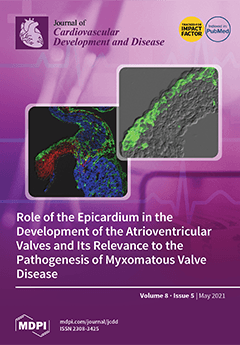
Cover Story (view full-size image): The epicardium and the epicardially derived cells (EPDCs) play an important role in heart development. A couple of years ago, the Wessels Lab established that EPDCs significantly contribute to those leaflets of the atrioventricular valves that are derived from the lateral atrioventricular cushions. In this article, dedicated to the memory of Dr. Adriana “Adri” Gittenberger-de Groot, we describe how the migration of EPDCs into the valves is spatiotemporally orchestrated and examine a number of molecular mechanisms that may play a role in regulating this process. Furthermore, we discuss the potential involvement of EPDCs in the pathogenesis of myxomatous valve disease (MVD), suggesting that this be taken into consideration when determining the genetic origin and pathogenesis of MVD. View this paper.
  • Issues are regarded as officially published after their release is announced to the table of contents alert mailing list.
  • You may sign up for e-mail alerts to receive table of contents of newly released issues.
  • PDF is the official format for papers published in both, html and pdf forms. To view the papers in pdf format, click on the "PDF Full-text" link, and use the free Adobe Reader to open them.
Order results
Result details
Section
Select all
Export citation of selected articles as:
20 pages, 1165 KiB  
Review
The Aging Vasculature: Glucose Tolerance, Hypoglycemia and the Role of the Serum Response Factor
by Hazel Aberdeen, Kaela Battles, Ariana Taylor, Jeranae Garner-Donald, Ana Davis-Wilson, Bryan T. Rogers, Candice Cavalier and Emmanuel D. Williams
J. Cardiovasc. Dev. Dis. 2021, 8(5), 58; https://doi.org/10.3390/jcdd8050058 - 17 May 2021
Cited by 6 | Viewed by 3286
Abstract
The fastest growing demographic in the U.S. at the present time is those aged 65 years and older. Accompanying advancing age are a myriad of physiological changes in which reserve capacity is diminished and homeostatic control attenuates. One facet of homeostatic control lost [...] Read more.
The fastest growing demographic in the U.S. at the present time is those aged 65 years and older. Accompanying advancing age are a myriad of physiological changes in which reserve capacity is diminished and homeostatic control attenuates. One facet of homeostatic control lost with advancing age is glucose tolerance. Nowhere is this more accentuated than in the high proportion of older Americans who are diabetic. Coupled with advancing age, diabetes predisposes affected subjects to the onset and progression of cardiovascular disease (CVD). In the treatment of type 2 diabetes, hypoglycemic episodes are a frequent clinical manifestation, which often result in more severe pathological outcomes compared to those observed in cases of insulin resistance, including premature appearance of biomarkers of senescence. Unfortunately, molecular mechanisms of hypoglycemia remain unclear and the subject of much debate. In this review, the molecular basis of the aging vasculature (endothelium) and how glycemic flux drives the appearance of cardiovascular lesions and injury are discussed. Further, we review the potential role of the serum response factor (SRF) in driving glycemic flux-related cellular signaling through its association with various proteins. Full article
Show Figures

Figure 1

10 pages, 1415 KiB  
Review
Sympathetic Activation and Arrhythmogenesis after Myocardial Infarction: Where Do We Stand?
by Konstantinos C. Zekios, Eleni-Taxiarchia Mouchtouri, Panagiotis Lekkas, Dimitrios N. Nikas and Theofilos M. Kolettis
J. Cardiovasc. Dev. Dis. 2021, 8(5), 57; https://doi.org/10.3390/jcdd8050057 - 15 May 2021
Cited by 9 | Viewed by 3669
Abstract
Myocardial infarction often leads to progressive structural and electrophysiologic remodeling of the left ventricle. Despite the widespread use of β-adrenergic blockade and implantable defibrillators, morbidity and mortality from chronic-phase ventricular tachyarrhythmias remains high, calling for further investigation on the underlying pathophysiology. Histological and [...] Read more.
Myocardial infarction often leads to progressive structural and electrophysiologic remodeling of the left ventricle. Despite the widespread use of β-adrenergic blockade and implantable defibrillators, morbidity and mortality from chronic-phase ventricular tachyarrhythmias remains high, calling for further investigation on the underlying pathophysiology. Histological and functional studies have demonstrated extensive alterations of sympathetic nerve endings at the peri-infarct area and flow-innervation mismatches that create a highly arrhythmogenic milieu. Such accumulated evidence, along with the previously well-documented autonomic dysfunction as an important contributing factor, has stirred intense research interest for pharmacologic and non-pharmacologic neuromodulation in post-infarction heart failure. In this regard, aldosterone inhibitors, sacubitril/valsartan and sodium-glucose cotransporter type 2 inhibitors have shown antiarrhythmic effects. Non-pharmacologic modalities, currently tested in pre-clinical and clinical trials, include transcutaneous vagal stimulation, stellate ganglion modulation and renal sympathetic denervation. In this review, we provide insights on the pathophysiology of ventricular arrhythmogenesis post-myocardial infarction, focusing on sympathetic activation. Full article
Show Figures

Figure 1

14 pages, 6743 KiB  
Article
Differential Spatio-Temporal Regulation of T-Box Gene Expression by microRNAs during Cardiac Development
by Mohamad Alzein, Estefanía Lozano-Velasco, Francisco Hernández-Torres, Carlos García-Padilla, Jorge N. Domínguez, Amelia Aránega and Diego Franco
J. Cardiovasc. Dev. Dis. 2021, 8(5), 56; https://doi.org/10.3390/jcdd8050056 - 14 May 2021
Cited by 4 | Viewed by 2668
Abstract
Cardiovascular development is a complex process that starts with the formation of symmetrically located precardiac mesodermal precursors soon after gastrulation and is completed with the formation of a four-chambered heart with distinct inlet and outlet connections. Multiple transcriptional inputs are required to provide [...] Read more.
Cardiovascular development is a complex process that starts with the formation of symmetrically located precardiac mesodermal precursors soon after gastrulation and is completed with the formation of a four-chambered heart with distinct inlet and outlet connections. Multiple transcriptional inputs are required to provide adequate regional identity to the forming atrial and ventricular chambers as well as their flanking regions; i.e., inflow tract, atrioventricular canal, and outflow tract. In this context, regional chamber identity is widely governed by regional activation of distinct T-box family members. Over the last decade, novel layers of gene regulatory mechanisms have been discovered with the identification of non-coding RNAs. microRNAs represent the most well-studied subcategory among short non-coding RNAs. In this study, we sought to investigate the functional role of distinct microRNAs that are predicted to target T-box family members. Our data demonstrated a highly dynamic expression of distinct microRNAs and T-box family members during cardiogenesis, revealing a relatively large subset of complementary and similar microRNA–mRNA expression profiles. Over-expression analyses demonstrated that a given microRNA can distinctly regulate the same T-box family member in distinct cardiac regions and within distinct temporal frameworks, supporting the notion of indirect regulatory mechanisms, and dual luciferase assays on Tbx2, Tbx3 and Tbx5 3′ UTR further supported this notion. Overall, our data demonstrated a highly dynamic microRNA and T-box family members expression during cardiogenesis and supported the notion that such microRNAs indirectly regulate the T-box family members in a tissue- and time-dependent manner. Full article
(This article belongs to the Section Basic and Translational Cardiovascular Research)
Show Figures

Figure 1

15 pages, 11289 KiB  
Systematic Review
Effect of Hydroxychloroquine on QTc in Patients Diagnosed with COVID-19: A Systematic Review and Meta-Analysis
by Angelos Arfaras-Melainis, Andreas Tzoumas, Damianos G. Kokkinidis, Maria Salgado Guerrero, Dimitrios Varrias, Xiaobo Xu, Luis Cerna, Ricardo Avendano, Cameron Kemal, Leonidas Palaiodimos and Robert T. Faillace
J. Cardiovasc. Dev. Dis. 2021, 8(5), 55; https://doi.org/10.3390/jcdd8050055 - 13 May 2021
Cited by 1 | Viewed by 2836
Abstract
Background: Hydroxychloroquine or chloroquine with or without the concomitant use of azithromycin have been widely used to treat patients with SARS-CoV-2 infection, based on early in vitro studies, despite their potential to prolong the QTc interval of patients. Objective: This is a systematic [...] Read more.
Background: Hydroxychloroquine or chloroquine with or without the concomitant use of azithromycin have been widely used to treat patients with SARS-CoV-2 infection, based on early in vitro studies, despite their potential to prolong the QTc interval of patients. Objective: This is a systematic review and metanalysis designed to assess the effect of hydroxychloroquine with or without the addition of azithromycin on the QTc of hospitalized patients with COVID-19. Materials and methods: PubMed, Scopus, Cochrane and MedRxiv databases were reviewed. A random effect model meta-analysis was used, and I-square was used to assess the heterogeneity. The prespecified endpoints were ΔQTc, QTc prolongation > 500 ms and ΔQTc > 60 ms. Results: A total of 18 studies and 7179 patients met the inclusion criteria and were included in this systematic review and meta-analysis. The use of hydroxychloroquine with or without the addition of azithromycin was associated with increased QTc when used as part of the management of patients with SARS-CoV-2 infection. The combination therapy with hydroxychloroquine plus azithromycin was also associated with statistically significant increases in QTc. Moreover, the use of hydroxychloroquine alone, azithromycin alone, or the combination of the two was associated with increased numbers of patients that developed QTc prolongation > 500 ms. Conclusion: This systematic review and metanalysis revealed that the use of hydroxychloroquine alone or in conjunction with azithromycin was linked to an increase in the QTc interval of hospitalized patients with SARS-CoV-2 infection that received these agents. Full article
Show Figures

Figure 1

18 pages, 4273 KiB  
Review
Role of the Epicardium in the Development of the Atrioventricular Valves and Its Relevance to the Pathogenesis of Myxomatous Valve Disease
by Renélyn Wolters, Ray Deepe, Jenna Drummond, Andrew B. Harvey, Emilye Hiriart, Marie M. Lockhart, Maurice J. B. van den Hoff, Russell A. Norris and Andy Wessels
J. Cardiovasc. Dev. Dis. 2021, 8(5), 54; https://doi.org/10.3390/jcdd8050054 - 12 May 2021
Cited by 3 | Viewed by 3885
Abstract
This paper is dedicated to the memory of Dr. Adriana “Adri” Gittenberger-de Groot and in appreciation of her work in the field of developmental cardiovascular biology and the legacy that she has left behind. During her impressive career, Dr. Gittenberger-de Groot studied many [...] Read more.
This paper is dedicated to the memory of Dr. Adriana “Adri” Gittenberger-de Groot and in appreciation of her work in the field of developmental cardiovascular biology and the legacy that she has left behind. During her impressive career, Dr. Gittenberger-de Groot studied many aspects of heart development, including aspects of cardiac valve formation and disease and the role of the epicardium in the formation of the heart. In this contribution, we review some of the work on the role of epicardially-derived cells (EPDCs) in the development of the atrioventricular valves and their potential involvement in the pathogenesis of myxomatous valve disease (MVD). We provide an overview of critical events in the development of the atrioventricular junction, discuss the role of the epicardium in these events, and illustrate how interfering with molecular mechanisms that are involved in the epicardial-dependent formation of the atrioventricular junction leads to a number of abnormalities. These abnormalities include defects of the AV valves that resemble those observed in humans that suffer from MVD. The studies demonstrate the importance of the epicardium for the proper formation and maturation of the AV valves and show that the possibility of epicardial-associated developmental defects should be taken into consideration when determining the genetic origin and pathogenesis of MVD. Full article
Show Figures

Figure 1

12 pages, 447 KiB  
Article
Sex Differences in Circadian Clock Genes and Myocardial Infarction Susceptibility
by Ivana Škrlec, Jasminka Talapko, Martina Juzbašić and Robert Steiner
J. Cardiovasc. Dev. Dis. 2021, 8(5), 53; https://doi.org/10.3390/jcdd8050053 - 8 May 2021
Cited by 12 | Viewed by 3431
Abstract
The growing body of evidence shows a significant difference in the circadian rhythm of cardiovascular disease based on biological sex. The incidence of cardiovascular disease varies between women and men. Additionally, biological sex is vital for the timely application of therapy—chronotherapy, which benefits [...] Read more.
The growing body of evidence shows a significant difference in the circadian rhythm of cardiovascular disease based on biological sex. The incidence of cardiovascular disease varies between women and men. Additionally, biological sex is vital for the timely application of therapy—chronotherapy, which benefits both sexes. This study aimed to examine the potential difference of single nucleotide polymorphisms (SNPs) of the circadian rhythm genes ARNTL, CLOCK, CRY2 and PER2 in women and men with myocardial infarction. A cross-sectional study was conducted, including 200 patients with myocardial infarction. Altogether, ten single nucleotide polymorphisms in the ARNTL, CLOCK, CRY2 and PER2 genes were analyzed. The Chi-square test yielded statistically significant differences in CLOCK gene rs11932595 polymorphism in a recessive genotype model between women and men with a p-value of 0.03 and an odds ratio 2.66, and a corresponding 95% confidence interval of 1.07 to 6.66. Other analyzed polymorphisms of the circadian rhythm genes ARNTL, CRY2, and PER2 did not significantly differ between the sexes. According to the study’s current results, the CLOCK gene’s genetic variability might affect myocardial infarction concerning biological sex. Full article
(This article belongs to the Section Genetics)
Show Figures

Figure 1

19 pages, 1102 KiB  
Review
Peptidic Connexin43 Therapeutics in Cardiac Reparative Medicine
by Spencer R. Marsh, Zachary J. Williams, Kevin J. Pridham and Robert G. Gourdie
J. Cardiovasc. Dev. Dis. 2021, 8(5), 52; https://doi.org/10.3390/jcdd8050052 - 5 May 2021
Cited by 20 | Viewed by 4863 | Correction
Abstract
Connexin (Cx43)-formed channels have been linked to cardiac arrhythmias and diseases of the heart associated with myocardial tissue loss and fibrosis. These pathologies include ischemic heart disease, ischemia-reperfusion injury, heart failure, hypertrophic cardiomyopathy, arrhythmogenic right ventricular cardiomyopathy, and Duchenne muscular dystrophy. A number [...] Read more.
Connexin (Cx43)-formed channels have been linked to cardiac arrhythmias and diseases of the heart associated with myocardial tissue loss and fibrosis. These pathologies include ischemic heart disease, ischemia-reperfusion injury, heart failure, hypertrophic cardiomyopathy, arrhythmogenic right ventricular cardiomyopathy, and Duchenne muscular dystrophy. A number of Cx43 mimetic peptides have been reported as therapeutic candidates for targeting disease processes linked to Cx43, including some that have advanced to clinical testing in humans. These peptides include Cx43 sequences based on the extracellular loop domains (e.g., Gap26, Gap 27, and Peptide5), cytoplasmic-loop domain (Gap19 and L2), and cytoplasmic carboxyl-terminal domain (e.g., JM2, Cx43tat, CycliCX, and the alphaCT family of peptides) of this transmembrane protein. Additionally, RYYN peptides binding to the Cx43 carboxyl-terminus have been described. In this review, we survey preclinical and clinical data available on short mimetic peptides based on, or directly targeting, Cx43, with focus on their potential for treating heart disease. We also discuss problems that have caused reluctance within the pharmaceutical industry to translate peptidic therapeutics to the clinic, even when supporting preclinical data is strong. These issues include those associated with the administration, stability in vivo, and tissue penetration of peptide-based therapeutics. Finally, we discuss novel drug delivery technologies including nanoparticles, exosomes, and other nanovesicular carriers that could transform the clinical and commercial viability of Cx43-targeting peptides in treatment of heart disease, stroke, cancer, and other indications requiring oral or parenteral administration. Some of these newly emerging approaches to drug delivery may provide a path to overcoming pitfalls associated with the drugging of peptide therapeutics. Full article
(This article belongs to the Special Issue Cardiomyopathy at the Sub-Cellular Level)
Show Figures

Figure 1

18 pages, 8303 KiB  
Systematic Review
Multimodality Imaging of the Anatomy of the Aortic Root
by Vera Lucia Paiocchi, Francesco F. Faletra, Enrico Ferrari, Susanne Anna Schlossbauer, Laura Anna Leo and Francesco Maisano
J. Cardiovasc. Dev. Dis. 2021, 8(5), 51; https://doi.org/10.3390/jcdd8050051 - 4 May 2021
Cited by 8 | Viewed by 3685
Abstract
The aortic root has long been considered an inert unidirectional conduit between the left ventricle and the ascending aorta. In the classical definition, the aortic valve leaflets (similar to what is perceived for the atrioventricular valves) have also been considered inactive structures, and [...] Read more.
The aortic root has long been considered an inert unidirectional conduit between the left ventricle and the ascending aorta. In the classical definition, the aortic valve leaflets (similar to what is perceived for the atrioventricular valves) have also been considered inactive structures, and their motion was thought to be entirely passive—just driven by the fluctuations of ventricular–aortic gradients. It was not until the advent of aortic valve–sparing surgery and of transcatheter aortic valve implantation that the interest on the anatomy of the aortic root again took momentum. These new procedures require a systematic and thorough analysis of the fine anatomical details of the components of the so-called aortic valve apparatus. Although holding and dissecting cadaveric heart specimens remains an excellent method to appreciate the complex “three-dimensional” nature of the aortic root, nowadays, echocardiography, computed tomography, and cardiac magnetic resonance provide excellent images of cardiac anatomy both in two- and three-dimensional format. Indeed, modern imaging techniques depict the aortic root as it is properly situated within the thorax in an attitudinally correct cardiac orientation, showing a sort of “dynamic anatomy”, which admirably joins structure and function. Finally, they are extensively used before, during, and after percutaneous structural heart disease interventions. This review focuses on the anatomy of the aortic root as revealed by non-invasive imaging techniques. Full article
Show Figures

Figure 1

13 pages, 3266 KiB  
Article
Deletion of a Hand1 lncRNA-Containing Septum Transversum Enhancer Alters lncRNA Expression but Is Not Required for Hand1 Expression
by Rajani M. George and Anthony B. Firulli
J. Cardiovasc. Dev. Dis. 2021, 8(5), 50; https://doi.org/10.3390/jcdd8050050 - 4 May 2021
Cited by 3 | Viewed by 2146
Abstract
We have previously identified a Hand1 transcriptional enhancer that drives expression within the septum transversum, the origin of the cells that contribute to the epicardium. This enhancer directly overlaps a common exon of a predicted family of long non-coding RNAs (lncRNA) that are [...] Read more.
We have previously identified a Hand1 transcriptional enhancer that drives expression within the septum transversum, the origin of the cells that contribute to the epicardium. This enhancer directly overlaps a common exon of a predicted family of long non-coding RNAs (lncRNA) that are specific to mice. To interrogate the necessity of this Hand1 enhancer, as well as the importance of these novel lncRNAs, we deleted the enhancer sequences, including the common exon shared by these lncRNAs, using genome editing. Resultant homozygous Hand1 enhancer mutants (Hand1ΔST/ΔST) present with no observable phenotype. Assessment of lncRNA expression reveals that Hand1ΔST/ΔST mutants effectively eliminate detectable lncRNA expression. Expression analysis within Hand1ΔST/ΔST mutant hearts indicates higher levels of Hand1 than in controls. The generation of Hand1 compound heterozygous mutants with the Hand1LacZ null allele (Hand1ΔST/LacZ) also did not reveal any observable phenotypes. Together these data indicate that deletion of this Hand1 enhancer and by consequence a family of murine-specific lncRNAs does not impact embryonic development in observable ways. Full article
Show Figures

Figure 1

24 pages, 2297 KiB  
Review
The Zebrafish Cardiac Endothelial Cell—Roles in Development and Regeneration
by Vanessa Lowe, Laura Wisniewski and Caroline Pellet-Many
J. Cardiovasc. Dev. Dis. 2021, 8(5), 49; https://doi.org/10.3390/jcdd8050049 - 1 May 2021
Cited by 12 | Viewed by 7168
Abstract
In zebrafish, the spatiotemporal development of the vascular system is well described due to its stereotypical nature. However, the cellular and molecular mechanisms orchestrating post-embryonic vascular development, the maintenance of vascular homeostasis, or how coronary vessels integrate into the growing heart are less [...] Read more.
In zebrafish, the spatiotemporal development of the vascular system is well described due to its stereotypical nature. However, the cellular and molecular mechanisms orchestrating post-embryonic vascular development, the maintenance of vascular homeostasis, or how coronary vessels integrate into the growing heart are less well studied. In the context of cardiac regeneration, the central cellular mechanism by which the heart regenerates a fully functional myocardium relies on the proliferation of pre-existing cardiomyocytes; the epicardium and the endocardium are also known to play key roles in the regenerative process. Remarkably, revascularisation of the injured tissue occurs within a few hours after cardiac damage, thus generating a vascular network acting as a scaffold for the regenerating myocardium. The activation of the endocardium leads to the secretion of cytokines, further supporting the proliferation of the cardiomyocytes. Although epicardium, endocardium, and myocardium interact with each other to orchestrate heart development and regeneration, in this review, we focus on recent advances in the understanding of the development of the endocardium and the coronary vasculature in zebrafish as well as their pivotal roles in the heart regeneration process. Full article
(This article belongs to the Special Issue Zebrafish Heart Development, Regeneration, and Disease Modelling)
Show Figures

Figure 1

14 pages, 1684 KiB  
Review
Regulation of Cardiac Conduction and Arrhythmias by Ankyrin/Spectrin-Based Macromolecular Complexes
by Drew Nassal, Jane Yu, Dennison Min, Cemantha Lane, Rebecca Shaheen, Daniel Gratz and Thomas J. Hund
J. Cardiovasc. Dev. Dis. 2021, 8(5), 48; https://doi.org/10.3390/jcdd8050048 - 29 Apr 2021
Cited by 7 | Viewed by 3748
Abstract
The cardiac conduction system is an extended network of excitable tissue tasked with generation and propagation of electrical impulses to signal coordinated contraction of the heart. The fidelity of this system depends on the proper spatio-temporal regulation of ion channels in myocytes throughout [...] Read more.
The cardiac conduction system is an extended network of excitable tissue tasked with generation and propagation of electrical impulses to signal coordinated contraction of the heart. The fidelity of this system depends on the proper spatio-temporal regulation of ion channels in myocytes throughout the conduction system. Importantly, inherited or acquired defects in a wide class of ion channels has been linked to dysfunction at various stages of the conduction system resulting in life-threatening cardiac arrhythmia. There is growing appreciation of the role that adapter and cytoskeletal proteins play in organizing ion channel macromolecular complexes critical for proper function of the cardiac conduction system. In particular, members of the ankyrin and spectrin families have emerged as important nodes for normal expression and regulation of ion channels in myocytes throughout the conduction system. Human variants impacting ankyrin/spectrin function give rise to a broad constellation of cardiac arrhythmias. Furthermore, chronic neurohumoral and biomechanical stress promotes ankyrin/spectrin loss of function that likely contributes to conduction disturbances in the setting of acquired cardiac disease. Collectively, this review seeks to bring attention to the significance of these cytoskeletal players and emphasize the potential therapeutic role they represent in a myriad of cardiac disease states. Full article
Show Figures

Figure 1

25 pages, 2297 KiB  
Review
The Role of Cell Tracing and Fate Mapping Experiments in Cardiac Outflow Tract Development, New Opportunities through Emerging Technologies
by Joshua C. Peterson, Tim P. Kelder, Marie José T. H. Goumans, Monique R. M. Jongbloed and Marco C. DeRuiter
J. Cardiovasc. Dev. Dis. 2021, 8(5), 47; https://doi.org/10.3390/jcdd8050047 - 26 Apr 2021
Cited by 4 | Viewed by 3569
Abstract
Whilst knowledge regarding the pathophysiology of congenital heart disease (CHDs) has advanced greatly in recent years, the underlying developmental processes affecting the cardiac outflow tract (OFT) such as bicuspid aortic valve, tetralogy of Fallot and transposition of the great arteries remain poorly understood. [...] Read more.
Whilst knowledge regarding the pathophysiology of congenital heart disease (CHDs) has advanced greatly in recent years, the underlying developmental processes affecting the cardiac outflow tract (OFT) such as bicuspid aortic valve, tetralogy of Fallot and transposition of the great arteries remain poorly understood. Common among CHDs affecting the OFT, is a large variation in disease phenotypes. Even though the different cell lineages contributing to OFT development have been studied for many decades, it remains challenging to relate cell lineage dynamics to the morphologic variation observed in OFT pathologies. We postulate that the variation observed in cellular contribution in these congenital heart diseases might be related to underlying cell lineage dynamics of which little is known. We believe this gap in knowledge is mainly the result of technical limitations in experimental methods used for cell lineage analysis. The aim of this review is to provide an overview of historical fate mapping and cell tracing techniques used to study OFT development and introduce emerging technologies which provide new opportunities that will aid our understanding of the cellular dynamics underlying OFT pathology. Full article
Show Figures

Figure 1

Previous Issue
Next Issue
Back to TopTop