
Outlining Potential Biomarkers of Exposure and Effect to Critical Minerals: Nutritionally Essential Trace Elements and the Rare Earth Elements

Abstract
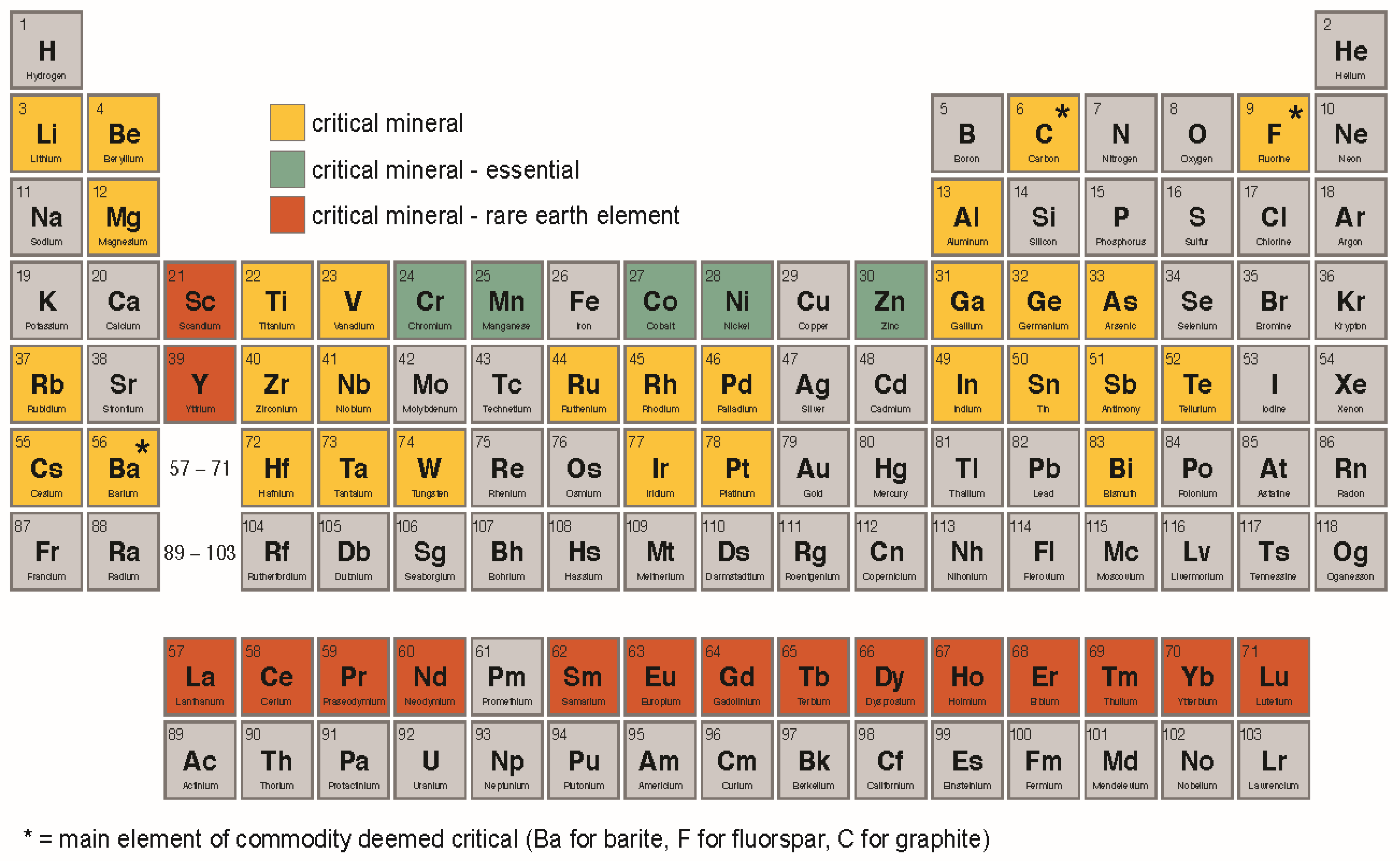
1. Introduction

2. Aquatic Studies and Environmental Parameter Considerations
- EPA national primary drinking water standards (maximum contaminant levels or MCLs), which are regulatory benchmarks for drinking water for protecting human health [37];
- EPA secondary drinking water standards, which are non-regulatory aesthetic benchmarks for drinking water [38];
- Non-regulatory health-based screening levels (HBSLs), developed by the USGS for contaminants without EPA standards or guidelines to place water quality data in a human health context [39];
- In the absence of any of the above, World Health Organization drinking water quality guidelines [40]; and
- Sediment quality guidelines (SQGs) for freshwater ecosystems [41] to provide environmental context for aquatic species; specifically, the Probable Effect Concentration [PEC], which is the concentration above which bed sediments are shown statistically to frequently have adverse effects on benthic biota.
3. Essential Trace Elements
3.1. Cobalt
3.2. Chromium
3.3. Manganese
3.4. Nickel
3.5. Zinc
4. Rare Earth Elements
5. Discussion
- At the cell surface, the critical first points of contact are lipids and biomembranes, with influences on bilipid membrane morphology and asymmetry.
- For the essential nutrients, the initial events that can be linked to an AOP can be considered to be potentially conserved across animal taxa [55].
- In fish, acute exposures target gills, which generally comprise over 50% of the surface area of the animal and are in intimate and continuous contact with the external water [19].
- The transport mechanisms into cells are varied. Metal-specific protein ion channels are needed for influx and efflux of Co, Mn, and Ni, and some others mediate their transport as well as allow influx of other divalent cations [42]. To excrete excess metals, various efflux mechanisms are upregulated so as to release them to the extracellular matrix [42]. Non-specific phosphate/sulfate anion transporters are involved in Cr(VI) transport [72,78], and divalent metal transporters (DMT) can transport Zn2+, Mn2+, Co2+, Cd2+, Cu2+, Ni2+, Pb2+, and Fe2+ [85]. Nickel can readily cross cell membranes via Ca channels and compete with Ca at its receptors [106]. The absorption of Zn is an active process facilitated by Zn transporters (ZIP4) [112].
- Cr(III) cannot cross cell membranes; Cr(VI), which can cross membranes, is more toxic to humans and is a carcinogen.
- In fish, compared with humans the molecular mechanisms of metal toxicity and subsequent physiological effects are under-studied [57]. Moreover, controlled studies with fish and CM can delineate molecular pathways and so too inform human AOP.
- By virtue of their ionic radii, the REEs may be capable of interacting with binding sites for Ca and heavy metals, disrupting normal protein function, or may deplete antioxidant protein pools [141].
- Because the REEs display variable modes of action, can be competitive with other metals at the cellular level, and display non-linear toxicological mechanisms, more studies with fish and humans can help to delineate AOP.
6. Summary
Author Contributions
Funding
Institutional Review Board Statement
Informed Consent Statement
Data Availability Statement
Acknowledgments
Conflicts of Interest
Appendix A

| CAS b | Substance Name | Listing c |
|---|---|---|
| 1309-64-4 | Antimony Trioxide | RAHC |
| 7440-38-2 | Arsenic | Known |
| 7440-41-7 | Beryllium | Known |
| 7787-47-5 | Beryllium Chloride | Known |
| 7787-56-6 | Beryllium Sulfate Tetrahydrate | Known |
| 13327-32-7 | Beryllium Hydroxide | Known |
| 13510-49-1 | Beryllium Sulfate | Known |
| 13598-00-0 | Beryllium Silicate | Known |
| 13598-15-7 | Beryllium Phosphate | Known |
| 25638-88-4 | Zinc Beryllium Silicate | Known |
| 66104-24-3 | Beryllium Carbonate | Known |
| 1333-82-0 | Chromium Trioxide | Known |
| 7775-11-3 | Sodium Chromate | Known |
| 7778-50-9 | Potassium Dichromate | Known |
| 7788-98-9 | Ammonium Chromate | Known |
| 7789-00-6 | Potassium Chromate | Known |
| 7789-06-2 | Strontium Chromate | Known |
| 7789-09-5 | Ammonium Dichromate | Known |
| 10588-01-9 | Sodium Dichromate | Known |
| 11119-70-3 | Lead Chromate | Known |
| 13530-65-9 | Zinc Chromate | Known |
| 13765-19-0 | Calcium Chromate | Known |
| 18540-29-9 | Chromium (VI) | Known |
| 71-48-7 | Cobalt Acetate | RAHC |
| 7440-48-4 | Cobalt | RAHC |
| 7646-79-9 | Cobalt Chloride | RAHC |
| 10026-24-1 | Cobalt Sulfate Heptahydrate | RAHC |
| 10124-43-3 | Cobalt Sulfate | RAHC |
| 10141-05-6 | Cobalt Nitrate | RAHC |
| 11104-61-3 | Cobalt Oxide | RAHC |
| 12653-56-4 | Cobalt Sulfide | RAHC |
| NA | Cobalt-Tungsten Carbide: Powders and Hard Metals | RAHC |
| 373-02-4 | Nickel Acetate | Known |
| 1313-99-1 | Nickel Monoxide | Known |
| 7440-02-0 | Metallic Nickel | RAHC |
| 7786-81-4 | Nickel Sulfate | Known |
| 11113-75-0 | Nickel Sulfide | Known |
| 12035-72-2 | Nickel Subsulfide | Known |
| 12054-48-7 | Nickel Hydroxide | Known |
| NA | Nickel Compounds | Known |
| Ranking | Substance Name | CAS b |
|---|---|---|
| 1 | Arsenic | 7440-38-2 |
| 14 | Chromium (VI) hexavalent | 18540-29-9 |
| 43 | Beryllium | 7440-41-7 |
| 58 | Chromium (VI) trioxide | 1333-82-0 |
| 70 | Cobalt | 7440-48-4 |
| 94 | Nickel | 7440-02-0 |
| 136 | Barium | 7440-39-3 |
| 150 | Chromium | 7440-47-3 |
| 155 | Fluorine | 7782-41-4 |
| 164 | Zinc | 7440-66-6 |
| 173 | Palladiumc | 7440-05-3 |
| 188 | Manganese | 7439-96-5 |
| 228 | Cesium-137 | 10045-97-3 |
| 244 | Antimony | 7440-36-0 |
| 277 | Vanadium | 7440-62-2 |
| 281 | Aluminum | 7429-90-5 |
| 351 | Titanium | 7440-32-6 |
| 359 | Lithium | 7439-93-2 |
| 360 | Chromium, trivalent | 16065-83-1 |
| 362 | Tin | 7440-31-5 |
| 376 | Cesium-134 | 13967-70-9 |
| 384 | Tungsten | 7440-33-7 |
| 448 | Indiumc | 7440-74-6 |
| 470 | Cerium | 7440-45-1 |
| 476 | Neodymium | 7440-00-8 |
| 479 | Germaniumc | 7440-56-4 |
| 492 | Bismuth-214 | 14733-03-0 |
| 494 | Bismuth-212 | 14913-49-6 |
| 510 | Ytterbium | 7440-64-4 |
| 511 | Scandium | 7440-20-2 |
| 513 | Gallium | 7440-55-3 |
| 514 | Niobium | 7440-03-1 |
| 519 | Tellurium | 13494-80-9 |
| 525 | Cobalt-60 | 10198-40-0 |
| 545 | Chromium (IV) | 15723-28-1 |
| 550 | Europium | 7440-53-1 |
| 551 | Dysprosium | 7429-91-6 |
| 551 | Praseodymium | 7440-10-0 |
| 551 | Samarium | 7440-19-9 |
| 566 | Aluminum oxide | 1344-28-1 |
| 578 | Cesium | 7440-46-2 |
| 578 | Lanthanum | 7439-91-0 |
| 578 | Magnesium | 7439-95-4 |
| 578 | Rubidium | 7440-17-7 |
| 578 | Yttrium | 7440-65-5 |
| 578 | Zirconium | 7440-67-7 |
| 692 | Tantalum c | 7440-25-7 |
References
- Nassar, N.T.; Brainard, J.; Gulley, A.; Manley, R.; Matos, G.; Lederer, G.; Bird, L.R.; Pineault, D.; Alonso, E.; Gambogi, J.; et al. Evaluating the mineral commodity supply risk of the U.S. manufacturing sector. Sci. Adv. 2020, 6, eaay8647. [Google Scholar] [CrossRef] [PubMed]
- FR 60835. Executive Order 13817 of Dec. 20, 2017; Executive Office of the President: Washington, DC, USA, 2017; pp. 60835–60837.
- Fortier, S.M.; Nassar, N.T.; Lederer, G.W.; Brainard, J.; Gambogi, J.; McCullough, E.A. Draft Critical Mineral List—Summary of Methodology and Background Information—U.S. Geological Survey Technical Input Document in Response to Secretarial Order No. 3359; U.S. Geological Survey Open-File Report 2018–1021; USGS: Reston, VA, USA, 2018; 15p. [CrossRef]
- Nassar, N.T.; Fortier, S.M. Methodology and Technical Input for the 2021 Review and Revision of the U.S. Critical Minerals List; U.S. Geological Survey Open-File Report 2021–1045; USGS: Reston, VA, USA, 2021; 31p. [CrossRef]
- Schulz, K.J.; DeYoung, J.H., Jr.; Seal, R.R., II; Bradley, D.C. Critical mineral resources of the United States—Economic and environmental geology and prospects for future supply. U.S. Geol. Surv. Prof. Pap. 2017, 1802, B1–B16. [Google Scholar] [CrossRef]
- Code of Federal Regulations. 2022 Final List of Critical Minerals. Fed. Reg. 2022, 87, 10381. Available online: https://www.govinfo.gov/app/details/FR-2022-02-24/2022-04027 (accessed on 24 February 2022).
- Wall, F. Rare earth elements. In Critical Metals Handbook; Gunn, G., Ed.; John Wiley & Sons: West Sussex, UK, 2014; pp. 312–339. ISBN 978-0-470-67171-9. [Google Scholar]
- Liu, Y.; Wu, M.; Zhang, L.; Bi, J.; Song, L.; Wang, L.; Liu, B.; Zhou, A.; Cao, Z.; Xiong, C.; et al. Prenatal exposure of rare earth elements cerium and ytterbium and neonatal thyroid stimulating hormone levels: Findings from a birth cohort study. Environ. Int. 2019, 133, 105222. [Google Scholar] [CrossRef]
- Garrels, R.M.; Mackenzie, F.T.; Hunt, C. Trace Elements. In Chemical Cycles and the Global Environment: Assessing Human Influences; William Kaufmann, Inc.: Los Altos, CA, USA, 1975; pp. 112–135. ISBN 0-913232-29-7. [Google Scholar]
- Liu, X.; Zhang, Y.; Piao, J.; Mao, D.; Li, Y.; Li, W.; Yang, L.; Yang, X. Reference Values of 14 Serum Trace Elements for Pregnant Chinese Women: A Cross-Sectional Study in the China Nutrition and Health Survey 2010–2012. Nutrients 2017, 9, 309. [Google Scholar] [CrossRef]
- Mehri, A. Trace Elements in Human Nutrition (II)—An Update. Int. J. Prev. Med. 2020, 11, 2. [Google Scholar]
- U.S. Environmental Protection Agency, Priority Pollutant List. 2014. Available online: https://www.epa.gov/sites/default/files/2015-09/documents/priority-pollutant-list-epa.pdf (accessed on 18 December 2022).
- National Toxicology Program. Report on Carcinogens, 15th ed.; U.S. Department of Health and Human Services PHS: Research Triangle Park: NC, USA, 2021. [CrossRef]
- ATSDR. Support Document to the 2019 Substance Priority List (Candidates for Toxicological Profiles); Agency for Toxic Substances and Disease Registry, Division of Toxicology and Human Health Sciences: Atlanta, GA, USA, 2020; p. 9. Available online: https://www.atsdr.cdc.gov/spl/index.html (accessed on 30 May 2022).
- Kroneck, P.M.; Sosa Torres, M.E. Metals, Microbes, and Minerals: The Biogeochemical Side of Life; Sigel, A., Freisinger, E., Sigel, R.K.O., Eds.; de Gruyter: Boston, MA, USA, 2021; Volume 21, 386p. [Google Scholar]
- Andreini, C.; Bertini, I.; Cavallaro, G.; Holliday, G.L.; Thornton, J.M. Metal ions in biological catalysis: From enzyme databases to general principles. JBIC J. Biol. Inorg. Chem. 2008, 13, 1205–1218. [Google Scholar] [CrossRef]
- Gutiérrez-González, E.; García-Esquinas, E.; de Larrea-Baz, N.F.; Salcedo-Bellido, I.; Navas-Acien, A.; Lope, V.; Gómez-Ariza, J.L.; Pastor, R.; Pollán, M.; Pérez-Gómez, B. Toenails as biomarker of exposure to essential trace metals: A review. Environ. Res. 2019, 179, 108787. [Google Scholar] [CrossRef]
- National Research Council, Committee on Diet and Health. Diet and Health: Implications for Reducing Chronic Disease Risk; The National Academies: Washington, DC, USA, 1989; ISBN 978-0-309-07474-2. [Google Scholar]
- Wood, C.M.; Farrell, A.P.; Brauner, C.J. Homeostasis and Toxicology of Essential Metals; Series Volume 31A; Academic Press: Boston, MA, USA, 2012; Volume 1, 497p. [Google Scholar]
- World Health Organization. Trace Elements in Human Nutrition and Human Health; WHO: Geneva, Switzerland, 1996; 343p, ISBN 9241561734. [Google Scholar]
- Van der Oost, R.; Beyer, J.; Vermeulen, N.P.E. Fish bioaccumulation and biomarkers in environmental risk assessment: A review. Environ. Toxicol. Pharmacol. 2003, 13, 57–149. [Google Scholar] [CrossRef]
- Authman, M.M.N.; Zaki, M.S.; Khallaf, E.A.; Abbas, H.H. Use of fish as bio-indicator of the effects of heavy metals pollution. J. Aquac. Res. Dev. 2015, 6, 4. [Google Scholar] [CrossRef]
- Jenkins, J.; Olivier, H.; Draugelis-Dale, R.; Eilts, B.; Torres, L.; Patiño, R.; Nilsen, E.; Goodbred, S. Assessing reproductive and endocrine parameters in male largescale suckers (Catostomus macrocheilus) along a contaminant gradient in the lower Columbia River, USA. Sci. Total. Environ. 2014, 484, 365–378. [Google Scholar] [CrossRef][Green Version]
- Jenkins, J.A.; Rosen, M.R.; Draugelis-Dale, R.O.; Echols, K.R.; Torres, L.; Wieser, C.M.; Kersten, C.A.; Goodbred, S.L. Sperm quality biomarkers complement reproductive and endocrine parameters in investigating environmental contaminants in common carp (Cyprinus carpio) from the Lake Mead National Recreation Area. Environ. Res. 2018, 163, 149–164. [Google Scholar] [CrossRef]
- Patiño, R.; Rosen, M.R.; Orsak, E.L.; Goodbred, S.L.; May, T.W.; Alvarez, D.; Echols, K.R.; Wieser, C.M.; Ruessler, S.; Torres, L. Patterns of metal composition and biological condition and their association in male common carp across an environmental contaminant gradient in Lake Mead National Recreation Area, Nevada and Arizona, USA. Sci. Total. Environ. 2012, 416, 215–224. [Google Scholar] [CrossRef]
- Velma, V.; Tchounwou, P.B. Chromium-induced biochemical, genotoxic and histopathologic effects in liver and kidney of goldfish, Carassius auratus. Mutat. Res. Toxicol. Environ. Mutagen. 2010, 698, 43–51. [Google Scholar] [CrossRef]
- Schmitt, C.J. Biomonitoring of Environmental Status and Trends (BEST) Program: Environmental Contaminants and Their Effects on Fish in the Mississippi River Basin; USGS/BRD/BSR-2002-0004; USGS: Reston, VA, USA, 2002; pp. 1–241. Available online: https://pubs.er.usgs.gov/publication/bsr020004 (accessed on 30 May 2022).
- Flora, S.J.S.; Sharma, A. Metals. In Biomarkers in Toxicology; Gupta, G., Ed.; Academic Press: San Diego, CA, USA, 2019; pp. 529–549. ISBN 978-0-12-814655-2. [Google Scholar]
- Blazer, V.; Gordon, S.; Walsh, H.; Smith, C. Perfluoroalkyl substances in plasma of smallmouth bass from the Chesapeake Bay watershed. Int. J. Environ. Res. Public Health 2021, 18, 5881. [Google Scholar] [CrossRef]
- National Academies of Sciences, Engineering, and Medicine; Division on Earth and Life Studies. Predicting Human Health Effects from Environmental Exposures: Applying Translatable and Accessible Biomarkers of Effect: Proceedings of a workshop in brief (2020); National Academies Press: Washington, DC, USA, 2020. [Google Scholar] [CrossRef]
- Jenkins, J.A.; Baudoin, B.A.; Johnson, D.; Fernie, K.J.; Stapleton, H.M.; Karouna-Renier, N.K. Establishment of baseline cytology metrics in nestling American kestrels (Falco sparverius): Immunomodulatory effects of the flame retardant isopropylated triarylphosphate isomers. Environ. Int. 2021, 157, 106779. [Google Scholar] [CrossRef]
- O’Mara, K.; Adams, M.; Burford, M.A.; Fry, B.; Cresswell, T. Uptake and accumulation of cadmium, manganese and zinc by fisheries species: Trophic differences in sensitivity to environmental metal accumulation. Sci. Total. Environ. 2019, 690, 867–877. [Google Scholar] [CrossRef]
- Wang, W.-X.; Fisher, N.S. Delineating metal accumulation pathways for marine invertebrates. Sci. Total. Environ. 1999, 237–238, 459–472. [Google Scholar] [CrossRef]
- Melake, B.A.; Nkuba, B.; Groffen, T.; De Boeck, G.; Bervoets, L. Distribution of metals in water, sediment and fish tissue. Consequences for human health risks due to fish consumption in Lake Hawassa, Ethiopia. Sci. Total. Environ. 2022, 843, 156968. [Google Scholar] [CrossRef]
- Pouil, S.; Teyssié, J.-L.; Rouleau, C.; Fowler, S.W.; Metian, M.; Bustamante, P.; Warnau, M. Comparative study of trophic transfer of the essential metals Co and Zn in two tropical fish: A radiotracer approach. J. Exp. Mar. Biol. Ecol. 2017, 486, 42–51. [Google Scholar] [CrossRef]
- Kumari, B.; Kumar, V.; Sinha, A.K.; Ahsan, J.; Ghosh, A.; Wang, H.; De Boeck, G. Toxicology of arsenic in fish and aquatic systems. Environ. Chem. Lett. 2016, 15, 43–64. [Google Scholar] [CrossRef]
- U.S. Environmental Protection Agency. National Primary Drinking Water Regulations: U.S. Environmental Protection Agency Web Page. Available online: https://www.epa.gov/ground-water-and-drinking-water/national-primary-drinking-water-regulations (accessed on 28 January 2022).
- U.S. Environmental Protection Agency. Secondary Drinking Water Standards—Guidance for Nuisance Chemicals: U.S. Environmental Protection Agency Web Page. Available online: https://www.epa.gov/sdwa/secondary-drinking-water-standards-guidance-nuisance-chemicals (accessed on 25 September 2022).
- Norman, J.E.; Toccalino, P.L.; Morman, S.A. Health-Based Screening Levels for Evaluating Water-Quality Data. In U.S. Geological Survey National Water Quality Assessment Program Web Page, 2nd ed.; USGS: Reston, VA, USA, 2018. [Google Scholar] [CrossRef]
- World Health Organization. Guidelines for Drinking-Water Quality: Fourth Edition Incorporating the First Addendum, 4th ed.; WHO: Geneva, Switzerland, 2017; 631p, Available online: https://www.who.int/publications/i/item/9789241549950 (accessed on 25 September 2022).
- MacDonald, D.D.; Ingersoll, C.G.; Berger, T.A. Development and evaluation of consensus-based sediment quality guidelines for freshwater ecosystems. Arch. Environ. Contam. Toxicol. 2000, 39, 20–31. [Google Scholar] [CrossRef] [PubMed]
- Sule, K.; Umbsaar, J.; Prenner, E.J. Mechanisms of Co, Ni, and Mn toxicity: From exposure and homeostasis to their interactions with and impact on lipids and biomembranes. Biochim. Biophys. Acta (BBA)—Biomembr. 2020, 1862, 183250. [Google Scholar] [CrossRef] [PubMed]
- Zoroddu, M.A.; Aaseth, J.; Crisponi, G.; Medici, S.; Peana, M.; Nurchi, V.M. The essential metals for humans: A brief overview. J. Inorg. Biochem. 2019, 195, 120–129. [Google Scholar] [CrossRef]
- Guzmán, D.C.; Olguín, H.J.; Brizuela, N.O.; Garcia, E.H.; Silva, M.L. The use of trace and essential elements in common clinical disorders: Roles in assessment of health and oxidative stress status. Nutr. Cancer 2019, 71, 13–20. [Google Scholar] [CrossRef]
- Broding, H.C.; Michalke, B.; Göen, T.; Drexler, H. Comparison between exhaled breath condensate analysis as a marker for cobalt and tungsten exposure and biomonitoring in workers of a hard metal alloy processing plant. Int. Arch. Occup. Environ. Health 2008, 82, 565–573. [Google Scholar] [CrossRef]
- Reinardy, H.C.; Syrett, J.R.; Jeffree, R.A.; Henry, T.B.; Jha, A.N. Cobalt-induced genotoxicity in male zebrafish (Danio rerio), with implications for reproduction and expression of DNA repair genes. Aquat. Toxicol. 2013, 126, 224–230. [Google Scholar] [CrossRef]
- Whitledge, G.W.; Kroboth, P.T.; Chapman, D.C.; Phelps, Q.E.; Sleeper, W.; Bailey, J.; Jenkins, J.A. Establishment of invasive Black Carp (Mylopharyngodon piceus) in the Mississippi River basin: Identifying sources and year classes contributing to recruitment. Biol. Invasions 2022, 24, 3885–3904. [Google Scholar] [CrossRef]
- Spilsbury, F.; McDonald, B.; Rankenburg, K.; Evans, N.J.; Grice, K.; Gagnon, M.M. Multivariate analysis of otolith microchemistry can discriminate the source of oil contamination in exposed fish. Comp. Biochem. Physiol. Part C Toxicol. Pharmacol. 2021, 254, 109253. [Google Scholar] [CrossRef]
- Arslan, Z.; Secor, D.H. Analysis of trace transition elements and heavy metals in fish otoliths as tracers of habitat use by American eels in the Hudson River estuary. Estuaries 2005, 28, 382–393. [Google Scholar] [CrossRef]
- Daros, F.A.; Condini, M.V.; Altafin, J.P.; Ferreira, F.D.O.; Hostim-Silva, M. Fish otolith microchemistry as a biomarker of the world’s largest mining disaster. Sci. Total. Environ. 2021, 807, 151780. [Google Scholar] [CrossRef]
- Viana, L.F.; Súarez, Y.R.; Cardoso, C.A.L.; Lima, S.M.; Andrade, L.H.D.C.; Lima-Junior, S.E. Use of fish scales in environmental monitoring by the application of Laser-Induced Breakdown Spectroscopy (LIBS). Chemosphere 2019, 228, 258–263. [Google Scholar] [CrossRef]
- Sun, P.L.; Hawkins, W.E.; Overstreet, R.M.; Brown-Peterson, N.J. Morphological deformities as biomarkers in fish from contaminated rivers in Taiwan. Int. J. Environ. Res. Public Health 2009, 6, 2307–2331. [Google Scholar] [CrossRef]
- Kousa, A.; Loukola-Ruskeeniemi, K.; Hatakka, T.; Kantola, M. High manganese and nickel concentrations in human hair and well water and low calcium concentration in blood serum in a pristine area with sulphide-rich bedrock. Environ. Geochem. Health 2021, 44, 3799–3819. [Google Scholar] [CrossRef]
- Squadrone, S.; Brizio, P.; Stella, C.; Favaro, L.; Da Rugna, C.; Florio, D.; Gridelli, S.; Abete, M.C. Feathers of Humboldt penguin are suitable bioindicators of Rare Earth Elements. Sci. Total. Environ. 2019, 678, 627–631. [Google Scholar] [CrossRef]
- Brix, K.V.; Schlekat, C.E.; Garman, E.R. The mechanisms of nickel toxicity in aquatic environments: An adverse outcome pathway analysis. Environ. Toxicol. Chem. 2017, 36, 1128–1137. [Google Scholar] [CrossRef]
- Barceloux, D.G. Cobalt. J. Toxicol. Clin. Toxicol. 1999, 37, 201–216. [Google Scholar] [CrossRef]
- Ziwa, G.; Crane, R.; Hudson-Edwards, K.A. Geochemistry, mineralogy and microbiology of cobalt in mining-affected environments. Minerals 2020, 11, 22. [Google Scholar] [CrossRef]
- Agency for Toxic Substances and Disease Registry (ATSDR). Toxicological Profile for Cobalt; U.S. Department of Health and Human Services, Public Health Service: Atlanta, GA, USA, 2004. Available online: https://www.atsdr.cdc.gov/toxprofiles/tp33.pdf (accessed on 18 December 2022).
- Gál, J.; Hursthouse, A.; Tatner, P.; Stewart, F.; Welton, R. Cobalt and secondary poisoning in the terrestrial food chain: Data review and research gaps to support risk assessment. Environ. Int. 2008, 34, 821–838. [Google Scholar] [CrossRef]
- Blust, R. Cobalt. In Homeostasis and Toxicology of Essential Metals; Wood, C.M., Farrell, A.P., Brauner, C.J., Eds.; Fish Physiology; Academic Press: Cambridge, MA, USA, 2012; Volume 31, Part A; pp. 291–326. [Google Scholar] [CrossRef]
- Hem, J.D. Study and Interpretation of the Chemical Characteristics of Natural Water; U.S. Geological Survey: Reston, VA, USA, 1985. [CrossRef]
- Dehaine, Q.; Tijsseling, L.T.; Glass, H.J.; Törmänen, T.; Butcher, A.R. Geometallurgy of cobalt ores: A review. Miner. Eng. 2020, 160, 106656. [Google Scholar] [CrossRef]
- Leyssens, L.; Vinck, B.; Van Der Straeten, C.; Wuyts, F.; Maes, L. Cobalt toxicity in humans—A review of the potential sources and systemic health effects. Toxicology 2017, 387, 43–56. [Google Scholar] [CrossRef] [PubMed]
- Okamoto, S.; Eltis, L.D. The biological occurrence and trafficking of cobalt. Metallomics 2011, 3, 963–970. [Google Scholar] [CrossRef] [PubMed]
- Di Giulio, R.T.; Newman, M.C. Ecotoxicology. In Casarett & Doull’s Essentials of Toxicology, 3rd ed.; Klaassen, C.D., Watkins, J.B., III, Eds.; McGraw Hill Medical: New York, NY, USA, 2015; pp. 441–451. ISBN 978-1-25-925535-9. [Google Scholar]
- Trindade, I.B.; Paquete, C.M.; Louro, R.O. Extracellular Redox Chemistry. In Metals, Microbes, and Minerals: The Biogeochemical Side of Life; Kroneck, P.M.H., Torres, M.E.S., Eds.; Metal Ions in Life Sciences; Walter de Gruyter GmbH: Boston, MA, USA, 2021; pp. 229–269. [Google Scholar]
- Ruttkay-Nedecky, B.; Nejdl, L.; Gumulec, J.; Zitka, O.; Masarik, M.; Eckschlager, T.; Stiborova, M.; Adam, V.; Kizek, R. The role of metallothionein in oxidative stress. Int. J. Mol. Sci. 2013, 14, 6044–6066. [Google Scholar] [CrossRef] [PubMed]
- Javed, M. Chronic effects of nickel and cobalt on fish growth. Int. J. Agric. Biol. 2013, 15, 575–579. [Google Scholar]
- Shanker, A.K.; Venkateswarlu, B. Chromium: Environmental pollution, health effects and mode of action. In Encyclopedia of Environmental Health; Elsevier: Amsterdam, The Netherlands, 2011; pp. 650–659. [Google Scholar]
- Alvarez, C.C.; Gómez, M.E.B.; Zavala, A.H. Hexavalent chromium: Regulation and health effects. J. Trace Elem. Med. Biol. 2021, 65, 126729. [Google Scholar] [CrossRef]
- Hamilton, E.M.; Young, S.D.; Bailey, E.H.; Watts, M.J. Chromium speciation in foodstuffs: A review. Food Chem. 2018, 250, 105–112. [Google Scholar] [CrossRef]
- O’Brien, T.J.; Ceryak, S.; Patierno, S.R. Complexities of chromium carcinogenesis: Role of cellular response, repair and recovery mechanisms. Mutat. Res. 2003, 533, 3–36. [Google Scholar] [CrossRef]
- Aslam, S.; Yousafzai, A.M. Chromium toxicity in fish: A review article. J. Entomol. Zool. Stud. 2017, 5, 1483–1488. [Google Scholar]
- Accordi, F.; Chimenti, C. Programmed cell death in the pancreas of Bufo bufo during metamorphosis. J. Anat. 2001, 199, 419–427. [Google Scholar] [CrossRef]
- Agency for Toxic Substances and Disease Registry (ATSDR). Toxicological Profile for Chromium; U.S. Department of Health and Human Services, Public Health Service: Atlanta, GA, USA, 2012. Available online: https://www.atsdr.cdc.gov/ToxProfiles/tp7.pdf (accessed on 18 December 2022).
- Sawicka, E.; Jurkowska, K.; Piwowar, A. Chromium (III) and chromium (VI) as important players in the induction of genotoxicity—Current view. Ann. Agric. Environ. Med. 2020, 28, 1–10. [Google Scholar] [CrossRef]
- Su, R.C.; Lad, A.; Breidenbach, J.D.; Kleinhenz, A.L.; Modyanov, N.; Malhotra, D.; Haller, S.T.; Kennedy, D.J. Assessment of diagnostic biomarkers of liver injury in the setting of microcystin-LR (MC-LR) hepatotoxicity. Chemosphere 2020, 257, 127111. [Google Scholar] [CrossRef]
- Ferreira, L.M.R.; Cunha-Oliveira, T.; Sobral, M.C.; Abreu, P.L.; Alpoim, M.C.; Urbano, A.M. Impact of carcinogenic chromium on the cellular response to proteotoxic stress. Int. J. Mol. Sci. 2019, 20, 4901. [Google Scholar] [CrossRef]
- Reid, S.D. Moybdenum and Chromium. In Homeostasis and Toxicology of Essential Metals; Wood, C.M., Farrell, A.P., Brauner, C.J., Eds.; Elsevier: New York, NY, USA, 2012; Volume 31A, pp. 375–415. [Google Scholar]
- Lewicki, S.; Zdanowski, R.; Krzyżowska, M.; Lewicka, A.; Dębski, B.; Niemcewicz, M.; Goniewicz, M. The role of Chromium III in the organism and its possible use in diabetes and obesity treatment. Ann. Agric. Environ. Med. 2014, 21, 331–335. [Google Scholar] [CrossRef]
- Roberts, A.P.; Oris, J. Multiple biomarker response in rainbow trout during exposure to hexavalent chromium. Comp. Biochem. Physiol. Part C Toxicol. Pharmacol. 2004, 138, 221–228. [Google Scholar] [CrossRef]
- Al-Sabti, K.; Metcalfe, C.D. Fish micronuclei for assessing genotoxicity in water. Mutat. Res. Toxicol. 1995, 343, 121–135. [Google Scholar] [CrossRef]
- Bjørklund, G.; Chartrand, M.S.; Aaseth, J. Manganese exposure and neurotoxic effects in children. Environ. Res. 2017, 155, 380–384. [Google Scholar] [CrossRef]
- Bal, B.; Ghosh, S.; Das, A.P. Microbial recovery and recycling of manganese waste and their future application: A review. Geomicrobiol. J. 2018, 36, 85–96. [Google Scholar] [CrossRef]
- Au, C.; Benedetto, A.; Aschner, M. Manganese transport in eukaryotes: The role of DMT1. Neurotoxicology 2008, 29, 569–576. [Google Scholar] [CrossRef]
- Agency for Toxic Substances and Disease Registry (ATSDR). Toxicological Profile for Manganese; U.S. Department of Health and Human Services, Public Health Service: Atlanta, GA, USA, 2012. Available online: https://www.atsdr.cdc.gov/ToxProfiles/tp151.pdf (accessed on 18 December 2022).
- Chen, P.; Culbreth, M.; Aschner, M. Exposure, epidemiology, and mechanism of the environmental toxicant manganese. Environ. Sci. Pollut. Res. 2016, 23, 13802–13810. [Google Scholar] [CrossRef]
- Ye, Q.; Park, J.E.; Gugnani, K.; Betharia, S.; Pino-Figueroa, A.; Kim, J. Influence of iron metabolism on manganese transport and toxicity. Metallomics 2017, 9, 1028–1046. [Google Scholar] [CrossRef] [PubMed]
- Gerber, G.; Léonard, A.; Hantson, P. Carcinogenicity, mutagenicity and teratogenicity of manganese compounds. Crit. Rev. Oncol. 2002, 42, 25–34. [Google Scholar] [CrossRef] [PubMed]
- McMahon, P.B.; Belitz, K.; Reddy, J.E.; Johnson, T.D. Elevated manganese concentrations in United States groundwater, role of land surface–soil–aquifer connections. Environ. Sci. Technol. 2018, 53, 29–38. [Google Scholar] [CrossRef] [PubMed]
- Aliko, V.; Qirjo, M.; Sula, E.; Morina, V.; Faggio, C. Antioxidant defense system, immune response and erythron profile modulation in gold fish, Carassius auratus, after acute manganese treatment. Fish Shellfish. Immunol. 2018, 76, 101–109. [Google Scholar] [CrossRef] [PubMed]
- Crossgrove, J.; Zheng, W. Manganese toxicity upon overexposure. NMR Biomed. 2004, 17, 544–553. [Google Scholar] [CrossRef] [PubMed]
- Takeda, A. Manganese action in brain function. Brain Res. Rev. 2003, 41, 79–87. [Google Scholar] [CrossRef]
- Tarale, P.; Chakrabarti, T.; Sivanesan, S.; Naoghare, P.; Bafana, A.; Krishnamurthi, K. Potential role of epigenetic mechanism in manganese induced neurotoxicity. BioMed Res. Int. 2016, 2016, 2548792. [Google Scholar] [CrossRef]
- Martins, A.C.; Krum, B.N.; Queiros, L.; Tinkov, A.A.; Skalny, A.V.; Bowman, A.B.; Aschner, M. Manganese in the diet: Bioaccessiblity, adequate intake, and neurotoxicological effects. J. Agric. Food Chem. 2020, 68, 12893–12903. [Google Scholar] [CrossRef]
- Zheng, W.; Fu, S.X.; Dydak, U.; Cowan, D.M. Biomarkers of manganese intoxication. Neurotoxicology 2011, 32, 1–8. [Google Scholar] [CrossRef]
- Agrawal, S.; Srivastava, A.K. Haematological responses in a fresh water fish to experimental manganese poisoning. Toxicology 1980, 17, 97–100. [Google Scholar] [CrossRef]
- Sharma, J.; Langer, S. Effect of manganese on haematological parameters of fish, Garra gotyla gotyla. J. Entomol. Zool. Stud. 2014, 2, 77–81. [Google Scholar]
- Duda-Chodak, A.; Blaszczyk, U. The impact of nickel on human health. J. Elem. 2008, 13, 685–696. [Google Scholar]
- Coman, V.; Robotin, B.; Ilea, P. Nickel recovery/removal from industrial wastes: A review. Resour. Conserv. Recycl. 2013, 73, 229–238. [Google Scholar] [CrossRef]
- Kasprzak, K.S.; Sunderman, F.W.; Salnikow, K. Nickel carcinogenesis. Mutat. Res. Mol. Mech. Mutagen. 2003, 533, 67–97. [Google Scholar] [CrossRef]
- Agency for Toxic Substances and Disease Registry (ATSDR). Toxicological Profile for Nickel; U.S. Department of Health and Human Services, Public Health Service: Atlanta, GA, USA, 2005. Available online: https://www.atsdr.cdc.gov/toxprofiles/tp15.pdf (accessed on 18 December 2022).
- Pyle, G.; Couture, P. Nickel. In Homeostasis and Toxicology of Essential Metals; Wood, C.M., Farrell, A.P., Brauner, C.J., Eds.; Elsevier: New York, NY, USA, 2012; Volume 31A, pp. 253–289. [Google Scholar]
- Cempel, M.; Nickel, G. Nickel: A review of its sources and environmental toxicology. Pol. J. Environ. Stud. 2006, 15, 375–382. [Google Scholar]
- Laulicht, F.; Brocato, J.; Ke, Q.; Costa, M. Carcinogenicity of Metal Compounds; Academic Press: Cambridge, MA, USA, 2015; pp. 351–378. [Google Scholar] [CrossRef]
- Forgacs, Z.; Massanyi, P.; Lukac, N.; Somosy, Z. Reproductive toxicology of nickel—Review. J. Environ. Sci. Health Part A 2012, 47, 1249–1260. [Google Scholar] [CrossRef]
- Poonkothai, M.; Vijayavathi, B.S. Nickel as an essential element and a toxicant. Int. J. Environ. Sci. 2012, 1, 285–288. [Google Scholar]
- Anke, M.; Groppel, B.; Kronemann, H.; Grun, M. Nickel—An essential element. IARC Sci. Publ. 1984, 53, 339–365. [Google Scholar] [PubMed]
- King, L.S.; Kozono, D.; Agre, P. From structure to disease: The evolving tale of aquaporin biology. Nat. Rev. Mol. Cell Biol. 2004, 5, 687–698. [Google Scholar] [CrossRef]
- Cerda, J.; Finn, R.N. Piscine aquaporins: An overview of recent advances. J. Exp. Zool. Part A: Ecol. Integr. Physiol. 2010, 313A, 623–650. [Google Scholar] [CrossRef]
- Bertolini, J.M.; Cipriano, R.C.; Pyle, S.W.; McLaughlin, J.J.A. Serological investigation of the fish pathogen Edwardsiella ictaluri, cause of enteric septicemia of catfish. J. Wildl. Dis. 1990, 26, 246–252. [Google Scholar] [CrossRef]
- Sandstead, H.H. Zinc. In Handbook on the Toxicology of Metals; Nordberg, G.F., Fowler, B.A., Norberg, M., Eds.; Academic Press: Cambridge, MA, USA, 2015; Volume II, pp. 1369–1385. [Google Scholar] [CrossRef]
- Agency for Toxic Substances and Disease Registry (ATSDR). Toxicological Profile for Zinc; U.S. Department of Health and Human Services, Public Health Service: Atlanta, GA, USA, 2005. Available online: https://www.atsdr.cdc.gov/toxprofiles/tp60.pdf (accessed on 12 February 2021).
- Wyszkowska, J.; Zaborowska, M.; Strachel, R. Deliberations on zinc—A trace mineral or a toxic element? J. Elem. 2016, 21, 625–639. [Google Scholar] [CrossRef][Green Version]
- Walsh, C.T.; Sandstead, H.H.; Prasad, A.S.; Newberne, P.M.; Fraker, P.J. Zinc: Health effects and research priorities for the 1990′s. Environ. Health Perspect. 1994, 102, 5–46. [Google Scholar] [PubMed]
- Sharma, R.; Garg, R.; Kumari, A. A review on biogenic synthesis, applications and toxicity aspects of zinc oxide nanoparticles. EXCLI J. 2020, 19, 1325–1340. [Google Scholar] [CrossRef] [PubMed]
- Yi, J.; Liang, P.; Liu, X.; Wu, K.; Liu, Y.; Wang, Y.; Xia, Y.; Zhang, J. Challenges, mitigation strategies and perspectives in development of zinc-electrode materials and fabrication for rechargeable zinc–air batteries. Energy Environ. Sci. 2018, 11, 3075–3095. [Google Scholar] [CrossRef]
- Hogstrand, C. Zinc. In Homeostasis and Toxicology of Essential Metals; Wood, C.M., Farrell, A.P., Brauner, C.J., Eds.; Academic Press: Cambridge, MA, USA, 2012; pp. 135–200. [Google Scholar]
- Vašák, M. Advances in metallothionein structure and functions. J. Trace Elem. Med. Biol. 2005, 19, 13–17. [Google Scholar] [CrossRef]
- Barber, L.B.; Keefe, S.H.; Antweiler, R.C.; Taylor, H.E.; Wass, R.D. Accumulation of contaminants in Fish from wastewater treatment wetlands. Environ. Sci. Technol. 2005, 40, 603–611. [Google Scholar] [CrossRef]
- Chasapis, C.T.; Ntoupa, P.-S.A.; Spiliopoulou, C.A.; Stefanidou, M.E. Recent aspects of the effects of zinc on human health. Arch. Toxicol. 2020, 94, 1443–1460. [Google Scholar] [CrossRef]
- Nordberg, M.; Nordberg, G.F. Trace element research-historical and future aspects. J. Trace Elem. Med. Biol. 2016, 38, 46–52. [Google Scholar] [CrossRef]
- Davis, S.R.; Cousins, R.J. Metallothionein expression in animals: A physiological perspective on function. J. Nutr. 2000, 130, 1085–1088. [Google Scholar] [CrossRef]
- Ilbäck, N.-G.; Glynn, A.W.; Wikberg, L.; Netzel, E.; Lindh, U. Metallothionein is induced and trace element balance changed in target organs of a common viral infection. Toxicology 2004, 199, 241–250. [Google Scholar] [CrossRef]
- Nordberg, M.; Nordberg, G.F. Toxicological aspects of metallothionein. Cell. Mol. Biol. 2000, 46, 451–463. [Google Scholar] [PubMed]
- Santonen, T.; Aitio, A.; Fowler, B.A.; Nordberg, M. Biological monitoring and biomarkers. In Handbook on the Toxicology of Metals; Academic Press: Cambridge, MA, USA, 2015; pp. 155–171. [Google Scholar]
- Clearwater, S.J.; Farag, A.; Meyer, J. Bioavailability and toxicity of dietborne copper and zinc to fish. Comp. Biochem. Physiol. Part C Toxicol. Pharmacol. 2002, 132, 269–313. [Google Scholar] [CrossRef]
- Niyogi, S.; Wood, C. Interaction between dietary calcium supplementation and chronic waterborne zinc exposure in juvenile rainbow trout, Oncorhynchus mykiss. Comp. Biochem. Physiol. Part C Toxicol. Pharmacol. 2006, 143, 94–102. [Google Scholar] [CrossRef]
- Hou, J.; Wu, Y.; Li, X.; Wei, B.; Li, S.; Wang, X. Toxic effects of different types of zinc oxide nanoparticles on algae, plants, invertebrates, vertebrates and microorganisms. Chemosphere 2018, 193, 852–860. [Google Scholar] [CrossRef]
- Wang, W.-Q.; Chen, H.-H.; Zhao, W.-J.; Fang, K.-M.; Sun, H.-J.; Zhu, F.-Y. Ecotoxicological assessment of spent battery extract using zebrafish embryotoxicity test: A multi-biomarker approach. Chemosphere 2021, 287, 132120. [Google Scholar] [CrossRef]
- Foley, M.; Askin, N.; Belanger, M.; Wittnich, C. Anadromous fish as biomarkers for the combined impact of marine and freshwater heavy metal pollution. Ecotoxicol. Environ. Saf. 2022, 230, 113153. [Google Scholar] [CrossRef]
- Gharib, A.A.; Abdel-Hamid, E.A.A.; Mousa, M.A.A.; Naiel, M.A.E. Improving water quality, growth performance, and modulating some stress physiological biomarkers in Cyprinus carpio using raw date nuclei as a zinc adsorbent agent. Appl. Water Sci. 2022, 12, 159. [Google Scholar] [CrossRef]
- Squadrone, S.; Brizio, P.; Stella, C.; Mantia, M.; Battuello, M.; Nurra, N.; Sartor, R.M.; Orusa, R.; Robetto, S.; Brusa, F.; et al. Rare earth elements in marine and terrestrial matrices of Northwestern Italy: Implications for food safety and human health. Sci. Total. Environ. 2019, 660, 1383–1391. [Google Scholar] [CrossRef]
- Reisman, D.; Weber, R.; McKernan, J.; Northeim, C. Rare Earth Elements: A Review of Production, Processing, Recycling, and Associated Environmental Issues; EPA/600/R-12/572; Office of Research and Development: Cincinnati, OH, USA, 2013; 135p. Available online: https://nepis.epa.gov/Exe/ZyNET.exe/P100EUBC.TXT?ZyActionD=ZyDocument&Client=EPA&Index=2011+Thru+2015&Docs=&Query=&Time=&EndTime=&SearchMethod=1&TocRestrict=n&Toc=&TocEntry=&QField=&QFieldYear=&QFieldMonth=&QFieldDay=&IntQFieldOp=0&ExtQFieldOp=0&XmlQuery=&File=D%3A%5Czyfiles%5CIndex%20Data%5C11thru15%5CTxt%5C00000005%5CP100EUBC.txt&User=ANONYMOUS&Password=anonymous&SortMethod=h%7C-&MaximumDocuments=1&FuzzyDegree=0&ImageQuality=r75g8/r75g8/x150y150g16/i425&Display=hpfr&DefSeekPage=x&SearchBack=ZyActionL&Back=ZyActionS&BackDesc=Results%20page&MaximumPages=1&ZyEntry=1&SeekPage=x&ZyPURL (accessed on 18 December 2022).
- Daumann, L.J.; Op den Camp, H.J.M. The Biochemistry of Rare Earth Elements. In Metals, Microbes, and Minerals: The Biogeochemical Side of Life; Kroneck, P.M., Sosa Torres, M.E., Sigel, A., Freisinger, E., Sigel, R.K.O., Eds.; Metal Ions in Life Sciences; Walter de Gruyter, GmbH: Boston, MA, USA, 2021; Volume 21, pp. 299–324. [Google Scholar] [CrossRef]
- Mayfield, D.B.; Fairbrother, A. Examination of rare earth element concentration patterns in freshwater fish tissues. Chemosphere 2015, 120, 68–74. [Google Scholar] [CrossRef]
- Li, X.; Chen, Z.; Chen, Z.; Zhang, Y. A human health risk assessment of rare earth elements in soil and vegetables from a mining area in Fujian Province, Southeast China. Chemosphere 2013, 93, 1240–1246. [Google Scholar] [CrossRef]
- Hanana, H.; Turcotte, P.; Dubé, M.; Gagnon, C.; Gagné, F. Response of the freshwater mussel, Dreissena polymorpha to sub-lethal concentrations of samarium and yttrium after chronic exposure. Ecotoxicol. Environ. Saf. 2018, 165, 662–670. [Google Scholar] [CrossRef] [PubMed]
- Tommasi, F.; Thomas, P.J.; Pagano, G.; Perono, G.A.; Oral, R.; Lyons, D.M.; Toscanesi, M.; Trifuoggi, M. Review of Rare Earth Elements as fertilizers and feed additives: A knowledge gap analysis. Arch. Environ. Contam. Toxicol. 2020, 81, 531–540. [Google Scholar] [CrossRef] [PubMed]
- Li, Y.; Li, P.; Yu, H.; Bian, Y. Recent advances (2010–2015) in studies of cerium oxide nanoparticles’ health effects. Environ. Toxicol. Pharmacol. 2016, 44, 25–29. [Google Scholar] [CrossRef] [PubMed]
- Hanana, H.; Kowalczyk, J.; André, C.; Gagné, F. Insights on the toxicity of selected rare earth elements in rainbow trout hepatocytes. Comp. Biochem. Physiol. Part C Toxicol. Pharmacol. 2021, 248, 109097. [Google Scholar] [CrossRef] [PubMed]
- Arêas, F.; Valadares, A.L.; Conde, D.M.; Costa-Paiva, L. The effect of vaginal erbium laser treatment on sexual function and vaginal health in women with a history of breast cancer and symptoms of the genitourinary syndrome of menopause. Menopause 2019, 26, 1052–1058. [Google Scholar] [CrossRef]
- Gagne, F. The wave nature of molecular responses in ecotoxicology. Curr. Top. Toxicol. 2016, 12, 11–24. [Google Scholar]
- Fagin, D. The learning curve. Nature 2012, 495, 465. [Google Scholar]
- Kaiser, J. HORMESIS: Sipping from a poisoned chalice. Science 2003, 302, 376–379. [Google Scholar] [CrossRef]
- Pagano, G.; Guida, M.; Tommasi, F.; Oral, R. Health effects and toxicity mechanisms of rare earth elements—Knowledge gaps and research prospects. Ecotoxicol. Environ. Saf. 2015, 115, 40–48. [Google Scholar] [CrossRef]
- Pagano, G.; Guida, M.; Siciliano, A.; Oral, R.; Koçbaş, F.; Palumbo, A.; Castellano, I.; Migliaccio, O.; Thomas, P.J.; Trifuoggi, M. Comparative toxicities of selected rare earth elements: Sea urchin embryogenesis and fertilization damage with redox and cytogenetic effects. Environ. Res. 2016, 147, 453–460. [Google Scholar] [CrossRef]
- Zhang, H.; Feng, J.; Zhu, W.; Liu, C.; Xu, S.; Shao, P.; Wu, D.; Yang, W.; Gu, J. Chronic toxicity of Rare-Earth Elements on human beings: Implications of blood biochemical indices in REE-high regions, South Jiangxi. Biol. Trace Element Res. 2000, 73, 1–18. [Google Scholar] [CrossRef]
- Rucki, M.; Kejlova, K.; Vlkova, A.; Jirova, D.; Dvorakova, M.; Svobodova, L.; Kandarova, H.; Letasiova, S.; Kolarova, H.; Mannerstrom, M.; et al. Evaluation of toxicity profiles of rare earth elements salts (lanthanides). J. Rare Earths 2020, 39, 225–232. [Google Scholar] [CrossRef]
- Vassal, M.; Rebelo, S.; Pereira, M. Metal oxide nanoparticles: Evidence of adverse effects on the male reproductive system. Int. J. Mol. Sci. 2021, 22, 8061. [Google Scholar] [CrossRef]
- Raemy, D.O.; Limbach, L.K.; Rothen-Rutishauser, B.; Grass, R.N.; Gehr, P.; Birbaum, K.; Brandenberger, C.; Günther, D.; Stark, W.J. Cerium oxide nanoparticle uptake kinetics from the gas-phase into lung cells in vitro is transport limited. Eur. J. Pharm. Biopharm. 2011, 77, 368–375. [Google Scholar] [CrossRef]
- Préaubert, L.; Tassistro, V.; Auffan, M.; Sari-Minodier, I.; Rose, J.; Courbiere, B.; Perrin, J. Very low concentration of cerium dioxide nanoparticles induce DNA damage, but no loss of vitality, in human spermatozoa. Toxicol. In Vitro 2018, 50, 236–241. [Google Scholar] [CrossRef]
- Qin, F.; Shen, T.; Li, J.; Qian, J.; Zhang, J.; Zhou, G.; Tong, J. SF-1 mediates reproductive toxicity induced by Cerium oxide nanoparticles in male mice. J. Nanobiotechnol. 2019, 17, 41. [Google Scholar] [CrossRef]
- Kim, Y.-S.; Lim, C.-H.; Shin, S.-H.; Kim, J.-C. Twenty-eight day repeated inhalation toxicity study of nan-sized neodymium oxide in male Sprague-Dawley rats. Toxicol. Res. 2017, 33, 239–253. [Google Scholar] [CrossRef]
- D’Haese, P.C.; Douglas, G.; Verhulst, A.; Neven, E.; Behets, G.J.; Vervaet, B.A.; Finsterle, K.; Lürling, M.; Spears, B. Human health risk associated with the management of phosphorus in freshwaters using lanthanum and aluminium. Chemosphere 2018, 220, 286–299. [Google Scholar] [CrossRef]
- Yang, X.; Yin, D.; Sun, H.; Wang, X.; Dai, L.; Chen, Y.; Cao, M. Distribution and bioavailability of rare earth elements in aquatic microcosm. Chemosphere 1999, 39, 2443–2450. [Google Scholar] [CrossRef]
- Guo, H.; Chen, L.; Wang, X.; Chen, Y. Physiological Responses of Carassius auratus to Ytterbium exposure. Ecotoxicol. Environ. Saf. 2002, 53, 312–316. [Google Scholar] [CrossRef]
- Klee, R.; Graedel, T. Elemental cycles: A status report on human or natural dominance. Annu. Rev. Environ. Resour. 2004, 29, 69–107. [Google Scholar] [CrossRef]
- Brown, A.C.; Fraser, T.R. On the connection between chemical constitution and physiological action. Part II—On the physiological action of the ammonium bases derived from Atropa and Conia. Earth Environ. Sci. Trans. R. Soc. Edinb. 1869, 25, 693–739. [Google Scholar] [CrossRef]
- Paquin, P.R.; Gorsuch, J.W.; Apte, S.; Batley, G.E.; Bowles, K.C.; Campbell, P.G.C.; Delos, C.G.; Di Toro, D.M.; Dwyer, R.L.; Glavez, F.; et al. The biotic ligand model: A historical overview. Comp. Biochem. Physiol. Part C Toxicol. Pharmacol. 2002, 133, 3–35. [Google Scholar] [CrossRef] [PubMed]
- Schmitt, C.J.; Brumbaugh, W.G. National contaminant biomonitoring program: Concentration of arsenic, cadmium, copper, lead, mercury, selenium, and zinc in U.S. freshwater fish, 1976–1984. Arch. Environ. Contam. Toxicol. 1990, 19, 731–747. [Google Scholar] [CrossRef]
- Prata, J.C. A One Health perspective on water contaminants. Water Emerg. Contam. Nanoplastics 2022, 1, 15. [Google Scholar] [CrossRef]
| Aluminum (Al) | Graphite (C) | Rubidium (Rb) |
| Antimony (Sb) | Hafnium (Hf) | Ruthenium (Ru) |
| Arsenic (As) | Holmium (Ho) | Samarium (Sm) |
| Barite (barium; Ba) | Indium (In) | Scandium (Sc) |
| Beryllium (Be) | Iridium (Ir) | Tantalum (Ta) |
| Bismuth (Bi) | Lanathanum (La) | Tellurium (Te) |
| Cerium (Ce)2 | Lithium (Li) | Terbium (Tb) |
| Cesium (Cs) | Lutetium (Lu) | Thulium (Tm) |
| * Chromium (Cr) | Magnesium (Mg) | Tin (Sn) |
| * Cobalt (Co) | * Manganese (Mn) | Titanium (Ti) |
| Dysprosium (Dy) | Neodymium (Nd) | Tungsten (W) |
| Erbium (Er) | Nickel (Ni) | Vanadium (V) |
| Europium (Eu) | Niobium (Nb) | Ytterbium (Yb) |
| Fluorspar (Fluorine; F) | Palladium (Pd) | Yttrium (Y) |
| Gadolinium (Gd) | Platinum (Pt) | * Zinc (Zn) |
| Gallium (Ga) | Praseodymium (Pr) | Zirconium (Zr) |
| Germanium (Ge) | Rhodium (Rh) |
Disclaimer/Publisher’s Note: The statements, opinions and data contained in all publications are solely those of the individual author(s) and contributor(s) and not of MDPI and/or the editor(s). MDPI and/or the editor(s) disclaim responsibility for any injury to people or property resulting from any ideas, methods, instructions or products referred to in the content. |
© 2023 by the authors. Licensee MDPI, Basel, Switzerland. This article is an open access article distributed under the terms and conditions of the Creative Commons Attribution (CC BY) license (https://creativecommons.org/licenses/by/4.0/).
Share and Cite
Jenkins, J.A.; Musgrove, M.; White, S.J.O. Outlining Potential Biomarkers of Exposure and Effect to Critical Minerals: Nutritionally Essential Trace Elements and the Rare Earth Elements. Toxics 2023, 11, 188. https://doi.org/10.3390/toxics11020188
Jenkins JA, Musgrove M, White SJO. Outlining Potential Biomarkers of Exposure and Effect to Critical Minerals: Nutritionally Essential Trace Elements and the Rare Earth Elements. Toxics. 2023; 11(2):188. https://doi.org/10.3390/toxics11020188
Chicago/Turabian StyleJenkins, Jill A., MaryLynn Musgrove, and Sarah Jane O. White. 2023. "Outlining Potential Biomarkers of Exposure and Effect to Critical Minerals: Nutritionally Essential Trace Elements and the Rare Earth Elements" Toxics 11, no. 2: 188. https://doi.org/10.3390/toxics11020188
APA StyleJenkins, J. A., Musgrove, M., & White, S. J. O. (2023). Outlining Potential Biomarkers of Exposure and Effect to Critical Minerals: Nutritionally Essential Trace Elements and the Rare Earth Elements. Toxics, 11(2), 188. https://doi.org/10.3390/toxics11020188