
Chair-Time During Polishing with Different Burs and Drills After Cement Customized Brackets Bonding: An In Vitro Comparative Study

 ,
,  , , and
Abstract
1. Introduction
2. Methodology
2.1. Trial Design
2.2. Sample Size and Inclusion Criteria
2.3. Sample Processes
2.4. Interventions
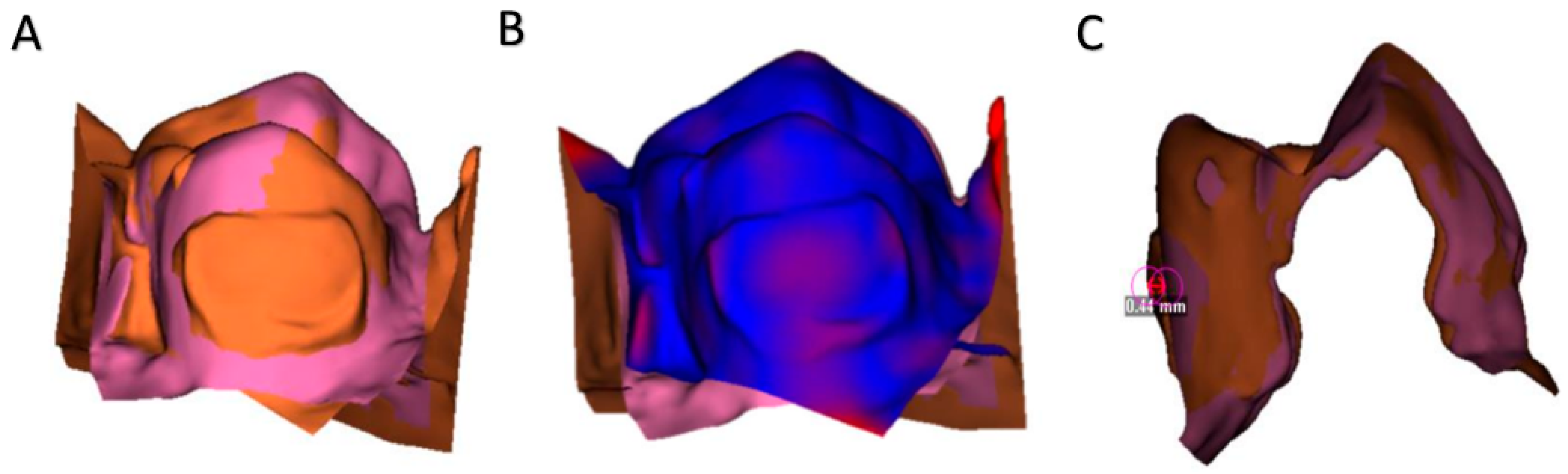
2.5. Methodology of Superimposition and Surface Assessment
2.6. Methodology of Measuring
2.7. Statistical Analysis
3. Results
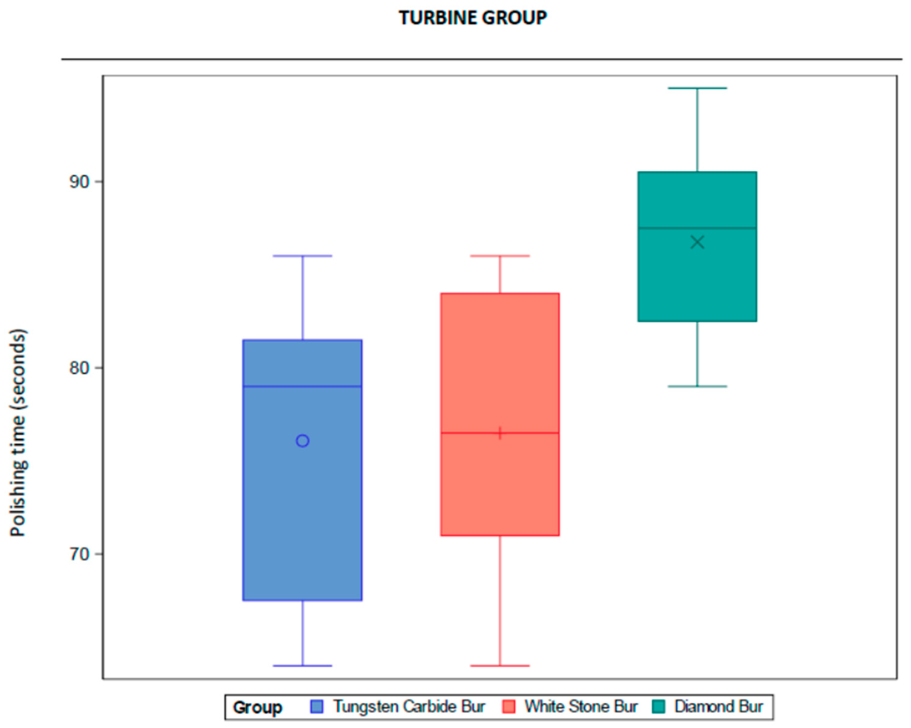
3.1. Turbine Group
3.1.1. Descriptive Analysis
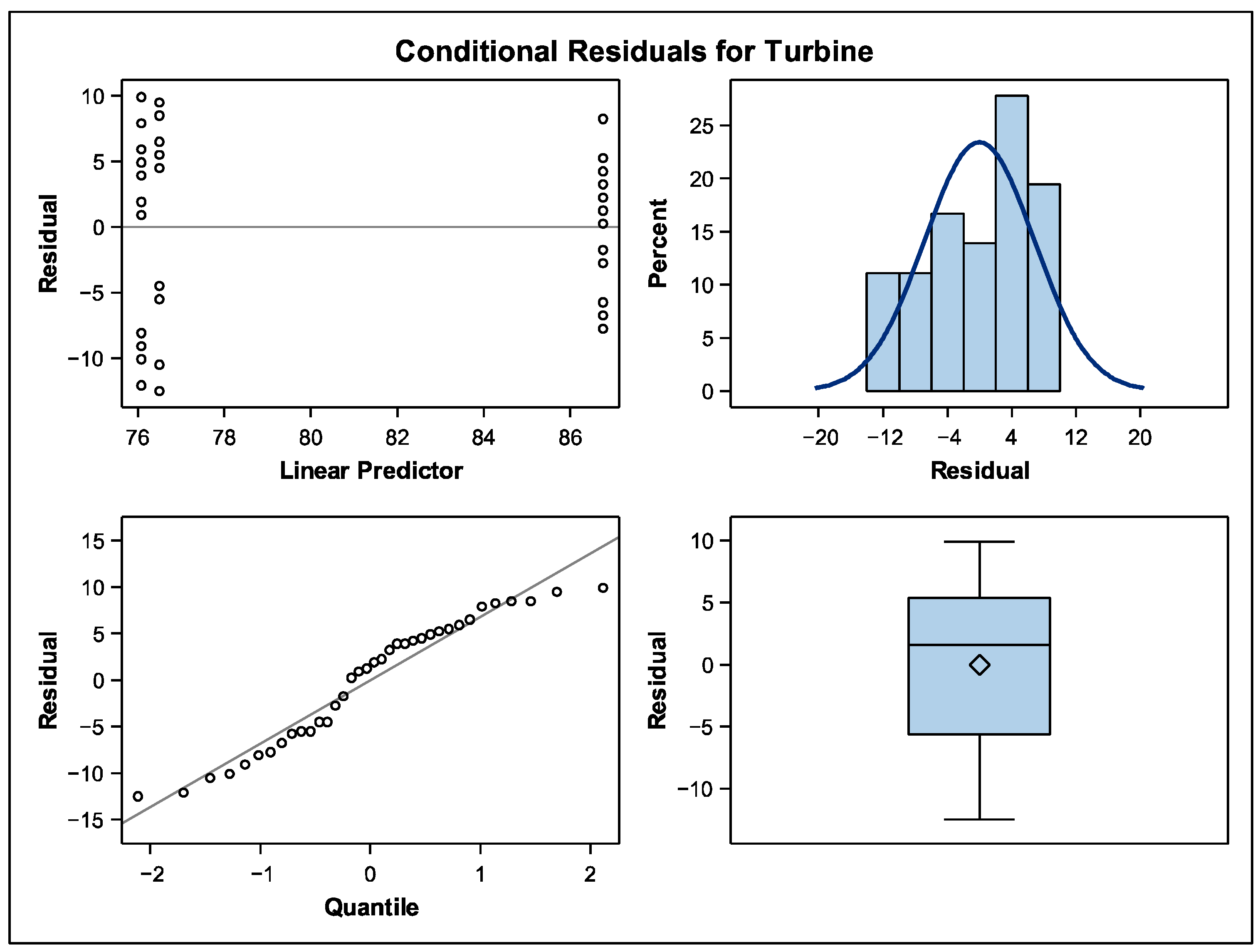
3.1.2. Comparative Analysis
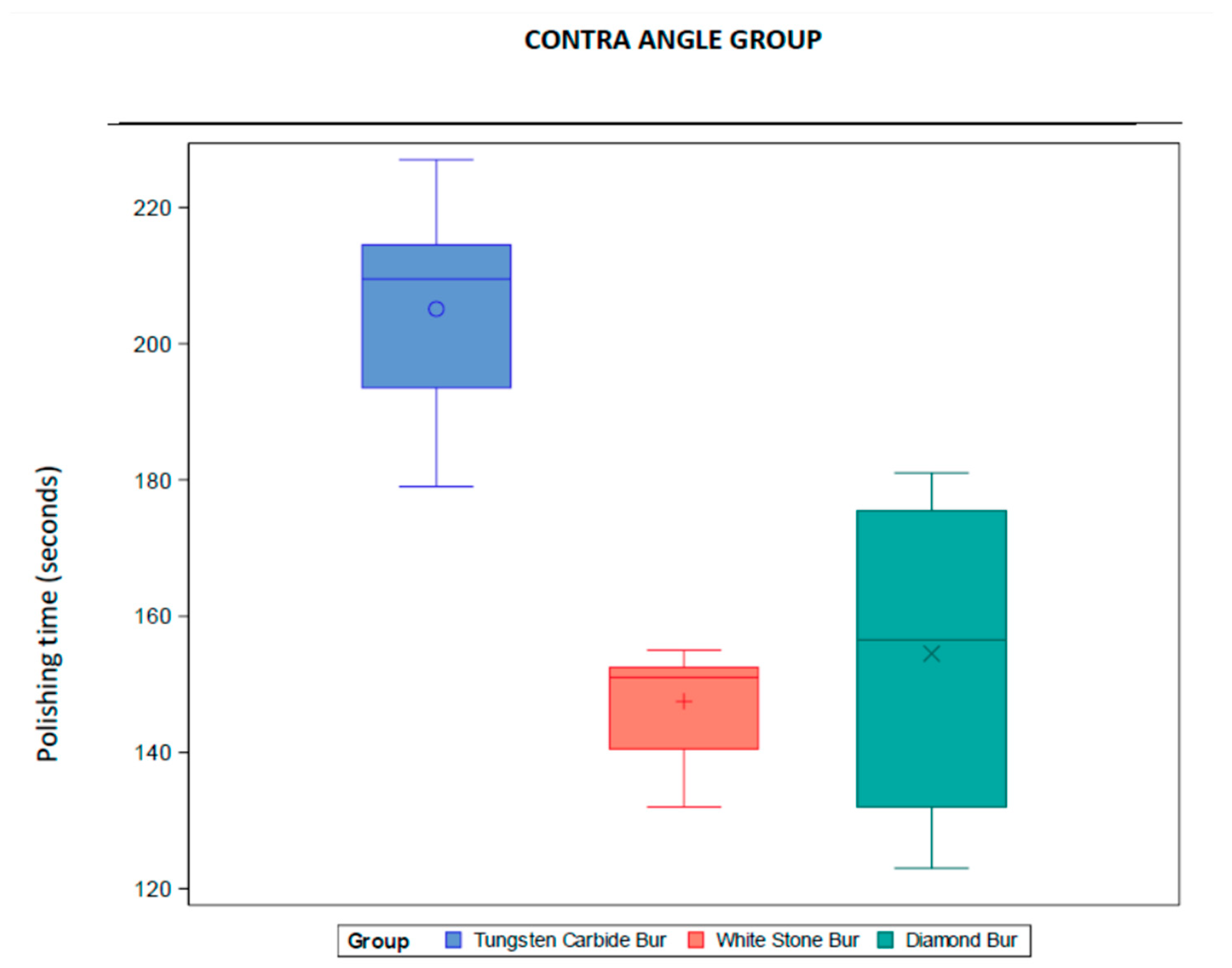
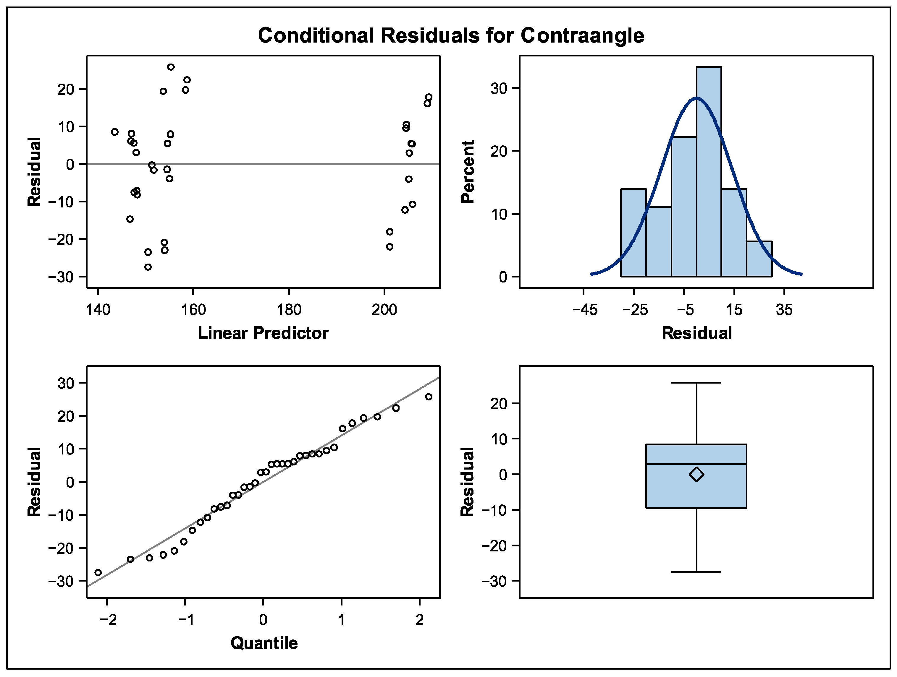
3.2. Contraangle Group
3.2.1. Descriptive Analysis
3.2.2. Comparative Analysis
4. Discussion
5. Conclusions
Author Contributions
Funding
Institutional Review Board Statement
Informed Consent Statement
Data Availability Statement
Conflicts of Interest
References
- Paolone, G.; Mandurino, M.; Baldani, S.; Paolone, M.G.; Goracci, C.; Scolavino, S.; Gherlone, E.; Cantatore, G.; Gastaldi, G. Quantitative Volumetric Enamel Loss after Orthodontic Debracketing/Debonding and Clean-Up Procedures: A Systematic Review. Appl. Sci. 2023, 13, 5369. [Google Scholar] [CrossRef]
- Oz, A.Z.; Oz, A.A.; Ural, C.; Kaleli, N.; Duran, I. Effectiveness of surface polishing after debonding of metal brackets from different CAD-CAM materials. J. Orofac. Orthop. 2024, 85 (Suppl. 1), 19–26. [Google Scholar] [CrossRef] [PubMed]
- Křivková, T.; Tichý, A.; Tycová, H.; Kučera, J. The Influence of Various Adhesive Systems and Polishing Methods on Enamel Surface Roughness after Debonding of Orthodontic Brackets: A Three-Dimensional In Vitro Evaluation. Materials 2023, 16, 5107. [Google Scholar] [CrossRef] [PubMed] [PubMed Central]
- Fahimi, M.A.; Azarbayejani, S.; Mohammadi-Bassir, M. Comparing the effect of two polishing systems on surface roughness of feldspathic, lithium disilicate, and translucent zirconia ceramics after orthodontic bracket debonding: An in vitro study. Int. Orthod. 2024, 22, 100923. [Google Scholar] [CrossRef] [PubMed]
- Nagasaki, R.; Ishikawa, R.; Ito, S.; Saito, T.; Iijima, M. Effects of polishing with paste containing surface pre-reacted glass-ionomer fillers on enamel remineralization after orthodontic bracket debonding. Microsc. Res. Tech. 2021, 84, 171–179. [Google Scholar] [CrossRef] [PubMed]
- Ibrahim, A.I.; Al-Hasani, N.R.; Al-Taai, N.T.; Ismail, H.M.; Abdulrahman, A.A.; Sauro, S. Enamel conservation orthodontic paradigm via treatment with newly developed remineralizing calcium-phosphate etchant pastes. J. Dent. 2025, 157, 105776. [Google Scholar] [CrossRef] [PubMed]
- Öncel, N.A.; Ulusoy, N.; Ulusoy, C. Comparison of shear bond strength and residual adhesive remnants on the enamel surface after debonding of three different orthodontic molar tubes: A scanning electron microscope study. J. Orofac. Orthop. 2024, 85 (Suppl. 1), 94–101. [Google Scholar] [CrossRef] [PubMed]
- Hodecker, L.D.; Scheurer, M.; Scharf, S.; Roser, C.J.; Fouda, A.M.; Bourauel, C.; Lux, C.J.; Bauer, C.A.J. Influence of Individual Bracket Base Design on the Shear Bond Strength of In-Office 3D Printed Brackets—An In Vitro Study. J. Funct. Biomater. 2023, 14, 289. [Google Scholar] [CrossRef] [PubMed] [PubMed Central]
- Scisciola, F.; Palone, M.; Scuzzo, G.; Scuzzo, G.; Huanca Ghislanzoni, L.T.; Lombardo, L. Accuracy of lingual straight-wire orthodontic treatment with passive self-ligating brackets and square slot: A retrospective study. Prog. Orthod. 2023, 24, 30. [Google Scholar] [CrossRef] [PubMed] [PubMed Central]
- Albertini, P.; Mazzanti, V.; Mollica, F.; Lombardo, L.; Siciliani, G. Comparative analysis of passive play and torque expression in self-ligating and traditional lingual brackets. J. Orofac. Orthop. 2022, 83, 13–22. [Google Scholar] [CrossRef] [PubMed]
- Sabbagh, H.; Khazaei, Y.; Baumert, U.; Hoffmann, L.; Wichelhaus, A.; Janjic Rankovic, M. Bracket Transfer Accuracy with the Indirect Bonding Technique-A Systematic Review and Meta-Analysis. J. Clin. Med. 2022, 11, 2568. [Google Scholar] [CrossRef] [PubMed] [PubMed Central]
- Belanche Monterde, A.; Albaladejo Martínez, A.; Curto, A.; Alonso Pérez-Barquero, J.; Guinot-Barona, C.; Zubizarreta-Macho, Á.; Calama González, R.M. Area and Volume of Remaining Cement and Enamel after Removal and Polishing of Buccal or Lingual Multibracket Appliances. Appl. Sci. 2021, 11, 1719. [Google Scholar] [CrossRef]
- Sung, J.-W.; Kwon, T.-Y.; Kyung, H.-M. Debonding forces of three different customized bases of a lingual bracket system. Korean J. Orthod. 2013, 43, 235–241. [Google Scholar] [CrossRef] [PubMed] [PubMed Central]
- Dudás, C.; Czumbel, L.M.; Kiss, S.; Gede, N.; Hegyi, P.; Mártha, K.; Varga, G. Clinical bracket failure rates between different bonding techniques: A systematic review and meta-analysis. Eur. J. Orthod. 2023, 45, 175–185. [Google Scholar] [CrossRef] [PubMed] [PubMed Central]
- Alakttash, A.M.; Fawzi, M.; Bearn, D. Adhesive precoated bracket systems and operator coated bracket systems: Is there any difference? A systematic review and meta-analysis. Angle Orthod. 2019, 89, 495–504. [Google Scholar] [CrossRef] [PubMed] [PubMed Central]
- Chandrasekharan, D.; Sourabh, C.; Deenadayalan; Praveen, K. Enamel surface roughness evaluation after bracket debonding: Comparison between light cure and self cure adhesive resin 3-Dimensional profilometric study. Indian J. Dent. Res. 2022, 33, 80–84. [Google Scholar] [CrossRef] [PubMed]
- Pinzan-Vercelino, C.R.M.; Souza Costa, A.C.; Gurgel, J.A.; Salvatore Freitas, K.M. Comparison of enamel surface roughness and color alteration after bracket debonding and polishing with 2 systems: A split-mouth clinical trial. Am. J. Orthod. Dentofac. Orthop. 2021, 160, 686–694. [Google Scholar] [CrossRef]
- Belanche Monterde, A.; Flores-Fraile, J.; Alonso Pérez-Barquero, J.; Peiro-Aubalat, A.; Mendieta Lasierra, P.; Zubizarreta-Macho, Á. Evaluation of Cement Remaining After Debonding and Polishing in Lingual Multibracket Appliance Using Planning Imaging 3D Software. Materials 2025, 18, 781. [Google Scholar] [CrossRef] [PubMed]
- Tosco, V.; Monterubbianesi, R.; Orilisi, G.; Procaccini, M.; Grandini, S.; Putignano, A.; Orsini, G. Effect of four different finishing and polishing systems on resin composites: Roughness surface and gloss retention evaluations. Minerva Stomatol. 2020, 69, 207–214. [Google Scholar] [CrossRef] [PubMed]
- Lippert, V.F.; Bresciani, E.; Mota, E.G.; Bittencourt, H.R.; Kramer, P.F.; Spohr, A.M. In vitro comparison of one-step, two-step, and three-step polishing systems on the surface roughness and gloss of different resin composites. J. Esthet. Restor. Dent. 2024, 36, 785–795. [Google Scholar] [CrossRef] [PubMed]
- Yousry, T.N.; Abolgheit, S.; Kassem, H.E. Enamel surface roughness following debonding resin clean up using high speed air-turbine and electric handpieces. In vitro study. BMC Oral Health 2024, 24, 609. [Google Scholar] [CrossRef] [PubMed] [PubMed Central]
- Madhyastha, P.S.; Hegde, S.; Srikant, N.; Kotian, R.; Iyer, S.S. Effect of finishing/polishing techniques and time on surface roughness of esthetic restorative materials. Dent. Res. J. 2017, 14, 326–330. [Google Scholar] [CrossRef] [PubMed] [PubMed Central]
- Thawaba, A.A.; Albelasy, N.F.; Elsherbini, A.M.; Hafez, A.M. Evaluation of enamel roughness after orthodontic debonding and clean-up procedures using zirconia, tungsten carbide, and white stone burs: An in vitro study. BMC Oral Health 2023, 23, 478. [Google Scholar] [CrossRef] [PubMed] [PubMed Central]
- Garg, R.; Dixit, P.; Khosla, T.; Gupta, P.; Kalra, H.; Kumar, P. Enamel Surface Roughness after Debonding: A Comparative Study using Three Different Burs. J. Contemp. Dent. Pract. 2018, 19, 521–526. [Google Scholar] [PubMed]
- Czolgosz, I.; Cattaneo, P.M.; Cornelis, M.A. Computer-aided indirect bonding versus traditional direct bonding of orthodontic brackets: Bonding time, immediate bonding failures, and cost-minimization. A randomized controlled trial. Eur. J. Orthod. 2021, 43, 144–151. [Google Scholar] [CrossRef] [PubMed]
- Palone, M.; Bizzocchi, C.; Guiducci, D.; Cremonini, F.; Pellitteri, F.; Spedicato, G.A.; Verducci, A.; Lombardo, L. Evaluation of effectiveness and efficiency of fixed orthodontic treatment comparing standard and computer-aided design and manufacturing conventional bracket systems using indirect bonding for both: A retrospective study. J. World Fed. Orthod. 2023, 12, 251–259. [Google Scholar] [CrossRef] [PubMed]
- Sezici, Y.L.; Önçağ, M.G. Conventional and self-ligating lingual orthodontic treatment outcomes in Class I nonextraction patients: A comparative study with the American Board of Orthodontics Objective Grading System. Am. J. Orthod. Dentofac. Orthop. 2023, 163, e106–e114. [Google Scholar] [CrossRef]
- Fernández-Serrano, J.; García-Espona, E.; Alarcón, J.A.; García-Espona, C.; García-Espona, I. Differences in the Ratios of General and Dental Specialists in Europe. Int. Dent. J. 2024, 74, 519–525. [Google Scholar] [CrossRef] [PubMed] [PubMed Central]
- de Oliveira, N.R.C.; Pigozzo, M.N.; Sesma, N.; Laganá, D.C. Clinical efficiency and patient preference of digital and conventional workflow for single implant crowns using immediate and regular digital impression: A meta-analysis. Clin. Oral Implants Res. 2020, 31, 669–686. [Google Scholar] [CrossRef] [PubMed]
- Jurasic, M.M.; Gibson, G.; Rich, S.; O’Toole, T.G.; Bestgen, S.; Arola, P.E.; Jones, J.A. Leading determinants of efficient dental care delivery. J. Public Health Dent. 2013, 73, 195–203. [Google Scholar] [CrossRef] [PubMed]

| Analysis Variable: Turbine | |||||||
|---|---|---|---|---|---|---|---|
| Group | N | N Miss | Mean | Median | Std Dev | Minimum | Maximum |
| Tungsten Carbide Bur | 12 | 0 | 76.08 | 79.00 | 7.69 | 64.00 | 86.00 |
| White Stone Bur | 12 | 0 | 76.50 | 76.50 | 7.95 | 64.00 | 86.00 |
| Diamond Bur | 12 | 0 | 86.75 | 87.50 | 5.05 | 79.00 | 95.00 |
| Type III Tests of Fixed Effects | |||||
|---|---|---|---|---|---|
| Effect | Num DF | Den DF | F Value | p Value | |
| Group | 2 | 22 | 8.89 | 0.0015 | * |
| Differences in Time Least Squares Means Adjustment for Multiple Comparisons: Tukey–Kramer | ||||||||
|---|---|---|---|---|---|---|---|---|
| Group | Group | Estimate | Standard Error | DF | t Value | p Value | Adj p | |
| Tungsten Carbide Bur | White Stone Bur | −0.4167 | 2.8658 | 22 | −0.15 | 0.8857 | 0.9884 | |
| Tungsten Carbide Bur | Diamond Bur | −10.6667 | 2.8658 | 22 | −3.72 | 0.0012 | 0.0033 | * |
| White Stone Bur | Diamond Bur | −10.2500 | 2.8658 | 22 | −3.58 | 0.0017 | 0.0046 | * |
| Analysis Variable: Contra angle | |||||||
|---|---|---|---|---|---|---|---|
| Group | N | N Miss | Mean | Median | Std Dev | Minimum | Maximum |
| Tungsten Carbide Bur | 12 | 0 | 205.08 | 209.50 | 15.35 | 179.00 | 227.00 |
| White Stone Bur | 12 | 0 | 147.50 | 151.00 | 7.28 | 132.00 | 155.00 |
| Diamond Bur | 12 | 0 | 154.50 | 156.50 | 21.70 | 123.00 | 181.00 |
| Type III Tests of Fixed Effects | |||||
|---|---|---|---|---|---|
| Effect | Num DF | Den DF | F Value | p Value | |
| Group | 2 | 22 | 51.85 | <0.0001 | * |
| Differences in Time Least Squares Means Adjustment for Multiple Comparisons: Tukey–Kramer | ||||||||
|---|---|---|---|---|---|---|---|---|
| Group | Group | Estimate | Standard Error | DF | t Value | p Value | Adj p | |
| Tungsten Carbide Bur | White Stone Bur | 57.5833 | 6.1709 | 22 | 9.33 | <0.001 | <0.001 | * |
| Tungsten Carbide Bur | Diamond Bur | 50.5833 | 6.1709 | 22 | 8.20 | <0.001 | <0.001 | * |
| White Stone Bur | Diamond Bur | −7.0000 | 6.1709 | 22 | −1.13 | 0.2689 | 0.5038 | |
Disclaimer/Publisher’s Note: The statements, opinions and data contained in all publications are solely those of the individual author(s) and contributor(s) and not of MDPI and/or the editor(s). MDPI and/or the editor(s) disclaim responsibility for any injury to people or property resulting from any ideas, methods, instructions or products referred to in the content. |
© 2025 by the authors. Licensee MDPI, Basel, Switzerland. This article is an open access article distributed under the terms and conditions of the Creative Commons Attribution (CC BY) license (https://creativecommons.org/licenses/by/4.0/).
Share and Cite
Flores-Fraile, J.; Belanche Monterde, A.; Alonso-Ezpeleta, O.; Galletti, C.; Zubizarreta-Macho, Á. Chair-Time During Polishing with Different Burs and Drills After Cement Customized Brackets Bonding: An In Vitro Comparative Study. Dent. J. 2025, 13, 347. https://doi.org/10.3390/dj13080347
Flores-Fraile J, Belanche Monterde A, Alonso-Ezpeleta O, Galletti C, Zubizarreta-Macho Á. Chair-Time During Polishing with Different Burs and Drills After Cement Customized Brackets Bonding: An In Vitro Comparative Study. Dentistry Journal. 2025; 13(8):347. https://doi.org/10.3390/dj13080347
Chicago/Turabian StyleFlores-Fraile, Javier, Alba Belanche Monterde, Oscar Alonso-Ezpeleta, Cosimo Galletti, and Álvaro Zubizarreta-Macho. 2025. "Chair-Time During Polishing with Different Burs and Drills After Cement Customized Brackets Bonding: An In Vitro Comparative Study" Dentistry Journal 13, no. 8: 347. https://doi.org/10.3390/dj13080347
APA StyleFlores-Fraile, J., Belanche Monterde, A., Alonso-Ezpeleta, O., Galletti, C., & Zubizarreta-Macho, Á. (2025). Chair-Time During Polishing with Different Burs and Drills After Cement Customized Brackets Bonding: An In Vitro Comparative Study. Dentistry Journal, 13(8), 347. https://doi.org/10.3390/dj13080347