Next Issue
Volume 10, September
Previous Issue
Volume 10, July


Dent. J., Volume 10, Issue 8 (August 2022) – 17 articles

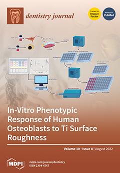
Cover Story (view full-size image): Despite the reported clinical advantages of the currently available surface-modified implants, some scientific reports suggest that surface roughness may play a key role in the accelerated development of bone-implant connection in the short term, and of peri-implant soft and hard tissue damage, which could be either reversible (peri-implant mucositis) or irreversible (peri-implantitis) in the long term. This study aims to investigate human osteoblast (HOB) response towards various Ti (Ti6Al-4V) implant surface roughness (created by a simple industrial milling machine), and to determine if surface roughness influences the early stage of HOB proliferation in the same way as other surface treatments, which, in turn, may potentially influence bone healing. View this paper
  • Issues are regarded as officially published after their release is announced to the table of contents alert mailing list.
  • You may sign up for e-mail alerts to receive table of contents of newly released issues.
  • PDF is the official format for papers published in both, html and pdf forms. To view the papers in pdf format, click on the "PDF Full-text" link, and use the free Adobe Reader to open them.
Order results
Result details
Section
Select all
Export citation of selected articles as:
9 pages, 248 KiB  
Article
Oral Hygiene Practices among Adults with Intellectual Disabilities—A Pilot Study
by Muhammad Afif Hakimin Abdullah, Ishak Nurul Sa’idah, Joe Knights, Sachinjeet Kaur Sodhi Dhaliwal, Long Chiau Ming and Jagjit Singh Dhaliwal
Dent. J. 2022, 10(8), 155; https://doi.org/10.3390/dj10080155 - 19 Aug 2022
Viewed by 1786
Abstract
It is known that the oral health status of people with intellectual disabilities (IDs) is likely to be compromised as compared with the general population. Until recently, the trend of oral hygiene among the adult population with intellectual disabilities in Brunei Darussalam had [...] Read more.
It is known that the oral health status of people with intellectual disabilities (IDs) is likely to be compromised as compared with the general population. Until recently, the trend of oral hygiene among the adult population with intellectual disabilities in Brunei Darussalam had yet to be studied. Thus, the aim of this study was to determine the oral hygiene practices, oral health knowledge and dental appointment patterns among the adult population with IDs in Brunei Darussalam. A cross-sectional study was conducted at different service providers in Brunei Darussalam for adults with IDs. An easy-to-read questionnaire was distributed to the participants. The responses of the questionnaire for IDs were analyzed. A total of 34 participants were recruited. It was found that all participants were practicing their daily oral hygiene routine. In terms of dental visits, more than half visit the dentist at least once a year. The majority of the participants agreed that visiting the dentist every 6 months was essential. This study showed that adults with IDs in Brunei Darussalam were aware of the importance of oral health and were maintaining their daily oral hygiene, although not with the ideal method. It is important for clinicians to not only educate the individuals with IDs but also their carer(s) when it comes to oral health knowledge, as carers play a key role in the oral health of the people under their care. Full article
11 pages, 1283 KiB  
Article
The Correlation of Swedish Snus, Nicotine Pouches and Other Tobacco Products with Oral Mucosal Health and Salivary Biomarkers
by Sintija Miluna, Ricards Melderis, Loreta Briuka, Ingus Skadins, Renars Broks, Juta Kroica and Dagnija Rostoka
Dent. J. 2022, 10(8), 154; https://doi.org/10.3390/dj10080154 - 17 Aug 2022
Cited by 3 | Viewed by 12684
Abstract
In society, tobacco products, such as e-cigarettes, and smokeless tobacco products, such as snus and nicotine pouches, are becoming more attractive. There is still a lack of information regarding the effects of these products on the oral mucosa and oral saliva biomarkers. The [...] Read more.
In society, tobacco products, such as e-cigarettes, and smokeless tobacco products, such as snus and nicotine pouches, are becoming more attractive. There is still a lack of information regarding the effects of these products on the oral mucosa and oral saliva biomarkers. The aim of this study is to evaluate oral mucosa and the presence of inflammatory biomarkers IL-6, IL-1, IL-8, TNF alpha and LRG-1 in saliva. Respondents were divided in four groups based on their tobacco product usage. Oral examination was carried out, saliva samples were taken, and the detection of IL-6, IL-8, IL-1, TNF alpha and LRG-1 levels in saliva was carried out. Out of the tobacco users, 30.8% were snus users, 48.7% were cigarette users and 20.5% were e-cigarette users. The control group was composed of respondents who did not use any tobacco products. E-cigarettes were used more by women, but snus was used more by men. Mucosal changes were seen in the group of snus users, and mucosal changes were only seen in men who had used 5–10 tobacco units per day for 5–10 years. Increased IL-6 levels in saliva were detected in respondents who also experienced mucosal changes. Mucosal changes were white, leathery and localized at the site where snus sachets were placed. Saliva, as an easily available biofluid, could be used as a first tool to detect potentially precancerous signs, but the LRG1 marker cannot be used as a prognostic marker. Full article
Show Figures

Graphical abstract

9 pages, 2195 KiB  
Article
Incorporation of Fluoride into Human Teeth after Immersion in Fluoride-Containing Solutions
by Jana Storsberg, Kateryna Loza and Matthias Epple
Dent. J. 2022, 10(8), 153; https://doi.org/10.3390/dj10080153 - 17 Aug 2022
Cited by 2 | Viewed by 2238
Abstract
Toothpastes and mouth rinses contain fluoride as a protective agent against caries. The aim of this study was to determine the degree of fluoride-uptake by human tooth mineral during immersion into fluoride-containing aqueous solutions as different pH. Human teeth were immersed in fluoride-containing [...] Read more.
Toothpastes and mouth rinses contain fluoride as a protective agent against caries. The aim of this study was to determine the degree of fluoride-uptake by human tooth mineral during immersion into fluoride-containing aqueous solutions as different pH. Human teeth were immersed in fluoride-containing solutions to assess the extent of fluoride incorporation into tooth enamel. A total of 16 extracted teeth from 11 patients were immersed at 37 °C for one minute into aqueous fluoride solutions (potassium fluoride; KF) containing either 250 ppm or 18,998 ppm fluoride (1-molar). Fluoride was dissolved either in pure water (neutral pH) or in a citrate buffer (pH 4.6 to 4.7). The elemental surface composition of each tooth was studied by energy-dispersive X-ray spectroscopy in combination with scanning electron microscopy and X-ray powder diffraction. The as-received teeth contained 0.17 ± 0.16 wt% fluoride on average. There was no significant increase in the fluoride content after immersion in 250 ppm fluoride solution at neutral or acidic pH values. In contrast, a treatment with a 1-molar fluoride solution led to significantly increased fluoride concentrations by 0.68 wt% in water and 9.06 wt% at pH 4.7. Although such fluoride concentrations are far above those used in mouth rinses or toothpastes, this indicates that fluoride can indeed enter the tooth surface, especially at a low pH where a dynamic dissolution-reprecipitation process may occur. However, precipitations of calcium fluoride (globuli) were detected in no cases. Full article
(This article belongs to the Special Issue Dentistry Journal: 10th Anniversary)
Show Figures

Graphical abstract

13 pages, 3224 KiB  
Article
Differences in Supragingival Microbiome in Patients with and without Full-Crown Prostheses
by Manli Guo, Zhidong Zhang, Jiyuan Lu, Di Wang, Yimin Yan, Shen Zhang, Xin Yu, Songhua Su, Lu Yuan, Zhige Li and Baoping Zhang
Dent. J. 2022, 10(8), 152; https://doi.org/10.3390/dj10080152 - 15 Aug 2022
Cited by 1 | Viewed by 1676
Abstract
Objectives: To characterize the microflora profile of supragingival biofilm in patients with and without full-crown prostheses. Methods: Plaque samples of full-crown prostheses and teeth in patients with porcelain-fused-to-metal crowns, all-ceramic crowns, and no prostheses were collected (three patients per group), using 16S rRNA [...] Read more.
Objectives: To characterize the microflora profile of supragingival biofilm in patients with and without full-crown prostheses. Methods: Plaque samples of full-crown prostheses and teeth in patients with porcelain-fused-to-metal crowns, all-ceramic crowns, and no prostheses were collected (three patients per group), using 16S rRNA high-throughput sequencing technology to conduct DNA sequencing on the samples and using Qiime, R, and PICRUSt2 software to perform bioinformatics analyses and functional analyses on sequencing data. Results: In total, 110,209 valid sequences were obtained in the experiment, corresponding to 11 phyla and 120 genera. The predominant species shared by the three groups were phyla Actinobacteria, Bacteroidetes, Firmicutes, Fusobacteria, and Proteobacteria and genera Rothia, Porphyromonas, Prevotella, Streptococcus, Veillonella, Leptotrichia, Neisseria, Citrobacter, and Pseudomonas. The species-difference analysis showed that genus Hameophilus significantly increased after the patient wore the dental prosthesis. Compared with the no-prosthesis samples, the functional analysis showed that cell motility increased in the samples from full-crown prostheses, while replication and repair, and translation decreased. Conclusions: This study reveals the changes in the oral microbial community of patients with full-crown prostheses, which could provide insights regarding the safety of materials for long-term use in the oral cavity. Full article
Show Figures

Figure 1

17 pages, 1977 KiB  
Systematic Review
Success Rates of Zygomatic Implants for the Rehabilitation of Severely Atrophic Maxilla: A Systematic Review
by Aleix Solà Pérez, David Pastorino, Carlos Aparicio, Marta Pegueroles Neyra, Rabia Sannam Khan, Simon Wright and Cemal Ucer
Dent. J. 2022, 10(8), 151; https://doi.org/10.3390/dj10080151 - 12 Aug 2022
Cited by 12 | Viewed by 3469
Abstract
Zygomatic implants are a treatment solution for patients with severe maxillary atrophy. This treatment option allows delivering immediate fixed teeth within 24 h. Numerous peer-reviewed publications have reported different success rates, resulting in a disagreement on the topic. Therefore, the overall efficacy and [...] Read more.
Zygomatic implants are a treatment solution for patients with severe maxillary atrophy. This treatment option allows delivering immediate fixed teeth within 24 h. Numerous peer-reviewed publications have reported different success rates, resulting in a disagreement on the topic. Therefore, the overall efficacy and predictability of this rehabilitation is still a matter of discussion. With this study, we aimed to identify the published literature on the use of zygomatic implants for the reconstruction of the severely atrophic maxilla and report the cumulative success rate (CSR) as a function of follow-up time. A systematic review of the literature on zygomatic implant for the treatment of severe maxillary atrophy was performed and 196 publications were included in the study. The cumulative success rate of zygomatic implants for the treatment of severe maxillary atrophy was 98.5% at less than 1 year, 97.5% between 1 and 3 years, 96.8% between 3 and 5 years and 96.1% after more than 5 years. The most commonly reported complications were soft tissue dehiscence, rhinosinusitis and prosthetic failures. The treatment of severe lack of bone in the upper maxilla with zygomatic implants is a safe procedure, reaching a cumulative success rate of 96.1% after more than 5 years. Full article
(This article belongs to the Special Issue Advanced Research on Oral Cancer and Dental Implants)
Show Figures

Figure 1

10 pages, 3233 KiB  
Article
Frequency and Local Etiological Factors of Impaction of Permanent Teeth among 1400 Patients in a Greek Population
by Kalliopi Siotou, Maria-Panagiota Kouskouki, Isidora Christopoulou, Apostolos I. Tsolakis and Ioannis A. Tsolakis
Dent. J. 2022, 10(8), 150; https://doi.org/10.3390/dj10080150 - 11 Aug 2022
Cited by 5 | Viewed by 3087
Abstract
Background: The purpose of this study is to analyze the frequency of impaction of permanent teeth, beyond the third molars, and to highlight the factors causing this condition. Methods: Panoramic radiographs of 1400 patients that sought orthodontic treatment in private practice were retrieved [...] Read more.
Background: The purpose of this study is to analyze the frequency of impaction of permanent teeth, beyond the third molars, and to highlight the factors causing this condition. Methods: Panoramic radiographs of 1400 patients that sought orthodontic treatment in private practice were retrieved and examined. All teeth that had not been erupted at the time of the examination while their root formation was completed were considered impacted. Results: In total, 212 out of 1400 patients had at least one impacted tooth (15.14%). The highest incidence of tooth impaction was in the canines of the maxilla, followed by the central incisors of the maxilla, the second molars of the mandible and the second premolars of the mandible. The most common etiological factors responsible for the impaction were the ectopic eruption pathway, loss of space in the arch, the ankylosis of the deciduous teeth and the presence of supernumerary teeth. Conclusions: Tooth impaction is frequently seen in everyday orthodontic practice. The upper canines are the teeth most frequently associated with impaction and failure of eruption. It is important to diagnose cases of impaction early on and identify the etiological factors in order to achieve immediate and effective treatment per patient. Full article
Show Figures

Figure 1

8 pages, 1976 KiB  
Article
Radiographic Changes to Silver Diamine Fluoride Treated Carious Lesions after a Rinsing Step
by Zaher Jabbour, Maryam Esmaeili, Marc Hayashi and Reuben Kim
Dent. J. 2022, 10(8), 149; https://doi.org/10.3390/dj10080149 - 09 Aug 2022
Cited by 3 | Viewed by 1823
Abstract
Silver diamine fluoride (SDF) is radiopaque. This in vitro study compares the changes in the radiopacity of carious lesions after SDF application, potassium iodide (PI) application, and water rinse. Ten recently extracted human teeth were sectioned and divided into two groups (n [...] Read more.
Silver diamine fluoride (SDF) is radiopaque. This in vitro study compares the changes in the radiopacity of carious lesions after SDF application, potassium iodide (PI) application, and water rinse. Ten recently extracted human teeth were sectioned and divided into two groups (n = 10 in each group): Group 1 = SDF, Group 2 = SDF + PI. Teeth in Group 1 received SDF for 1 min and rinsed with 15 mL water. Group 2 received the same protocol with the addition of PI application for 1 min after SDF application. All samples were scanned with micro-computed tomography before SDF application, after SDF application, after PI application (group 2) and after water rinse. The radiopacity of the carious lesions increased significantly after SDF application in Group 1 and 2 (p < 0.017, p < 0.008, respectively). A significant increase in radiopacity after PI application was also observed in Group 2 (p < 0.008). Water rinsing significantly decreased the radiopacity in Group 1 and 2 (p < 0.017, p < 0.008, respectively), but the radiopacity remained significantly higher than the preoperative values (Group 1 p < 0.017, Group 2 p < 0.008). The radiopacity of carious lesions increases after SDF and SDF + PI applications. Water rinsing could reduce the radiopacity of SDF and SDF + PI treated carious lesions, and might reduce the content of SDF in carious lesions. Full article
(This article belongs to the Special Issue Preventive Dental Care, Chairside and Beyond)
Show Figures

Figure 1

13 pages, 1662 KiB  
Article
Can the Concentration of Citric Acid Affect Its Cytotoxicity and Antimicrobial Activity?
by Luciano Giardino, Luigi Generali, Paolo Savadori, Mirela Cesar Barros, Leticia Lobo de Melo Simas, Jolanta Pytko-Polończyk, Wojciech Wilkoński, Vasudev Ballal and Flaviana Bombarda de Andrade
Dent. J. 2022, 10(8), 148; https://doi.org/10.3390/dj10080148 - 09 Aug 2022
Cited by 2 | Viewed by 2512
Abstract
Background: There has been no unanimity concerning the ideal concentration of citric acid for safe use in clinical practice. This study evaluated the cytotoxicity and the antibacterial activity in infected dentinal tubules of 10% and 1% citric acid (CA) solutions. Methods: The cytotoxicity [...] Read more.
Background: There has been no unanimity concerning the ideal concentration of citric acid for safe use in clinical practice. This study evaluated the cytotoxicity and the antibacterial activity in infected dentinal tubules of 10% and 1% citric acid (CA) solutions. Methods: The cytotoxicity of CA solutions in DMEM (diluted 1/10, 1/100) was assessed in L-929 fibroblasts. A broth macrodilution method (MIC and MBC) was used to assess CA antibacterial concentration. The antimicrobial activity of CA solutions was also evaluated after their final rinse inside root canals in previously Enterococcus faecalis-contaminated dentinal tubules. Ten infected dentine samples were rinsed for 5 min with 5% NaOCl and subsequently with 1% citric acid for 3 min. Another 10 were rinsed with 5% NaOCl and 10% citric acid for 3 min; the remaining four specimens were utilized as positive controls. Two uncontaminated specimens were used as negative controls. After LIVE/DEAD BacLight staining, the samples were assessed using CLSM to analyze the percentage of residual live and dead cells. Results: Both undiluted and diluted CA solutions showed severe toxicity; no changes from normal morphology were displayed when diluted 1/100. The MIC and MBC of CA were 6.25 mg/mL and 12.50 mg/mL, respectively. CA solutions demonstrated significantly low levels of bacterial counts than the positive control group, reporting a value of 9.3% for the 10% solution versus the 1% solution (35.2%). Conclusions: Despite its valuable antimicrobial properties, the cytotoxic effects of citric acid should be considered during endodontic treatment. Full article
(This article belongs to the Special Issue Endodontics and Restorative Sciences)
Show Figures

Figure 1

9 pages, 588 KiB  
Article
The Potential of Digital Impression in Orthodontics
by Sabina Saccomanno, Stefano Saran, Valeria Vanella, Rodolfo Francesco Mastrapasqua, Luca Raffaelli and Luca Levrini
Dent. J. 2022, 10(8), 147; https://doi.org/10.3390/dj10080147 - 08 Aug 2022
Cited by 5 | Viewed by 2534
Abstract
Background: Over the past 20 years, there have been many innovations in orthodontic diagnosis and therapy. Among the innovations, there is the taking of dental impressions (DIs). Dental impressions are the negative imprint of hard and soft tissues of one or both arches, [...] Read more.
Background: Over the past 20 years, there have been many innovations in orthodontic diagnosis and therapy. Among the innovations, there is the taking of dental impressions (DIs). Dental impressions are the negative imprint of hard and soft tissues of one or both arches, and they allow a plaster model to be formed, i.e., a positive reproduction. Traditional dental impressions can be made of different materials, such as alginate, while digital impression is captured by an intra-oral scanner. Digital impression, despite the evident advantages, has not yet replaced the conventional impression. The aim of this study is to evaluate which dental impressions are the most used by dentists. For this purpose, we considered 120 questionnaires sent electronically to patients of different dental private practices from different countries, where the dentists can use both techniques. The results highlighted that the kind of impression adopted is very much influenced by the type of therapy and orthodontic devices used in the treatment. We can conclude that, despite the advent of digital technology, conventional impressions are still used for fixed devices, while digital impressions are more adopted for orthodontic customized devices and therapies with clear aligners, that are very widespread among adult patients. Full article
(This article belongs to the Special Issue Orthodontics and New Technologies)
Show Figures

Figure 1

10 pages, 239 KiB  
Concept Paper
Leadership and Managerial Skills in Dentistry: Characteristics and Challenges Based on a Preliminary Case Study
by Maria Antoniadou
Dent. J. 2022, 10(8), 146; https://doi.org/10.3390/dj10080146 - 05 Aug 2022
Cited by 3 | Viewed by 3943
Abstract
Dentistry is changing rapidly in a dynamic and challenging mode and is incorporating digital technology, communication, and managerial skills for profitable enterprises. On the new dental horizon, the profession requires engaging and inspiring role models and leaders. Ιdentifying and developing human soft skills [...] Read more.
Dentistry is changing rapidly in a dynamic and challenging mode and is incorporating digital technology, communication, and managerial skills for profitable enterprises. On the new dental horizon, the profession requires engaging and inspiring role models and leaders. Ιdentifying and developing human soft skills can improve quality issues and guarantee a sustainable dental business. The concept of leadership is a very complex and multidimensional phenomenon as observed from the current literature. In different commercial environments, there has been a huge discussion on the specific characteristics of an effective leader. In dentistry, the subject needs further investigation. This article aims to bring up the importance of dental leadership and highlights the need of identifying dental leaders committed to excellency. It also challenges the need for educational shift on dental leadership management. Finally, it aims to support and develop educational transformations based on positive preliminary data observed by incorporating a new, relevant subject in the dental curriculum. Full article
22 pages, 13182 KiB  
Review
Bioengineering Tools Applied to Dentistry: Validation Methods for In Vitro and In Silico Analysis
by Jefferson David Melo de Matos, Daher Antonio Queiroz, Leonardo Jiro Nomura Nakano, Valdir Cabral Andrade, Nathália de Carvalho Ramos Ribeiro, Alexandre Luiz Souto Borges, Marco Antonio Bottino and Guilherme da Rocha Scalzer Lopes
Dent. J. 2022, 10(8), 145; https://doi.org/10.3390/dj10080145 - 04 Aug 2022
Cited by 7 | Viewed by 2216
Abstract
This study aimed to evaluate the use of bioengineering tools, finite element analysis, strain gauge analysis, photoelastic analysis, and digital image correlation, in computational studies with greater validity and reproducibility. A bibliographic search was performed in the main health databases PUBMED and Scholar [...] Read more.
This study aimed to evaluate the use of bioengineering tools, finite element analysis, strain gauge analysis, photoelastic analysis, and digital image correlation, in computational studies with greater validity and reproducibility. A bibliographic search was performed in the main health databases PUBMED and Scholar Google, in which different studies, among them, laboratory studies, case reports, systematic reviews, and literature reviews, which were developed in living individuals, were included. Therefore, articles that did not deal with the use of finite element analysis, strain gauge analysis, photoelastic analysis, and digital image correlation were excluded, as well as their use in computational studies with greater validity and reproducibility. According to the methodological analysis, it is observed that the average publication of articles in the Pubmed database was 2.03 and with a standard deviation of 1.89. While in Google Scholar, the average was 0.78 and the standard deviation was 0.90. Thus, it is possible to verify that there was a significant variation in the number of articles in the two databases. Modern dentistry finds in finite element analysis, strain gauge, photoelastic and digital image correlation a way to analyze the biomechanical behavior in dental materials to obtain results that assist to obtain rehabilitations with favorable prognosis and patient satisfaction. Full article
(This article belongs to the Special Issue Advances in Oral Implant Health)
Show Figures

Figure 1

14 pages, 6928 KiB  
Article
Characterization of SIBLING Proteins in the Mineralized Tissues
by Sandeep Dab, Nancy Abdelhay, Carlos Alberto Figueredo, Seema Ganatra and Monica Prasad Gibson
Dent. J. 2022, 10(8), 144; https://doi.org/10.3390/dj10080144 - 04 Aug 2022
Cited by 2 | Viewed by 1932
Abstract
The SIBLING proteins are a family of non-collagenous proteins (NCPs) previously thought to be expressed only in dentin but have been demonstrated in other mineralized and non-mineralized tissues. They are believed to play vital roles in both osteogenesis and dentinogenesis. Since they are [...] Read more.
The SIBLING proteins are a family of non-collagenous proteins (NCPs) previously thought to be expressed only in dentin but have been demonstrated in other mineralized and non-mineralized tissues. They are believed to play vital roles in both osteogenesis and dentinogenesis. Since they are tightly regulated lifelong processes and involve a peak of mineralization, three different age groups were investigated. Fifteen wild-type (WT) mice were euthanized at ages 1, 3, and 6 months. Hematoxylin and eosin staining (H&E) was performed to localize various microscopic structures in the mice mandibles and tibias. The immunostaining pattern was compared using antibodies for dentin sialoprotein (DSP), dentin matrix protein 1 (DMP1), bone sialoprotein (BSP), and osteopontin (OPN). Immunostaining of DSP in tibia showed its most noticeable staining in the 3-month age group. DSP was expressed in alveolar bone, cellular cementum, and PDL. A similar expression of DMP1 was seen in the tibia and dentin. BSP was most noticeably detected in the tibia and acellular cementum. OPN was mainly expressed in the bone. A lower level of OPN was observed at all age groups in the teeth. The immunostaining intensity was the least detected for all proteins in the 6-month tibia sample. The expression patterns of the four SIBLING proteins showed variations in their staining intensity and temporospatial patterning concordant with skeletal and dental maturity. These findings suggest some role in this tightly regulated mineralization process. Full article
Show Figures

Figure 1

14 pages, 2552 KiB  
Article
In Vitro and In Vivo Anti-Inflammatory Effects of TEES-10®, a Mixture of Ethanol Extracts of Ligularia stenocephala Matsum. & Koidz. and Secale cereale L. Sprout, on Gingivitis and Periodontitis
by Seungah Lee, In Hye Kim, Junkee Hong, Byung-Ju Jeon, Sung-Su Kim, Ji-Won Lee, Jin-Young Park, Seong-Ho Choi, Tae-Kyeong Lee, Jae-Kook Cha and Moo-Ho Won
Dent. J. 2022, 10(8), 143; https://doi.org/10.3390/dj10080143 - 02 Aug 2022
Viewed by 2292
Abstract
Gingivitis and periodontitis are inflammatory disorders caused by dental plaque and calculus. These disorders often lead to tooth loss if not treated properly. Although antibiotics can be used, it is hard to treat them due to the difficulty in supplying effective doses of [...] Read more.
Gingivitis and periodontitis are inflammatory disorders caused by dental plaque and calculus. These disorders often lead to tooth loss if not treated properly. Although antibiotics can be used, it is hard to treat them due to the difficulty in supplying effective doses of antibiotics to lesion areas and side effects associated with long-term use of antibiotics. In the present study, attempts were made to provide in vitro and in vivo evidence to support anti-inflammatory activities of TEES-10®, a mixture of ethanol extracts of Ligularia stenocephala (LSE) and Secale cereale L. sprout (SCSE) toward gingivitis and periodontitis by performing the following experiments. TEES-10® with a ratio of 6:4 (LSE:SCSE) showed the best effects in both stimulating the viability and inhibiting the cytotoxicity. In in vitro experiments, TEES-10® showed an ability to scavenge 2,2-diphenyl-1-picrylhydrazyl and superoxide radicals and remove ROS generated in periodontal ligament cells treated with lipopolysaccharide. TEES-10® also enhanced the viability of stem cells from human exfoliated deciduous teeth and stimulated the osteogenic differentiation of deciduous teeth cells. In in vivo experiments using rats with induced periodontitis, TEES-10® significantly decreased inflammatory cell infiltration and the numbers of osteoclasts, increased alveolar process volume and the numbers of osteoblasts, decreased serum levels of IL-1β and TNF-α (pro-inflammatory cytokines), and increased serum levels of IL-10 and IL-13 (anti-inflammatory cytokines). These results strongly support the theory that TEES-10® has the potential to be developed as a health functional food that can treat and prevent gingival and periodontal diseases and improve dental health. Full article
Show Figures

Graphical abstract

9 pages, 1124 KiB  
Article
Comparative Dissemination of Aerosol and Splatter Using Suction Device during Ultrasonic Scaling: A Pilot Study
by Nutthawadee Engsomboon, Praewpat Pachimsawat and Bhornsawan Thanathornwong
Dent. J. 2022, 10(8), 142; https://doi.org/10.3390/dj10080142 - 01 Aug 2022
Cited by 3 | Viewed by 2224
Abstract
Objective: This study compared the aerosol and splatter diameter and count numbers produced by a dental mouth prop with a suction holder device and a saliva ejector during ultrasonic scaling in a clinical setting. Methodology: Fluorescein dye was placed in the dental equipment [...] Read more.
Objective: This study compared the aerosol and splatter diameter and count numbers produced by a dental mouth prop with a suction holder device and a saliva ejector during ultrasonic scaling in a clinical setting. Methodology: Fluorescein dye was placed in the dental equipment irrigation reservoirs with a mannequin, and an ultrasonic scaler was employed. The procedures were performed three times per device. The upper and bottom board papers were placed on the laboratory platform. All processes used an ultrasonic scaler to generate aerosol and splatter. A dental mouth prop with a suction holder and a saliva ejector were also tested. Photographic analysis was used to examine the fluorescein samples, followed by image processing in Python and assessment of the diameter and count number. For device comparison, statistics were used with an independent t-test. Result: When using the dental mouth prop with a suction holder, the scaler produced aerosol particles that were maintained on the upper board paper (mean ± SD: 1080 ± 662 µm) compared to on the bottom board paper (1230 ± 1020 µm). When the saliva ejector was used, it was found that the diameter of the aerosol on the upper board paper was 900 ± 580 µm, and the diameter on the bottom board paper was 1000 ± 756 µm. Conclusion: There was a significant difference in the aerosol and splatter particle diameter and count number between the dental mouth prop with a suction holder and saliva ejector (p < 0.05). Furthermore, the results revealed that there was a statistically significant difference between the two groups on the upper and bottom board papers. Full article
Show Figures

Graphical abstract

12 pages, 1604 KiB  
Article
Bruxism’s Implications on Fixed Orthodontic Retainer Adhesion
by Anca Labuneț, Adriana Objelean, Oana Almășan, Andreea Kui, Smaranda Buduru and Sorina Sava
Dent. J. 2022, 10(8), 141; https://doi.org/10.3390/dj10080141 - 01 Aug 2022
Cited by 1 | Viewed by 2011
Abstract
Background: Fixed retainers assist in maintaining the outcomes of orthodontic treatment. Fixed retention may be affected by bruxism. Objective: Evaluate two adhesives (an ormocer and a flowable composite) used for fixed orthodontic retention in simulated bruxism settings, compared to regular mastication, using a [...] Read more.
Background: Fixed retainers assist in maintaining the outcomes of orthodontic treatment. Fixed retention may be affected by bruxism. Objective: Evaluate two adhesives (an ormocer and a flowable composite) used for fixed orthodontic retention in simulated bruxism settings, compared to regular mastication, using a dual axis chewing simulator. Methods: Eighty human teeth were used. Periodontal tissues were simulated and exposed to 120,000 mechanical cycles, corresponding to 6 months of clinical service. Each set of two teeth was supplied with a pre-shaped, fixed, multi-braided, stainless steel wire retainer, in 1.5 cm portions, to establish passive contact with the lingual surface of the teeth. The Adhesive Remnant Index (ARI) was used to evaluate the shear bond strength. A stereomicroscope was used to assess the micro-infiltration. Results: There was no significant difference in the mean value of micro-infiltration between adhesives in the mastication group but in the bruxism group. During testing, one composite sample (ARI score 1) was broken in the mastication group, while three ormocer samples (ARI score 2) and one composite sample (ARI score 1) were broken in the bruxism group. Conclusions: The mean value for micro-infiltration in composite (0.31) was more than double that in ormocer (0.13). Full article
(This article belongs to the Special Issue Dentistry Journal: 10th Anniversary)
Show Figures

Figure 1

14 pages, 2102 KiB  
Article
In-Vitro Phenotypic Response of Human Osteoblasts to Different Degrees of Titanium Surface Roughness
by Muataz A. Osman, Rasha A. Alamoush, Evgeny Kushnerev, Kevin G. Seymour, Susan Shawcross and Julian M. Yates
Dent. J. 2022, 10(8), 140; https://doi.org/10.3390/dj10080140 - 29 Jul 2022
Cited by 4 | Viewed by 1852
Abstract
Objectives: This study aimed to investigate human osteoblast (HOB) responses towards different degrees of titanium (Ti) implant surface roughness. Methods: Four degrees of Ti surface roughness were investigated on a micrometer roughness scale: smooth (S: 0.08–0.1 µm), minimally rough (MM: 0.3–0.5 µm), moderately [...] Read more.
Objectives: This study aimed to investigate human osteoblast (HOB) responses towards different degrees of titanium (Ti) implant surface roughness. Methods: Four degrees of Ti surface roughness were investigated on a micrometer roughness scale: smooth (S: 0.08–0.1 µm), minimally rough (MM: 0.3–0.5 µm), moderately rough (MR: 1.2–1.4 µm), and rough (R: 3.3–3.7 µm). HOB cells were cultured, expanded, and maintained according to the supplier’s protocol. Cell proliferation and cytotoxicity were assessed at day 1, 3, 5, and 10 using alamarBlue and lactate dehydrogenase colorimetric assays. Data were analyzed with one-way ANOVA, two-way ANOVA, and Tukey’s post hoc test (p = 0.05 for all tests). Results: There was no significant difference in the cell proliferation or cytotoxicity of the HOB cells in contact with the different degrees of Ti surface roughness. There was, however, a significant time effect on cell proliferation (p < 0.0001) with different exposure durations for each roughness degree. Furthermore, a positive correlation (non-significant) between proliferation and cytotoxicity was observed for all investigated degrees of surface roughness. Conclusion: All investigated roughness degrees showed comparable HOB proliferation, with the MR surface presenting the highest percentage, followed by the R, MM, ad S, surfaces, respectively. The S surface showed the highest cytotoxic effect on HOBs; however, it did not reach the cytotoxic level suggested by the ISO for any medical device to be considered cytotoxic. Full article
(This article belongs to the Section Dental Implantology)
Show Figures

Graphical abstract

11 pages, 11472 KiB  
Article
Clinical Outcome of Fully Digital Workflow for Single-Implant-Supported Crowns: A Retrospective Clinical Study
by Francesco Gianfreda, Paolo Pesce, Erich Marcano, Valeria Pistilli, Patrizio Bollero and Luigi Canullo
Dent. J. 2022, 10(8), 139; https://doi.org/10.3390/dj10080139 - 27 Jul 2022
Cited by 2 | Viewed by 2172
Abstract
A digital workflow by means of intraoral scanners and computer tomography has been used in dental implantology, allowing clinicians to be potentially more accurate and precise. Computer-Aided Design and Computer-Aided Manufacturing (CAD-CAM) and 3D models facilitate the process from treatment planning to the [...] Read more.
A digital workflow by means of intraoral scanners and computer tomography has been used in dental implantology, allowing clinicians to be potentially more accurate and precise. Computer-Aided Design and Computer-Aided Manufacturing (CAD-CAM) and 3D models facilitate the process from treatment planning to the surgical procedure, up to the implant placement and final prosthesis. The aim of the present retrospective study was to evaluate a fully digital workflow for single-tooth implant rehabilitation. A total of 19 patients (22 implants) were included in the present study, with a mean follow-up time of 2 years. A fully digital workflow was performed on each patient through the planning, design and printing of a surgical guide, following a digital impression made with an intraoral scanner, computer-tomography-guided implant placement and, finally, with the delivery of a CAD-CAM crown. The two-year follow-up results were satisfactory in terms of the aesthetic yield and precision of the prosthesis. In single-implant-supported restorations, due to digital protocols and digital planning, a reduced number of clinical sessions was registered and the treatment plan results were more predictable. Future studies are needed to understand the application of fully digital protocols in cases of partially or totally edentulous patients. Full article
(This article belongs to the Collection Bio-Logic Approaches to Implant Dentistry)
Show Figures

Graphical abstract

Previous Issue
Back to TopTop