Article

Four New Terpenoids from Fufang Yinhua Jiedu Granules and Their Anti-Influenza A Virus Activity

Academy of Military Medical Sciences, Beijing 100850, China
*
Authors to whom correspondence should be addressed.
Separations 2026, 13(4), 105; https://doi.org/10.3390/separations13040105
Submission received: 13 February 2026 / Revised: 12 March 2026 / Accepted: 18 March 2026 / Published: 26 March 2026

Abstract

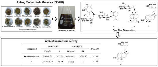
Fufang Yinhua Jiedu Granules (FFYHG) is usually applied to treat influenza and the common cold. However, there is no available report concerning the effects of chemical constituents in FFYHG on antiviral activity. In our study, four new terpenoid derivatives (14) and seventeen known compounds were isolated from FFYHG. Their structures and absolute configurations were determined by various techniques, including high-resolution mass spectrometry analysis, 1/2-dimensional (1D/2D) nuclear magnetic resonance (NMR) analysis, comparative electronic circular dichroism (ECD) studies (experiment vs. calculation), and acid hydrolysis. In addition, the inhibitory effects of twenty-one isolated compounds against influenza A viruses (H1N1) including A/California/07/2009 (CA07) and A/WSN/1933 (WSN) strains were evaluated in vitro, and compound 4 exhibited a moderate inhibitory effect on CA07 strain, with a half maximal inhibitory concentration (IC50) value of 37.10 ± 1.35 μM. This study enhanced the understanding of the active ingredients in FFYHG against influenza virus, providing a foundation for further research on the material basis and quality control of FFYHG.

Graphical Abstract

1. Introduction

Influenza is an acute respiratory disease first identified in the 16th century, which spreads rapidly through communities during epidemics [1]. There are hundreds of thousands of respiratory deaths each year related to seasonal influenza globally [2,3,4]. As a major respiratory pathogen, influenza A viruses (IAV) contain different subtypes due to the different neuraminidase (NA) and hemagglutinin (HA) compositions, including H1N1, H3N2, H5N1, H7N9, etc. [5]. Among them, H1N1 is the most common influenza virus that infects humans, causing seasonal, periodic, and unpredictable pandemics [6,7]. At present, chemical drugs acting on different single targets, including oseltamivir, amantadine, rimantadine and zanamivir, are the main antiviral therapies in clinical practice [8,9]. However, the growing resistance of influenza A virus to chemical drugs poses an increasing global public health concern, particularly in Europe and North America [8,10]. Thus, the discovery of new effective anti-influenza virus agents is of utmost urgency.
Fufang Yinhua Jiedu Granules (FFYHG) is derived from the formula of ‘Yinqiao San’, which acts to disperse wind, relieve the exterior, clear heat, and detoxify. Modern clinical applications demonstrated that FFYHG exerts significant therapeutic effects on wind-heat type influenza and common cold, addressing symptoms like fever, headache, runny nose, cough, sore throat, and general body aches. It is composed of ten Chinese medicinal herbs: Forsythia suspensa Vahl (Oleaceae), Artemisia annua L. (Asteraceae), Lonicerae similis Flos (Caprifoliaceae), Chrysanthemum indicum L. (Asteraceae), Isatis indigotica Fort. (Brassicaceae), Commelina communis L. (Commelinaceae), Glycine max (L.) Merr. (Fabaceae), Schizonepeta tenuifolia Briq. (Lamiaceae), Peucedanum praeruptorum Dunn (Apiaceae), and Mentha haplocalyx Briq (Lamiaceae). Pharmacological experiments evidenced the inhibitory effect of FFYHG on H1N1, H3N2, H5N1, H7N9, and H9N2 subtype influenza viruses in vitro [11]. Meanwhile, the influenza A-infected mice model indicated that FFYHG regulated the Toll-like receptor 7-myeloid differentiation primary response 88 (TLR7-MyD88) signaling pathway to display anti-influenza A activity in vivo [12]. To date, studies on the chemical constituents of FFYHG have primarily focused on the characterization and tentative structural identification of its constituents, using analytical techniques such as high-performance liquid chromatography ultraviolet detector (HPLC-UV) [11] and ultra-high performance liquid chromatography-quadrupole time-of-flight mass spectrometry (UHPLC-QTOF-MS) [13]. However, studies concerning the isolation and purification of compounds from FFYHG remain limited. Therefore, it is necessary to carry out systematic phytochemical separation studies on FFYHG to obtain compounds, laying a foundation for subsequent quality control and mechanism investigation.
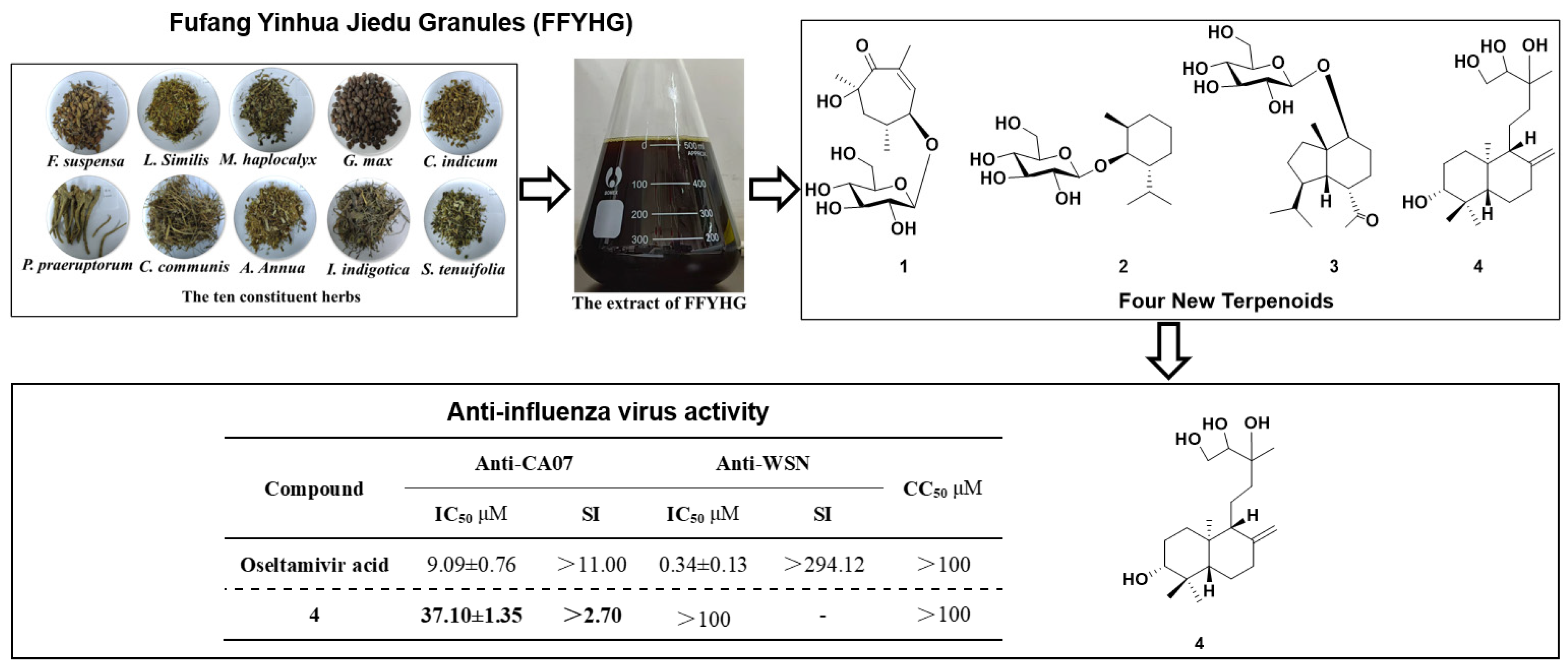
In this present study, the chemical compositions of FFYHG were investigated using various column chromatographic methods, and twenty-one compounds were obtained, including four undescribed compounds (14) (Figure 1). These compounds were evaluated for their inhibitory activities on two H1N1 strains, A/California/07/2009 (CA07) and A/WSN/1933 (WSN). Herein, we report the purification, structural elucidation, and biological investigation of twenty-one compounds from FFYHG.

2. Results and Discussion

2.1. Isolation and Structure Elucidation

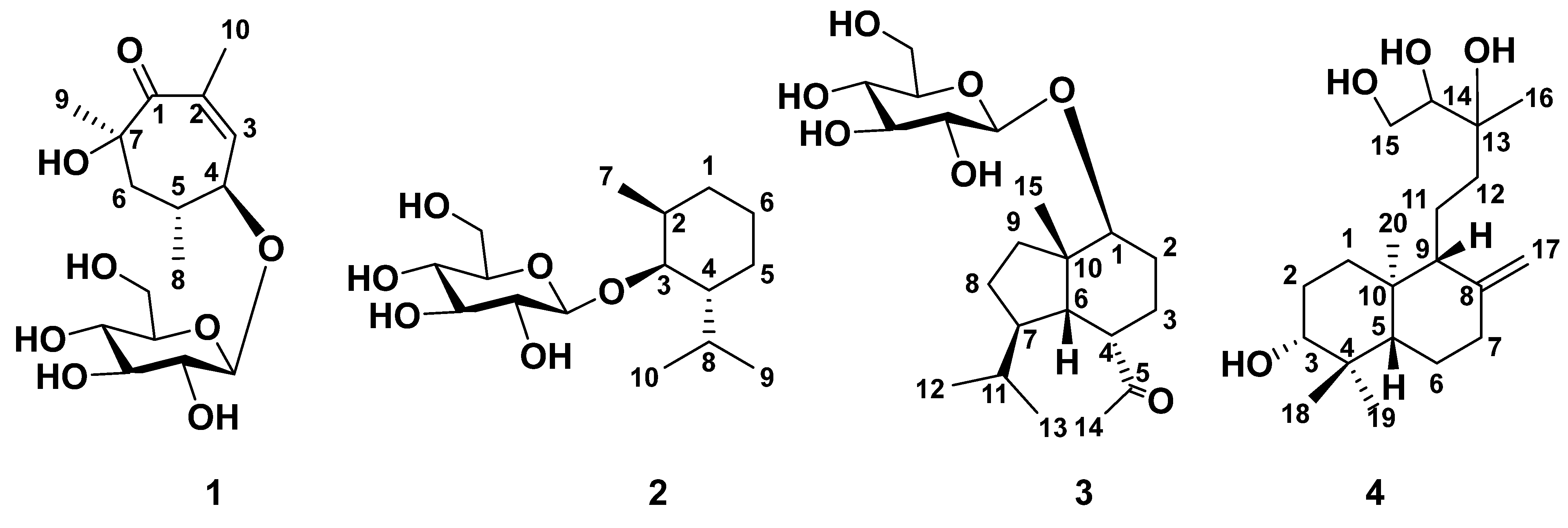
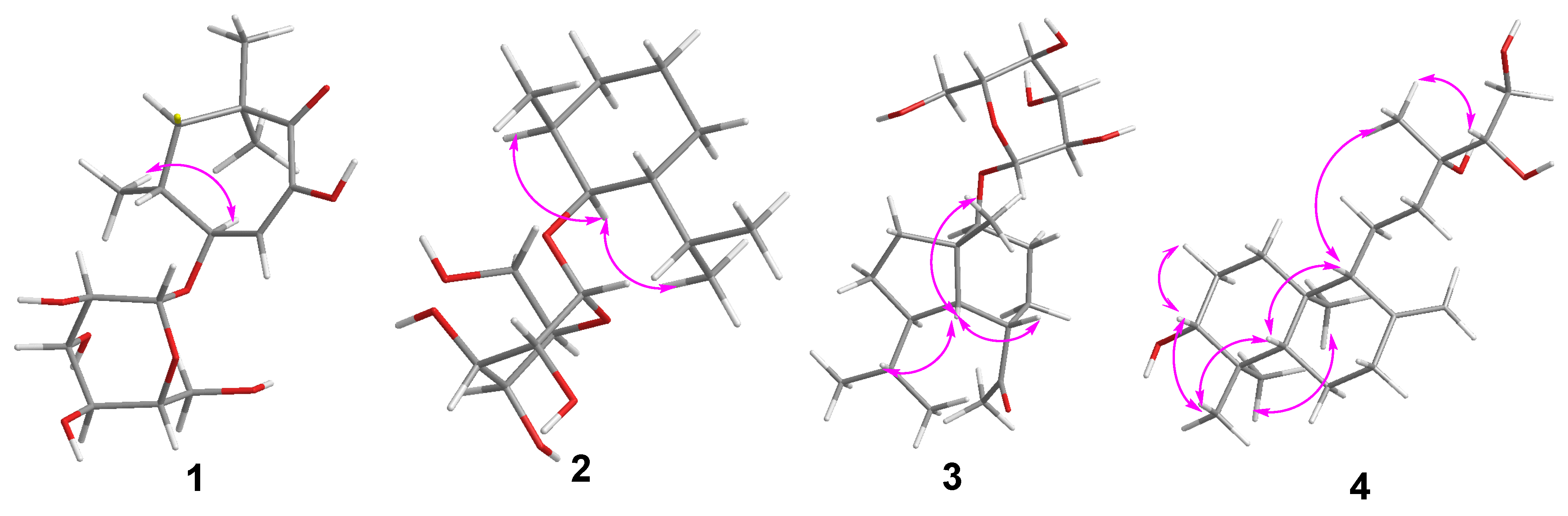
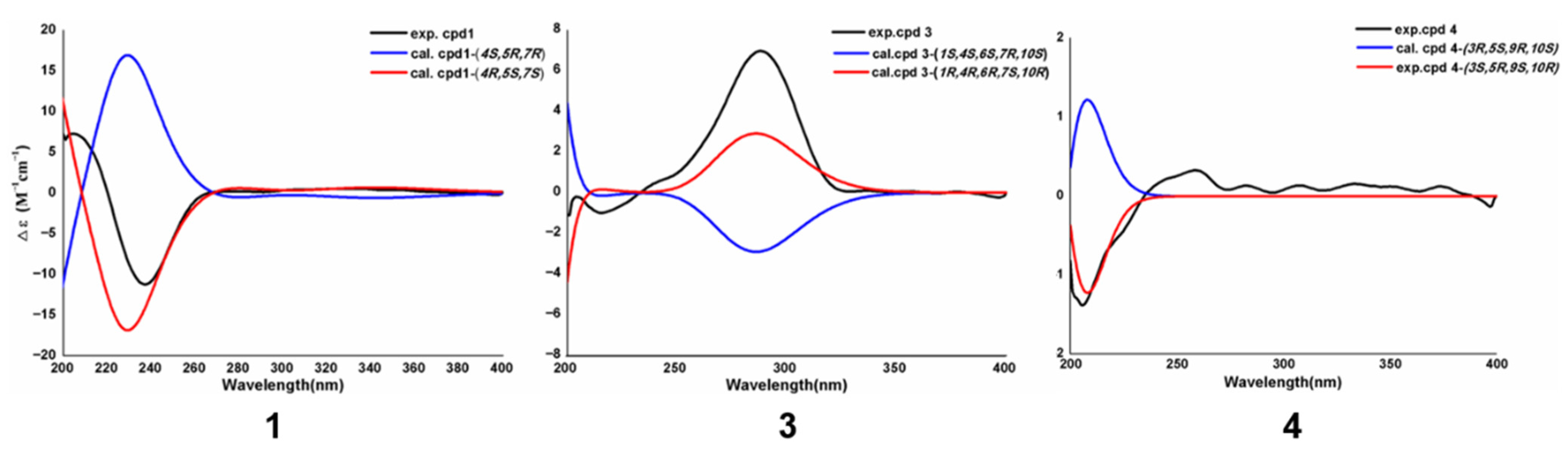
Compound 1 was obtained in the form of white amorphous solid, and its molecular formula was determined to be C16H26O8 by the high-resolution electrospray ionization mass spectrometry (HR-ESI-MS) peak at m/z 391.1599 [M+HCOO]. The 1H nuclear magnetic resonance (NMR) spectral data exhibited the presence of one olefinic hydrogen [δH 7.22 (d, J = 2.1 Hz, 1H, H-3)], three methyl groups [δH 1.39, 1.37, 1.14 (each 3H, s, H-9, H-10, H-8)], one methylene group [δH 2.48 (dd, J = 15.3, 3.1 Hz, 1H); 2.24 (d, J = 12.9 Hz, 1H), H-6], two methines (one of them was oxygenated) [δH 4.39 (dd, J = 9.1, 2.1 Hz, 1H, H-4); 2.17 (dtd, J = 12.8, 6.2, 3.2 Hz, 1H, H-5)]. The 13C NMR spectrum in Table 1 showed 16 carbon signals including one carbonyl carbon (δC 200.8, C-1), two olefinic carbons (δC 146.4, 145.7, C-2, C-3), six glycosylated carbons (δC 102.2, 78.3, 78.2, 75.1, 71.8, 63.0), two oxygenated carbons (δC 79.5, 72.2, C-4, C-7) and three methyl groups (δC 29.2, 29.0, 18.2), one methine carbon (δC 38.1, C-5), and one methylene carbon (δC 46.6, C-6). The above features indicated that 1 had a structure similar to that of a monoterpenoid glycoside, Magnoliaterpenoid C [14]. The heteronuclear multiple bond correlation (HMBC) correlations of compound 1 from H-10 to C-2, and H-9 to C-7, and from H-3 to C-7/C-5/C-1 and from H-8 to C-6/C-5/C-4 (Figure 2), combined with 1H-1H correlation spectroscopy (COSY) correlations of H-6 and H-5/H-4/H-3, confirmed the monoterpenoid unit. Differences in the two compounds are the locations of the methyl groups and the sugar unit. The HMBC spectrum of 1 showed the correlations of H-10 to C-2, H-8 to C-5, H-9 to C-7, suggesting the methylation site at C-2, C-5 and C-7. The glycosylation site was deduced at C-4 from the HMBC correlation from H-1′ (δH 4.50) to C-4. The relative configuration of compound 1 was assigned by the analysis of nuclear overhauser effect spectroscopy (NOESY) correlations of H-8 with H-4/H-9 (Figure 3). Its absolute configuration was definitively confirmed as (4R,5S,7S) by comparing the experimental and calculated electronic circular dichroism (ECD) spectra (Figure 4). Thus, compound 1 was determined as (4R,5S,7S)-2,5,7-trimethyl-7-hydroxycyclohept-2-en-1-one-4-β-D-glucopyranoside.
Compound 2 appeared as an amorphous white powder with a molecular formula of C16H30O6 by the HR-ESI-MS peak at m/z 363.2016 [M+HCOO]. The 1H NMR spectrum of compound 2 showed a set of characteristic proton signals of oxygenated cyclohexane scaffold [δH 3.57 (td, J = 10.7, 4.3 Hz, 1H, H-3); 2.11 (m, 1H, H-2); 1.66 (ddt, J = 13.7, 10.1, 3.2 Hz, 2H, H-1β, H-5β); 1.36 (m, 2H, H-6); 1.04 (qd, J = 13.3, 12.8, 3.8 Hz, 1H, H-1α); 1.23 (ddt, J = 13.1, 10.5, 3.1 Hz, 1H, H-4); 0.90 (overlapped, 1H, H-5α)], a group of diagnostic isopropyl signals [δH 2.31 (pd, J = 7.0, 2.5 Hz, 1H, H-8); 0.88 (d, J = 7.2 Hz, 3H, H-9); 0.81 (d, J = 6.9 Hz, 3H, H-10)], one methyl protons [δH 0.93 (d, J = 6.6 Hz, 3H, H-7)], and a anomeric proton [δH 4.36 (d, J = 7.8 Hz, 1H, H-1′)]. Correspondingly, the 13C NMR spectrum gave 16 characteristic carbon signals, of which six carbons (δC 101.3, 78.2, 77.7, 75.1, 71.9, 63.1) were assigned to be the sugar substitution. The 1-dimensional (1D) NMR data revealed that compound 2 was an m-menthane glycoside [15], which was further supported by the 1H-1H COSY combinations of H-6/H-5/H-4/H-3/H-2/H-1 and HMBC correlations from H-7 to C-2 (δC 41.7) and from H-9 to C-4 (δC 49.0). The positions of the methyl and isopropyl unit were determined at C-2 and C-4, respectively, based on the observed HMBC correlations from H-7 to C-2 and H-9 to C-4/C-8/C-10. In addition, the β-D-glucose was identified at C-3 by the coupling constant (J = 7.8 Hz) and the HMBC correlation from H-1′ to C-3. The relative configuration of 2 was assigned as rel-2R,3R,4R by the analysis of NOESY, H-2 with H-3 and H-3 with H-10 (Figure 3). Thus, compound 2 was determined as rel-(2R,3R,4R)-m-menthane-3-O-β-D-glucopyranoside.
Compound 3, a white amorphous powder, possessed a molecular formula of C21H36O7 by the HR-ESI-MS peak at m/z 445.2446 [M+HCOO]. The characteristic proton signals at δH 2.19, (s, 3H, H-14); 0.94 (d, J = 6.8 Hz, 3H, H-12); 0.90 (d, J = 7.2 Hz, 6H, H-13, H-15); 2.15 (ddd, J = 12.2, 8.4, 3.3 Hz, 1H, H-3β); 1.70 (dt, J = 9.0, 3.0 Hz, 2H, H-2); 1.53 (ddt, J = 14.1, 10.8, 5.5 Hz, 1H, H-3α); 2.07 (dd, J = 10.1, 2.7 Hz, 2H, H-9α); 1.74 (m, 1H, H-8α); 1.44 (dddd, J = 13.4, 9.5, 5.7, 3.3, 1H, H-8β); 1.11 (m, 1H, H-9β); 3.67 (td, J = 10.9, 10.0, 5.6 Hz, 1H, H-1); 2.45 (dt, J = 5.6, 3.0 Hz, 1H, H-4); 2.07 (dd, J = 10.1, 2.7 Hz, 2H, H-6); 1.87 (tt, J = 10.0, 5.2 Hz, 1H, H-7); 1.79 (m, 1H, H-11) in the 1H-NMR spectrum, along with the related 15 carbon signals (Table 2) containing a keto carbonyl carbon [δC 214.5, C-5] and one oxygenated carbon (δC 78.8, C-1), indicated the existence of a 6(5→4)-abeo-salvionane sesquiterpenoid unit [16], further supporting by the HMBC cross-peaks of H-14 with C-4, H-12 and H-13 with C-7, H-15 with C-10 and the 1H-1H COSY cross-peaks of H-4/H-6/H-7 (Figure 2). The NMR spectrum of compound 3 resembled that of Artemerioside E [16]. The sugar unit was deduced at C-1 based on the remaining six carbons [δC 101.9, 78.2, 77.6, 75.0, 71.9, 63.0] and the characteristic anomeric proton [δH 4.26 (d, J = 7.8 Hz, 1H, H-1′)], and the HMBC cross-peaks from H-1′ to C-1. The main difference between the two compounds is the absence of the crotonyl substituent at C-6′, which was confirmed by the HMBC cross-peaks of H-6′ and C-4′/5′, as well as the acid hydrolysis experiment. The relative configuration of 3 was assigned by the NOESY correlation of H-4 with H-6, and H-6 with H-15 (Figure 3). The configuration of compound 3 was deduced as 1R,4R,6R,7S,10R by comparing the experimental ECD spectrum with the calculated spectrum (Figure 4). Thus, 3 was (1R,4R,6R,7S,10R)-6(5→4)-abeo-salvionane-1-O-β-D-glucopyranoside.
Compound 4, a white amorphous substance, was deduced to have a molecular formula of C20H36O4 on the basis of the HR-ESI-MS ion peak at m/z 385.2581 [M+HCOO]. The 1H NMR spectral data showed the resonances for one olefinic hydrogen at δH 4.83 (q, J = 1.6 Hz, 1H, H-17), four methyl groups at δH 1.12, 0.97, 0.76, 0.72, (each 3H, s, H-16, H-18, H-19, H-20), seven methylenes (two of them were oxygenated) at [δH 3.76 (dd, J = 11.1, 3.2 Hz, 1H, H-15α); 3.50 (dd, J = 11.1, 8.0 Hz, 1H, H-15β), 2.40 (ddd, J = 12.7, 4.3, 2.4 Hz, 1H, H-7α); 1.98 (td, J = 13.0, 5.1 Hz, 1H, H-7β); 1.86 (dt, J = 13.2, 3.6 Hz, 1H, H-1α); 1.76 (ddt, J = 12.8, 5.3, 2.6 Hz, 1H, H-6α); 1.67 (m, 2H, H-11α/12α); 1.63 (dt, J = 12.5, 3.3 Hz, 2H, H-2); 1.42 (m, 2H, H-6β/11β); 1.29 (dd, J = 11.1, 2.4 Hz, 1H, H-12β); 1.23 (ddt, J = 17.6, 8.4, 4.3 Hz, 1H, H-1β)]; as well as signals at [δH 3.43 (dd, J = 8.0, 3.3 Hz, 1H, H-14); 3.19 (m, 1H, H-3); 1.55 (d, J = 10.7 Hz, 1H, H-9); 1.13 (d, J = 2.9 Hz, 1H, H-5)] belonging to four methines, were also observed, and the 13C NMR spectrum gave the ten resonances, including two olefinic carbons (δC 149.7, 107.3), four methyl groups (δC 28.9, 22.1, 16.1, 15.1) and four oxygenated carbons (δC 79.6, 78.5, 75.2, 64.0). The above NMR data of 4 were similar to those of Pierisjaponin C [17], a known labdane diterpenoid scaffold. Comparison of the spectroscopic data of compound 4 with those of Pierisjaponin C suggested that they have the same skeleton but a slight difference in chemical shifts in the side chain attached at C-9 due to the different substitutions. The double bond between C-14 and C-15 in Pierisjaponin C was replaced by two hydroxyl groups in compound 4, which was supported by the HMBC correlations of H-12 (δH 1.29) to C-13 (δC 75.2), H-14 (δH 3.43) to C-15 (δC 64.0) and H-16 (δH 1.12) to C-12 (δC 39.1)/C-14 (δC 78.5), as well as the combination with element analysis. The relative configuration of 4 was deduced by the NOESY correlation of H-3 with H-18, and H-3 with H-5, and H-5 with H-9, and H-19 with H-20, and H-9 with H-16, and H-16 with H-14 (Figure 3). The absolute configuration of the labdane diterpenoid scaffold was confirmed as 3S,5R,9S,10R by comparing the experimental ECD spectrum with the calculated spectrum (Figure 4). Thus, 4 was determined as (3S,5R,9S,10R)-3,13,14,15-tetrahydroxy-ent-labda-8(17)-ene.
The seventeen known compounds were characterized to be cyclo(L-pro-L-Asp) (5) [18], rengyol (6) [19], guanosine (7) [20], 4-[2-(β-D-glucopyranosyloxy)ethyl]-4-hydroxy-2-cyclohexen-1-one (8) [21], rengyoside B (9) [22], rengyoside A (10) [23], rosmarinic acid (11) [24,25], apterin (12) [26], lcariside F2 (13) [27], scopolin (14) [28,29], citrusin c (15) [30], annphenone (16) [31], forsythenside L (17) [32], hesperidin (18) [33], apigenin-7-O-β-D-glucopyranoside (19) [34], kamiohnoyneosides A (20) [35], gingerglycolipid A (21) [36] by comparing with previously documented NMR spectral data. Their structures are shown in the Supplementary Materials, Figure S1.
By comparing with literature [13] and HR-MS data on the individual herbs, the sources of isolated compounds were further discussed. Compound 1 is derived from I. indigotica. Compound 2 originates from M. haplocalyx. Particularly, a structurally similar known compound, rel-(1R,2S,3R,4R) p-menthane-1,2,3-triol 3-O-β-D-glucopyranoside, has also been reported from M. haplocalyx [15], further supporting its chemotaxonomic relevance. Compound 3 is believed to be from F. suspensa. Compound 4 is thought to be from P. praeruptorum. Among known compounds, two flavonoids (compounds 18 and 19) have been reported from L. similis [13]. Two polyacetylene glycosides (compounds 20 and 21) have been isolated from C. indicum [13]. Consequently, the herbal sources of the above-mentioned compounds are diverse.

2.2. The Inhibitory Activities of Compounds on Influenza A Viruses

IAV are major respiratory pathogens that pose significant threats to public health worldwide due to their high mutation rates and potential to cause pandemics. Among various subtype subtypes, the H1N1 subtype demonstrates significant virulence. The continuous emergence of drug-resistant strains necessitates the search for new antiviral agents from natural sources. In this study, two representative H1N1 strains, WSN strain (first isolated from humans in 1933, one of the earliest human influenza viruses to be isolated in history) [37] and CA07 strain (the novel swine-origin A influenza virus that emerged in 2009) [38], were selected for antiviral evaluation.
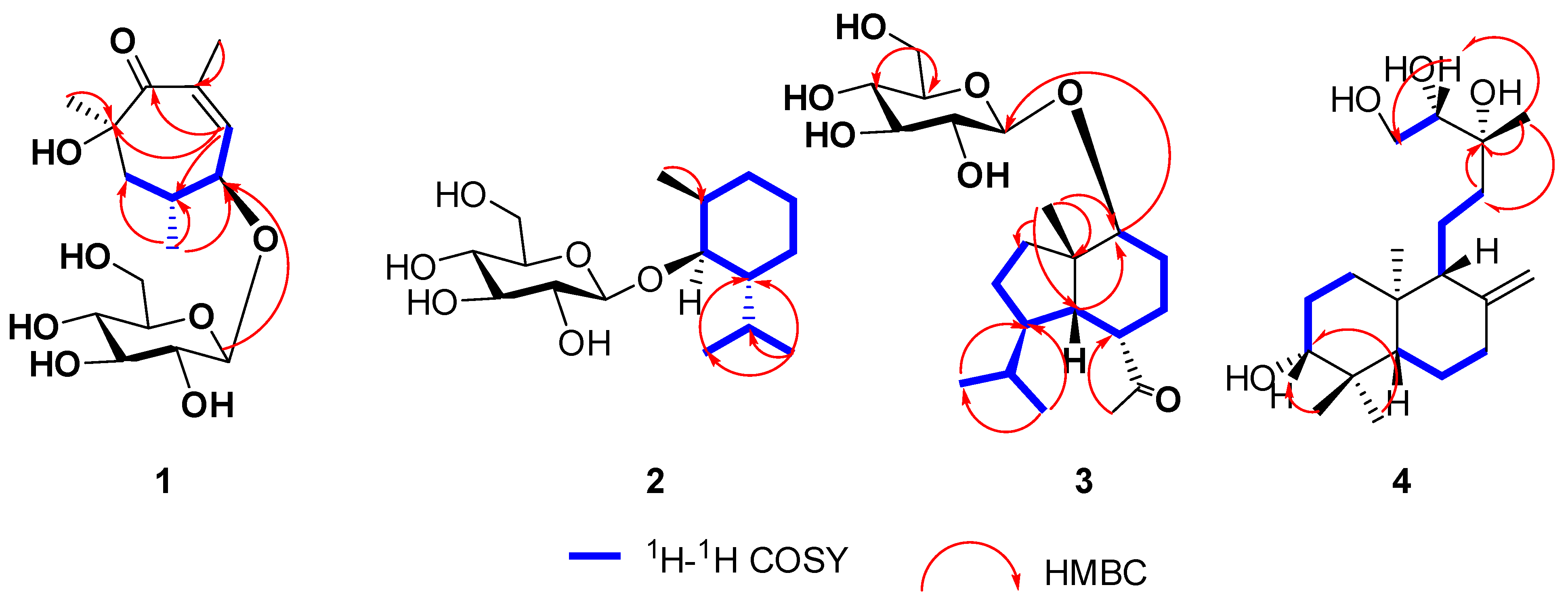
The activities of compounds 121 against CA07 and WSN were tested on Madin-Darby Canine Kidney (MDCK) cells, using oseltamivir acid as the positive control. As presented in Table 3, only the new compound 4 demonstrated potent protective effects against CA07 virus in MDCK cells, with a half maximal inhibitory concentration (IC50) value of 37.10 μM. It has been reported that the parent nucleus of 4, labdane diterpenoid, possesses anti-Zika virus activity [39], further supporting the anti-virus potential of compound 4. However, the remaining 20 compounds, including the other new compounds 13, were inactive (IC50 > 100 µM). The complete bioactivity data for all isolates are available in the Supplementary Materials, Table S1.

3. Material and Methods

3.1. General Experimental Procedures

Column chromatography (CC) was experimented using macroporous resin HP-20 (Anhui Sanxing Resin Technology Co., Ltd., Bengbu, China) and silica gel (200–300 mesh, from Qingdao Marine Chemical Co., Ltd., Qingdao, China), under the guidance of thin-layer chromatography (TLC) analysis on silica gel plates (GF254, Tianjin Silida Technology Co., Ltd., Tianjin, China). Medium pressure preparative chromatography (MPLC) was conducted by BUCHI Flash Pure C-850 system coupled with Flash Pure ECOFLEX Columns (C18, 220 g or 800 g, BUCHI Labortechnik AG, Flawil, Switzerland; polyamide, 100–200 mesh, Changfeng Chemical Co., Ltd., Chongqing, China). Analytical HPLC was executed on Vanquish Flex UPLC instrument equipped with a detector named charged aerosol detection (CAD) from Thermo Fisher Scientific (Waltham, MA, USA) using analytical columns, including an Agela Venusil ASB C18 column (5 μm, 250 × 4.6 mm, Agela Technologies, Tianjin, China), an Agela Venusil XBP C18 column (5 μm, 250 × 4.6 mm, Agela Technologies, Tianjin, China), a XUnion C18 column (5 μm, 250 × 4.6 mm, Acchrom, Wenling, China), and a Welch Ultimate XB Phenyl column (5 μm, 250 × 4.6 mm, Welch Materials, Inc., West Haven, CT, USA). Semi-preparative HPLC was conducted on an Elite P3500 High-Pressure Constant Current Pump (Elite Analytical Instruments Co., Ltd., Dalian, China) connected with a refractive index detector (RID) detector (Shodex 20A, Showa Denko, Kawasaki, Japan) using chromatographic columns of semi-preparative scale corresponding to the analytical scale columns listed above. Liquid chromatography system (Agilent 1100 series, Santa Clara, CA, USA) connected with a UV detector (Agilent Technology, Santa Clara, CA, USA) and HPLC system (Vanquish core, Thermo Fisher Scientific, Waltham, MA, USA) equipped with a CAD detector, a flow-splitting valve, and an automatic fraction collection system were used to purify low-volume fractions. HR-ESI-MS spectra were acquired on a Q-TOF mass spectrometer (Waters Synapt G2-S, Waters Co., Milford, MA, USA). NMR data were collected using a Bruker Avance III 600 NMR spectrometer (Billerica, MA, USA). Measurements of optical rotation were carried out with an Anton Paar MCP 200 polarimeter (Anton Paar Co., Graz, Austria). Circular dichroism (CD) spectra were acquired using a Chirascan spectropolarimeter (Applied Photophysics Ltd., Leatherhead, Surrey, UK).

3.2. Experimental Material

Fufang Yinhua Jiedu Granules (FFYHG) extract was provided by Yifan Pharmaceutical Co., Ltd. (Hangzhou, China). Solvents for NMR spectroscopy (methanol-d4, DMSO-d6) were supplied by Cambridge Isotope Laboratories, Inc. (Tewksbury, MA, USA). Material for acid hydrolysis experiment, including D-glucose, O-tolyl isothiocyanate, L-cysteine methyl ester hydrochloride, anhydrous pyridine and trifluoroacetic acid, were purchased from Shanghai Macklin Biochemical Co., Ltd. (Shanghai, China); Sinopharm chemical reagent Co., Ltd. (Shanghai, China) and American Thermo Scientific, respectively. Methanol, acetonitrile and formic acid (Chromatography grade) for HPLC were also ordered from American Thermo Fisher Scientific. Ethanol (analytical grade), trichloromethane (analytical grade), methanol (analytical and preparative grade), and acetonitrile (preparative grade) for isolation and purification were supplied by Sinopharm chemical reagent Co., Ltd. (Shanghai, China).
Two H1N1 strains, A/California/07/2009 (CA07) and A/WSN/1933 (WSN), and MDCK cells were cultured by BSL-2 Laboratory of the Academy of Military Medical Sciences. Oseltamivir acid (CAS: 187227-45-8) was purchased from MedChemExpress LLC (Monmouth Junction, NJ, USA).

3.3. Extraction and Isolation

After dissolving in deionized water, the supernatant of FFYHG extract (2 kg) was separated on a macroporous resin column (HP-20) and eluted with EtOH-H2O (0:100, 10:90, 35:65, 70:30 and 95:5, v/v) to yield four fractions (HP10, HP35, HP70, and HP95).
HP10 (25 g) was subjected to a Flash Pure column (polyamide, 100–200 mesh, 800 mL) for preparative MPLC and eluted with EtOH-H2O containing 0.03% formic acid, gradually increased from 0 to 100% within 120 min (v/v, 30 mL/min) to give two fractions (HP10-P1–HP10-P2). HP10-P1 (10 g) was fractioned on a Flash Pure column (FP ECOFLEX C18, 800 g) for preparative MPLC and eluted with MeOH-H2O (ranging from 1:99 to 13:97, v/v, 30 mL/min) to obtain ten fractions (HP10-P1-C-1–HP10-P1-C-10). HP10-P1-C4 was separated on the XBP C18 column by semi-preparative HPLC using CH3CN-H2O containing 0.03% formic acid (5:95, v/v, 4.7 mL/min) to yield ten fractions (HP10-P1-C4-X1–HP10-P1-C4-X10). HP10-P1-C4-X2 was separated on the XBP C18 column by semi-preparative HPLC using MeOH-H2O containing 0.046% formic acid (2:98, v/v, 2.38 mL/min) to afford compound 5 (1.42 mg, tR 15 min, purity 100.00%), 6 (6.77 mg, tR 20 min, purity 94.43%), 7 (3.01 mg, tR 22 min, purity 94.70%), 8 (4.31 mg, tR 23 min, purity 97.64%), 9 (20.02 mg, tR 27.5 min, purity 99.74%), and 10 (6.55 mg, tR 30 min, purity 93.62%).
HP35 (90 g) was subjected to silica gel CC eluting with CHCl3-MeOH (100:0, 9:1, 8.5:1.5, 7.5:2.5, 7:3, 6:4 and 0:100, v/v) to give five fractions (HP35-S1–HP35-S5). HP35-S2 (10 g) was fractioned on a Flash Pure column (FP ECOFLEX C18, 800 g) by preparative MPLC using MeOH-H2O (ranging from 1:99 to 100:0, v/v, 25 mL/min) to obtain seven fractions (HP35-S2-B-1–HP35-S2-B-7). HP35-S2-B4 afforded compound 11 (10 mg, purity 95.27%) upon precipitation with methanol. HP35-S2-B5 was separated on the XBP C18 column by semi-preparative HPLC using MeOH-H2O containing 0.03% formic acid (40:60, v/v, 2.38 mL/min) to afford compound 12 (24.97 mg, tR 31 min, purity 100.00%) and yield ten fractions (HP35-S2-B5-X1–HP35-S2-B5-X10). HP35-S2-B5-X7 was separated on the XB Phenyl column by semi-preparative HPLC using CH3CN-H2O containing 0.03% formic acid (10:90, v/v, 4.7 mL/min) to afford compound 13 (2 mg, tR 40 min, purity 100.00%). HP35-S2-B5-X3 was separated on the XB Phenyl column by semi-preparative HPLC using CH3CN-H2O (8:92, v/v, 4.7 mL/min) to afford compound 1* (1.22 mg, tR 12 min, purity 98.10%) and compound 14 (7.06 mg, tR 17 min, purity 98.17%).
HP70 (100 g) was loaded onto the silica gel column and eluted with CHCl3-MeOH (100:0, 15:1, 10:1, 6:1, 3:1, 2:1, and 0:100, v/v) to give twenty-one fractions (HP70-S1–HP70-S21). HP70-S9 (4 g) was fractioned on a Flash Pure column (FP ECOFLEX C18, 220 g) by preparative MPLC using MeOH-H2O (35:85, v/v, 50 mL/min) to obtain ten fractions (HP70-S9-B-1–HP70-S9-B-10). HP70-S9-B4 was separated on the XUnion C18 column by semi-preparative HPLC using CH3CN-H2O (21:79, v/v, 4.9 mL/min) to obtain compound 15 (14 mg, tR 20 min, purity 100.00%). HP70-S9-B8 was separated on the ASB C18 column by semi-preparative HPLC using CH3CN-H2O (28:72, v/v, 4.9 mL/min) to yield eight fractions (HP70-S9-B8-A7–HP70-S9-B8-A8). HP70-S9-B8-A7 was separated on the XB Phenyl column by HPLC using CH3CN-H2O (25:75, v/v, 1.0 mL/min) to afford compound 2* (1.57 mg, tR 26 min, purity 100.00%) and compound 3* (3.39 mg, tR 30 min, purity 100.00%). HP70-S9-B8-A8 was separated on the XB Phenyl column by semi-preparative HPLC using CH3CN-H2O (25:75, v/v, 1.0 mL/min) to afford compound 4* (1 mg, tR 32 min, purity 100.00%). HP70-S10 (4 g) was fractioned on a Flash Pure column (FP ECOFLEX C18, 220 g) by preparative MPLC using MeOH-H2O (20:80, v/v, 50 mL/min) to obtain eight fractions (HP70-S10-B-1–HP70-S10-B-8). HP70-S10-B2 was separated on the ASB C18 column by semi-preparative HPLC using CH3CN-H2O (15:85, v/v, 4.9 mL/min) to obtain compound 16 (9.74 mg, tR 21 min, purity 100.00%). HP70-S13 (4 g) was fractioned on a Flash Pure column (FP ECOFLEX C18, 220 g) by preparative MPLC using MeOH-H2O; the percentage of MeOH was gradually increased from 20% to 70% v/v within 60 min (50 mL/min) to obtain several fractions (HP70-S13-B-1–HP70-S13-B-8). HP70-S13-B3 was separated on the ASB C18 column by semi-preparative HPLC using CH3CN-H2O (23:77, v/v, 4.7 mL/min) to yield nine fractions (HP70-S13-B3-A1–HP70-S13-B3-A9). HP70-S13-B3-A6 was separated on the XB Phenyl column by semi-preparative HPLC using CH3CN-H2O (17:83, v/v, 4.7 mL/min) to afford compound 17 (23.68 mg, tR 20 min, purity 100.00%). HP70-S13-B4 was separated on the ASB C18 column by semi-preparative HPLC using CH3CN-H2O (20:80, v/v, 4.7 mL/min) to yield sixteen fractions (HP70-S13-B4-A1–HP70-S13-B4-A16). HP70-S13-B4-A4 was separated on the XB Phenyl column by semi-preparative HPLC using CH3CN-H2O (18:82, v/v, 4.7 mL/min) to afford compound 18 (16 mg, tR 30 min, purity 98.80%). HP70-S13-B4-A5 was separated on the XB Phenyl column by semi-preparative HPLC using CH3CN-H2O (16:84, v/v, 4.7 mL/min) to afford compound 19 (3.09 mg, tR 42 min, purity 97.06%). HP70-S13-B4-A7 was separated on the XB Phenyl column by semi-preparative HPLC using CH3CN-H2O (16:84, v/v, 4.7 mL/min) to afford compound 20 (1.67 mg, tR 52 min, purity 93.73%). HP70-S14 (5.7 g) was fractioned on a Flash Pure column (FP ECOFLEX C18, 800 g) by preparative MPLC using MeOH-H2O (ranging from 20:80 to 60:40, v/v, 50 mL/min) to yield eleven fractions (HP70-S14-B-1–HP70-S14-B-11). HP70-S14-B11 was separated on the ASB C18 column by semi-preparative HPLC using CH3CN-H2O (43:57, v/v, 4.7 mL/min) to afford compound 21 (10.94 mg, tR 35 min, purity 92.97%).

3.4. Physicochemical Properties and Spectroscopic Data of New Compounds

(4R,5S,7S)-2,5,7-trimethyl-7-hydroxycyclohept-2-en-1-one-4-β-D-glucopyranoside (1): white amorphous powder; [ α ] D 25 −73.99 (c 0.12, MeOH); 1H and 13C NMR data, see Table 1; HR-ESI-MS m/z 391.1599 [M+HCOO] (calcd for C17H27O10, 391.1604).
Rel-(2R,3R,4R)-m-menthane-3-O-β-D-glucopyranoside (2): white amorphous powder; [ α ] D 25 −35.99 (c 0.11, MeOH); 1H and 13C NMR data, see Table 1; HR-ESI-MS m/z 363.2016 [M+HCOO] (calcd for C17H31O8, 363.2019).
(1R,4R,6R,7S,10R)-6(5→4)-abeo-salvionane-1-O-β-D-glucopyranoside (3): white amorphous powder; [ α ] D 25 −5 (c 0.34, MeOH); 1H and 13C NMR data, see Table 2; HR-ESI-MS m/z 445.2446 [M+HCOO] (calcd for C22H37O9, 445.2438).
(3S,5R,9S,10R)-3,13,14,15-tetrahydroxy-ent-labda-8(17)-ene (4): white amorphous powder; [ α ] D 25 +31.99 (c 0.10, MeOH); 1H and 13C NMR data, see Table 2; HR-ESI-MS m/z 385.2581 [M+HCOO] (calcd for C21H37O6, 385.2590).

3.5. Anti-Influenza A Virus (H1N1) Activity Inhibition Test

3.5.1. Pretreatment of Compounds

Compounds 121 and the positive control (oseltamivir acid) were formulated as 100 mM stock solutions in DMSO.

3.5.2. Cytopathic Effect (CPE) Inhibition Assay

Anti-influenza viral assay was conducted following methods established previously [40,41]. MDCK cells were plated in 96-well plates at a density of 1.5 × 10⁴ cells per well and maintained for 24 h at 37 °C in a CO2 incubator. Using virus growth medium, the tested compounds 121 and positive control were diluted to 400 μM, followed by 3-fold serial dilutions to obtain 10 concentrations, from 400 μM to 0.02 μM. The culture medium was changed to virus growth medium (100 μL/well). The diluted samples (50 μL/well) were added to the 96-well plate in ascending order of concentration, while the wells of cell control and virus control received 50 μL/well of virus growth medium. Finally, 50 μL/well of virus growth medium containing virus diluted to 100 median tissue culture infectious dose 50 (TCID50) was added, resulting in a final volume of 200 μL in each well. The highest final concentration of the sample in the white 96-well plate was 100 μM, followed by 3-fold serial dilutions to yield 10 concentration gradients from 100 μM to 0.005 μM, and the final DMSO concentration in the highest test concentration was 0.1% (v/v), decreasing proportionally in subsequent dilutions. The plates were incubated in a CO2 incubator at 37 °C for 72 h, and cellular CPE were observed daily. The inhibitory effects of the compounds on influenza virus were measured using the CellTiter-Glo® luminescent cell (Promega corporation, Madison, WI, USA) viability assay by reading the luminescence in a SpectraMax M5 multi-mode microplate reader (Molecular Devices, San Jose, CA, USA). Each experiment was performed independently twice, with three replicate wells per concentration for each sample in each experiment.

3.5.3. Cytotoxicity Assay

The procedure of the cytotoxicity assay was similar to that of the CPE inhibition assay described in Section 3.5.2; the difference was that the virus inoculum was changed to the medium supplemented. The IC50 values of the tested compounds were calculated by S-curve fitting on the inhibition rate-concentration data using Origin 8.0 software. The median toxic dose (TD50) value was calculated by the same method, and then the selectivity index SI = TD50/IC50 was calculated using Origin 8.0 software.

4. Conclusions

In conclusion, a phytochemical investigation of FFYHG was conducted for the first time in the present study, leading to the isolation and identification of four undescribed terpenoid derivatives and seventeen known compounds. Among them, compound 1 is a seven-membered ring monoglycoside. Compound 2 belongs to the m-menthane glycosides. Compound 3 possesses the core structure of a 6(5→4)-abeo-salvionane sesquiterpenoid unit. Compound 4 is classified as a labdane diterpenoid. Notably, compound 4 exhibited significant antiviral activity against CA07. The above findings not only enhance the chemical understanding of FFYHG but also provide a scientific rationale for the application of FFYHG in treating viral infections.

Supplementary Materials

The following supporting information can be downloaded at: https://www.mdpi.com/article/10.3390/separations13040105/s1, Experimental methods: refer to the detailed procedure of optimization of chromatographic conditions and acid hydrolysis experiment; Figure S1: Structures of compounds 521; Figure S2: Identification of sugars; Figures S3–S30: NMR spectra of compounds 1, 2, 3, 4; Figure S31: Optimization of chromatographic conditions for compound 4; Table S1: Anti-CA07 and Anti-WSN activity, cytotoxicity, and selective index of compounds 121.

Author Contributions

Writing—original draft, validation, writing—review and editing, X.W.; methodology, formal analysis, supervision, X.-J.C., Q.S. and J.S.; resources, formal analysis, investigation, validation, writing—review and editing, supervision, H.-Z.L. and B.-P.M. All authors have read and agreed to the published version of the manuscript.

Funding

This work was supported by the National Key Research and Development Program of China (Grant No. 2023YFC3502804).

Institutional Review Board Statement

Not applicable.

Informed Consent Statement

Not applicable.

Data Availability Statement

The experimental data presented in the current study are included in the article and Supplementary Materials.

Conflicts of Interest

The authors declare that they have no known competing financial interests or personal relationships that could have appeared to influence the work reported in this paper.

References

  1. Stevaert, A.; Naesens, L. The influenza virus polymerase complex: An update on its structure, functions, and significance for antiviral drug design. Med. Res. Rev. 2016, 36, 1127–1173. [Google Scholar] [CrossRef] [Scilit] [PubMed]
  2. Azziz-Baumgartner, E.; Cheng, P.Y.; Dawood, F.; Foppa, I.; Olsen, S.; Haber, M.; Jeffers, C.; Macintyre, C.R.; Newall, A.T.; Wood, J.G. Estimates of global seasonal influenza-associated respiratory mortality: A modelling study. Lancet 2017, 391, 10127. [Google Scholar] [CrossRef] [Scilit]
  3. Park, M.; Wu, P.; Goldstein, E.; Kim, W.J.; Cowling, B.J. Influenza-associated excess mortality in South Korea. Am. J. Prev. Med. 2016, 50, e111–e119. [Google Scholar] [CrossRef] [Scilit] [PubMed]
  4. Cohen, C.; Walaza, S.; Treurnicht, F.K.; McMorrow, M.; Madhi, S.A.; McAnerney, J.M.; Tempia, S. In- and out-of-hospital mortality associated with seasonal and pandemic influenza and respiratory syncytial virus in South Africa, 2009–2013. Clin. Infect. Dis. 2018, 66, 95–103. [Google Scholar] [CrossRef] [Scilit]
  5. Palese, P. Influenza: Old and new threats. Nat. Med. 2004, 10, S82–S87. [Google Scholar] [CrossRef] [Scilit]
  6. McKimm-Breschkin, J.L. Influenza neuraminidase inhibitors: Antiviral action and mechanisms of resistance. Influenza Other Respir. Viruses 2013, 7, 25–36. [Google Scholar] [CrossRef] [Scilit]
  7. Medina, R.; García-Sastre, A. Influenza A viruses: New research developments. Nat. Rev. Microbiol. 2011, 9, 590–603. [Google Scholar] [CrossRef] [Scilit]
  8. Hanshaoworakul, W.; Simmerman, J.M.; Narueponjirakul, U.; Sanasuttipun, W.; Shinde, V.; Kaewchana, S.; Areechokechai, D.; Levy, J.; Ungchusak, K. Severe human influenza infections in Thailand: Oseltamivir treatment and risk factors for fatal outcome. PLoS ONE 2009, 4, e6051. [Google Scholar] [CrossRef] [Scilit]
  9. Lackenby, A.; Hungnes, O.; Dudman, S.G.; Meijer, A.; Paget, W.J.; Hay, A.J.; Zambon, M.C. Emergence of resistance to oseltamivir among influenza A (H1N1) viruses in Europe. Euro Surveill. 2008, 13, 8026. [Google Scholar] [CrossRef] [Scilit]
  10. Meijer, A.; Lackenby, A.; Hungnes, O.; Lina, B.; van der Werf, S.; Schweiger, B.; Opp, M.; Paget, J.; van de Kassteele, J.; Hay, A.; et al. Oseltamivir-resistant influenza virus A (H1N1), Europe, 2007–08 season. Emerg. Infect. Dis. 2009, 15, 552–560. [Google Scholar] [CrossRef]
  11. Zhang, Y.Q.; Wang, R.H.; Shi, W.Q.; Zheng, Z.H.; Wang, X.Q.; Li, C.; Zhang, S.F.; Zhang, P.H. Antiviral effect of fufang yinhua jiedu (FFYH) granules against influenza A virus through regulating the inflammatory responses by TLR7/MyD88 signaling pathway. J. Ethnopharmacol. 2021, 275, 114063. [Google Scholar] [CrossRef] [Scilit]
  12. Zheng, Z.H.; Zhang, Y.Q.; Li, M.; Wang, W.L.; Wei, H.L.; Su, Q.; Wang, R.H.; Shi, W.Q.; Wang, L.; Wu, J.X.; et al. In vitro and in vivo antiviral effect of Fufang Yinhua Jiedu granules (FFYH) combined with oseltamivir against influenza A virus. Phytomed. Plus 2023, 3, 100386. [Google Scholar] [CrossRef] [Scilit]
  13. Yao, L.; Wang, X.; Nan, Y.; Liang, H.Z.; Wang, M.Y.; Song, J.; Chen, X.J.; Ma, B.P. Exploring the chemical compositions of Fufang Yinhua Jiedu granules based on ultra-high-performance liquid chromatography coupled to quadrupole time-of-flight mass spectrometry combined with multistage intelligent data annotation strategy. J. Chromatogr. A 2024, 1728, 465010. [Google Scholar] [CrossRef] [Scilit] [PubMed]
  14. Feng, W.S.; He, Y.H.; Zheng, X.K.; Wang, J.C.; Cao, Y.G.; Zhang, Y.L.; Song, K. Four new monoterpenoid glycosides from the flower buds of Magnolia biondii. Molecules 2016, 21, 728. [Google Scholar] [CrossRef] [Scilit] [PubMed]
  15. She, G.M.; Xu, C.; Liu, B. New monocyclic monoterpenoid glycoside from Mentha haplocalyx Briq. Chem. Cent. J. 2012, 6, 37. [Google Scholar] [CrossRef] [Scilit]
  16. He, X.F.; Wang, M.F.; Ma, Y.B.; Li, T.Z.; Chen, J.J. Artemeriosides A-F, the first examples of natural sesquiterpenoids substituted by a 6′-O-crontonyl β-glucopyranoside from Artemisia annua. Fitoterapia 2023, 169, 105619. [Google Scholar] [CrossRef] [Scilit]
  17. Zheng, G.J.; Jin, P.F.; Huang, L.; Zhang, Q.H.; Meng, L.K.; Yao, G.M. Structurally diverse diterpenoids from Pieris japonica as potent analgesics. Bioorg. Chem. 2020, 99, 103794. [Google Scholar] [CrossRef] [Scilit]
  18. Challa, C.; Kumar, N.; John, M.; Lankalapalli, R.S. A comparative study of antimicrobial properties of cyclo(l-Pro-l-Asp) with its 2-ketopiperazine analog. Med. Chem. Res. 2014, 23, 2377–2385. [Google Scholar] [CrossRef] [Scilit]
  19. Abdullahi, H.; Nyandat, E.; Galeffi, C.; Messana, I.; Nicoletti, M.; Bettolo, G.B.M. Cyclohexanols of Halleria lucida. Phytochemistry 1986, 25, 2821–2823. [Google Scholar] [CrossRef] [Scilit]
  20. Wang, C.Y.; Han, L.; Kang, K.; Shao, C.L.; Wei, Y.X.; Zheng, C.J.; Guan, H.S. Secondary metabolites from green algae Ulva pertusa. Chem. Nat. Compd. 2010, 46, 828–830. [Google Scholar] [CrossRef] [Scilit]
  21. Hase, T.; Kawamoto, Y.; Ohtani, K.; Kasai, R.; Yamasaki, K.; Picheansoonthon, C. Cyclohexylethanoids and related glucosides from Millingtonia hortensis. Phytochemistry 1995, 39, 235–241. [Google Scholar] [CrossRef] [Scilit]
  22. Wu, J.; Zhou, X.L.; Huang, S.; Wang, C.J. Chemical constituents from flowers of Incarvillea younghusbandii. Chin. Herb. Med. 2012, 43, 55–59. [Google Scholar]
  23. Kutluay, V.M.; Makino, T.; Inoue, M.; Saracoglu, I. New knowledge about old drugs; a cardenolide type glycoside with cytotoxic effect and unusual secondary metabolites from Digitalis grandiflora Miller. Fitoterapia 2019, 134, 73–80. [Google Scholar] [CrossRef] [Scilit] [PubMed]
  24. Lee, J.H.; Park, K.H.; Le, M.H.; Kim, H.T.; Seo, W.D.; Kim, J.Y.; Baek, I.Y.; Jang, D.S.; Ha, T.J. Identification, characterisation, and quantification of phenolic compounds in the antioxidant activity-containing fraction from the seeds of Korean perilla (Perilla frutescens) cultivars. Food Chem. 2013, 136, 843–852. [Google Scholar] [CrossRef] [Scilit]
  25. Begum, S.; Perwaiz, S.; Siddiqui, B.S.; Khan, S.; Fayyaz, S.; Ramzan, M. Chemical constituents of Cordia latifolia and their nematicidal activity. Chem. Biodivers. 2011, 8, 850–861. [Google Scholar] [CrossRef] [Scilit]
  26. Tian, Y.J.; Li, J.N.; Feng, J.L.; Wang, Q.; Zhang, C.Y.; Zhou, H.G.; Chen, W.Q.; Yang, C. Chemical constituents from Notopterygium root. J. Liaoning Univ. Tradit. Chin. Med. 2013, 15, 42–44. [Google Scholar]
  27. Wang, L.B.; Wang, J.W.; Wang, C.; Sun, S.C.; Xu, B.; Wu, L.J. Chemical constituents in the lipid-lowering fraction of flos Helichrysum arenarium (III). Chin. J. Med. Chem. 2012, 22, 220–226. [Google Scholar]
  28. Kwon, O.S.; Choi, J.S.; Islam, M.N.; Kim, Y.S.; Kim, H.P. Inhibition of 5-lipoxygenase and skin inflammation by the aerial parts of Artemisia capillaris and its constituents. Arch. Pharm. Res. 2011, 34, 1561–1569. [Google Scholar] [CrossRef] [Scilit]
  29. Luyen, B.T.T.; Trang, B.T.T. New solasodine-type glycoalkaloids isolated from Solanum nigrum and their cytotoxic activity. Chem. Biodivers. 2024, 21, e202400872. [Google Scholar] [CrossRef] [Scilit]
  30. Wang, M.; Shao, Y.; Huang, T.-C.; Wei, G.-J.; Ho, C.-T. Isolation and structural elucidation of aroma constituents bound as glycosides from sage (Salvia officinalis). J. Agric. Food Chem. 1998, 46, 2509–2511. [Google Scholar] [CrossRef] [Scilit]
  31. Li, X.Y.; Long, C.L.; Wang, Y.H.; Guo, R. Chemical constituents of Oxyria digyna. Nat. Prod. Res. 2008, 20, 816–820. [Google Scholar] [CrossRef]
  32. Li, C.; Dai, Y.; Zhang, S.X.; Duan, Y.H.; Liu, M.L.; Chen, L.Y.; Yao, X.S. Quinoid glycosides from Forsythia suspensa. Phytochemistry 2014, 104, 105–113. [Google Scholar] [CrossRef] [Scilit] [PubMed]
  33. Yusifova, D.Y.; Movsumov, I.S.; Garaev, E.A.; Mahiou-Leddet, V.; Herbette, F.M.G.; Baghdikian, B.; Ollivier, E. Biologically active compounds from Lepidium campestre and pulp from lemon-juice production. Chem. Nat. Compd. 2015, 51, 964–965. [Google Scholar] [CrossRef] [Scilit]
  34. Donga, X.W.; Huang, H.P.; Wang, R.; Luo, S.Y.; Mi, Y.H.; Pan, Y.Q.; Shen, W.; Cui, J.M.; Hua, X.L.; Cheng, X.X.; et al. High-speed counter-current chromatography assisted preparative isolation of phenolic compounds from the flowers of Chrysanthemum morifolium cv. Fubaiju. J. Sep. Sci. 2023, 46, 2300172. [Google Scholar] [CrossRef] [Scilit] [PubMed]
  35. Kurimoto, S.I.; Fujita, H.; Kawaguchi, S.; Sasaki, Y.F.; Nakamura, T.; Kubota, T. Kamiohnoyneosides A and B, two new polyacetylene glycosides from flowers of edible Chrysanthemum “Kamiohno”. J. Nat. Med. 2021, 75, 167–172. [Google Scholar] [CrossRef] [Scilit]
  36. Yoshikawa, M.; Yamaguchi, S.; Kunimi, K.; Matsuda, H.; Okuno, Y.; Yamahara, J.; Murakami, N. Stomachic principles in ginger. III. An anti-ulcer Principle, 6-gingesulfonic acid, and three monoacyldigalactosylglycerols, gingerglycolipids A, B, and C, from Zingiberis rhizoma originating in Taiwan. Chem. Pharm. Bull. 1994, 42, 1226–1230. [Google Scholar] [CrossRef] [Scilit]
  37. Kristensson, K. Avian influenza and the brain—Comments on the occasion of resurrection of the Spanish flu virus. Brain Res. Bull. 2006, 68, 406–413. [Google Scholar] [CrossRef] [Scilit]
  38. Kiseleva, I.; Larionova, N.; Kuznetsov, V.; Rudenko, L. Phenotypic characteristics of novel swine-origin influenza A/California/07/2009 (H1N1) virus. Influenza Other Resp. 2009, 4, 1–5. [Google Scholar] [CrossRef] [Scilit]
  39. Wu, X.W.; Qiao, G.R.; Zhao, X.R.; Li, Q.; Ze, M.A.; Li, X.L.; Zheng, C.B.; Xiao, W.L. 3,4-seco-Prenyllabdane sesterterpenoids and 3,4-seco-labdane diterpenoids with Zika virus inhibitory potential from Callicarpa nudiflora. Phytochemistry 2025, 236, 114488. [Google Scholar] [CrossRef] [Scilit]
  40. An, L.W.; Liu, R.; Tang, W.; Wu, J.G.; Chen, X.L. Screening and identification of inhibitors against influenza A virus from a US drug collection of 1280 drugs. Antivir. Res. 2014, 109, 54–63. [Google Scholar] [CrossRef] [Scilit]
  41. Zhang, J.; Guo, X.J.; Chen, X.J.; Zhang, R.R.; Ma, B.P.; Liu, Z.Q. Ent-atisane diterpenoids from Euphorbia wallichii and their anti-influenza A virus activity. Phytochemistry 2024, 220, 113996. [Google Scholar] [CrossRef] [Scilit]
Figure 1. Structures of newly isolated compounds 14.
Separations 13 00105 g001
Figure 2. Key 1H-1H correlation spectroscopy (COSY) and heteronuclear multiple bond correlation (HMBC) correlations of compounds 14.
Separations 13 00105 g002
Figure 3. Key nuclear overhauser effect spectroscopy (NOESY) correlations of compounds 14. Red arrows and red lines depict spatial proximity, corresponding to through-space dipolar couplings where hydrogen atoms are within approximately 5 Å of each other.
Separations 13 00105 g003
Figure 4. Experimental and calculated electronic circular dichroism (ECD) spectra of compounds 1, 3, and 4.
Separations 13 00105 g004
Table 1. 1H (600 MHz) and 13C (150 MHz) nuclear magnetic resonance (NMR) chemical shifts for 1 and 2 in methanol-d4.
NO.1 2
δH (J in Hz)δCδH (J in Hz)δC
1 200.81.04 qd (13.3, 12.8, 3.8)
1.66 ddt (13.7, 10.1, 3.2)
24.2
2 146.42.11 m41.7
37.22 d (2.1)145.73.57 td (10.7, 4.3)78.2
44.39 dd (9.1, 2.1)79.51.23 ddt (13.1, 10.5, 3.1)49.0
52.17 dtd (12.8, 6.2, 3.2)38.10.90 overlapped
1.66 ddt (13.7, 10.1, 3.2)
35.7
62.48 dd (15.3, 3.1)
2.24 d (12.9)
46.61.36 m32.8
7 72.20.93 d (6.6)22.7
81.14 d (6.1)18.22.31 pd (7.0, 2.5)26.2
91.39 s29.20.88 d (7.2)21.5
101.37 s29.00.81 d (6.9)16.3
1′4.50 d (7.8)102.24.36 d (7.8)101.3
2′3.22 dd (9.2, 7.8)75.13.14 dd (9.1, 7.8)75.1
3′3.39 ddd (9.0, 5.5, 2.9)78.23.35 t (8.9)78.2
4′3.29 m71.83.3 overlapped71.9
5′3.29 m78.33.23 ddd (9.5, 5.5, 2.5)77.7
6′3.89 dd (11.8, 1.5)
3.66 ddd (11.8, 4.0, 1.7)
63.03.84 dd (11.6, 2.5)
3.67 dd (11.6, 5.5)
63.1
Table 2. 1H (600 MHz) and 13C (150 MHz) NMR chemical shifts for 3 and 4 in methanol-d4.
NO.3 4
δH (J in Hz)δCδH (J in Hz)δC
13.67 td (10.9, 10.0, 5.6)78.81.86 dt (13.2, 3.6)
1.23 ddt (17.6, 8.4, 4.3)
38.4
21.70 dt (9.0, 3.0)24.71.63 dt (12.5, 3.3)28.7
31.53 ddt (14.1, 10.8, 5.5)
2.15 ddd (12.2, 8.4, 3.3)
22.23.19 m79.6
42.45 dt (5.6, 3.0)49.8 40.2
5 214.51.13 d (2.9)56.1
62.07 dd (10.1, 2.7)51.51.76 ddt (12.8, 5.3, 2.6)
1.42 m
25.3
71.87 tt (10.0, 5.2)49.92.40 ddd (12.7, 4.3, 2.4)
1.98 td (13.0, 5.1)
39.4
81.74 m
1.44 dddd (13.4, 9.5, 5.7, 3.3)
23.4 149.7
92.07 dd (10.1, 2.7)
1.11 m
37.91.55 d (10.7)58.7
10 47.4 40.7
111.79 m31.01.67 m
1.42 m
18.1
120.94 d (6.8)22.81.67 m
1.29 dd (11.1, 2.4)
39.1
130.90 d (7.2)17.2 75.2
142.19 s28.33.43 dd (8.0, 3.3)78.5
150.90 d (7.2)21.13.76 dd (11.1, 3.2)
3.50 dd (11.1, 8.0)
64.0
16 1.12 s22.1
17 4.83 q (1.6)107.3
18 0.97 s28.9
19 0.76 s16.1
20 0.72 s15.1
1′4.26 d (7.8)101.9
2′3.14 dd (9.0, 7.8)75.0
3′3.34 t (8.9)78.2
4′3.30 overlapped71.9
5′3.23 ddd (9.4, 5.6, 2.4)77.6
6′3.85 dd (11.7, 2.4)
3.67 td (10.9, 10.0, 5.6)
63.0
Table 3. The inhibitory activities on CA07 and WSN, cytotoxicity, and selective index of compounds 14 a.
CompoundAnti-CA07Anti-WSNCC50 d μM
IC50 b μMSI cIC50 μMSI
Oseltamivir acid9.09 ± 0.76>11.000.34 ± 0.13>294.12>100
1>100->100->100
2>100->100->100
3>100->100->100
437.10 ± 1.35>2.70>100->100
a The activity data of 17 known compounds are listed in the Supplementary Materials; b The half-maximal inhibitory concentration; c The selectivity index, SI = CC50/EC50; d The concentration of the 50% cytotoxic effect.
Disclaimer/Publisher’s Note: The statements, opinions and data contained in all publications are solely those of the individual author(s) and contributor(s) and not of MDPI and/or the editor(s). MDPI and/or the editor(s) disclaim responsibility for any injury to people or property resulting from any ideas, methods, instructions or products referred to in the content.