Abstract
Recently, intensive efforts have been undertaken to find new, superior biomaterial solutions in the field of hybrid inorganic–organic materials. In our studies, biomicroconcretes containing hydroxyapatite (HAp)–chitosan (CTS) granules dispersed in an α tricalcium phospahate (αTCP) matrix were investigated. The influence of CTS content and the size of granules on the physicochemical properties of final bone implant materials (setting time, porosity, mechanical strength, and phase composition) were evaluated. The obtained materials were found to be promising bone substitutes for use in non-load bearing applications.
1. Introduction
Development of new bioactive, hybrid-type biomaterials may limit the application of auto- and allografts, which are nowadays commonly used in bone substitution. Hybrid materials are composed of at least two chemically bonded constituents, usually inorganic and organic, interpenetrating at the submicron level [1]. The above hybrids may provide numerous favorable physicochemical and biological properties necessary for bone tissue regeneration. These types of biomaterials, if supplemented with bone morphogenetic proteins or mesenchymal stem cells, are called biohybrids [2].
Among polymeric components of hybrid materials for medicine, chitosan (CTS), which belongs to the group of polysaccharides, deserves special attention. It is characterized by biocompatibility and fast biodegradability as well as hemostatic, antibacterial, and fungicidal properties. It also accelerates the wound healing process [3]. The combination of chitosan with calcium phosphate based bioceramics (hydroxyapatite—HAp, tricalcium phosphate—TCP) is an interesting material solution in biomaterials engineering [4,5,6,7,8]. Hybrids composed of calcium phosphate bone cement and chitosan belong to this group of biomaterials. The new HAp-based, self-hardening composites with high strength and resorbability may be promising for moderate stress-bearing craniofacial, dental and orthopedic repairs [9,10]. A HAp–CTS composite can serve as a delivery system for drugs and biologically active agents, including human bone morphogenetic proteins (e.g., rhBMP-2) [7,11]. The combination of bioresorbable polymer and biocompatible, bioactive ceramic material can mimic the natural function of bone. Chemical interactions between CTS and HAp generate a possibility to form coordination bonds between the –NH2 of chitosan and the Ca2+ of hydroxyapatite. Despite many composite materials having been recently developed, to date, only a few fulfil the medical requirements. One of the reasons for this is that producing a favorable microstructure with uniformly dispersed inorganic and organic constituents still remains a challenge. Furthermore, materials often do not fulfill the requirements regarding mechanical properties, which strongly depend on the material’s composition (e.g., HAp:CTS ratio) and method of manufacture [12,13].
Previous studies have considered less complicated HAp–CTS composites for the reconstruction and regeneration of bone. Different methods may be applied to the manufacture of such HAp–CTS composite implants in the form of blocks, granules, membranes, coatings, and laminates of varying porosity and pore size distribution [5,12,14]. Nowadays, scientists are focused on more sophisticated materials containing HAp–CTS hybrids. It should be noted that the existing knowledge about HAp–CTS composites is largely unordered and sometimes controversial. Biomicroconcretes based on well-established calcium phosphate bioceramics (CaPs) and calcium phosphate bone cements (CPCs), including α-TCP based biomaterials, which have been applied in medicine for years, seem to be one of the most innovative solutions in this field. Composites consisting of a CPC matrix and HAp as well as BCP (biphasic calcium phosphate) granules were obtained and examined in one study [15,16]. Furthermore, biomicroconcretes composed of α-TCP based bone cement and biodegradable calcium sulphate granules were investigated [17]. The factors influencing the properties of hybrid-type biomaterials, including biomicroconcretes, have yet not been clearly identified.
The most promising form of the potential implant materials may be biomicroconcretes containing porous hydroxyapatite–chitosan (HAp–CTS) granules (acting as the aggregate) and α-tricalcium phosphate (α-TCP)-based bone cement as a binding agent. This more complex system was the subject of our research. The combination of hybrid granules and cement in one material is supposed to ensure the handiness of the implant preparation and its good adaptation to the size and shape of a bone defect. The aim of our studies was to determine the influence of the size of hybrid HAp–CTS granules and the content of chitosan within them on the physicochemical properties of final bone implant materials, such as setting time, microstructure, porosity, mechanical strength, and phase composition.
2. Materials and Methods
2.1. Preparation of the Materials
α-Tricalcium phosphate (α-TCP) powder was synthesized by the wet chemical method [18]. Hydroxyapatite (HAp)–chitosan (CTS) hybrid materials were obtained by a method described previously [9]. The following substrates were used: Ca(OH)2 (≥99.5 wt.%, Merck), H3PO4 (85.0 wt.%, POCH), and medium-molecular-weight chitosan (~100,000 kDa, DD ≥ 75.0 wt.%, Sigma-Aldrich, NJ, USA). HAp–CTS hybrids were applied in the form of granules, playing the role of aggregates in the biomicroconcrete. Three different sizes of granules were prepared: 0.3–0.6 mm; 0.6–0.8 mm; 0.8–1.0 mm, with 17 wt. % of chitosan (Series A) and 23 wt. % of chitosan (Series B). The biomicroconcretes were prepared by mixing the solid phase, i.e., HAp–CTS granules and αTCP powder in a 2:3 weight ratio (ball mill MM400, Retsch, Haan, Germany). The liquid phase (1.0 wt. % of chitosan solution in 0.5 wt. % of acetic acid solution, POCH, Gliwice, Poland) was then introduced into the powder phase (Figure 1).
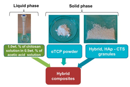
Figure 1.
Schematic representation of the content of hybrid biomicroconcrete-type composites.
The liquid to powder ratio (L/P) that produced the appropriate consistency of pastes was established to be between 0.52–0.56 g/g (Series A) and 0.56–0.62 g/g (Series B) (Table 1).

Table 1.
The composition of solid and liquid phases of biomicroconcretes.
The procedure applied during the preparation of biomicroconcretes is shown in Figure 2.
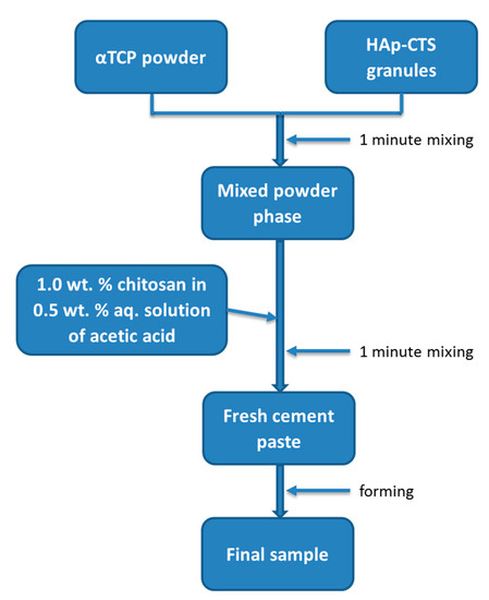
Figure 2.
The flow chart of the preparation procedure.
2.2. Phase Composition
Phase composition was examined via the X-ray diffraction method (XRD). The crystalline phases of initial α-TCP powder and set biomicroconcretes were analyzed via X-ray diffraction (D-2 Phaser, Bruker, Karlsruhe, Niemcy), within the 2θ range of 10°–90° (scanning speed of 1 °/min). Before measurements were obtained, materials were crushed and sieved to below 63 µm. The crystalline phases were identified by comparing the experimental X-ray diffractograms with the PDF (powder diffraction file) of the ICDD (International Center of Diffraction Data). Phase quantification was done using the Rietveld method (Diffrac Suite Topas Rietveld Analysis Software, Bruker, Karlsruhe, Germany, 2011). Quality fits were done by refining the zero offset, scale factors, and lattice parameters and applying a pseudo-Voigt peak shape model. All measurements were made in triplicate.
2.3. Setting Times
The setting times (initial and final) were measured according to the ASTM C266-08 standard using Gillmore Needles (Syl&Ant Instruments, Poniszowice, Poland) [19]. Results are presented as the average value of six measurements ± standard deviation (SD).
2.4. Microstructure
The microstructures of developed materials was studied using a scanning electron microscope (Nova NanoSem 200, JEOL JSM 5400, Fei Europe Company, Eindhoven, The Netherlands). All samples were coated with a thin layer of carbon and then imaged using SEM. Chemical compositions in microareas were determined via energy-dispersive X-ray spectroscopy (EDX, Oxford Instruments, Oxford, UK). The accelerating voltage was equal to 18kV.
2.5. Porosity
Mercury intrusion porosimetry was applied to measure the open porosity and pore size distribution of set and hardened biomicroconcretes (AutoPore IV 9500 Porosimeter, Micromeritics, Norcross, GA, USA). The examined samples were dried before being introduced into the penetrometer of the apparatus. All measurements were made in triplicate.
2.6. Compressive Strength
Cylindrical samples of 6 mm in diameter and 12 mm height were tested using a universal testing machine Instron 3345, with a crosshead displacement rate equal to 1.0 mm min−1. The compressive strength parameters were calculated by averaging 10 measurements and are presented as the mean ± standard deviation (SD).
2.7. Statistics
Data points in graph figures represent the mean, with error bars representing the standard deviation (SD). Comparative studies were performed using one-way ANOVA. Furthermore, Tukey’s HSD post hoc tests were used.
3. Results and Discussion
A new generation of biomaterials combining hybrid hydroxyapatite–chitosan granules with α-TCP based bone cement was obtained. α-TCP is a degradable, self-setting phase and acted as a binder for hybrid granules. X-ray examinations of the synthesized α-TCP powder were carried out. The initial α-TCP powder was composed mainly of α-TCP phase (91 ± 2 wt. %) and a small amount of hydroxyapatite (9 ± 2 wt. %). Our previous studies showed that the hybrid HAp–CTS granules were composed of hydroxyapatite as the only crystalline phase [9]. In the case of biomicroconcretes, 7 days after setting and hardening, two crystalline phases, i.e., α-TCP and hydroxyapatite, were detected (Figure 3).
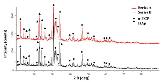
Figure 3.
XRD patterns of the biomicroconcretes.
The quantitative analysis of phase composition showed that the amount of individual phases changed after setting and hardening (Table 2).
Table 2.
The phase composition of biomicroconcretes (7 days after setting and hardening).
The hydroxyapatite contained in the material comes from: (1) hybrid granules (HAp–CTS) and (2) hydrolysis of α-TCP. An increase in hydroxyapatite content and a decrease in α-TCP content, in comparison to the composition of initial powder batches (Table 1), were noticed in both series (Table 2). This indicated a high reactivity of α-TCP powder, which to a large extent hydrolyzed to hydroxyapatite (Equation (1)). This reaction plays a key role in the setting process of calcium phosphate bone cements [6,17,20].
3 α-Ca3(PO4)2 + H2O→ Ca9(HPO4) (PO4)5(OH)
α-TCP + H2O → calcium-deficient hydroxyapatite
In vivo, the two-phasic α-TCP/hydroxyapatite matrix is supposed to be selectively resorbed and replaced by newly formed bone tissue. The quantitative phase composition differed for materials from Series A and B. A higher amount of hydroxyapatite was detected in the case of composites belonging to Series A (60–70 wt. %) in comparison to Series B (48–52 wt. %). These results indicate that hybrid granules HAp–CTS inhibited the hydrolysis of α-TCP to HAp. The process was more intensive for granules with a higher content of chitosan. It was probably due to the water uptake by chitosan present in the granules. Water is necessary for α-TCP hydrolysis (Equation (1)), and a lack of an appropriate amount of H2O can inhibit the process. The absorption of water molecules by chitosan-containing composites was observed inter alia by Correlo et al. [21].
Developed biomicroconcretes, regardless of granule size, possessed initial setting times between 7 and 10 min (Table 3). The final setting times of biomaterials from both series were longer than recommended in the literature (<15 min).
Table 3.
The setting times of the biomicroconcretes.
Materials containing medium granules (0.6–0.8mm) and small granules (0.3–0.6) revealed final setting times between 23–29 min. These biomicroconcretes set in a period of time that was acceptable from a medical point of view, and would allow the surgeon to place material in a bone void and adjust its shape to the shape of the defect. The influence of granule size and the content of chitosan in hybrid granules was observed. The granules with larger size (0.8–1.0 mm) and higher chitosan content negatively delayed the final setting time, up to 37 min. A significant shortening of the setting times of biomicroconcretes in comparison to cement based on α-TCP without granules was noticed. According to Czechowska et al. [6], α-TCP-based bone cement possesses an initial setting time equal to 7 ± 1 min and a final setting time equal to 41 ± 3 min (L/P = 0.48 g/g). Thus, it can be concluded that the presence of hybrid granules generally reduces the final setting times of composites in comparison to a single component, α-TCP-based biomaterial. The observed shortening of the setting times may have been connected to the presence of hydroxyapatite, introduced with hybrid granules (HAp–CTS). Precipitated hydroxyapatite is often used as a crystallization nucleus, accelerating the setting process of calcium phosphate bone cements [22]. Overlong setting times may result in a continuation of the setting process after wound closure. Considering the requirements for cement-type bone substitutes (initial setting time: 3–8 min and final setting time: <15 min) [23], the results obtained for Series A, with lower chitosan content in hybrid HAp–CTS (17 wt. %), were found to be the most favorable.
SEM observations revealed that hybrid HAp–CTS granules possessed a compact, homogenous microstructure with the polymeric bridges visible on their surfaces. The presence of polymer bridges allowed granules to be easily distinguished from the calcium phosphate matrix. The granules were evenly distributed in the biomicroconcretes. The presence of chitosan ensured good cohesion of granules and final materials, preventing their disintegration. Good adhesion between the matrix and hybrid granules was observed (Figure 4).
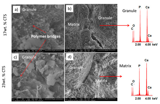
Figure 4.
SEM microphotographs of cross-sections of materials from Series A (a,b) and B (c,d), and results of EDS analysis.
The total open porosity of the samples from both series was similar and equal to 45 ± 5 vol. % (Series A) and 50 ± 5 vol. % (Series B). A multimodal pore size distribution was obtained for all biomicroconcrestes, with a pore size distribution in the range of 0.005–40 µm (Figure 5).
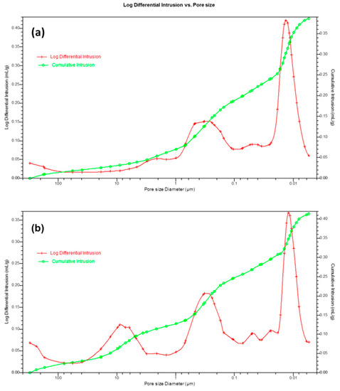
Figure 5.
Pore size distribution of biomicroconcretes of materials from Series A (a) and Series B (b).
In the case of composites with lower chitosan content (17 wt. %—Series A), pores between 0.005 and 4 µm were visible. The peaks corresponding to lower pore size values were connected with the existence of pores located between CaP grains and agglomerates in the cement matrix [6]. Peaks occurring above 1 µm were connected with the presence of hybrid granules in the material. Biomicroconcretes with higher chitosan content (23 wt. %—Series B) possessed pores with diameters ranging from 0.005 µm up to ~40 µm. Thus, a higher amount of chitosan in hybrid granules contributed to the formation of pores of a larger diameter, which, in combination with higher porosity, caused a significant reduction of the mechanical strength of biomicroconcretes for this series. This was confirmed by the outcomes of the compressive strength measurements (Figure 6).
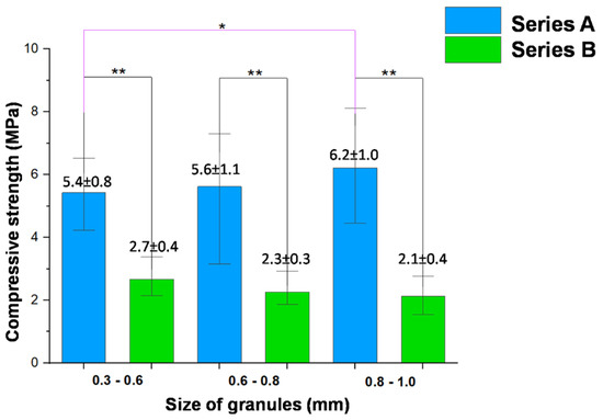
Figure 6.
Compressive strength of biomicroconcretes after 7 days of setting and hardening in the air (statistically significant difference: * p < 0.05, ** p < 0.0001).
The results obtained for biomicroconcretes containing granules with 23 wt. % chitosan (Series B, 2.1 ± 0.4 MPa–2.7 ± 0.4 MPa) were even two or three times lower compared to the materials composed of granules with 17 wt. % content of chitosan (Series A). Composites from Series A possessed mechanical strength ranging from 5.4 ± 0.8 MPa to 6.2 ± 1.0 MPa. These values are similar to compressive strength of trabecular bone (4–12 MPa) [24]. The obtained results proved that the use of granules with a larger content of chitosan adversely affected the mechanical strength of the final biomicroconcretes. In addition, it was demonstrated that the size of granules only slightly affected the mechanical properties of HAp–CTS–α-TCP composites.
The developed biomaterials combining hybrid hydroxyapatite–chitosan granules and α-TCP-based bone cement, with their phase composition, microstructure, and surgical handiness, will probably meet the requirements of regenerative medicine [6,25].
4. Summary
New microconcrete-type implant materials based on α-TCP–HAp–chitosan were obtained. The influence of chitosan content and the size of the hybrid HAp–CTS granules on the physicochemical properties of final potential bone substitutes were investigated. The obtained biomaterials, after 7 days of setting and hardening in the air, were composed of two crystalline phases: HAp (~60–70 wt. %) and α-TCP (~30–40 wt. %). Regardless of the chitosan content and the size of granules, a significant decrease in the amount of α-TCP phase compared to the initial composition was observed. This indicated the high reactivity of α-TCP powder, which hydrolyzed to non-stoichiometric hydroxyapatite (CDHAp). Our research revealed that generally, the presence of granules caused a shortening of setting times of composites, in comparison to the single component, α-TCP-based material. Biomicroconcretes containing smaller granules with 17 wt. % of chitosan possessed satisfying setting times, similar to those recommended in the literature. Such values allow for the preparation of implant material in operating room conditions and a relatively simple procedure for its introduction into the bone defect. Higher sizes of granules adversely extended the final setting times of materials. The developed biomaterials possessed high open porosity equal to ~45–50 vol. % and multimodal pore size distribution in the range of 0.005 to 40 µm. SEM observations confirmed good adhesion between matrix and hybrid granules. The compressive strength (2–6 MPa) of the obtained complex composites was close to the values corresponding to spongy bone. The amount of chitosan in hybrid HAp–CTS granules influenced the mechanical strength of studied materials. Higher compressive strength was noticed for biomicroconcretes containing granules with a lower amount of chitosan (17 wt. %). The influence of granule size in the range of 0.3 to 1.0 mm on the mechanical strength of the studied biomicroconcretes was not studied. The promising physicochemical properties of developed biomicroconcretes and their potential as novel bone substitutes need to be confirmed in biological studies.
Author Contributions
A.Ś., A.Z., J.C. conceived and planned the experiments. A.Ś. supervised the project. A.Z., J.C., E.C. carried out the experiments. All authors have read and agreed to the published version of the manuscript.
Funding
This research was supported by the National Science Centre, Poland Grant No. 2017/27/B/ST8/01173.
Conflicts of Interest
The authors declare no conflict of interest.
References
- Kickelbick, G. Hybrid materials-past, present and future. Hybrid Mater. 2014, 1, 39–51. [Google Scholar] [CrossRef]
- Muzzarelli, R. Chitosan composites with inorganics, morphogenetic proteins and stem cells, for bone regeneration. Carbohydr. Polym. 2011, 83, 1433–1445. [Google Scholar] [CrossRef]
- Dorozhkin, S.V. Medical Application of Calcium Orthophosphate Bioceramics. BIO 2011, 1, 1–51. [Google Scholar] [CrossRef]
- Venkatesan, J.; Kim, S.K. Chitosan composites for bone tissue engineering—An overview. Mar. Drugs 2010, 8, 2252–2266. [Google Scholar] [CrossRef] [PubMed]
- Peniche, C.; Solís, Y.; Davidenko, N.; García, R. Chitosan/hydroxyapatite-based composites. Biotecnol. Apl. 2010, 27, 202–210. [Google Scholar]
- Czechowska, J.; Zima, A.; Paszkiewicz, Z.; Lis, J.; Ślósarczyk, A. Physicochemical properties and biomimetic behaviour of α-TCP-chitosan based materials. Ceram. Int. 2014, 40, 5523–5532. [Google Scholar] [CrossRef]
- Lai, Y.L.; Cheng, Y.M.; Yen, S.K. Doxorubicin-chitosan-hydroxyapatite composite coatings on titanium alloy for localized cancer therapy. Mater. Sci. Eng. C 2019, 104, 109953. [Google Scholar] [CrossRef]
- Cichoń, E.; Ślósarczyk, A.; Zima, A. Influence of selected surfactants on physicochemical properties of calcium phosphate bone cements. Langmuir 2019, 35, 13656–13662. [Google Scholar] [CrossRef]
- Zima, A. Hydroxyapatite-chitosan based bioactive hybrid biomaterials with improved mechanical strength. Spectrochim. Acta Part A 2018, 193, 175–184. [Google Scholar] [CrossRef]
- Sun, L.; Xu, H.H.K.; Takagi, S.; Chow, I.C. Fast setting calcium phosphate cement -chitosan composite mechanical properites and sdissolution rates. J. Biomater. Appl. 2007, 21, 299–315. [Google Scholar] [CrossRef]
- Kashiwazaki, H.; Yamaguchi, K.; Harada, N.; Akazawa, T.; Murata, M.; Iizuka, T.; Ikoma, T.; Tanaka, J.; Inoue, N. In vivo evaluation of a novel chitosan/HAp composite biomaterial as a carrier of rhBMP-2. J. Hard Tissue Biol. 2010, 19, 181–186. [Google Scholar] [CrossRef]
- Li, H.; Zhou, C.R.; Zhu, M.Y.; Tian, J.H.; Rong, J.H. Preparation and characterization of homogeneous hydroxyapatite/chitosan composite scaffolds via in-situ hydration. J. Biomater. Nanobiotechnol. 2010, 1, 42. [Google Scholar] [CrossRef]
- Im, K.H.; Park, J.H.; Kim, K.N.; Kim, K.M.; Choi, S.H.; Kim, C.K.; Lee, Y.K. Organic-Inorganic hybrids of hydroxyapatite with chitosan. In Key Engineering Materials; Trans Tech Publications Ltd.: Baech, Switzerland, 2005; Volume 284, pp. 729–732. [Google Scholar]
- Reves, B.T.; Jennings, J.A.; Bumgardner, J.D.; Haggard, W.O. Preparation and functional assessment of composite chitosan-nano-hydroxyapatite scaffolds for bone regeneration. J. Funct. Biomater. 2012, 3, 114–130. [Google Scholar] [CrossRef] [PubMed]
- Son, Y.J.; Chung, T.J.; Oh, K.S. Brushite Bone Cement Prepared from Granular Hydroxyapatite and β-Tricalcium Phosphate. In Key Engineering Materials; Trans Tech Publications Ltd.: Baech, Switzerland, 2017; Volume 720, pp. 153–156. [Google Scholar]
- Lee, G.H.; Makkar, P.; Paul, K.; Lee, B. Incorporation of BMP-2 loaded collagen conjugated BCP granules in calcium phosphate cement based injectable bone substitutes for improved bone regeneration. Mater. Sci. Eng. C 2017, 77, 713–724. [Google Scholar] [CrossRef] [PubMed]
- Czechowska, J.; Zima, A.; Ślósarczyk, A. Comparative study on physicochemical properties of alpha-TCP/calcium sulphate dihydrate biomicroconcretes containing chitosan, sodium alginate or methylcellulose. Acta Bioeng. Biomech. 2020, in press. [Google Scholar] [CrossRef]
- Kolmas, J.; Kaflak, A.; Zima, A.; Ślósarczyk, A. Alpha-tricalcium phosphate synthesized by two different routes: Structural and spectroscopic characterization. Ceram. Int. 2015, 41, 5727–5733. [Google Scholar] [CrossRef]
- Standard test method for time setting of hydraulic-cement paste by gillmore needles, 19428–2959. In ASTM Annual Book of Standards; ASTM C266-15; American Society for Testing and Materials: West Conshohocken, PA, USA, 2015.
- Weichhold, J.; Gbureck, U.; Goetz-Neunhoeffer, F.; Hurle, K. Setting mechanism of a CDHA forming α-TCP cement modified with sodium phytate for improved injectability. Materials 2019, 12, 2098. [Google Scholar] [CrossRef]
- Correlo, V.M.; Pinho, E.D.; Pashkuleva, I.; Bhattacharya, M.; Neves, N.M.; Reis, R.L. Water absorption and degradation characteristics of chitosan-based polyesters and hydroxyapatite composites. Macromol. Biosci. 2007, 7, 354–363. [Google Scholar] [CrossRef]
- Fernandez, E.; Ginebra, M.P.; Boltong, M.G.; Driessens, F.C.M.; Planell, J.A.; Ginebra, J.; De Maeyer, E.A.P.; Verbeeck, R.M.H. Kinetic study of the setting reaction of a calcium phosphate bone cement. J. Biomed. Mater. Res. 1996, 32, 367–374. [Google Scholar] [CrossRef]
- Ginebra, M.P.; Fernández, E.; Boltong, M.G.; Bermúdez, O.; Planell, J.A.; Driessens, F.C.M. Compliance of an apatitic calcium phosphate cement with the short-term clinical requirements in bone surgery, orthopaedics and dentistry. Clin. Mater. 1994, 17, 99–104. [Google Scholar] [CrossRef]
- Seidi, A.; Ramalingam, M.; Elloumi-Hannachi, I.; Ostrovidov, S.; Khademhosseini, A. Gradient biomaterials for soft-to-hard interface tissue engineering. Acta Biomater. 2011, 7, 1441–1451. [Google Scholar] [CrossRef] [PubMed]
- Pilia, M.; Guda, T.; Appleford, M. Development of composite scaffolds for load-bearing segmental bone defects. BioMed Res. Int. 2013, 2013, 458253. [Google Scholar] [CrossRef] [PubMed]


© 2020 by the authors. Licensee MDPI, Basel, Switzerland. This article is an open access article distributed under the terms and conditions of the Creative Commons Attribution (CC BY) license (http://creativecommons.org/licenses/by/4.0/).