Article

Malus baccata var. gracilis and Malus toringoides Bark Polyphenol Studies and Antioxidant, Antimicrobial and Anticancer Activities

1
Plant Production Department, College of Food and Agricultural Sciences, King Saud University, P.O. Box 2455, Riyadh 11451, Saudi Arabia
2
Floriculture, Ornamental Horticulture, and Garden Design Department, Faculty of Agriculture (El-Shatby), Alexandria University, Alexandria 21545, Egypt
3
Department of Geography, Environmental Management, and Energy Studies, University of Johannesburg, APK Campus, 2006, Johannesburg, South Africa
4
Department of Pharmaceutical Botany, Medical College, Jagiellonian University, ul. Medyczna 9, 30-688 Kraków, Poland
5
Precision Agriculture Laboratory, Department of Pomology, Faculty of Agriculture (El-Shatby), Alexandria University, Alexandria 21545, Egypt
*
Author to whom correspondence should be addressed.
Processes 2020, 8(3), 283; https://doi.org/10.3390/pr8030283
Submission received: 24 January 2020 / Revised: 13 February 2020 / Accepted: 19 February 2020 / Published: 2 March 2020
(This article belongs to the Special Issue Extraction, Characterization and Pharmacology of Natural Products)

Abstract

:
Exploring new sources of polyphenols with biological activities that work against human diseases is the target of natural product studies. This study determined the polyphenol composition of the bark of Malus species M. baccata var. gracilis (Rehder) T.C.Ku and M. toringoides (Rehder) Hughes, using high-performance liquid chromatography with a diode-array detector (HPLC-DAD) analysis. The antiproliferative, cytotoxic, antioxidant and antimicrobial applications of these extracts, as well as the identified phenol, were studied. The HPLC-DAD analysis confirmed three polyphenols in the extracts out of the 21 screened compounds: protocatechuic acid, gallic acid, and catechin. The major constituents in M. baccata and M. toringoides were protocatechuic acid, at 3.16 and 7.15 mg 100 g−1 dry weight (DW), respectively, and catechin, at 5.55 and 6.80 mg 100 g−1 DW, respectively. M. baccata and M. toringoides bark extracts showed antioxidant activities using 2,2-diphenyl-1-picrylhydrazyl (DPPH), β-carotene bleaching, and ferric-reducing antioxidant power (FRAP) assays, which were attributed to the dominance of protocatechuic acid. The highest antiproliferative and cytotoxic effects were against Jurkat cells. Against MCF-7 and Hela cells, there was necrotic cell accumulation in the early apoptotic as well as the late apoptotic phase. The bark extracts showed noticeable antibacterial effects against Listeria monocytogenes, Bacillus cereus, and Escherichia coli. Protocatechuic acid showed comparable results to bark extracts. There were antifungal effects against Aspergillus ochraceus, A. niger, and Candida albicans, and the activities were higher than the commercial reagent. M. baccata and M. toringoides could be considered as a new source of phenolic acids, including protocatechuic acid with anticancer, antibacterial antifungal, and antioxidant-promising effects.

1. Introduction

Polyphenol groups (e.g., phenolic acids) are important secondary plant metabolites that contain a large group of compounds with extremely important pharmacological applications. They are widely used for the control of neurodegenerative diseases [1], cancers [2,3,4,5], inflammation cardiovascular health [6,7], type 2 diabetes [8], obesity [9], insect control [10], antibacterial agents [11,12], antioxidants [13], food processing, and the production of functional foods [14]. Polyphenols are abundant in plants, and specific phenols have strong anticancer activities [15]. Recent trends indicate that a balanced lifestyle with a polyphenol-rich diet can control cancers such as prostate cancer, and prevent the future occurrence of the disease [16]. Polyphenols act on tumors by different mechanisms, including the induction of detoxification enzymes and the activation of the antioxidant mechanism, antiproliferation, apoptosis through cell-cycle arrest, and molecular regulation of genes associated with cancer [2,17,18,19,20]. Tree barks are promising sources of these polyphenols; they contain phenolic acids, flavonoids, and catechins, as well as sugars [2,12,19,21,22]. Phenolic acids are an important group of phenols as they act as antioxidant agents and have, for example, anticancer and antimicrobial properties [3,23,24,25,26,27].
Microbial infections with Staphylococcus aureus cause serious human diseases such as soft tissue infection, pneumonia, sepsis, and peritonitis [28,29]. Other serious diseases, such as listeriosis (which leads to preterm birth and miscarriage) and mild gastroenteritis of immunocompetent individuals, are caused by the bacterial foodborne pathogen Listeria monocytogenes [30]. The outbreak of such diseases may cause a high number of deaths, especially with the rising issue of greater resistance to antibiotics [31]. These facts are propelling the search for natural alternative sources of antimicrobial agents, such as those found in tree barks. Recent studies have described the antifungal effects of many polyphenols against plant pathogenic fungi such as Botrytis cinerea and Fusarium oxysporum [32,33], as well as human pathogenic fungi such as Candida albicans, and others [34,35].
The genus Malus (Rosaceae) consists of 30 species, including the cultivated apple (Malus pumila Mill). Malus baccata and Malus toringoides (Figure 1) are wild taxa that are considered as sources of variation for apple breeding [36]. M. baccata is used as rootstock for commercial apple cultivars and is distributed in Northern America and Eurasia [37]. Few reports have described the chemical composition and biological activities of the fruits [38,39]. Furthermore, no reports have investigated the biological activities of bark extracts. M. toringoides is a drought-resistant wild species [40], and no previous investigations have been performed on the chemical composition of the bark.
Malus baccata and Malus toringoides are widely distributed species; however, experimental studies regarding the chemical composition and biological activities of bark extracts are limited. In this regard, the polyphenol profiles were evaluated qualitatively and quantitatively using the high-performance liquid chromatography with a diode-array detector (HPLC-DAD) method for the first time. Furthermore, the antiproliferative, cytotoxic, antioxidant, antibacterial, and antifungal effects of the bark were explored.

2. Results

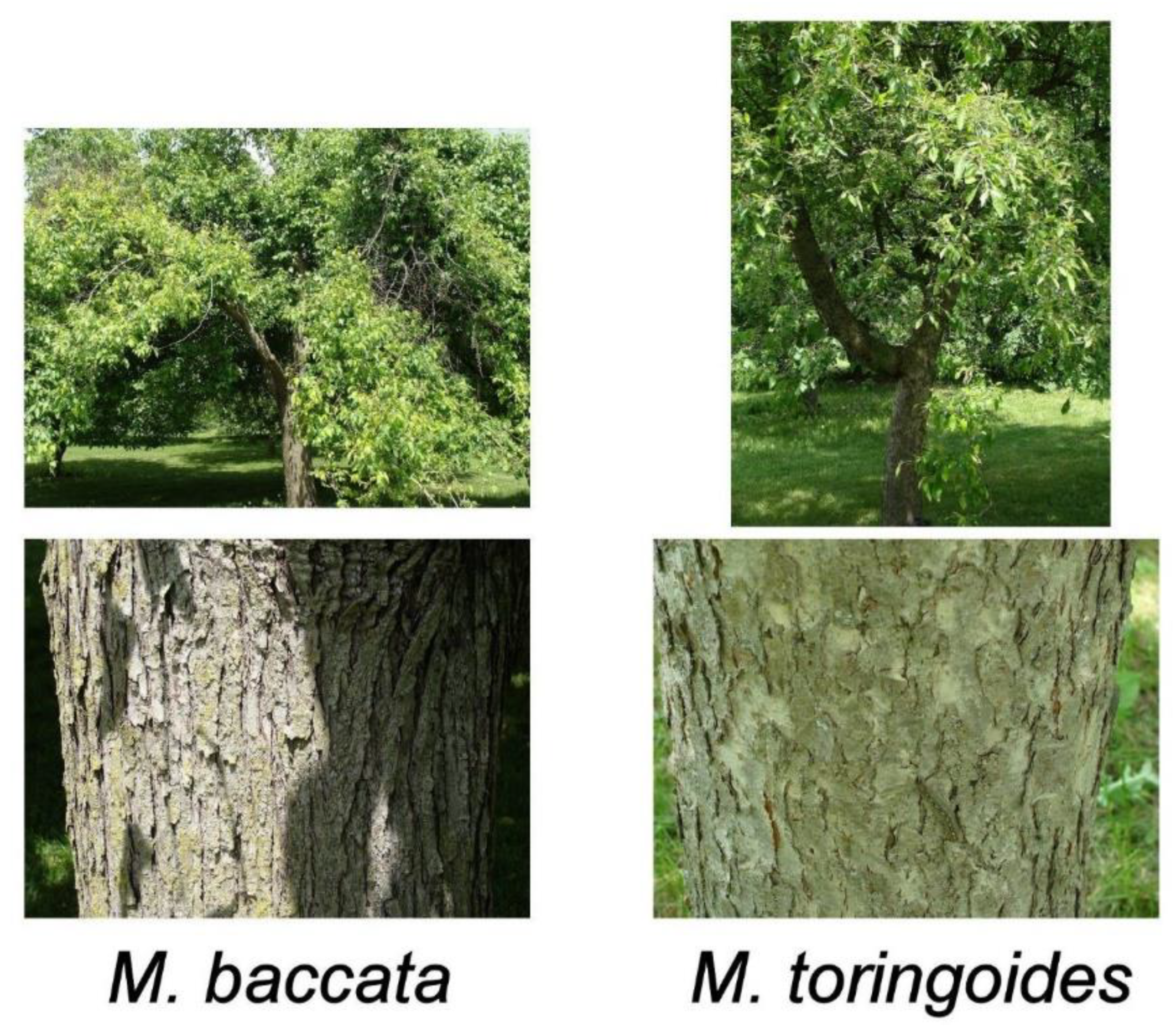
2.1. Polyphenol Profiling of M. baccata and M. toringoides Bark

M. baccata and M. toringoides bark methanolic extracts were studied using the high-performance liquid chromatography with a diode-array detector (DAD-HPLC) method (Figure 2). Three polyphenols out of the 21 screened were determined in both bark extracts: protocatechuic acid, gallic acid, and catechin (see the Materials and Methods section) (Table 1). The amount of protocatechuic acid for M. baccata and M. toringoides was 3.16 and 7.15 mg 100 g−1 (dry weight) DW, respectively. The lower amounts of gallic acid were found in both species: 0.29 mg 100 g−1 DW and 0.44 mg 100 g−1 DW, respectively. Catechin was estimated in considerable amounts: 5.55 and 6.80 mg 100 g−1 DW, respectively (Table 1).

2.2. Antioxidant Effects

The M. baccata and M. toringoides bark extracts showed antioxidant activities by several methods (Table 2). M. toringoides showed higher antioxidant effects than M. baccata using β-carotene bleaching, DPPH, and ferric-reducing antioxidant power (FRAP) assays. A Butylated hydroxytoluene (BHT) standard showed the lowest IC50 value compared to the two examined species and the phenolic standard (protocatechuic acid).

2.3. Anticancer Effects

The bark extracts of M. baccata and M. toringoides had noticeable antiproliferative activities against cancer cells, as shown in Table 3. The highest effects were against Jurkat cells. MCF-7 and Hela cells were affected moderately by bark extracts. The lowest anticancer effects were against HT-29 cells, using both extracts as well as the phenolic standard.
The apoptotic assay of M. baccata and M. toringoides is shown in Figure 3. Necrotic cell accumulation was found in the early and late apoptotic phases.

2.4. Antibacterial Activities

The bark extracts of M. baccata and M. toringoides had noticeable antibacterial activities against some bacteria such as Bacillus cereus, Escherichia coli and Listeria monocytogenes. Other bacteria, such as Micrococcus flavus and Staphylococcus aureus, were more resistant (Table 4).
The phenolic standard protocatechuic acid showed comparable results to the bark extracts. The highest effects of bark extracts were against Bacillus cereus.

2.5. Antifungal Effects

The bark extracts of M. baccata and M. toringoides had noticeable antifungal effects against fungi (Table 5). The lowest IC50 values were against Aspergillus ochraceus, A. niger, and Candida albicans. However, there was low activity against Penicillium funiculosum and P. ochrochloron, which were more resistant. The activities of the extracts were higher than the commercial reagent ketoconazole (KTZ). The antifungal effects of protocatechuic acid were comparable to those of M. baccata and M. toringoides.

3. Discussion

In our study, M. baccata and M. toringoides bark extracts were shown to contain three polyphenols: protocatechuic acid, gallic acid, and catechin (Table 1 and Figure 2). Protocatechuic acid and catechin were the most quantitatively dominant compounds. A previous investigation on the fruit pulps and seeds of M. baccata found amino acids, fructose, sucrose, and fatty acids, and suggested that the fruits are a good source of phenolic compounds [38]. Further, the analyses of the fruit skin and flesh of different apple cultivars revealed the presence of several triterpenes, including ursolic and oleanolic [39], and showed anti-inflammatory activities. A preliminary study using two-dimensional chromatograms on Whatman No. 1 paper indicated the presence of few flavonoids (e.g., quercetin) in some Malus cultivars in the United Kingdom, and the results have been used in the classification of this species [41]. However, to our knowledge, this is the first report exploring the phenolic content and biological activities of the tree bark of M. baccata and M. toringoides extracts. Protocatechuic acid and catechin were detected in low quantities, but they are essential antioxidant and anticancer compounds [42,43,44]. Undoubtedly, a more thorough chemical composition analysis will be needed, for example, one using more modern methods, in order to obtain more detailed knowledge about the composition of M. baccata and M. toringoides bark extracts.
M. baccata and M. toringoides bark extracts showed antiradical activities, and M. toringoides was higher as an antioxidant. These activities could be attributed to the phenolic profile detected. The major compound detected was protocatechuic acid, which was strongly associated with antioxidant activities in all assays and was relatively comparable to standard antioxidants. Protocatechuic acid is a strong antioxidant, as several studies previous studies have found [42,45]. Other tree bark extracts showed noticeable antiradical activities which were attributed to several phenols including protocatechuic acid [2].
M. baccata and M. toringoides had noticeable antiproliferative activities against cancer, and the highest effects were against the Jurkat cells MCF 7 and Hela. The apoptotic assay of M. baccata and M. toringoides showed necrotic cells increase in the early and late apoptotic phases in the flow cytometry assay. Such effects are associated with high concentrations of protocatechuic acid. Previous investigations revealed that protocatechuic acid inhibited the chemical carcinogenesis and showed proapoptotic and antiproliferative activities [42]. Protocatechuic acid inhibits the metastasis of B16/F10 cancer cells by the expression and control of Ras/Akt/NF-κB and MMP-2 production [46]. It also has toxic effects against gastric epithelial cancer cells, and works as a natural urease inhibitor [47].
The extracts of M. baccata and M. toringoides bark had antibacterial activities against some bacteria such as B. cereus, E. coli, and L. monocytogenes. Further, protocatechuic acid showed comparable results to bark extracts. The antibacterial activities of bark extracts are attributed to the availability of phenolic acids such as protocatechuic acid. A previous investigation revealed that protocatechuic acid revealed antibacterial activities against S. aureus [48]. Further, protocatechuic acid and other phenols in mushrooms were responsible for the antimicrobial and antifungal activities against several organisms [49].
The bark extracts of M. baccata and M. toringoides had noticeable antifungal activities, and the lowest IC50 values were against A. ochraceus, A. niger, and C. albicans. The antifungal activities of protocatechuic acid were comparable to those of M. baccata and M. toringoides. The antifungal activities of these bark extracts are attributed to their phenolic profile, including protocatechuic acid. Protocatechuic acid has known antifungal activities against several fungi [50], and had a strong antifungal effect against Botrytis cinerea [51].

4. Materials and Methods

4.1. Plant Material and Preparation

The barks of Malus species M. baccata var. gracilis (Rehder) T.C.Ku and M. toringoides (Rehder) Hughes were obtained in the spring from the arboretum of the University of Guelph, Canada, and vouchered in both Alexandria University, Egypt (Hosam0001050–1052) and the University of Guelph. Bark samples (3 replicates) were dried by lyophilization (Labconco, California, USA) and powdered [3,52]. Of the dried powder (DW), 0.2 g was sonicated in a methanol extraction (10 mL, Chempur, Lublin, Poland) for 2 × 30 min at 30 °C (Sonic-2, POLSONIC, Poland). The filtered extracts (filtered by Whatman paper) were left at room temperature to lose the methanol (for about 24 h). The methanol (1 mL) was added to the residue and mixed until dissolved completely, and the samples were kept in deep freezing at −80 °C. Before using the samples for bioassays, the methanol was eliminated using a rotary evaporator (40 °C). The bacterial and fungal isolates were obtained from Alexandria University, Egypt. Breast adenocarcinoma cultures (MCF-7), T-cell lymphoblast like (Jurkat), colon adenocarcinoma (HT-29), cervical adenocarcinoma (HeLa), and HEK-293 (normal human cells) were obtained from an institutional collection of the American Type Culture Collection (ATCC).

4.2. Chemicals

A collection of high-performance liquid chromatograph (HPLC) standards were used for the qualification and quantification of phenolics, including cinnamic acid and its derivatives (caffeic acid, o-coumaric acid, m-coumaric acid, p-coumaric acid, ferulic acid, hydrocaffeic acid, isoferulic acid, and sinapic acid); benzoic acid and its derivatives (3,4-dihydroxyphenylacetic acid, ellagic acid, gallic acid, gentisic acid, p-hydroxybenzoic acid, protocatechuic acid, salicylic acid, syringic acid, and vanillic acid); and depsides (chlorogenic acid, neochlorogenic acid, and rosmarinic acid). The flavonoids were quantified using the standards of aglycone (myricetin, quercetin, kaempferol, rhamnetin, and luteolin) and glycoside (apigetrin, cynaroside, hyperoside, quercitrin, robinin, rutoside, isoquercetin, trifolin, and vitexin). The catechin and its derivatives (epigallocatechin gallate epicatechin, epigallocatechin, and epicatechin gallate) were also screened. All chemicals were purchased from Sigma-Aldrich, Berlin, Germany.

4.3. Analyses of Phenolic Compounds

Bark extracts were subjected to HPLC-DAD analysis [2,3,53,54] using a Merck-Hitachi liquid chromatograph (LaChrom Elite) containing a diode-array detection (DAD) detector (L-2455). A Purospher RP-18e column was used (5 μm, 250 × 4 mm; Merck, Berlin, Germany). A flow rate was set at 1 mL min−1 at 25 °C. The extract volume of 20 µL was used for the injection. The compounds were detected at 254 nm by the validated HPLC method [53,54]. Compound identification was accomplished by comparing UV retention times and spectra of reference substances. Co-chromatography was also used for identification. The quantification was determined using calibration curves [53,54,55].

4.4. Anticancer Activities

The antiproliferative and cytotoxic activities of each bark were examined against HeLa, MCF-7, Jurkat, and HT-29 [2,3,33]. HEK-293 (normal cells) were also used. The 3--2,5-diphenyltetrazolium bromide (MTT) assay was used. Briefly, the cells were grown in Minimum Essential Medium (MEM) medium with 10% FBS, 0.1 mM non-essential amino acids, 17.8 mM NaHCO3, and 1 mM sodium pyruvate in 75 cm2 flasks. Then, microtiter plates (96-well) were used for seeding cells at 4  ×  10−4 cells per µL in 270  µL medium at 37 °C, 5% CO2 for 48 h. Extracts were added to the culture media in the plates until reaching 50, 100, 200, 300, and 400 µg/mL final concentration. Then, the Phosphate-buffered saline (PBS) was used to wash the extract traces, and 12 mM MTT dissolved in PBS was added to the medium. Isoprobanol (0.04 N HCl) was added to each well, and the mixture was left for 40 min. Vinblastine sulfate and taxol served as positive controls; a negative control was also used (untreated cells). A microplate reader was used at a 570 nm wavelength (Thermo Scientific, California, USA). The inhibition activity percentage was determined in triplicate:
I A A =   ( A B 570 n m ) C ( A B 570 n m ) s ( A B 570 n m ) C × 100
where AB is the absorbance, and ( A B 570 n m ) C and ( A B 570 n m ) s are Abs.570 nm of control and sample, respectively.
By plotting the percentage of viable cells against extract concentration, the IC50 values were calculated in µg mL−1 using flow cytometry (FAC Scan, Becton Dickinson, Iowa, USA) as described in [2,33,56], and untreated cells were considered control.

4.5. Antioxidant Activity

Bark extracts were subjected to β-carotene bleaching as well as FRAP and DPPH assays in triplicate replications [3,57,58,59,60,61]. A wavelength of 470 nm was used in the β-carotene bleaching experiment, while a 517 nm wavelength was used in the DPPH to measure absorbance. The bark extract amounts that scavenged 50% of the β-carotene or DPPH were considered as the IC50 value and expressed as µg mL−1. This was determined by plotting the extract concentration against the inhibition percentage. The FRAP reagent was prepared in the experiment as in, e.g., [43]. The FRAP reagent (3 mL) was mixed with 100 μL of bark extracts as well as Trolox. The mixture was incubated for 30 min at 37 °C and the absorbance was determined at 593 nm. The calibration procedure was accomplished using serial dilutions of Trolox (0–0.5 mmol/L).

4.6. Antibacterial Activity

The bark extracts’ bacterial control against Listeria monocytogenes (clinical isolate), Staphylococcus aureus (ATCC 6538), Escherichia coli (ATCC 35210), Bacillus cereus (ATCC 14579), Pseudomonas aeruginosa (ATCC 27853) and Micrococcus flavus (ATCC 10240), were tested using the micro-dilution protocol [5,34,62,63] in microtiter plates (96-well). Serial concentrations of bark extracts were mixed with 1.0 × 10⁴ colony forming unit (CFU) bacterial inoculum in each well, with 100 μL tryptic soy broth per well, then incubated for one day at 37 °C on a rotary shaker. The lowest concentration of extracts that caused no visible growth (using a binocular microscope) was considered as the minimum inhibitory concentration (MIC). To determine the minimum bactericidal concentration (MBC), serial sub-culturing of bark extracts (2 μL) was used, and the lowest concentration causing elimination of 99.5% of the inoculum was considered as the MBC. The optical density was determined using 655 nm wavelength. Streptomycin (positive control) was used at 0.01–10 mg/mL), as well as DMSO (negative control, 1%).

4.7. Antifungal Activity

The antifungal activities of Malus sp. bark extracts were determined against various fungi, including Penicillium ochrochloron (ATCC 48663), A. ochraceus (ATCC 12066), Candida albicans (ATCC 12066), A. niger (ATCC 6275), Aspergillus flavus (ATCC 9643), and Penicillium funiculosum (ATCC 56755). The microdilution method was employed in this assay [34,56,62]. The minimum inhibitory and fungicidal concentrations (MIC and MFC, respectively) were determined. The minimal concentration inhibiting fungi growth by stereomicroscope was considered as MIC. Extracts’ serial dilution (of 2 µL) was used in sub-cultures (at 28 °C for 72 h) of the fungi in microtiter plates containing 100 µL of broth medium and inoculum to determine the MFC. Positive and negative controls were used, including ketoconazole (KTZ, 1–3500 µg/mL) and DMSO (1%), respectively.

4.8. Research Ethics

The study was conducted in accordance with the Declaration of Helsinki, and the protocol was approved by the Ethics Committee of Alexandria University (63663-937) on 1 December 2017.

4.9. Statistical Analyses

The least significance difference (LSD) was determined using SPSS software.

5. Conclusions

This is the first report investigating the phenolic profile of M. baccata bark. In addition, this is the first report on the antiproliferative, cytotoxic, antioxidant, antibacterial, and antifungal effects of M. baccata and M. toringoides bark extracts. Out of the 21 screened, the HPLC study revealed the availability of three different polyphenols: protocatechuic acid, gallic acid, and catechin. The major compounds were protocatechuic acid and catechin. M. baccata and M. toringoides bark extracts showed antioxidant activities using β-carotene bleaching, DPPH, and FRAP assays, which were attributed to the dominance of protocatechuic acid. The highest antiproliferative and cytotoxic effects were against Jurkat cells. Against MCF-7 and Hela cells, there was an accumulation of necrotic cells in the apoptotic phases. The bark extracts showed noticeable antibacterial effects against Listeria monocytogenes, Bacillus cereus, and Escherichia coli. The protocatechuic acid showed comparable results to bark extracts. There were antifungal effects against Aspergillus ochraceus, A. niger, and Candida albicans, and the effects were higher than the commercial reagent. M. baccata can be considered as a newly explored source of phenols that have antioxidant and other biological activities.

Author Contributions

Conceptualization, H.O.E., A.S., H.E., and A.A.B. Data curation, H.O.E., A.S., P.K., and F.A.A.-M. Formal analysis, H.O.E., A.S., P.K., and H.E. Funding acquisition, A.S., F.A.A.-M., and D.O.E.-A. Investigation, H.O.E., A.S., P.K., F.A.A.-M., and D.O.E.-A. Methodology, H.O.E., P.K., and D.O.E.-A. Project administration, H.E. Resources, H.E. Visualization, A.S. Writing—original draft, H.O.E., A.S., and F.A.A.-M. Writing—review and editing, H.O.E. and A.S. All authors have read and agreed to the published version of the manuscript.

Funding

The work was supported by King Saud University, Researchers Supporting Project number (RSP-2019/118).

Acknowledgments

The authors extend their appreciation to King Saud University, Researchers Supporting Project for funding this work through research group (RSP-2019/118).

Conflicts of Interest

The authors declare no conflict of interest.

References

  1. Ebrahimi, A.; Schluesener, H. Natural polyphenols against neurodegenerative disorders: Potentials and pitfalls. Ageing Res. Rev. 2012, 11, 329–345. [Google Scholar] [CrossRef]
  2. Elansary, H.O.; Szopa, A.; Kubica, P.; Al-Mana, F.A.; Mahmoud, E.A.; El-Abedin, T.K.A.Z.; Mattar, M.A.; Ekiert, H. Phenolic Compounds of Catalpa speciosa, Taxus cuspidata, and Magnolia acuminata have Antioxidant and Anticancer Activity. Molecules 2019, 24, 412. [Google Scholar] [CrossRef] [Green Version]
  3. Elansary, H.O.; Szopa, A.; Kubica, P.; Ekiert, H.; Mattar, M.A.; Al-Yafrasi, M.A.; El-Ansary, D.O.; Zin El-Abedin, T.K.; Yessoufou, K. Polyphenol Profile and Pharmaceutical Potential of Quercus spp. Bark Extracts. Plants 2019, 8, 486. [Google Scholar] [CrossRef] [PubMed] [Green Version]
  4. Elansary, H.O. Tree Bark Phenols Regulate the Physiological and Biochemical Performance of Gladiolus Flowers. Processes 2020, 8, 71. [Google Scholar] [CrossRef] [Green Version]
  5. Elansary, H.O.; Szopa, A.; Klimek-Szczykutowicz, M.; Jafernik, K.; Ekiert, H.; Mahmoud, E.A.; Barakat, A.A.; El-Ansary, D.O. Mammillaria Species—Polyphenols Studies and Anti-Cancer, Anti-Oxidant, and Anti-Bacterial Activities. Molecules 2019, 25, 131. [Google Scholar] [CrossRef] [PubMed] [Green Version]
  6. Tangney, C.C.; Rasmussen, H.E. Polyphenols, inflammation, and cardiovascular disease. Curr. Atheroscler. Rep. 2013, 15, 324. [Google Scholar] [CrossRef]
  7. Magrone, T.; Magrone, M.; Russo, A.M.; Jirillo, E. Recent Advances on the Anti-Inflammatory and Antioxidant Properties of Red Grape Polyphenols: In Vitro and In Vivo Studies. Antioxidants 2019, 9, 35. [Google Scholar] [CrossRef] [Green Version]
  8. Cao, H.; Ou, J.; Chen, L.; Zhang, Y.; Szkudelski, T.; Delmas, D.; Daglia, M.; Xiao, J. Dietary polyphenols and type 2 diabetes: Human Study and Clinical Trial. Crit. Rev. Food Sci. Nutr. 2019, 59, 3371–3379. [Google Scholar] [CrossRef]
  9. Wang, S.; Moustaid-Moussa, N.; Chen, L.; Mo, H.; Shastri, A.; Su, R.; Bapat, P.; Kwun, I.; Shen, C.-L. Novel insights of dietary polyphenols and obesity. J. Nutr. Biochem. 2014, 25, 1–18. [Google Scholar] [CrossRef] [Green Version]
  10. El-Sabrout, M.A.; Salem, Z.M.M.; Bin-Jumah, M.; Allam, A.A. Toxicological Activity of Some Plant Essential Oils Against Tribolium castaneum and Culex pipiens Larvae. Processes 2019, 7, 933. [Google Scholar] [CrossRef] [Green Version]
  11. Okla, K.M.; Alamri, A.S.; Salem, Z.M.M.; Ali, M.H.; Behiry, I.S.; Nasser, A.R.; Alaraidh, A.I.; Al-Ghtani, M.S.; Soufan, W. Yield, Phytochemical Constituents, and Antibacterial Activity of Essential Oils from the Leaves/Twigs, Branches, Branch Wood, and Branch Bark of Sour Orange (Citrus aurantium L.). Processes 2019, 7, 363. [Google Scholar] [CrossRef] [Green Version]
  12. Salem, M.Z.M.; Elansary, H.O.; Ali, H.M.; El-Settawy, A.A.; Elshikh, M.S.; Abdel-Salam, E.M.; Skalicka-Wozniak, K. Bioactivity of essential oils extracted from Cupressus macrocarpa branchlets and Corymbia citriodora leaves grown in Egypt. BMC Complement. Altern. Med. 2018, 18, 23. [Google Scholar] [CrossRef] [PubMed]
  13. Halagarda, M.; Groth, S.; Popek, S.; Rohn, S.; Pedan, V. Antioxidant Activity and Phenolic Profile of Selected Organic and Conventional Honeys from Poland. Antioxidants 2020, 9, 44. [Google Scholar] [CrossRef] [PubMed] [Green Version]
  14. Khan, M.; Siddiqui, S.A. Concurrent chemoradiotherapy with or without induction chemotherapy for the management of cervical lymph node metastasis from unknown primary tumor. J. Cancer Res. Ther. 2018, 14, 1117–1120. [Google Scholar] [CrossRef]
  15. Abdal Dayem, A.; Choi, H.Y.; Yang, G.-M.; Kim, K.; Saha, S.K.; Cho, S.-G. The Anti-Cancer Effect of Polyphenols against Breast Cancer and Cancer Stem Cells: Molecular Mechanisms. Nutrients 2016, 8, 581. [Google Scholar] [CrossRef]
  16. Costea, T.; Nagy, P.; Ganea, C.; Szöllősi, J.; Mocanu, M.-M. Molecular Mechanisms and Bioavailability of Polyphenols in Prostate Cancer. Int. J. Mol. Sci. 2019, 20, 1062. [Google Scholar] [CrossRef] [Green Version]
  17. Alvarado-Sansininea, J.J.; Sánchez-Sánchez, L.; López-Muñoz, H.; Escobar, M.L.; Flores-Guzmán, F.; Tavera-Hernández, R.; Jiménez-Estrada, M. Quercetagetin and Patuletin: Antiproliferative, Necrotic and Apoptotic Activity in Tumor Cell Lines. Molecules 2018, 23, 2579. [Google Scholar] [CrossRef] [Green Version]
  18. Sezer, E.D.; Oktay, L.M.; Karadadaş, E.; Memmedov, H.; Selvi Gunel, N.; Sözmen, E. Assessing Anticancer Potential of Blueberry Flavonoids, Quercetin, Kaempferol, and Gentisic Acid, Through Oxidative Stress and Apoptosis Parameters on HCT-116 Cells. J. Med. Food 2019, 22, 1118–1126. [Google Scholar] [CrossRef]
  19. Salem, M.Z.M.; Mansour, M.M.A.; Elansary, H.O. Evaluation of the effect of inner and outer bark extracts of sugar maple (Acer saccharum var. saccharum) in combination with citric acid against the growth of three common molds. J. Wood Chem. Technol. 2019, 39, 136–147. [Google Scholar] [CrossRef]
  20. Saleh, A.; ElFayoumi, H.M.; Youns, M.; Barakat, W. Rutin and orlistat produce antitumor effects via antioxidant and apoptotic actions. N-S Arch. Pharmacol. 2019, 392, 165–175. [Google Scholar] [CrossRef]
  21. Andrade, C.; Ferreres, F.; Gomes, G.M.N.; Duangsrisai, S.; Srisombat, N.; Vajrodaya, S.; Pereira, M.D.; Gil-Izquierdo, A.; Andrade, B.P.; Valentão, P. Phenolic Profiling and Biological Potential of Ficus curtipes Corner Leaves and Stem Bark: 5-Lipoxygenase Inhibition and Interference with NO Levels in LPS-Stimulated RAW 264.7 Macrophages. Biomolecules 2019, 9, 400. [Google Scholar] [CrossRef] [PubMed] [Green Version]
  22. Dróżdż, P.; Pyrzynska, K.J. Assessment of polyphenol content and antioxidant activity of oak bark extracts. Eur. J. Wood Prod. 2018, 76, 793–795. [Google Scholar] [CrossRef] [Green Version]
  23. Cueva, C.; Moreno-Arribas, M.V.; Martín-Álvarez, P.J.; Bills, G.; Vicente, M.F.; Basilio, A.; Rivas, C.L.; Requena, T.; Rodríguez, J.M.; Bartolomé, B. Antimicrobial activity of phenolic acids against commensal, probiotic and pathogenic bacteria. Res. Microbiol. 2010, 161, 372–382. [Google Scholar] [CrossRef] [PubMed]
  24. Khadem, S.; Marles, R.J. Monocyclic Phenolic Acids; Hydroxy- and Polyhydroxybenzoic Acids: Occurrence and Recent Bioactivity Studies. Molecules 2010, 15, 7985–8005. [Google Scholar] [CrossRef]
  25. Wojdyło, A.; Nowicka, P.; Grimalt, M.; Legua, P.; Almansa, S.M.; Amorós, A.; Carbonell-Barrachina, A.Á; Hernández, F. Polyphenol Compounds and Biological Activity of Caper (Capparis spinosa L.) Flowers Buds. Plants 2019, 8, 539. [Google Scholar]
  26. Fejér, J.; Kron, I.; Pellizzeri, V.; Pľuchtová, M.; Eliašová, A.; Campone, L.; Gervasi, T.; Bartolomeo, G.; Cicero, N.; Babejová, A.; et al. First Report on Evaluation of Basic Nutritional and Antioxidant Properties of Moringa Oleifera Lam. from Caribbean Island of Saint Lucia. Plants 2019, 8, 537. [Google Scholar] [CrossRef] [Green Version]
  27. Bazzicalupo, M.; Burlando, B.; Denaro, M.; Barreca, D.; Trombetta, D.; Smeriglio, A.; Cornara, L. Polyphenol Characterization and Skin-Preserving Properties of Hydroalcoholic Flower Extract from Himantoglossum robertianum (Orchidaceae). Plants 2019, 8, 502. [Google Scholar] [CrossRef] [Green Version]
  28. Kim, H.K.; Missiakas, D.; Schneewind, O. Mouse models for infectious diseases caused by Staphylococcus aureus. J. Immunol. Methods 2014, 410, 88–99. [Google Scholar] [CrossRef]
  29. Mahmoud, E.A.; Elansary, H.O.; El-Ansary, D.O.; Al-Mana, F.A. Elevated Bioactivity of Ruta graveolens against Cancer Cells and Microbes Using Seaweeds. Processes 2020, 8, 75. [Google Scholar] [CrossRef] [Green Version]
  30. Desai, A.N.; Anyoha, A.; Madoff, L.C.; Lassmann, B. Changing epidemiology of Listeria monocytogenes outbreaks, sporadic cases, and recalls globally: A review of ProMED reports from 1996 to 2018. Int. J. Infect. Dis. 2019, 84, 48–53. [Google Scholar] [CrossRef] [Green Version]
  31. Ventola, C.L. The antibiotic resistance crisis: Part 1: Causes and threats. Pharm. Ther. 2015, 40, 277–283. [Google Scholar]
  32. Aguirre-Joya, J.A.; Pastrana-Castro, L.; Nieto-Oropeza, D.; Ventura-Sobrevilla, J.; Rojas-Molina, R.; Aguilar, C.N. The physicochemical, antifungal and antioxidant properties of a mixed polyphenol based bioactive film. Heliyon 2018, 4, e00942. [Google Scholar] [CrossRef] [PubMed] [Green Version]
  33. Yessoufou, K.; Elansary, H.O.; Mahmoud, E.A.; Skalicka-Wozniak, K. Antifungal, antibacterial and anticancer activities of Ficus drupacea L. stem bark extract and biologically active isolated compounds. Ind. Crop. Prod. 2015, 74, 752–758. [Google Scholar] [CrossRef]
  34. Elansary, H.O.; Szopa, A.; Kubica, P.; Ekiert, H.; Ali, H.M.; Elshikh, M.S.; Abdel-Salam, E.M.; El-Esawi, M.; El-Ansary, D.O. Bioactivities of Traditional Medicinal Plants in Alexandria. Evid. Based Complement. Altern. Med. 2018, 2018, 1463579. [Google Scholar] [CrossRef] [PubMed] [Green Version]
  35. Elansary, H.O.; Salem, M.Z.M.; Ashmawy, N.A.; Yessoufou, K.; El-Settawy, A.A.A. In vitro antibacterial, antifungal and antioxidant activities of Eucalyptus spp. leaf extracts related to phenolic composition. Nat. Prod. Res. 2017, 31, 2927–2930. [Google Scholar] [CrossRef] [PubMed]
  36. Cornille, A.; Giraud, T.; Smulders, M.J.M.; Roldán-Ruiz, I.; Gladieux, P. The domestication and evolutionary ecology of apples. Trends Genet. 2014, 30, 57–65. [Google Scholar] [CrossRef]
  37. Volk, G.M.; Chao, C.T.; Norelli, J.; Brown, S.K.; Fazio, G.; Peace, C.; McFerson, J.; Zhong, G.-Y.; Bretting, P. The vulnerability of US apple (Malus) genetic resources. Genet. Resour. Crop. Evol. 2015, 62, 765–794. [Google Scholar] [CrossRef]
  38. Dadwal, V.; Agrawal, H.; Sonkhla, K.; Joshi, R.; Gupta, M. Characterization of phenolics, amino acids, fatty acids and antioxidant activity in pulp and seeds of high altitude Himalayan crab apple fruits (Malus baccata). J. Food Sci. Technol. 2018, 55, 2160–2169. [Google Scholar] [CrossRef]
  39. Andre, C.M.; Greenwood, J.M.; Walker, E.G.; Rassam, M.; Sullivan, M.; Evers, D.; Perry, N.B.; Laing, W.A. Anti-Inflammatory Procyanidins and Triterpenes in 109 Apple Varieties. J. Agric. Food Chem. 2012, 60, 10546–10554. [Google Scholar] [CrossRef]
  40. Wang, N.; Jiang, S.; Zhang, Z.; Fang, H.; Xu, H.; Wang, Y.; Chen, X. Malus sieversii: The origin, flavonoid synthesis mechanism, and breeding of red-skinned and red-fleshed apples. Hortic. Res. Engl. 2018, 5, 70. [Google Scholar] [CrossRef] [Green Version]
  41. Williams, A.H. Chemical evidence from the flavonoids relevant to the classification of Malus species. Bot. J. Linn. Soc. 1982, 84, 31–39. [Google Scholar] [CrossRef]
  42. Kakkar, S.; Bais, S. A review on protocatechuic acid and its pharmacological potential. ISRN Pharmacol. 2014, 9, 952943. [Google Scholar] [CrossRef] [PubMed] [Green Version]
  43. Chen, C. Sinapic Acid and Its Derivatives as Medicine in Oxidative Stress-Induced Diseases and Aging. J. Oxid. Med. Cel. Longevity. 2016, 2016, 10. [Google Scholar] [CrossRef] [PubMed] [Green Version]
  44. Chandrasekar, V.; Belur, P.D.; Regupathi, I. Effect of hydroxybenzoic acids antioxidants on the oxidative stability of sardine oil. Resource Effic. Technol. 2016, 2, S114–S118. [Google Scholar] [CrossRef]
  45. Semaming, Y.; Pannengpetch, P.; Chattipakorn, S.C.; Chattipakorn, N. Pharmacological Properties of Protocatechuic Acid and Its Potential Roles as Complementary Medicine. Evid. Based Complement. Altern. Med. 2015, 2015, 593902. [Google Scholar] [CrossRef] [Green Version]
  46. Lin, H.-H.; Chen, J.-H.; Chou, F.-P.; Wang, C.-J. Protocatechuic acid inhibits cancer cell metastasis involving the down-regulation of Ras/Akt/NF-κB pathway and MMP-2 production by targeting RhoB activation. Br. J. Pharmacol. 2011, 162, 237–254. [Google Scholar] [CrossRef] [Green Version]
  47. Hassan, S.T.S.; Švajdlenka, E. Biological Evaluation and Molecular Docking of Protocatechuic Acid from Hibiscus sabdariffa L. as a Potent Urease Inhibitor by an ESI-MS Based Method. Molecules 2017, 22, 1696. [Google Scholar] [CrossRef] [Green Version]
  48. Miklasińska, M.; Kępa, M.; Wojtyczka, R.D.; Idzik, D.; Zdebik, A.; Orlewska, K.; Wąsik, T.J. Antibacterial Activity of Protocatechuic Acid Ethyl Ester on Staphylococcus aureus Clinical Strains Alone and in Combination with Antistaphylococcal Drugs. Molecules 2015, 20, 13536–13549. [Google Scholar] [CrossRef] [Green Version]
  49. Heleno, S.A.; Barros, L.; Martins, A.; Morales, P.; Fernández-Ruiz, V.; Glamoclija, J.; Sokovic, M.; Ferreira, I.C.F.R. Nutritional value, bioactive compounds, antimicrobial activity and bioaccessibility studies with wild edible mushrooms. LWT Food Sci. Technol. 2015, 63, 799–806. [Google Scholar] [CrossRef] [Green Version]
  50. Yessoufou, K.; Daru, B.H.; Tafirei, R.; Elansary, H.O.; Rampedi, I. Integrating biogeography, threat and evolutionary data to explore extinction crisis in the taxonomic group of cycads. Ecol. Evol. 2017, 7, 2735–2746. [Google Scholar] [CrossRef] [Green Version]
  51. Nguyen, X.H.; Naing, K.W.; Lee, Y.S.; Moon, J.H.; Lee, J.H.; Kim, K.Y. Isolation and characteristics of protocatechuic acid from Paenibacillus elgii HOA73 against Botrytis cinerea on strawberry fruits. J. Basic Microbiol. 2015, 55, 625–634. [Google Scholar] [CrossRef] [PubMed]
  52. Salem, M.Z.M.; El-Hefny, M.; Nasser, R.A.; Ali, H.M.; El-Shanhorey, N.A.; Elansary, H.O. Medicinal and biological values of Callistemon viminalis extracts: history, current situation and prospects. Asian Pac. J. Trop. Med. 2017, 10, 229–237. [Google Scholar] [CrossRef] [PubMed]
  53. Sulkowska-Ziaja, K.; Maslanka, A.; Szewczyk, A.; Muszynska, B. Physiologically Active Compounds in Four Species of Phellinus. Nat. Prod. Commun. 2017, 12, 363–366. [Google Scholar] [CrossRef] [PubMed] [Green Version]
  54. Szopa, A.; Kokotkiewicz, A.; Bednarz, M.; Luczkiewicz, M.; Ekiert, H. Studies on the accumulation of phenolic acids and flavonoids in different in vitro culture systems of Schisandra chinensis (Turcz.) Baill. using a DAD- HPLC method. Phytochem. Lett. 2017, 20, 462–469. [Google Scholar] [CrossRef]
  55. Szopa, A.; Kokotkiewicz, A.; Kubica, P.; Banaszczak, P.; Wojtanowska-Krośniak, A.; Krośniak, M.; Marzec-Wróblewska, U.; Badura, A.; Zagrodzki, P.; Bucinski, A.; et al. Comparative analysis of different groups of phenolic compounds in fruit and leaf extracts of Aronia sp.: A. melanocarpa, A. arbutifolia, and A. ×prunifolia and their antioxidant activities. Eur. Food Res. Technol. 2017, 243, 1645–1657. [Google Scholar] [CrossRef] [Green Version]
  56. Elansary, H.O.; Abdelgaleil, S.A.M.; Mahmoud, E.A.; Yessoufou, K.; Elhindi, K.; El-Hendawy, S. Effective antioxidant, antimicrobial and anticancer activities of essential oils of horticultural aromatic crops in northern Egypt. BMC Complement. Altern. Med. 2018, 18, 214. [Google Scholar] [CrossRef]
  57. Elansary, H.O.; Yessoufou, K.; Abdel-Hamid, A.M.E.; El-Esawi, M.A.; Ali, H.M.; Elshikh, M.S. Seaweed Extracts Enhance Salam Turfgrass Performance during Prolonged Irrigation Intervals and Saline Shock. Front. Plant. Sci. 2017, 8, 8. [Google Scholar] [CrossRef] [Green Version]
  58. Ferreira, J.P.A.; Miranda, I.; Sousa, V.B.; Pereira, H. Chemical composition of barks from Quercus faginea trees and characterization of their lipophilic and polar extracts. PLoS ONE 2018, 13, e0197135. [Google Scholar] [CrossRef]
  59. El-Esawi, A.M.; Elkelish, A.; Soliman, M.; Elansary, O.H.; Zaid, A.; Shabir, W.H. Serratia marcescens BM1 Enhances Cadmium Stress Tolerance and Phytoremediation Potential of Soybean Through Modulation of Osmolytes, Leaf Gas Exchange, Antioxidant Machinery, and Stress-Responsive Genes Expression. Antioxidants 2020, 9, 43. [Google Scholar] [CrossRef] [Green Version]
  60. Elansary, H.O.; Szopa, A.; Klimek-Szczykutowicz, M.; Ekiert, H.; Barakat, A.A.; Al-Mana, F.A. Antiproliferative, Antimicrobial, and Antifungal Activities of Polyphenol Extracts from Ferocactus Species. Processes 2020, 8, 138. [Google Scholar] [CrossRef] [Green Version]
  61. Elansary, H.O.; Mahmoud, E.A. Basil cultivar identification using chemotyping still favored over genotyping using core barcodes and possible resources of antioxidants. J. Essent. Oil Res. 2015, 27, 82–87. [Google Scholar] [CrossRef]
  62. Elansary, H.O.; Yessoufou, K.; Shokralla, S.; Mahmoud, E.A.; Skaicka-Wozniak, K. Enhancing mint and basil oil composition and antibacterial activity using seaweed extracts. Ind. Crop. Prod. 2016, 92, 50–56. [Google Scholar] [CrossRef]
  63. Abd El-Kareem, M.S.M.; Mohamed, A.R.; Elansary, H.O.; Al-Mana, F.A. Mass Spectral Fragmentation of Pelargonium graveolens Essential Oil Using GC–MS Semi-Empirical Calculations and Biological Potential. Processes 2020, 8, 128. [Google Scholar] [CrossRef] [Green Version]
Figure 1. The morphological appearance of Malus baccata and Malus toringoides bark.
Processes 08 00283 g001
Figure 2. The chromatographic separation of methanolic bark extracts of Malus baccata (A) and Malus toringoides (B) by the high-performance liquid chromatography with a diode-array detector (HPLC-DAD) method; 1—gallic acid, 2—protocatechuic acid, 3—catechin.
Processes 08 00283 g002
Figure 3. Flow cytometry of bark extracts after 48 h. Necrotic cell accumulation was found in the early and late apoptotic phases using IC50 concentration of bark extracts and protocatechuic acid. Protocat. acid: protocatechuic acid.
Processes 08 00283 g003
Table 1. The quantitative estimations of phenols in Malus baccata and Malus toringoides bark extracts (mg 100 g−1 (dry weight) DW ± SD).
Protocatechuic AcidGallic AcidCatechin
M. baccata3.16 ± 0.440.29 ± 0.075.55 ± 0.91
M. toringoides7.15 ± 0.400.44 ± 0.136.80 ± 1.12
Table 2. β-carotene bleaching, DPPH, and FRAP of Malus baccata and Malus toringoides bark extracts and protocatechuic acid.
β-Carotene-Bleaching Assay (IC50, µg mL−1)DPPH (IC50, µg mL−1)FRAP (IC50, mM TEAC/g Extract)
M. baccata7.5 ± 0.3c5.8 ± 0.2c9.3 ± 0.1e
M. toringoides6.2 ± 0.2d5.2 ± 0.1cd7.5 ± 0.3e
protocatechuic acid9.1 ± 0.3d7.3 ± 0.1d11.5 ± 0.7f
BHT3.3 ± 0.1e2.7 ± 0.1e-
Trolox--3.3 ± 0.1g
Different letters within a column indicate significant differences (p ≤ 0.05). TEAC: Trolox equivalents antioxidant.
Table 3. Antiproliferative activity [IC50 (µg mL−1)] of Malus baccata and Malus toringoides bark extracts, (µg mL−1) as well as protocatechuic acid, on cancer cells. Values are the means of 3 replicates.
HeLaHT-29MCF-7JurkatHEK-293
M. baccata64.2 ± 0.2104.62 ± 4.841.3 ± 2.138.3 ± 1.1˃400
M. toringoides55.31 ± 1.389.59 ± 2.530.35 ± 1.231.25 ± 1.4˃400
protocatechuic acid40.58 ± 2.397.54 ± 4.919.65 ± 1.745.32 ± 3.1˃400
vinblastine sulfate2.3 ± 0.08 18.23 ± 0.70.1 ± 0.0145.5 ± 0.9
taxol0.07 ± 0.009
Table 4. Malus baccata and Malus toringoides bark extracts and protocatechuic acid antibacterial activities by means of minimum inhibitory (MIC) and bactericidal concentration (MBC). Values are means of 3 replicates.
B. Cereus MIC MBCP. Aeruginosa MIC MBCL. Monocytogenes MIC MBCE. Coli MIC MBCM. Flavus MIC MBCS. Aureus MIC MBC
M. baccata0.21 ± 0.030.36 ± 0.020.32 ± 0.030.41 ± 0.050.79 ± 0.050.47 ± 0.03
0.56 ± 0.050.87 ± 0.030.92 ± 0.051.29 ± 0.112.13 ± 0.271.12 ± 0.17
M. toringoides0.15 ± 0.030.24 ± 0.030.30 ± 0.030.37 ± 0.030.98 ± 0.070.31 ± 0.03
0.43 ± 0.050.73 ± 0.050.83 ± 0.051.13 ± 0.152.01 ± 0.311.03 ± 0.13
protocatechuic acid0.23 ± 0.020.35 ± 0.030.25 ± 0.010.27 ± 0.010.55± 0.030.26± 0.03
0.65 ± 0.050.83 ± 0.040.67 ± 0.050.71 ± 0.031.37 ± 0.130.79 ± 0.05
streptomycin0.09 ± 0.010.08 ± 0.010.12 ± 0.020.10 ± 0.010.11 ± 0.010.16 ± 0.02
0.19 ± 0.020.17 ± 0.010.27 ± 0.030.21 ± 0.020.21 ± 0.010.31 ± 0.03
Table 5. Minimum inhibitory (MIC) and fungicidal concentration (MFC) of Malus baccata and Malus toringoides bark extracts as well as protocatechuic acid. Values are means of 3 replicates.
A. Flavus MIC MFCA. Ochraceus MIC MFCA. Niger MIC MFCC. Albicans MIC MFCP. Funiculosum MIC MFCP. Ochrochloron MIC MFC
M. baccata0.43 ± 0.030.37± 0.030.39 ± 0.030.47 ± 0.050.51± 0.011.89 ± 0.13
0.95 ± 0.050.69 ± 0.030.75 ± 0.051.34 ± 0.211.23 ± 0.23N.D.
M. toringoides0.29 ± 0.030.25 ± 0.020.29 ± 0.050.34 ± 0.050.45 ± 0.051.13 ± 0.02
0.57 ± 0.050.49 ± 0.030.69 ± 0.031.23 ± 0.131.11 ± 0.09N.D.
protocatechuic acid0.37 ± 0.030.29 ± 0.030.33 ± 0.020.59 ± 0.70.59 ± 0.011.37 ± 0.15
0.86 ± 0.050.53 ± 0.030.61 ± 0.031.11 ± 0.161.73 ± 0.31N.D.
KTZ0.19 ± 0.010.20 ± 0.010.11± 0.010.21± 0.012.04 ± 0.150.20 ± 0.03
0.41 ± 0.030.43 ± 0.030.21 ± 0.030.42 ± 0.033.73 ± 0.090.41 ± 0.03
N.D.: Not detected.

