Article

Antidiabetic and Antilipidemic Activity of Root Extracts of Salacia oblonga against Streptozotocin-Induced Diabetes in Wistar Rats

by
Kakara Gift Kumar Deepak
1,
Surekha Challa
1,*,
Ganta Suhasin
2,
Neelapu Nagesewara Rao Reddy
1,
Hosam O. Elansary
3 and
Diaa O. El-Ansary
4
1
Department of Biochemistry & Bioinformatics, Institute of Science and GITAM (Deemed to be University); Visakhapatnam, Andhra Pradesh 530045, India
2
GITAM Institute of Pharmacy, GITAM (Deemed to be University) Visakhapatnam 530 045, Andhra Pradesh, India
3
Plant Production Department, College of Food and Agricultural Sciences, King Saud University, P.O. Box 2455, Riyadh 11451, Saudi Arabia
4
Precision Agriculture Laboratory, Department of Pomology, Faculty of Agriculture (El-Shatby), Alexandria University, Alexandria 21544, Egypt
*
Author to whom correspondence should be addressed.
Processes 2020, 8(3), 301; https://doi.org/10.3390/pr8030301
Submission received: 3 February 2020 / Revised: 25 February 2020 / Accepted: 28 February 2020 / Published: 5 March 2020
(This article belongs to the Special Issue Extraction, Characterization and Pharmacology of Natural Products)

Abstract

:
Salacia oblonga is used to treat diabetes, hypocholesteremia, gonorrhea, rheumatism, asthma, inflammation, oxidative stress, etc. In the present study, the antidiabetic activity of S. oblonga methanolic root extracts collected from three geographical locations, viz., Eleshwaram (Andhra Pradesh), Thoothukudi (Tamil Nadu), and Karwar (Karnataka), was studied in vitro. Among the three extracts, S. oblonga root extracts from Eleshwaram showed maximum α-glucosidase and α-amylase inhibitory activities, indicating better antidiabetic activity. Acute toxicity studies of S. oblonga carried out in Albino Wistar rats showed no toxicity. Further, in vivo studies showed antidiabetic and antilipidemic activities in Albino Wistar rats with induced type II diabetes mellitus. Type II diabetes mellitus was induced in the experimental rats by intraperitoneal injection of nicotinamide and streptozotocin. The rats were orally fed different doses (ranging from 0 to 400 mg/kg body weight) of S. oblonga root extracts from Eleshwaram for 14 days. Blood glucose, lipid, bilirubin, and creatinine levels were analyzed on day 0, 7, and 14. The S. oblonga root extract from Eleshwaram decreased the glucose levels in a dose-dependent manner as well as the lipid, creatinine, and bilirubin levels in diabetic rats. Thus, the present study demonstrates antidiabetic and antilipidemic properties of S. oblonga root extracts.

1. Introduction

Type-II diabetes mellitus (TIIDM) is a metabolic disorder characterized by increased blood glucose levels and, eventually, insulin resistance, leading to hyperglycemia and hypercholesterolemia [1,2,3]. Hyperglycemia is due to insufficient insulin secretion or improper action of insulin, whereas hypercholesterolemia is due to various fat metabolic alterations and insufficient insulin function [4,5]. The various risk factors contributing to TIIDM are genetic factors (family history and genetic differences in different ethnic groups), aging, lack of physical activity, and obesity (excess of body fat). Individuals with obesity are much prone to TIIDM, with a sevenfold risk increase [6]. The precise mechanisms of TIIDM development in relation to obesity and diabetes are unclear, but excess fat deposition in the upper body and ectopic fat deposition make insulin ineffective [7]. Increased fat deposition (obesity) prevents the entry of glucose into cells, and thus glucose gets accumulated in the blood. At the same time, increased fat deposition also increases the levels of free fatty acids in plasma. The above two conditions increase the release of insulin in some instances, leading to insulin resistance. The increase in the plasma free fatty acids also increases insulin resistance, and this relationship is reciprocal [8].
Liver and kidneys metabolize free fatty acids, phospholipids, and triglycerides. Liver, an insulin-dependent tissue, plays a major role in detoxification and in the metabolism of carbohydrates and lipids. Generally, the degradation of protein and nucleic acid results in the formation of creatinine, a non-protein nitrogenous compound; however, hyperglycemia induces the elevation of creatinine and bilirubin. Creatinine and bilirubin are considered as markers of renal and liver diseases, respectively [9]. A high blood glucose level causes diabetic nephropathy (kidney damage) and is one of the important causes of increased death in individuals with diabetes. Rehman et al. (2005) showed that the risk of diabetic nephropathy is increased by 33% in diabetic individuals [10].
Salacia oblonga (Celastraceae) is a hard, woody medicinal plant used in Indian Ayurveda to treat diabetes [11]. The stems and roots of S. oblonga contain active principles such as salacinol, kotalanol, kotalgenin-16-acetate, and many more [12]. Salacinol and kotalanol are potent α-glucosidase inhibitors that competitively bind to α-glucosidases in the small intestine. The competitive binding of these inhibitors helps lower the amount of blood glucose released into the blood stream and, thus, maintains optimum blood sugar levels [11]. S. oblonga is listed as a vulnerable species in International Union for Conservation of Nature (IUCN) red list of threatened species (2016), due to its overexploitation and the extensive use of its roots and stems. Apart from its antidiabetic activity, S. oblonga is also reported to have antilipidemic, antimalarial, anti-inflammatory, antimicrobial, antiperoxidative, antileukemic, and astringent properties [13,14,15,16,17,18,19].
Medicinal plants growing in different geographical locations have different phytochemical constituents, that are distinct in type and proportions according to the environmental differences. Phytochemicals are produced as a result of the integrated interaction of plants with the environment. During plant growth, changes in environmental conditions along with genetic factors strongly influence the synthesis of phytochemicals [20,21,22]. Plant species spread across different environments and different geographical locations selectively accumulate phytochemicals and, thus, differ in their medicinal properties [23].
The present study identifies various phytochemicals present in S. oblonga roots and analyzes in vivo the antidiabetic and antilipidemic properties of S. oblonga root extracts in streptozotocin (STZ)-induced diabetic rats

2. Materials and Methods

2.1. Chemicals

Streptozotocin, nicotinamide, dextrose, picric acid, and α-amylase were procured from HiMedia, India. Ethanol, hydrochloric acid, sulphuric acid, methanol, diethyl ether, Tween-80 were procured from Merck. Acarbose and α-glucosidase (EC number 3.2.1.20) were procured from Sigma-Aldrich, St. Louis, MO, USA.

2.2. Plant Material

The roots of S. oblonga growing in the wild were collected from three different geographical places in India, viz., Eleshwaram (E) of Andhra Pradesh (17.2833° N 82.1000° E); Thoothukudi (T) of Tamil Nadu (8.764166° N 78.134836° E), and Karwar (K) of Karnataka (14.818481° N 74.141613° E). The plant species were authenticated by Dr. Harasreeralu, Department of Biotechnology, Dr. V.S. Krishna Govt. P.G. College, Visakhapatnam, India.

2.2.1. Plant Extraction

The air-dried roots of S. oblonga were finely ground using a mechanical blender. Five hundred grams of each root powder was subjected to hot-reflux extraction separately, using 1500 mL of 80% methanol (methanol/water). Each cycle was repeated 3 times for a period of 3 h (initial volume, ~500 mL), and fresh solvent (~250 mL of 80% methanol) was added to the same after filtering through Whatman filter paper No.1. The solvent from 3 cycles was evaporated to 2/3rd of its initial volume using a rotatory evaporator (Superfit Continental Private Limited, Model: R/150/01). The rest of the solvent was removed using a lyophilizer. The powdered extracts obtained after lyophilization were labeled as SREE (S. oblonga root extract of Eleshwaram), SRET (S. oblonga root extract of Thoothukudi), and SREK (S. oblonga root extract of Karwar) and stored at −20 °C until further use. The extracts were dissolved in DMSO to get a final concentration of 10 mg/mL before use.

2.2.2. Phytochemical Analysis

The phytochemicals, such as alkaloids, phenols, flavonoids, saponins, glycosides, steroids, present in the powdered root extracts were qualitatively analyzed as mentioned, using standard methods [24].

Test for Alkaloids

Two milliliters of plant extract was mixed with 1% diluted HCl, steamed, and filtered. To this, 2 drops of Meyer’s reagent were added along the walls of the test tube. The presence of alkaloids in the extract was detected on the basis of the appearance and intensity of a white or creamy precipitate.

Test for Flavonoids

The plant extract was treated with few drops of a sodium hydroxide solution to obtain an intense yellow color. The addition of few drops of a diluted acid revealed the presence of flavonoids.

Test for Phenols

Fifty milligrams of plant extract was dissolved in distilled water and filtered. Then, 2 mL of filtrate was taken and mixed with few drops of 5% FeCl3. The appearance of a green, blue, or violet color indicated the presence of phenols.

Test for Saponins

A small amount of plant extract was mixed with 20 mL of distilled water and vigorously shaken for 15 min in a graduated cylinder. The presence of a stable 2 cm foam for 10 min indicated the presence of saponins.

Test for Cardiac Glycosides

Two milliliters of plant extract was mixed with a little amount of glacial acetic acid, 0.1% ferric chloride, and concentrated H2SO4. The appearance of a blue precipitate indicated the presence of cardiac glycosides.

Test for Steroids

Concentrated sulphuric acid was added along the walls of the test tube to 100 mg of plant extract dissolved in 2 mL of chloroform. The formation of a reddish-brown color ring at the interface between the extract and the acid indicated the presence of steroids.

Test for Terpenoids

To 500 µL of plant extract dissolved in 2 mL chloroform, 3 mL of H2SO4 was added dropwise along the walls of the test tube. The appearance of a reddish-brown coloration at the interface between the extract and the acid indicated the presence of terpenoids.

Determination of Total Phenolic Content

A plant extract (250 µg/mL) was added to 0.5 mL of freshly prepared 1000-fold diluted Folin–Ciocalteu reagent and incubated at 22 °C for 5 min. To this solution, 0.06% Na2CO3 was added, and the mixture was incubated at 22 °C for 90 min. The absorbance was measured at 725 nm, and the results were expressed as µg of gallic acid equivalents (GAE).

Determination of Total Flavonoid Content

A plant extract (250 µg/mL) was mixed with 0.3 mL of distilled water and 0.03 mL of 5% sodium nitrite (NaNo2) and was incubated for 5 min. After incubation, 0.03 mL of 10% aluminum chloride was added, and incubation was carried out for 5 min. This was followed by the addition of 0.2 mL of 1 mM sodium hydroxide (NaOH). The reaction mixture was finally diluted to 1 mL with distilled water. The change in absorbance was measured at 510 nm. Quercetin was used as a standard, and the results were expressed as µg of quercetin equivalents (QE) fresh weight.

2.3. In Vitro Antidiabetic Activity of S. oblonga Root Extracts

The antidiabetic activity of S. oblonga root extracts was determined by estimating the inhibition of enzyme activities (α-glucosidase and α-amylase).

2.3.1. α-Glucosidase Inhibition Activity

Two hundred fifty microliters of root extracts containing 50, 100, 150, 200, and 250 µg of plant extract in DMSO was individually mixed with 100 µL of phosphate buffer (0.1 M, pH 6.9) and incubated for 5 min at 25 °C with the respective controls (only DMSO). After incubation, 100 µL of p-nitrophenyl-α-D-glucopyranoside (5 mM) was added, and the mixture was incubated for 10 min at 25 °C. The change in absorbance was measured spectrophotometrically at 405 nm and was compared with that of standard acarbose [25]. The percentage inhibition of α-glucosidase was calculated using the following formula: percentage inhibition of the enzyme α-glucosidase (%) = [(A0 − A1)/(A1)] × 100. where A0 is the absorbance of the negative control (containing all reagents except the root extracts), and A1 is the absorbance of samples containing the root extracts. Fifty percent inhibitory concentration (IC50) was calculated in a graph (linear regression) using MS Excel software and was confirmed using ED50 calculator software, version 10. We plotted in the graph percentage inhibition (%) on the X-axis versus concentration of root extract (µg/mL) on the Y-axis, using the equation Y = mx + C, where ‘Y’ is the inhibition percentage, ‘x’ is the concentration of root extract, ‘C’ is a constant, and m is the coefficient.

2.3.2. α-Amylase Inhibition Activity

Two hundred fifty microliters of root extract containing 50, 100, 150, 200, and 250 µg of plant extract in DMSO were individually mixed with 100 µL sodium phosphate buffer (0.02 M, pH 6.9) and 100 µL of α-amylase (4.5 units/mL/min) and incubated at 25 °C for 10 min with the respective controls (only DMSO). After the incubation, 100 µL of starch solution (1%) was added to the above mixture, and incubation was continued for 30 min at 25 °C. Then, 1 mL of dinitrosalicyclic acid was added to arrest the reaction. The test tubes were boiled for 5 min and cooled at room temperature. Then, the reaction mixture was diluted 10-fold with distilled water. The change in absorbance was recorded using a spectrophotometer at 540 nm and was compared with the that induced by acarbose [25]. The percentage inhibition was calculated using the formula: Percentage inhibition of the enzyme α-amylase (%) = [(A0 − A1)/(A1)] × 100.

2.4. In Vivo Antidiabetic and Antilipidemic Activities

2.4.1. Animals

Both male and female Albino Wistar rats (weight 180–220 g and age 10 weeks old) were procured from Gosh Enterprises, Kolkata, West Bengal, India, and maintained at the animal house of GITAM, Visakhapatnam, Andhra Pradesh. The rats were acclimatized for one week in the animal house, which was maintained at 25–30 °C and relative humidity of 45%–55%, with a 12 h dark/light cycle. Three rats per cage were kept in polypropylene cages sized 42 cm in length, 28 cm in width, and 14 cm in height. Paddy husk was used for bedding and changed on alternative days, and the cages were washed using a mild disinfectant. The rats were provided with a standard diet (feed pellets supplied by Sindhu Labs, Hyderabad, Telangana, India) and water ad libitum. The animal studies in the present work were reviewed and approved by the Institutional Animal Ethical Committee of Institute of Science, GITAM (Deemed to be University), Visakhapatnam, Andhra Pradesh, India (GU/GIS/IAEC/2013/Protocol No. 04/2013).

2.4.2. Acute Toxicity Study

The acute toxicity of SREE was tested in young female albino Wistar rats (200–220 g) according to the Organization for Economic Co-operation and Development (OECD) 423 guidelines. The female rats (five per group) were orally fed with 500, 1000, and 2000 mg/kg of body weight (bw) of SREE as a single dose and checked for lethality. The treated animals were closely monitored in the first 30 min, 4 h, and for the next 14 days to check for signs of toxicity.

2.5. Experimental Design

All animals of both sexes were stabilized in the animal house for one week before starting the experiment. TIIDM was induced in the experimental rats by administering an intraperitoneal (IP) injection of nicotinamide (110 mg/kg bw in 0.9% NaCl) followed by STZ (60 mg/kg bw in 0.1 M sodium citrate buffer, pH 4.5). Control rats were injected with saline (0.9% NaCl). Subsequent to the administration of STZ, the animals were provided with feed and 10% dextrose after one hour and 4 h, respectively, to prevent hypoglycemia. After 72 h, the blood glucose levels were determined, and the onset of diabetes was confirmed in the animal groups with increased glucose levels (>250 mg/dL). Once the onset of TIIDM was confirmed, the animals were divided into 7 groups, with 6 animals in each group (n = 6):
  • Group I: Normal control rats (NC)
  • Group II: STZ-induced diabetic control (DC)
  • Group III: STZ-induced diabetic group which were administered 2 mg/kg bw acarbose (STD)
  • Group IV: STZ-induced diabetic group administered orally 50 mg/kg bw of SREE (A)
  • Group V: STZ-induced diabetic group administered orally 100 mg/kg bw of SREE (B)
  • Group VI: STZ-induced diabetic group administered orally 200 mg/kg bw of SREE (C)
  • Group VII: STZ-induced diabetic group administered orally 400 mg/kg bw of SREE (D)
A fresh suspension of SREE was prepared using 0.05% Tween 80 and was administered by intragastric route twice a day. Blood was collected on the 0th, 7th, and 14th day from the retro-orbital plexus, and plasma was separated to carry out the analyses.

2.6. Estimation of Blood Glucose, Lipid, Bilirubin, and Creatinine Levels

Blood glucose (fasting and post-prandial), lipid [High-density lipoprotein (HDL), total cholesterol, and triglycerides], bilirubin, and creatinine levels were estimated using commercial kits procured from Excel Diagnostics Pvt. Ltd., Hyderabad, India.

2.7. Statistical Analysis

Data from all experiments are presented as mean ± SD (n = 6). One-way ANOVA followed by Dunnett’s test was performed to compare multiple treatments (Group III to VII) with controls (Group I and II). The values were considered significant when p was less than 0.05, 0.01, 0.001. The data were statistically analyzed using MATLAB R2014b (8.4.0.150421), license number 1061579.

3. Results

3.1. Phytochemical Analysis of S. oblonga Root Extracts

The phytochemical analysis of S. oblonga root extracts (SREE, SRET, and SREK) revealed the presence of alkaloids, flavonoids, phenols, saponins, steroids, and terpenoids in varying concentrations (Table 1). The phytochemicals were qualitatively analyzed and, therefore, expressed as ‘+, ++, +++’ based on the physical appearance (color intensity) of the reaction mixture. In each test, ‘+’ indicates presence of phytochemicals, ‘++’ indicates high concentration of phytochemicals, and ‘+++’ indicates very high concentration of phytochemicals, whereas ‘−’ indicates absence of phytochemicals.
The total phenolic content of SREE, SRET, and SREK were 78.9 ± 2.1, 42.1 ± 2.7, and 38.6 ± 2.7 µg GAE per 250 µg of extract, respectively.
The total flavonoid content was expressed as µg of quercetin per 250 µg extract (Table 2). The total flavonoid content was the highest in SREE, followed by SRET and SREK, with 83.2 ± 3.5, 50.9 ± 3.3, and 48.8 ± 3.8 µg QE per 250 µg plant extract, respectively

3.2. In Vitro Antidiabetic Activity of S. oblonga Root Extracts

3.2.1. α-Glucosidase Inhibition Activity

S. oblonga root extracts (SREE, SRET, SREK) and a standard (acarbose) showed minimum α-glucosidase inhibition at the concentration of 50 µg/mL. There was an increase in the inhibition of α-glucosidase as the concentration of SREE increased from 100 to 250 µg, which was found to be similar to the inhibition induced by the standard. The inhibition of the enzyme α-glucosidase at the highest extract concentration (250 µg) was 73.6%, 17.1%, and 29.4% for SREE, SRET and SREK, respectively, as shown in Figure 1A. The IC50 values of SREE, SRET, SREK and the standard were 147.3 ± 2.3 µg/mL, 764.5 ± 4.2 µg/mL, 427.0 ± 2.3 µg/mL, and 141.7 ± 1.8 µg/mL, respectively (Table 2).

3.2.2. α-Amylase Inhibition Activity

The inhibition of α-amylase by different root extracts increased as the extract concentration increased from 50 to 250 µg. Among the root extracts, the IC50 value of SREK was the lowest (189.6 ± 2.5 μg/mL), followed by those of SREE (193.7 ± 1.6 μg/mL) and SRET (199.4 ± 2.3 μg/mL), as shown in Figure 1B. The IC50 value of the standard was 169.9 ± 1.9 µg/mL (Table 3).

3.2.3. Acute Toxicity Studies

In this study, no abnormal behavior or mortality was observed even at the highest extract dose (2000 mg/kg bw). Therefore, SREE was considered to be safe, and the experimental dosage of SREE (50, 100, 200, 400 mg/kg bw) tested in the present study was fixed for further studies.

3.2.4. Effect of SREE on Blood Glucose Levels in STZ-Induced Diabetic Rats

Random and postprandial blood glucose levels were assessed in all animal groups. An increase in random blood glucose was observed in group II (DC) from the initial (day 0) to the final day (day 14), when compared to the group I (NC). The animal groups (IV–VII) treated with SREE (50, 100, 200, 400 mg/kg bw) showed a gradual decrease in the random and postprandial blood glucose levels from day 7 to day 14 in a dose-dependent manner, as indicated in Table 4. All doses of SREE (50–400 mg/kg bw) significantly (p < 0.001) lowered random and postprandial blood glucose levels, when compared to the DC (group II). The maximum decrease in plasma blood glucose was observed at 400 mg/kg bw of S. oblonga (group VII). Significant differences were observed between the controls, the standard-treated, and the SREE-treated groups (p < 0.05) and were supported by Dunnett’s test.

3.2.5. Effect of SREE on Lipid Levels in STZ-Induced Diabetic Rats

The effect of SREE on lipid levels is shown in Table 5. Total cholesterol and triglyceride levels significantly increased in DC (group II) when compared to NC (group I). Groups (III–VII) treated with SREE (50–400 mg/kg bw) and acarbose showed a significant reduction in total cholesterol and triglyceride levels when compared to DC (group II). The effect of SREE on total cholesterol and triglycerides was found to be dose-dependent.
The HDL–cholesterol level in DC (group II) decreased in comparison to NC (group I). The groups (III–VII) treated with SREE (50–400 mg/kg bw) and acarbose showed a significant increase in the levels of HDL–cholesterol when compared to DC (group II). The animal group (VII) treated with 400 mg/kg bw of SREE showed the maximum increase in HD– cholesterol. Significant differences were observed between the controls, the standard-treated, and the extract-treated groups (p < 0.05) and were supported by Dunnett’s test.

3.2.6. Effect of SREE on Creatinine and Total Bilirubin Levels in STZ-Induced Diabetic Rats

In DC (group II), creatinine and bilirubin levels increased significantly when compared to their levels in NC, whereas in the SREE-treated groups, creatinine and bilirubin levels significantly decreased when the animals were treated with the high extract concentrations of 200 mg/kg bw (group VI) and 400 mg/kg bw (group VII) on day 14, as shown in Table 6. A similar trend was also observed in the standard group. Significant differences were observed between the controls, the standard-treated, and the extract-treated groups (p < 0.05) and ere supported by Dunnett’s test.

4. Discussion

TIIDM is a metabolic disorder characterized by insulin resistance or insulin deficiency leading to increased blood glucose levels. Type II diabetes is often associated with hyperlipidemia, obesity, and oxidative stress. In the present study, the effect of SREE on blood glucose, lipid, bilirubin, and creatinine levels was evaluated.
The root extracts of S. oblonga collected from different Indian geographical regions (Eleshwaram, Thoothukudi, and Karwar) inhibited the enzymes α-amylase and α-glucosidase. Among the three root extracts, SREE showed the maximum inhibition of the enzyme α-glucosidase, similar to that of the standard acarbose. In contrast, no difference in α-amylase inhibition was observed for the three root extracts. Therefore, further in vivo antidiabetic and antilipidemic studies were carried out using SREE. SREE was also found to be safe in acute toxicity studies.
STZ-induced diabetic rats treated with different doses of SREE decreased random and post-prandial blood glucose levels. All animals showed a decrease in blood glucose levels, but animals treated with 200 mg/kg bw (group VI) and 400 mg/kg bw (group VII) showed the maximum decrease of blood glucose levels when compared to the DC group (II). SREE probably inhibited intestinal enzymes such as α-glucosidase and α-amylase, leading to the breakdown of starch, dextrin, maltose, and sucrose into monosaccharides. The activity of SREE could be due to the presence of active principles such as salacinol and kotalanol [12].
STZ-induced diabetic rats when administered SREE orally (for 14 days) presented a significant decrease of blood glucose levels to almost normal values. These results are in accordance with previous results of antidiabetic studies conducted by various groups [15,26]. Krishnakumar et al. carried out antidiabetic studies on Sprague–Dawley rats using different-solvent extracts of S. oblonga roots [15]. The rats were fed 250 mg/kg bw of extracts each day, and the extracts lowered their blood glucose levels. Bhat et al. [27] carried out antidiabetic studies on albino Wistar rats using S. oblonga root extracts at concentrations of 50, 100, and 500 mg/kg bw and observed increased insulin and decreased Hb1Ac levels. Kushwaha et al. carried out antidiabetic studies using hydroalcoholic root extracts of S. oblonga and reported similar effects in rats [26]. The other possible mechanism lowering blood glucose levels is the potentiation of β cells of pancreatic islets, leading to the secretion of insulin. This increases insulin-mediated cell uptake of glucose or promotes a synergistic action with various active principles present in the crude extract.
Lipids play a vital role in the pathogenesis of diabetes mellitus. High levels of lipids in diabetic individuals are mainly due to an upsurge in the mobilization of free fatty acids from peripheral fat depots. This is possible as insulin inhibits the hormone-sensitive lipase. The most common lipid abnormalities in diabetic individuals are hypercholesteremia and hypertriglyceridemia. In the present study, we observed an increase in cholesterol and triglyceride levels, whereas, in diabetic rats treated with SREE, total cholesterol and triglycerides levels decreased. At the same time, there was a significant rise in HDL–cholesterol in SREE-treated groups (IV–VII). The decrease was similar to that seen in rats treated with the standard control (acarbose). Previous studies on S. oblonga root extracts reported a decrease in total cholesterol and triglyceride levels, and our results are in agreement with these reports [26,27]. Thus, S. oblonga root is considered a good source of antidiabetic and antilipidemic agents, and these compounds are free from side effects [28].
Phytochemical studies have identified chemical constituents like phenols, flavonoids, alkaloids, saponins, steroids, and terpenoids in S. oblonga root. It may be concluded that the synergistic effects of various phytochemicals such as salacinol, kotalanol, aldose reductase, and other constituents may be responsible for the antidiabetic and antilipidemic activity of S. oblonga root. One of the major complications with diabetes is progressive kidney damage (diabetic nephropathy). The progression of diabetic nephropathy is correlated with increased bilirubin and creatinine levels in the blood. Creatinine is readily filtered by the kidneys, but higher levels of creatinine in the blood indicate decreased kidney function. In our present study, the root extracts of S. oblonga lowered bilirubin and creatinine levels when compared to the levels in the diabetic control (DC) group. The decrease in the levels of bilirubin and creatinine was similar to those reported in previous studies [29]. Thus, this study shows that root extracts of S. oblonga can be used to delay complications such as diabetic nephropathy which is associated with diabetes.

5. Conclusions

The present study demonstrates the antidiabetic and antilipidemic role of S. oblonga root extracts. Even at low concentrations, the root extracts showed positive effects (maintaining blood glucose levels). The decrease in blood glucose levels can be due to the presence of α-glucosidase inhibitors in the root extracts, like salacinol and kotalanol. In addition to antidiabetic and antilipidemic activities, the root extracts of S. oblonga may delay various complications associated with diabetes.

Author Contributions

S.C., G.S. designed the study, and K.G.K.D., performed the experiments. S.C., N.N.R.R., D.O.E.-A., and H.O.E. analyzed the data and drafted the manuscript. All authors have read and agreed to the published version of the manuscript.

Funding

The work was supported by King Saud University, Researchers Supporting Project number (RSP-2019/118).

Acknowledgments

The authors extend their appreciation to King Saud University, Researchers Supporting Project for funding this work through research group (RSP-2019/118). K.G.K.D., G.S., N.N.R., and C.S. are thankful to the GITAM (Deemed to be University), Visakhapatnam, India, for providing the facility and support. The authors thank DST-FIST lab, Life Sciences, for providing facilities. C.S. is thankful to the Department of Science and Technology (DST) and the University Grants Commission (UGC), New Delhi, for funding the project. K.G.K.D. is thankful to DST, New Delhi, for Junior Research Fellowship and Senior Research Fellowship for the project SR/FT/LS-32/2010. The authors would like to thank K. Siva Teja for his timely help to carry out the animal studies.

Conflicts of Interest

The authors declare that they have no competing interests.

References

  1. Pandurangan, M.; Kim, D.H. Therapeutic potential of cyanobacteria against streptozotocin-induced diabetic rats. 3 Biotech 2016, 6, 94. [Google Scholar] [CrossRef] [PubMed] [Green Version]
  2. Chinsembu, K.C. Diabetes mellitus and nature’s pharmacy of putative antidiabetic plants. J. Herb. Med. 2019, 15, 100230. [Google Scholar] [CrossRef]
  3. Odeyemi, S.; Dewar, J. In Vitro Antidiabetic Activity Affecting Glucose Uptake in HepG2 Cells Following Their Exposure to Extracts of Lauridia tetragona (Lf) RH Archer. Processes 2020, 8, 33. [Google Scholar] [CrossRef] [Green Version]
  4. Bouskila, M.; Pajvani, U.B.; Scherer, P.E. Adiponectin: A relevant player in PPARgamma-agonist-mediated improvements in hepatic insulin sensitivity? Int. J. Obes. (Lond.) 2005, 29 (Suppl. 1), S17–S23. [Google Scholar] [CrossRef] [Green Version]
  5. Shulman, G.I. Ectopic fat in insulin resistance, dyslipidemia, and cardiometabolic disease. N. Engl. J. Med. 2014, 371, 1131–1141. [Google Scholar] [CrossRef]
  6. Dabelea, D.; Snell-Bergeon, J.K.; Hartsfield, C.L.; Bischoff, K.J.; Hamman, R.F.; McDuffie, R.S. Increasing prevalence of gestational diabetes mellitus (GDM) over time and by birth cohort: Kaiser Permanente of Colorado GDM Screening Program. Diabetes Care 2005, 28, 579–584. [Google Scholar] [CrossRef] [Green Version]
  7. Sohn, E.; Kim, J.; Kim, C.-S.; Jo, K.; Lee, Y.M.; Kim, J.S. Root of Polygonum cuspidatum extract reduces progression of diabetes-induced mesangial cell dysfunction via inhibition of platelet-derived growth factor-BB (PDGF-BB) and interaction with its receptor in streptozotocin-induced diabetic rats. BMC Complement Altern. Med. 2014, 14, 1–9. [Google Scholar] [CrossRef] [Green Version]
  8. Boden, G. Endoplasmic reticulum stress: Another link between obesity and insulin resistance/inflammation? Diabetes 2009, 58, 518–519. [Google Scholar] [CrossRef] [Green Version]
  9. Almdal, T.P.; Vilstrup, H. Effects of streptozotocin-induced diabetes and diet on nitrogen loss from organs and on the capacity of urea synthesis in rats. Diabetologia 1987, 30, 952–956. [Google Scholar] [CrossRef] [Green Version]
  10. Rehman, G.; Khan, S.A.; Hamayun, M. Studies on diabetic nephropathy and secondary diseases in type 2 diabetes. Int. J. Diabetes Dev. Ctries. 2005, 25, 25–29. [Google Scholar] [CrossRef]
  11. Deepak, K. Nageswara Rao Reddy n, Surekha C Role of Antidiabetic Compounds on Glucose Metabolism–A Special Focus on Medicinal Plant: Salacia sps. Med. Chem. 2014, 4, 373–381. [Google Scholar]
  12. Matsuda, H.; Murakami, T.; Yashiro, K.; Yamahara, J.; Yoshikawa, M. Antidiabetic principles of natural medicines. IV. Aldose reductase and qlpha-glucosidase inhibitors from the roots of Salacia oblonga Wall. (Celastraceae): Structure of a new friedelane-type triterpene, kotalagenin 16-acetate. Chem. Pharm. Bull. (Tokyo) 1999, 47, 1725–1729. [Google Scholar] [CrossRef] [PubMed] [Green Version]
  13. Karunanayake, E.; Welihinda, J.; Sirimanne, S.; Adorai, G.S. Oral hypoglycaemic activity of some medicinal plants of Sri Lanka. J. Ethnopharmacol. 1984, 11, 223–231. [Google Scholar] [CrossRef]
  14. Yoshikawa, M.; Murakami, T.; Yashiro, K.; Matsuda, H. Kotalanol, a potent alpha-glucosidase inhibitor with thiosugar sulfonium sulfate structure, from antidiabetic ayurvedic medicine Salacia reticulata. Chem. Pharm. Bull. (Tokyo) 1998, 46, 1339–1340. [Google Scholar] [CrossRef] [PubMed] [Green Version]
  15. Krishnakumar, K.; Augusti, K.T.; Vijayammal, P.L. Anti-peroxidative and hypoglycaemic activity of salacia oblonga extract in diabetic rats. Pharm. Biol. 2000, 38, 101–105. [Google Scholar] [CrossRef]
  16. Li, Y.; Peng, G.; Li, Q.; Wen, S.; Huang, T.H.; Roufogalis, B.D.; Yamahara, J. Salacia oblonga improves cardiac fibrosis and inhibits postprandial hyperglycemia in obese Zucker rats. Life Sci. 2004, 75, 1735–1746. [Google Scholar] [CrossRef]
  17. Collene, A.L.; Hertzler, S.R.; Williams, J.A.; Wolf, B.W. Effects of a nutritional supplement containing Salacia oblonga extract and insulinogenic amino acids on postprandial glycemia, insulinemia, and breath hydrogen responses in healthy adults. Nutrition 2005, 21, 848–854. [Google Scholar] [CrossRef]
  18. Surekha, C. Salacia as an ayurvedic medicine with multiple targets in diabetes and obesity. Ann. Phytomed. 2015, 4, 46–53. [Google Scholar]
  19. Deepak, K.G.; Suneetha, G.; Surekha, C. A simple and effective method for vegetative propagation of an endangered medicinal plant Salacia oblonga Wall. J. Nat. Med. 2016, 70, 115–119. [Google Scholar] [CrossRef]
  20. Liu, W.; Yin, D.; Li, N.; Hou, X.; Wang, D.; Li, D.; Liu, J. Influence of Environmental Factors on the Active Substance Production and Antioxidant Activity in Potentilla fruticosa L. and Its Quality Assessment. Sci. Rep. 2016, 6, 28591. [Google Scholar] [CrossRef]
  21. Penuelas, J.; Llusià, J. Effects of carbon dioxide, water supply, and seasonality on terpene content and emission by Rosmarinus officinalis. J. Chem. Ecol. 1997, 23, 979–993. [Google Scholar] [CrossRef] [Green Version]
  22. Liu, W.; Liu, J.; Yin, D.; Zhao, X. Influence of ecological factors on the production of active substances in the anti-cancer plant Sinopodophyllum hexandrum (Royle) TS Ying. PLoS ONE 2015, 10, e0122981. [Google Scholar]
  23. Dong, J.; Ma, X.; Wei, Q.; Peng, S.; Zhang, S. Effects of growing location on the contents of secondary metabolites in the leaves of four selected superior clones of Eucommia ulmoides. Ind. Crops Prod. 2011, 34, 1607–1614. [Google Scholar] [CrossRef]
  24. Swarna, J.; Ravindhran, R. Pharmacognostical and phytochemical evaluation of Talinum triangulare (Jacq.) Willd. Int. J. Pharm. Pharm. Sci. 2013, 5, 1–8. [Google Scholar]
  25. Nagarani, G.; Abirami, A.; Siddhuraju, P. A comparative study on antioxidant potentials, inhibitory activities against key enzymes related to metabolic syndrome, and anti-inflammatory activity of leaf extract from different Momordica species. Food Sci. Hum. Wellness 2014, 3, 36–46. [Google Scholar] [CrossRef] [Green Version]
  26. Kushwaha, P.S.; Singh, A.K.; Keshari, A.K.; Maity, S.; Saha, S. An Updated Review on the Phytochemistry, Pharmacology, and Clinical Trials of Salacia oblonga. Pharmacogn. Rev. 2016, 10, 109–114. [Google Scholar] [PubMed] [Green Version]
  27. Bhat, B.M.; Raghuveer, C.V.; D’Souza, V.; Manjrekar, P.A. Antidiabetic and hypolipidemic effect of salacia oblonga in streptozotocin induced diabetic rats. J. Clin. Diagn. Res. 2012, 6, 1685–1687. [Google Scholar]
  28. Leach, M.J.; Lauche, R.; Zhang, A.L.; Cramer, H.; Adams, J.; Langhorst, J.; Dobos, G. Characteristics of herbal medicine users among internal medicine patients: A cross-sectional analysis. J. Herb. Med. 2017, 10, 59–63. [Google Scholar] [CrossRef]
  29. El-Demerdash, F.; Yousef, M.I.; El-Naga, N.A. Biochemical study on the hypoglycemic effects of onion and garlic in alloxan-induced diabetic rats. Food Chem. Toxicol. 2005, 43, 57–63. [Google Scholar] [CrossRef]
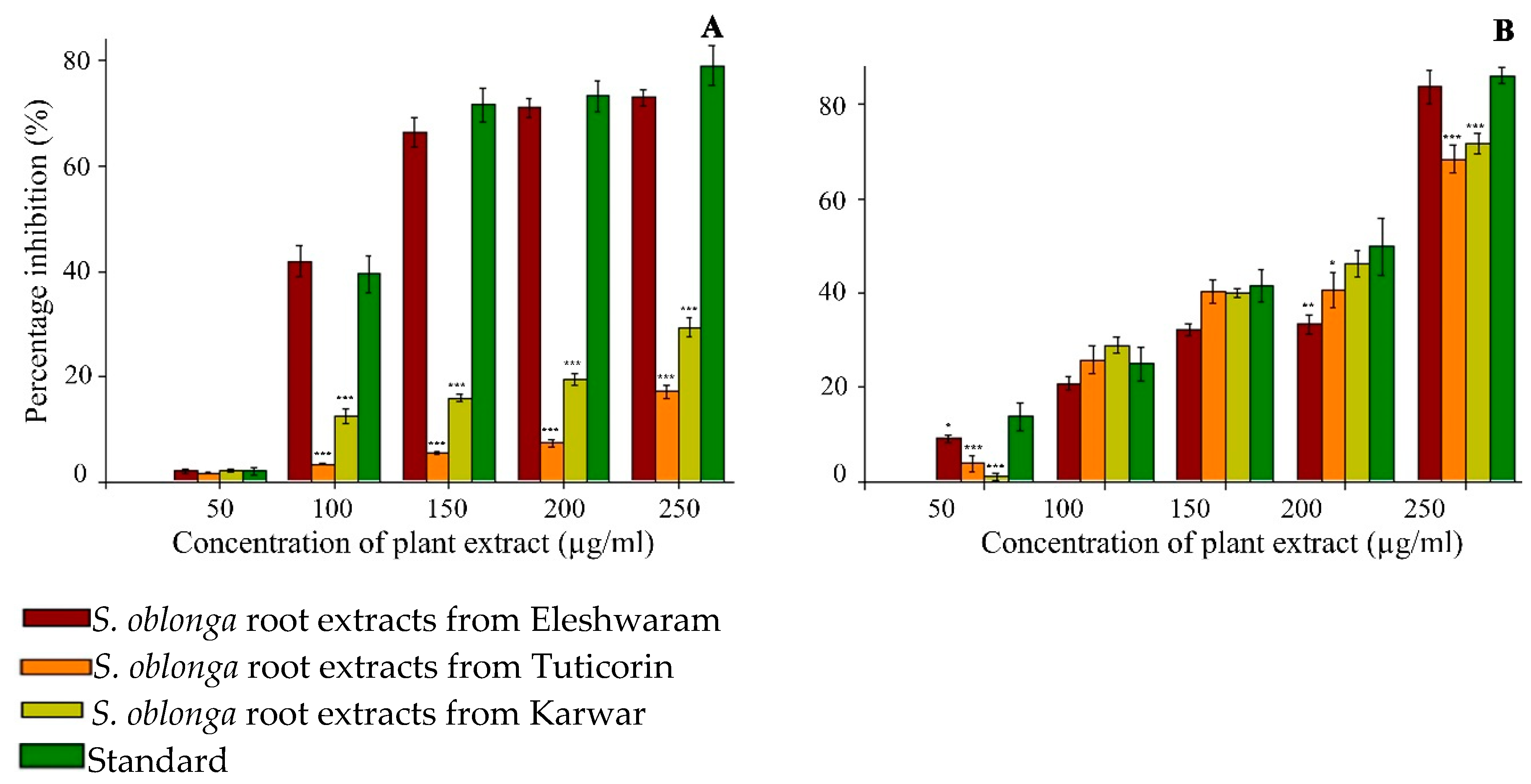
Figure 1. Alpha-glucosidase and α-amylase inhibition activities of root extracts of S. oblonga. (A) Alpha-glucosidase inhibition assay; (B) Alpha-amylase inhibition assay. Values are expressed s mean ± SD (n = 6). * p < 0.05 vs. Control; ** p < 0.01 vs. Control; *** p < 0.001 vs. Control.
Processes 08 00301 g001
Table 1. Phytochemical analysis of root extracts of Salacia oblonga.
PhytochemicalSREESRETSREK
Phenols+++++
Flavonoids+++
Alkaloids++++
Saponins+++++
Glycosides
Steroids++++
Terpenoids++++
SREE: S. oblonga root extracts from Eleshwaram, SRET: S. oblonga root extracts from Tuticorin, SREK: S. oblonga root extracts from Karwar. −: negative/absent; +: positive/present; ++: high concentration; +++: very high concentration.
Table 2. Total phenolic and flavonoid content in S. oblonga root extracts.
Plant Extract (250 µg/mL)Total Phenols (µg GAE/250 µg Extract)Total Flavonoids (µg QE/250 µg Extract)
SREE78.9 ± 2.183.2 ± 3.5
SRET42.1 ± 2.750.9 ± 3.3
SREK38.6 ± 2.748.8 ± 3.8
Values are expressed as mean ± SD (n = 6); GAE: gallic acid equivalents; QE: quercetin equivalents.
Table 3. Effects of root extracts of S. oblonga on α-glucosidase and α-amylase inhibition activities.
IC50 Values of Carbohydrate-Hydrolyzing Enzymes Inhibition Activities (µg/mL)
Plant Extractα-Glucosidase Inhibitionα-Amylase Inhibition
SREE147.3 ± 2.3 *193.7 ± 1.6 *
SRET764.5 ± 4.2 *199.4 ± 2.3 *
SREK427.0 ± 2.3 *189.6 ± 2.5 *
Acarbose141.7 ± 1.8169.9 ± 1.9
Values are presented as mean ± SD (n = 6); * p < 0.05 using one-way ANOVA (Dunnett’s Method) vs. standard; *** p < 0.001 vs. standard.
Table 4. Random and postprandial blood glucose levels in different groups treated with SREE.
Concentration of Random Blood Glucose (mg/dL)
0th day7th day14th day
NC69.7 ± 6.979.1 ± 10.380.3 ± 6.6
DC385.2 ± 17.4379.6 ± 12.2369.5 ± 40.9
Std302.4 ± 37.4 **94.6 ± 26.7 ***90.8 ± 29.9 ***
A315.5 ± 45.1 *169.2 ± 34.3 ***122.8 ± 24.4 ***
B346.2 ± 25.7 *166.8 ± 29.8 ***150.4 ± 31.6 ***
C337.5 ± 64.6123.9 ± 20.5 ***129.7 ± 29.3 ***
D364.4 ± 46.3107.1 ± 20.7 ***108.4 ± 11.0 ***
Concentration of Postprandial Blood Glucose (mg/dL)
0th day7th day14th day
NC106.5 ± 14.598.1 ± 16.596.6 ± 21.0
DC401.3 ± 15.2388.6 ± 10.0385.3 ± 21.6
Std325.4 ± 22.3 *101.6 ± 21.0 *102.0 ± 27.2 *
A324.1 ± 37.2 *182.4 ± 21.3 *136.7 ± 23.2 *
B349.8 ± 20.4 *176.5 ± 24.1 *157.2 ± 33.3 *
C356.8 ± 48.9 ***134.5 ± 15.0 *137.1 ± 22.5 *
D370.7 ± 40.0119.7 ± 13.5 *114.4 ± 10.3 *
NC: normal control; DC: diabetic control; Std: streptozotocin (STZ)-induced diabetic rats administered 2 mg/kg of acarbose; A: STZ-induced diabetic rats administered 50 mg/kg bw of SREE; B: STZ-induced diabetic rats administered 100 mg/kg bw of SREE; C: STZ-induced diabetic rats administered 200 mg/kg bw of SREE; D: STZ-induced diabetic rats administered 400 mg/kg bw of SREE; data expressed as mean ± SD (n = 6). * p < 0.05 using one-way ANOVA (Dunnett’s Method) vs. DC; ** indicates highly significance; *** p < 0.001 vs. DC.
Table 5. Concentration of total cholesterol, High-density lipoprotein (HDL–cholesterol), and triglycerides in various groups treated with SREE.
Total Cholesterol (mg/dL)HDL–Cholesterol (mg/dL)Triglycerides (mg/dL)
0th day7th day14th day0th day7th day14th day0th day7th day14th day
NC25.9 ± 3.442.1 ± 9.144.6 ± 11.028.9 ± 3.331.6 ± 4.833.3 ± 6.6132.8 ± 14.0124.3 ± 27.5105.1 ± 36.7
DC44.8 ± 8.461.7 ± 8.564.1 ± 9.038.5 ± 7.536.5 ± 11.834.2 ± 6.5363.2 ± 22.8183.7 ± 56.8162.9 ± 33.2
Std39.8 ± 19.642.5 ± 7.6 **36.3 ± 7.0 **29.3 ± 3.130.7 ± 3.2 *33.2 ± 4.3378.3 ± 32.8128.1 ± 39.5113.7 ± 28.2
A38.2 ± 15.666.6 ± 14.743.3 ± 10.4 *36.7 ± 14.131.22 ± 3.833.6 ± 5.4320.8 ± 22.2119.4 ± 37.5102.0 ± 33.2
B51.6 ± 4.043.1 ± 5.7 **41.5 ± 7.2 **32.1 ± 2.936.9 ± 13.233.8 ± 5.6389.1 ± 32.380.1 ± 16.4 *82.1 ± 16.4 *
C44.8 ± 6.539.6 ± 7.6 **38.6 ± 4.3 **30.4 ± 7.435.8 ± 10.333.9 ± 6.7 **331.2 ± 49.6164.5 ± 43.779.9 ± 6.9 *
D42.8 ± 17.044.0 ± 21.034.3 ± 9.2 **34.8 ± 5.737.6 ± 12.940.7 ± 7.9385.3 ± 22.1129.8 ± 31.075.9 ± 5.6 *
Data expressed as mean ± SD (n = 6); * p < 0.05 using one-way ANOVA (Dunnett’s Method) vs. DC; ** p < 0.01 vs. DC; *** p < 0.001 vs. DC.
Table 6. Concentration of creatinine and total bilirubin (mg/dL) in different groups treated with SREE.
Creatinine (mg/dL)Bilirubin (mg/dL)
0th day7th day14th day0th day7th day14th day
NC0.47 ± 0.10.54 ± 0.00.54 ± 0.10.51 ± 0.030.57 ± 0.00.75 ± 0.1 ***
DC0.42 ± 0.11.04 ± 0.20.84 ± 0.20.52 ± 0.041.74 ± 0.11.16 ± 0.1
Std0.44 ± 0.10.73 ± 0.1 *0.64 ± 0.1 *0.54 ± 0.060.84 ± 0.1 *0.71 ± 0.0 ***
A0.54 ± 0.10.59 ± 0.1 *0.55 ± 0.1 *0.51 ± 0.051.10 ± 0.10.84 ± 0.1 ***
B0.45 ± 0.00.81 ± 0.1 *0.61 ± 0.1 *0.53 ± 0.040.84 ± 0.1 **0.8 ± 0.0 ***
C0.47 ± 0.10.60 ± 0.1 *0.50 ± 0.1 *0.49 ± 0.030.9 ± 0.2 *0.73 ± 0.1***
D0.48 ± 0.10.58 ± 0.1 *0.55 ± 0.1 *0.49 ± 0.030.71 ± 0.1 ***0.62 ± 0.0 ***
Data expressed as mean ± SD (n = 6); * p < 0.05 using one-way ANOVA (Dunnett’s Method) vs. DC; ** p < 0.01 vs. DC; *** p < 0.001 vs. DC.