
Extraction of His Bundle Pacing Lead: More Difficult than Coronary Sinus Lead Extraction: An Analysis of 3897 Lead Extraction Procedures Including 27 His and 253 Coronary Sinus Lead Removals

 , ,
, ,  and
and
Abstract

1. What’s New?
2. Introduction
3. Goal of the Study
4. Methods
4.1. Study Population
4.2. Lead Extraction Procedure
4.2.1. Definitions
4.2.2. Techniques of HBP Lead Extraction
4.3. Dataset and Statistical Methods
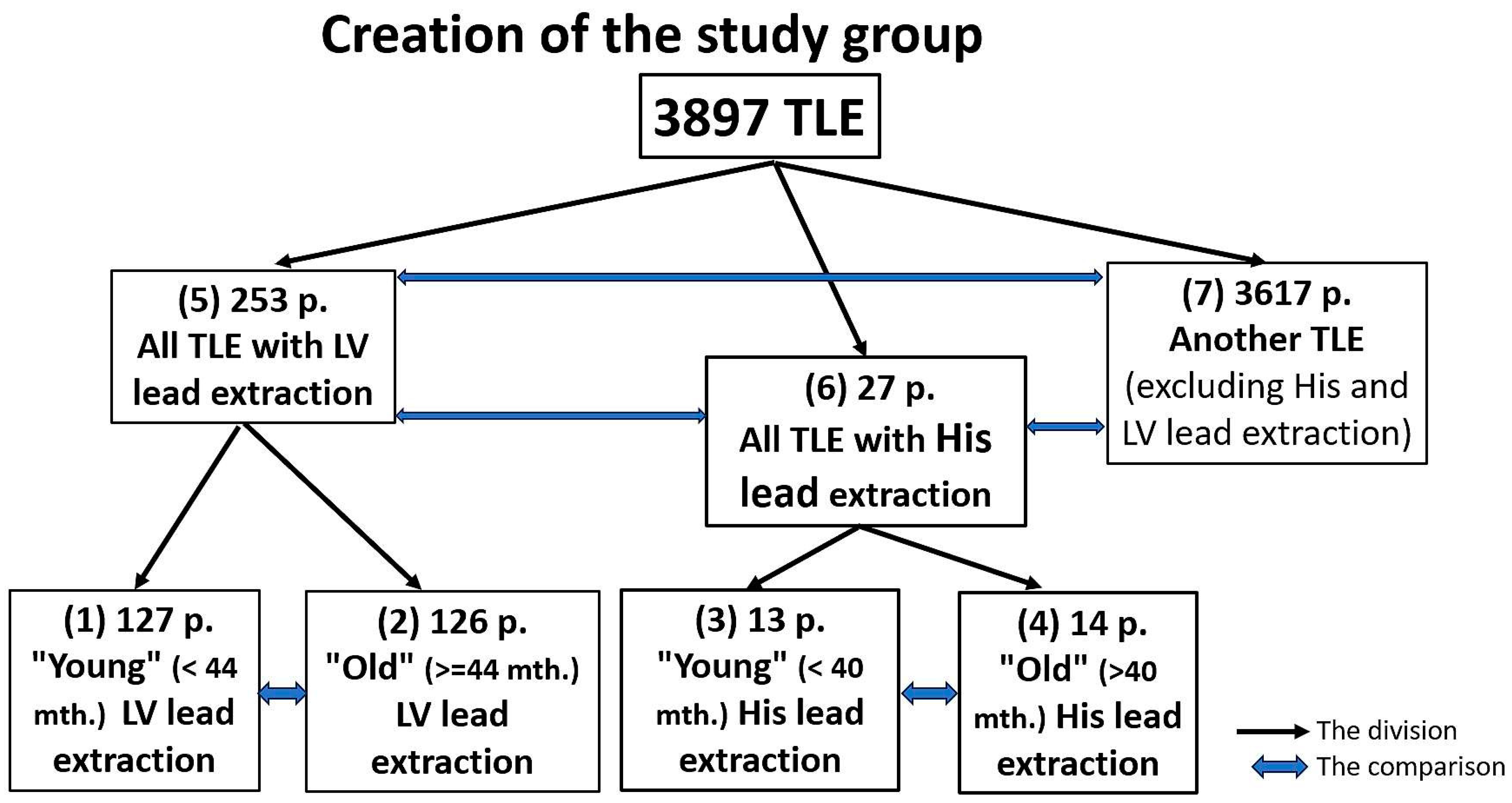
4.3.1. Creation of Subgroups for Analysis
4.3.2. Statistical Analysis
4.3.3. Approval of the Bioethics Committee
5. Results
6. Discussion
7. Conclusions
- HBP is used in CRT-D systems for resynchronisation of the failing heart in 33.33%.
- The extraction of HBP leads is most frequently performed for non-infectious indications (59.26%) and most often because of lead dysfunction (33.33%).
- The extraction of “old” (>40 months) HBP leads is longer (8.57 vs. 3.87 min) and more difficult than the removal of “young” leads due to unexpected procedure difficulties (14.29%) and the use of second line/advanced tools (28.57%), but it does not entail the risk of major complications and procedure-related death and is comparable to those encountered in the extraction of LVP leads of a similar age.
- Survival after lead extraction was comparable between HBP and LVP groups but shorter compared to patients who underwent the removal of other leads.
Study Limitations
Author Contributions
Funding
Institutional Review Board Statement
Informed Consent Statement
Data Availability Statement
Conflicts of Interest
References
- Deshmukh, P.; Casavant, D.A.; Romanyshyn, M.; Anderson, K. Permanent, direct His-bundle pacing: A novel approach to cardiac pacing in patients with normal His-Purkinje activation. Circulation 2000, 101, 869–877. [Google Scholar] [CrossRef] [PubMed]
- Sharma, P.S.; Dandamudi, G.; Herweg, B.; Wilson, D.; Singh, R.; Naperkowski, A.; Koneru, J.N.; Ellenbogen, K.A.; Vijayaraman, P. Permanent His-bundle pacing as an alternative to biventricular pacing for cardiac resynchronization therapy: A multicenter experience. Heart Rhythm 2018, 15, 413–420. [Google Scholar] [CrossRef] [PubMed]
- Abdelrahman, M.; Subzposh, F.A.; Beer, D.; Durr, B.; Naperkowski, A.; Sun, H.; Oren, J.W.; Dandamudi, G.; Vijayaraman, P. Clinical outcomes of his bundle pacing compared to right ventricular pacing. J. Am. Coll. Cardiol. 2018, 71, 2319–2330. [Google Scholar] [CrossRef] [PubMed]
- Jastrzębski, M.; Moskal, P.; Bednarek, A.; Kiełbasa, G.; Czarnecka, D. His-bundle pacing as a standard approach in patients with permanent atrial fibrillation and bradycardia. Pacing Clin. Electrophysiol. 2018, 41, 1508–1515. [Google Scholar] [CrossRef]
- Zanon, F.; Abdelrahman, M.; Marcantoni, L.; Naperkowski, A.; Subzposh, F.A.; Pastore, G.; Baracca, E.; Boaretto, G.; Raffagnato, P.; Tiribello, A.; et al. Long term performance and safety of His bundle pacing: A multicenter experience. J. Cardiovasc. Electrophysiol. 2019, 30, 1594–1601. [Google Scholar] [CrossRef] [PubMed]
- Keene, D.; Arnold, A.D.; Jastrzębski, M.; Burri, H.; Zweibel, S.; Crespo, E.; Chandrasekaran, B.; Bassi, S.; Joghetaei, N.; Swift, M.; et al. His bundle pacing, learning curve, procedure characteristics, safety, and feasibility: Insights from a large international bservational study. J. Cardiovasc. Electrophysiol. 2019, 30, 1984–1993. [Google Scholar] [CrossRef]
- De Leon, J.; Seow, S.C.; Boey, E.; Soh, R.; Tan, E.; Gan, H.H.; Lee, J.Y.; Teo, L.J.T.; Yeo, C.; Tan, V.H.; et al. Adopting permanent His bundle pacing: Learning curves and medium-term outcomes. Europace 2022, 24, 606–613. [Google Scholar] [CrossRef] [PubMed]
- Frausing, M.H.J.P.; Bæk, A.L.; Kristensen, J.; Gerdes, C.; Nielsen, J.C.; Kronborg, M.B. Long-term follow-up of selective and non-selective His bundle pacing leads in patients with atrioventricular block. J. Interv. Card. Electrophysiol. 2023; online ahead of print. [Google Scholar] [CrossRef]
- Jastrzębski, M.; Moskal, P.; Huybrechts, W.; Curila, K.; Sreekumar, P.; Rademakers, L.M.; Ponnusamy, S.S.; Herweg, B.; Sharma, P.S.; Bednarek, A.; et al. Left bundle branch-optimized cardiac resynchronization therapy (LOT-CRT): Results from an international LBBAP collaborative study group. Heart Rhythm 2022, 19, 13–21. [Google Scholar] [CrossRef]
- Zhang, W.; Huang, J.; Qi, Y.; Wang, F.; Guo, L.; Shi, X.; Wu, W.; Zhou, X.; Li, R. Cardiac resynchronization therapy by left bundle branch area pacing in patients with heart failure and left bundle branch block. Heart Rhythm 2019, 16, 1783–1790. [Google Scholar] [CrossRef]
- Vijayaraman, P.; Ponnusamy, S.; Cano, Ó.; Sharma, P.S.; Naperkowski, A.; Subsposh, F.A.; Moskal, P.; Bednarek, A.; Dal Forno, A.R.; Young, W.; et al. Left Bundle Branch Area Pacing for Cardiac Resynchronization Therapy: Results From the International LBBAP Collaborative Study Group. JACC Clin. Electrophysiol. 2021, 7, 135–147. [Google Scholar] [CrossRef]
- Jastrzębski, M.; Kiełbasa, G.; Cano, O.; Curila, K.; Heckman, L.; De Pooter, J.; Chovanec, M.; Rademakers, L.; Huybrechts, W.; Grieco, D.; et al. Left bundle branch area pacing outcomes: The multicentre European MELOS study. Eur. Heart J. 2022, 43, 4161–4173. [Google Scholar] [CrossRef] [PubMed]
- Keene, D.; Anselme, F.; Burri, H.; Pérez, Ó.C.; Čurila, K.; Derndorfer, M.; Foley, P.; Gellér, L.; Glikson, M.; Huybrechts, W.; et al. Conduction system pacing, a European survey: Insights from clinical practice. Europace 2023, 25, euad019. [Google Scholar] [CrossRef]
- Burri, H.; Jastrzebski, M.; Cano, Ó.; Čurila, K.; de Pooter, J.; Huang, W.; Israel, C.; Joza, J.; Romero, J.; Vernooy, K.; et al. EHRA clinical consensus statement on conduction system pacing implantation: Executive summary. Endorsed by the Asia-Pacific Heart Rhythm Society (APHRS), Canadian Heart Rhythm Society (CHRS) and Latin-American Heart Rhythm Society (LAHRS). Europace 2023, 25, 1537–1548. [Google Scholar] [CrossRef]
- Wijesuriya, N.; Elliott, M.K.; Mehta, V.; Behar, J.M.; Niederer, S.; Wilkoff, B.L.; Rinaldi, C.A. Transvenous lead extraction in conduction system pacing. Front. Physiol. 2022, 13, 993604. [Google Scholar] [CrossRef]
- Kusumoto, F.M.; Schoenfeld, M.H.; Wilkoff, B.; Berul, C.I.; Birgersdotter-Green, U.M.; Carrillo, R.; Cha, Y.M.; Clancy, J.; Deharo, J.C.; Ellenbogen, K.A.; et al. 2017 HRS expert consensus statement on cardiovascular implantable electronic device lead management and extraction. Heart Rhythm 2017, 14, e503–e551. [Google Scholar] [CrossRef]
- Garnreiter, J.; Whitaker, P.; Pilcher, T.; Etheridge, S.; Saarel, E. Lumenless pacing leads: Performance and extraction in pediatrics and congenital heart disease. Pacing Clin. Electrophysiol. 2015, 38, 42–47. [Google Scholar] [CrossRef] [PubMed]
- Perez, A.A.; Woo, F.W.; Tsang, D.C.; Carrillo, R.G. Transvenous lead extractions: Current approaches and future trends. Arrhythm. Electrophysiol. Rev. 2018, 7, 210–217. [Google Scholar] [CrossRef] [PubMed]
- Krainski, F.; Miller, J.P.; Pretorius, V.; Birgersdotter-Green, U. What goes in may need to come out: Considerations in the extraction of a lumenless, fixed-screw permanent pacemaker lead. Heart Rhythm. O2 2020, 1, 160–163. [Google Scholar] [CrossRef]
- Vijayaraman, P.; Subzposh, F.A.; Naperkowski, A. Extraction of the permanent His bundle pacing lead: Safety outcomes and feasibility of reimplantation. Heart Rhythm 2019, 16, 1196–1503. [Google Scholar] [CrossRef]
- Boczar, K.; Ząbek, A.; Dębski, M.; Gajek, J.; Lelakowski, J.; Małecka, B. Transvenous extraction of His bundle pacing lead: New challenge in the field of lead extraction. Cardiol. J. 2019, 26, 805. [Google Scholar] [CrossRef]
- Migliore, F.; Dall’Aglio, P.; Falzone, P.V.; Bertaglia, E.; Zanon, F. Extraction of a very old His bundle pacing lead: A safe and effective procedure? Pacing Clin. Electrophysiol. 2021, 44, 1464–1465. [Google Scholar] [CrossRef] [PubMed]
- Boczar, K.; Ząbek, A.; Golińska-Grzybała, K.; Gajek, J.; Holcman, K.; Kostkiewicz, M.; Lelakowski, J. Initial experience with transvenous lead extraction of His bundle pacing leads. Kardiol. Pol. 2023, 81, 1015–1017. [Google Scholar] [CrossRef] [PubMed]
- Vijayaraman, P. Extraction of left bundle branch pacing lead. JACC Clin. Electrophysiol. 2020, 6, 903–904. [Google Scholar] [CrossRef] [PubMed]
- Ponnusamy, S.S.; Vijayaraman, P. Late dislodgement of left bundle branch pacing lead and successful extraction. J. Cardiovasc. Electrophysiol. 2021, 32, 2346–2349. [Google Scholar] [CrossRef] [PubMed]
- Migliore, F.; Aruta, P.; Cecchetto, A.; Iliceto, S.; Gerosa, G.; Catanzariti, D. Extraction of left bundle branch pacing lead: A safe procedure? Europace 2021, 23, 1921. [Google Scholar] [CrossRef]
- le Polain de Waroux, J.B.; Wielandts, J.Y.; Gillis, K.; Hilfiker, G.; Sorgente, A.; Capulzini, L.; Geerts, B.; Knecht, S.; Duytschaever, M.; Tavernier, R. Repositioning and extraction of stylet-driven pacing leads with extendable helix used for left bundle branch area pacing. J. Cardiovasc. Electrophysiol. 2021, 32, 1464–1466. [Google Scholar] [CrossRef] [PubMed]
- Ayala Valani, L.; Dion, D.; Daneault, B.; Ayala-Paredes, F. Deep septal perforation during left bundle area pacing resulting in left ventricular capture and delayed systemic embolism. Can. J. Cardiol. 2023, 39, 1432–1435. [Google Scholar] [CrossRef] [PubMed]
- Agudo, C.A.; Jaén, E.G.; Sánchez, D.J.; Urda, V.C.; Ramos, J.T.; Lozano, I.F. Extraction of a fractured pacemaker lead in the left bundle branch area using a snare via a femoral approach. J. Interv. Card. Electrophysiol. 2023, 66, 239–240. [Google Scholar] [CrossRef] [PubMed]
- Bongiorni, M.G.; Burri, H.; Deharo, J.C.; Starck, C.; Kennergren, C.; Saghy, L.; Rao, A.; Tascini, C.; Lever, N.; Kutarski, A.; et al. 2018 EHRA expert consensus statement on lead extraction: Recommendations on definitions, endpoints, research trial design, and data collection requirements for clinical scientific studies and registries: Endorsed by APHRS/HRS/LAHRS. Europace 2018, 20, 1217. [Google Scholar] [CrossRef]
- Jacheć, W.; Polewczyk, A.; Polewczyk, M.; Tomasik, A.; Kutarski, A. Transvenous Lead Extraction SAFeTY Score for Risk Stratification and Proper Patient Selection for Removal Procedures Using Mechanical Tools. J. Clin. Med. 2020, 9, 361. [Google Scholar] [CrossRef] [PubMed]
- Sidhu, B.S.; Ayis, S.; Gould, J.; Elliott, M.K.; Mehta, V.; Kennergren, C.; Butter, C.; Deharo, J.-C.; Kutarski, A.; Maggioni, A.P.; et al. Risk stratification of patients undergoing transvenous lead extraction with the ELECTRa Registry Outcome Score (EROS): An ESC EHRA EORP European lead extraction ConTRolled ELECTRa registry analysis. Europace 2021, 23, 1462–1471. [Google Scholar] [CrossRef]
- Bontempi, L.; Curnis, A.; Della Bella, P.; Cerini, M.; Radinovic, A.; Inama, L.; Melillo, F.; Salghetti, F.; Marzi, A.; Gargaro, A.; et al. The MB score: A new risk stratification index to predict the need for advanced tools in lead extraction procedures. Europace 2020, 22, 613–621. [Google Scholar] [CrossRef]
- Bontempi, L.; Vassanelli, F.; Cerini, M.; Inama, L.; Salghetti, F.; Giacopelli, D.; Gargaro, A.; Raweh, A.; Curnis, A. Predicting the difficulty of a transvenous lead extraction procedure: Validation of the LED index. J. Cardiovasc. Electrophysiol. 2017, 28, 811–818. [Google Scholar] [CrossRef]
- Mazzone, P.; Tsiachris, D.; Marzi, A.; Ciconte, G.; Paglino, G.; Sora, N.; Sala, S.; Vergara, P.; Gulletta, S.; Della Bella, P. Predictors of advanced lead extraction based on a systematic stepwise approach: Results from a high-volume center. Pacing Clin. Electrophysiol. 2013, 36, 837–844. [Google Scholar] [CrossRef] [PubMed]
- Jacheć, W.; Nowosielecka, D.; Ziaja, B.; Polewczyk, A.; Kutarski, A. LECOM (Lead Extraction COMplexity): A New Scoring System for Predicting a Difficult Procedure. J. Clin. Med. 2023, 12, 7568. [Google Scholar] [CrossRef]
- Kutarski, A.; Jacheć, W.; Nowosielecka, D.; Polewczyk, A. Unexpected Procedure Difficulties Increasing the Complexity of Transvenous Lead Extraction: The Single Centre Experience with 3721 Procedures. J. Clin. Med. 2023, 15, 2811. [Google Scholar] [CrossRef]


| Patient-Related Risk Factors and Coexisting Indications for TLE (in 3897 Patients) | “Young” (≤40 mths) His Lead Extraction | “Old” (>40 mths) His Lead Extraction | “Young” (≤44 mths) LV Lead Extraction | “Old” (>44 mths) LV Lead Extraction | All TLEs with LV Lead Extraction | All TLEs with His Lead Extraction | TLE of Other than His and LV Leads |
|---|---|---|---|---|---|---|---|
| Group number/number of patients | Group 1 n = 13 mean ± SD n (%) | Group 2 n = 14 mean ± SD n (%) P: 1 vs. 2 | Group 3 n = 127 mean ± SD n (%) | Group 4 n = 126 mean ± SD n (%) P: 3 vs. 4 | Group 5 n = 253 mean ± SD n (%) | Group 6 n = 27 mean ± SD n (%) P: 5 vs. 6 | Group 7 n = 3617 mean ± SD n (%) 5 vs. 7 6 vs. 7 |
| Patient age during TLE [years] | 68.38 ± 9.24 | 72.50 ± 7.43 p = 0.131 | 68.43 ± 10.58 | 69.30 ± 9.34 p = 0.560 | 68.87 ± 9.97 | 70.52 ± 8.45 p = 0.603 | 65.84 ± 16.05 p = 0.370 p = 0.167 |
| Patient age at first system implantation [years] | 64.92 ± 9.72 | 63.64 ± 8.03 p = 0.961 | 64.57 ± 11.49 | 61.60 ± 9.58 p = 0.012 | 63.09 ± 10.66 | 64.26 ± 8.73 p = 0.971 | 57.13 ± 17.51 p = 0.092 p = 0.001 |
| Female | 3 (23.08) | 5 (35.71) p = 0.767 | 27 (21.26) | 26 (20.63) p = 0.974 | 53 (20.95) | 8 (29.63) p = 0.428 | 1424 (39.37) p = 0.001 p = 0.404 |
| Coronary heart disease | 10 (76.93) | 6 (42.86) p = 0.159 | 63 (49.67) | 55 (57.47) p = 0.410 | 118 (46.64) | 15 (55.56) p = 0.498 | 2041 (56.43) p = 0.003 p = 0.917 |
| NYHA FC III or IV | 4 (30.77) | 8 (57.14) p = 0.505 | 63 (49.61) | 49 (38.89) p = 0.112 | 112 (44.27) | 12 (44.44) p = 1.000 | 491 (13.58) p = 0.001 p = 0.001 |
| LVEF [%] | 43.31 ± 15.90 | 44.14 ± 11.03 p = 0.699 | 33.80 ± 13.95 | 33.05 ± 12.55 p = 0.929 | 33.43 ± 13.25 | 43.74 ± 13.33 p = 0.001 | 50.65 ± 14.82 p = 0.009 p = 0.001 |
| Charlson co-morbidity index [points] | 5.77 ± 3.42 | 6.57 ± 3.99 p = 0.980 | 6.54 ± 3.91 | 5.99 ± 3.53 p = 0.403 | 6.26 ± 3.73 | 6.19 ± 3.68 p = 0.972 | 4.81 ± 3.68 p = 0.030 p = 0.001 |
| PADIT score [points] | 4.69 ± 3.38 | 3.07 ± 2.84 p = 0.230 | 5.52 ± 2.81 | 5.98 ± 3.04 p = 0.154 | 5.75 ± 2.93 | 3.85 ± 3.16 p = 0.003 | 3.54 ± 2.82 p = 0.648 p = 0.001 |
| Main indications for TLE—(primary/predominant) | |||||||
| Infective endocarditis with or without pocket infection | 4 (30.77) | 5 (35.71) p = 0.892 | 60 (47.24) | 63 (50.00) p = 0.755 | 123 (48.62) | 9 (33.33) p = 0.190 | 722 (19.96) p = 0.001 p = 0.309 |
| Local (isolated) pocket infection | 2 (15.38) | 0 (0.00) p = 0.430 | 20 (15.75) | 14 (11.11) p = 0.370 | 34 (13.44) | 2 (7.41) p = 0.130 | 330 (9.12) p = 0.001 p = 0.835 |
| Mechanical lead damage (electrical failure) | 2 (15.38) | 2 (14.29) p = 0.644 | 9 (7.09) | 21 (16.67) p = 0.031 | 30 (11.86) | 4 (14.81) p = 0.891 | 1026 (28.37) p = 0.001 p = 0.179 |
| Lead dysfunction (exit/ entry block, dislodgement, perforation, extracardiac pacing) | 5 (38.46) | 4 (28.57) p = 0.892 | 35 (27.56) | 14 (11.11) p = 0.002 | 49 (19.37) | 9 (33.33) p = 0.164 | 809 (22.37) p = 0.025 p = 0.516 |
| Change of pacing mode/upgrading, downgrading and other non- infectious indications * | 0 (0.00) | 2 (14.29) p = 0.496 | 3 (2.36) | 14 (11.11) p = 0.012 | 17 (6.72) | 3 (11.11) p = 0.636 | 730 (20.18) p = 0.001 p = 0.201 |
| System-Related Risk Factors for TLE Complexity and Major Complications of TLE | “Young” (≤40 mths) His Lead Extraction | “Old” (>40 mths) His Lead Extraction | “Young” (≤44 mths) LV Lead Extraction | “Old” (>44 mths) LV Lead Extraction | All TLEs with LV Lead Extraction | All TLEs with His Lead Extraction | TLE of Other than His and LV Leads |
|---|---|---|---|---|---|---|---|
| Number of patients/group number | Group 1 n = 13 mean ± SD n (%) | Group 2 n = 14 mean ± SD n (%) P: 1 vs. 2 | Group 3 n = 127 mean ± SD n (%) | Group 4 n = 126 mean ± SD n (%) P: 3 vs. 4 | Group 5 n = 253 mean ± SD n (%) | Group 6 n = 27 mean ± SD n (%) P: 5 vs. 6 | Group 7 n = 3617 mean ± SD n (%) 5 vs. 7 6 vs. 7 |
| System and history of pacing | |||||||
| Type of CIED | |||||||
| Pacemaker | 7 (53.85) | 11 (78.57) p = 0.341 | 35 (27.56) | 38 (30.16) p = 0.571 | 73 (28.85) | 18 (66.67) p < 0.001 | 2678 (74.04) p = 0.001 p = 0.516 |
| Implantable cardioverter defibrillator | 0 (0.00) | 0 (0.00) NA | 0 (0.00) | 3 (2.38) p = 0.243 | 3 (1.19) | 0 (0.00) p = 0.679 | 845 (23.36) p = 0.001 p = 0.008 |
| Cardiac resynchronisation cardioverter defibrillator | 6 (46.15) | 3 (21.43) p = 0.341 | 92 (72.44) | 85 (67.46) p = 0.467 | 177 (69.96) | 9 (33.33) p = 0.001 | 94 (2.60) p < 0.001 p < 0.001 |
| Dwell time of oldest extracted lead [months] | 41.31 ± 35.00 | 105.93 ± 42.40 <0.001 | 47.87 ± 45.20 | 93.24 ± 44.32 p < 0.001 | 70.46 ± 50.13 | 74.81 ± 50.47 p = 0.576 | 105.19 ± 77.55 p = 0.059 p = 0.001 |
| Global lead dwell time before TLE [years] | 6.24 ± 5.33 | 22.01 ± 9.33 <0.001 | 8.79 ± 7.78 | 21.68 ± 9.90 p < 0.001 | 15.21 ± 10.98 | 14.41 ± 11.00 p = 0.680 | 15.51 ± 13.15 p = 0.833 p = 0.369 |
| HBP and LVP lead implant duration [months] | 13.15 ± 8.38 | 92.93 ± 38.22 <0.001 | 21.94 ± 16.09 | 78.45 ± 26.98 p < 0.001 | 50.20 ± 35.96 | 54.52 ± 49.12 p = 0.568 | NA NA NA |
| Abandoned lead before TLE | 1 (7.69) | 1 (7.14) p = 0.496 | 14 (11.02) | 20 (15.87) p = 0.344 | 34 (13.44) | 2 (7.41) p = 0.557 | 382 (10.56) p = 0.118 p = 0.828 |
| Number of leads in the heart before TLE before TLE | 2.38 ± 0.87 | 2.64 ± 0.50 p = 0.590 | 2.97 ± 0.62 | 3.10 ± 0.58 p = 0.175 | 3.04 ± 0.60 | 2.52 ± 0.70 p = 0.003 | 1.87 ± 0.68 p = 0.001 p = 0.001 |
| Number of procedures before lead extraction | 1.77 ± 0.60 | 2.08 ± 0.76 p = 0.294 | 1.91 ± 1.33 | 2.27 ± 0.98 p < 0.001 | 2.09 ± 1.18 | 1.92 ± 0.69 p = 0.830 | 1.84 ± 1.07 p = 0.146 p = 0.001 |
| Scores for evaluation of risk factors for major complications or procedure complexity | |||||||
| Safety score estimated risk of MC (risk of MC) [%] | 0.60 ± 0.50 | 1.94 ± 1.96 p = 0.019 | 0.98 ± 2.72 | 2.08 ± 2.22 p < 0.001 | 1.54 ± 2.53 | 1.33 ± 1.61 p = 0.837 | 1.73 ± 2.98 p = 0.682 p = 0.697 |
| 2 and 3 EROS score [risk of MC] | 4 (36.36) | 4 (50.00) p = 0.767 | 65 (51.18) | 68 (53.97) p = 0.853 | 133 (52.57) | 16 (59.26) p = 0.646 | 1399 (38.70) p = 0.001 p = 0.047 |
| MB score (need for advanced tools) [points] | 1.85 ± 1.46 | 3.21 ± 0.58 p = 0.024 | 2.16 ± 1.39 | 3.45 ± 0.93 p < 0.001 | 2.80 ± 1.35 | 2.56 ± 1.28 p = 0.260 | 2.58 ± 1.24 p = 0.782 p = 0.005 |
| LED index (predicted fluoroscopy time) [points] | 5.54 ± 3.10 | 10.79 ± 3.36 p = 0.001 | 6.28 ± 4.16 | 10.32 ± 3.68 p < 0.001 | 8.29 ± 4.42 | 8.26 ± 4.15 p = 0.961 | 10.23 ± 6.53 p = 0.193 p = 0.001 |
| Advanced LE (ALET—need for advanced TLE techniques) scale (3 and 4 points) | 6 (46.15) | 6 (42.86) p = 0.830 | 68 (53.64) | 96 (76.19) p < 0.001 | 164 (64.82) | 12 (44.44) p = 0.061 | 1217 (33.65) p = 0.001 p = 0.328 |
| LECOM score (expected procedure complexity) [%] | 16.19 ± 10.30 | 22.39 ± 15.70 p = 0.843 | 23.33 ± 18.49 | 30.88 ± 19.11 p < 0.001 | 27.09 ± 19.14 | 19.41 ± 13.50 p = 0.026 | 19.81 ± 18.29 p = 0.391 p = 0.001 |
| TLE-related potential risk factors for major complications and procedure complexity | |||||||
| Number of extracted leads per patient | 2.23 ± 0.83 | 2.14 ± 0.86 p = 0.393 | 2.46 ± 1.01 | 2.75 ± 0.88 p = 0.044 | 2.60 ± 0.96 | 2.19 ± 0.83 p = 0.012 | 1.59 ± 0.66 p = 0.004 p = 0.001 |
| Extraction of abandoned lead(s) (any) | 1 (7.69) | 1 (7.14) p = 0.496 | 13 (10.24) | 18 (14.29) p = 0.453 | 31 (12.25) | 2 (7.41) p = 0.668 | 351 (9.70) p = 0.228 p = 0.940 |
| Extraction of passive- fixation lead (excluding LV lead) | 7 (53.85) | 10 (71.43) p = 0.585 | 125 (98.43) | 124 (98.41) p = 0.620 | 249 (98.42) | 17 (62.96) p < 0.001 | 1985 (54.88) p = 0.001 p = 0.518 |
| Age of extracted HBP/LVP leads [months] | 13.15 ± 8.38 | 92.93 ± 38.22 p = 0.000 | 21.94 ± 16.09 | 78.45 ± 26.98 p < 0.001 | 50.20 ± 35.96 | 54.52 ± 49.12 p = 0.568 | NA NA NA |
| Oldest extracted lead per patient [months] | 41.31 ± 35.00 | 105.93 ± 42.40 <0.001 | 45.68 ± 44.85 | 90.74 ± 41.08 p < 0.001 | 68.12 ± 48.50 | 74.81 ± 50.47 p = 0.458 | 103.49 ± 76.54 p = 0.076 p = 0.001 |
| Patient-Related Risk Factors and Coexisting Indications for TLE (in 3897 Patients) | “Young” (≤40 mths) His Lead Extraction | “Old” (>40 mths) His Lead Extraction | “Young” (≤44 mths) LV Lead Extraction | “Old” (>44 mths) LV Lead Extraction | All TLEs with LV Lead Extraction | All TLEs with His Lead Extraction | TLE of Other than His and LV Leads |
|---|---|---|---|---|---|---|---|
| Group number/number of patients | Group 1 n = 13 mean ± SD n (%) | Group 2 n = 14 mean ± SD n (%) P: 1 vs. 2 | Group 3 n = 127 mean ± SD n (%) | Group 4 n = 126 mean ± SD n (%) P: 3 vs. 4 | Group 5 n = 253 mean ± SD n (%) | Group 6 n = 27 mean ± SD n (%) P: 5 vs. 6 | Group 7 n = 3617 mean ± SD n (%) 5 vs. 7 6 vs. 7 |
| Procedure duration (sheath-to-sheath) [minutes] | 7.85 ± 3.16 | 19.43 ± 17.82 p = 0.057 | 14.35 ± 17.97 | 25.38 ± 27.56 p < 0.001 | 19.85 ± 23.85 | 13.85 ± 14.08 p = 0.052 | 14.71 ± 23.25 p = 0.354 p = 0.001 |
| Mean time of single lead extraction (sheath-to-sheath/number of extracted leads) [minutes] | 3.87 ± 1.90 | 8.57 ± 6.97 p = 0.015 | 5.85 ± 5.94 | 9.31 ± 9.58 p < 0.001 | 7.57 ± 8.14 | 6.31 ± 5.63 p = 0.452 | 9.02 ± 13.00 p = 0.161 p = 0.043 |
| Time of single HBP/LVP lead extraction [minutes] | 2.15 ± 2.23 | 4.14 ± 1.96 p = 0.021 | 2.07 ± 2.24 | 4.05 ± 5.19 p < 0.001 | 3.05 ± 4.10 | 3.19 ± 2.29 p = 0.868 | NA NA NA |
| Removal of HBP/LVP lead with simple traction | 4 (30.77) | 0 (0.00) p = 0.025 | 63 (49.61) | 10 (7.94) p < 0.001 | 73 (28.97) | 4 (14.81) p = 0.185 | NA NA NA |
| Extraction of HBP/LVP lead using mechanical dilation | 9 (69.23) | 14 (100.0) p = 0.088 | 64 (50.39) | 116 (91.27) p < 0.001 | 179 (70.75) | 23 (85.19) p = 0.127 | NA NA NA |
| Number of unexpected procedure difficulties (“technical problems”) per patient | 0.00 ± 0.00 | 1.75 ± 0.96 p = 0.045 | 1.57 ± 0.85 | 1.50 ± 0.67 p = 0.049 | 1.52 ± 0.72 | 1.75 ± 0.96 p = 0.800 | 1.45 0.77 p = 0.753 p = 0.908 |
| CID-TLE score [points] | 0.00 | 0.93 ± 1.33 p = 0.004 | 0.43 ± 0.94 | 0.98 ± 1.23 p < 0.001 | 0.70 ± 1.12 | 0.48 ± 1.05 p = 0.620 | 0.54 ± 1.12 p = 0.773 p = 0.012 |
| CID-TLE score ≥ 2 points | 0 (0.00) | 5 (35.71) p = 0.059 | 22 (17.32) | 49 (38.89) p < 0.001 | 71 (28.06) | 5 (18.52) p = 0.405 | 662 (18.30) p = 0.001 p = 0.825 |
| Use of additional tools | |||||||
| Evolution (old and new) or TightRail | 0 (0.00) | 1 (7.14) p = 0.970 | 1 (0.79) | 2 (1.59) p = 1.000 | 3 (1.19) | 1 (3.70) p = 0.845 | 61 (1.69) p = 0.001 p = 0.952 |
| Metal sheath, lasso-basket catheter | 0 (0.00) | 3 (21.43) p = 0.247 | 8 (6.30) | 22 (17.46) p = 0.011 | 30 (11.86) | 3 (11.11) p = 0.842 | 499 (13.80) p = 0.440 p = 0.902 |
| TLE efficacy and complications | |||||||
| Major complications (any) | 0 (0.00) | 0 (0.00) NA | 1 (0.79) | 1 (0.79) p = 0.481 | 2 (0.79) | 0 (0.00) p = 0.460 | 78 (2.16) p = 0.212 p = 0.917 |
| Haemopericardium | 0 (0.00) | 0 (0.00) NA | 1 (0.79) | 0 (0.00) p = 0.997 | 1 (0.40) | 0 (0.00) p = 0.171 | 45 (1.24) p = 0.366 p = 0.771 |
| Rescue cardiac surgery | 0 (0.00) | 0 (0.00) NA | 1 (0.79) | 1 (0.79) p = 1.000 | 2 (0.79) | 0 (0.00) p = 0.460 | 38 (1.05) p = 0.941 p = 0.678 |
| Tricuspid valve damage during TLE (severe) | 0 (0.00) | 0 (0.00) NA | 0 (0.00) | 0 (0.00) NA | 0 (0.00) | 0 (0.00) NA | 25 (0.69) p = 0.357 p = 0.461 |
| Death, procedure-related (intra-, post-procedural) | 0 (0.00) | 0 (0.00) NA | 1 (0.79) | 0 (0.00) p = 0.997 | 1 (0.40) | 0 (0.00) p = 0.171 | 5 (0.14) p = 0.859 p = 0.847 |
| Partial radiographic success (retained tip or <4 cm lead fragment) | 0 (0.00) | 0 (0.00) NA | 0 (0.00) | 8 (6.35) p = 0.012 | 8 (3.16) | 0 (0.00) p = 0.742 | 145 (4.01) p = 0.616 p = 0.573 |
| Procedural success | 13 (100.0) | 14 (100.0) NA | 125 (98.43) | 118 (93.65) p = 0.104 | 243 (96.05) | 27 (100.0) p = 0.613 | 3444 (95.22) p = 0.654 p = 0.978 |
| Mortality after TLE (death for each time interval) | |||||||
| All model log rank p < 0.001 | |||||||
| Survivors during follow-up | 7 (53.85) | 8 (57.14) p = 0.830 | 47 (37.01) | 59 (46.83) p = 0.146 | 106 (41.90) | 15 (55.56) p = 0.247 | 2264 (62.59) p = 0.001 p = 0.580 |
| Deaths during follow-up | 6 (45.15) | 6 (42.86) p = 0.830 | 80 (62.99) | 67 (53.18) p = 0.146 | 147 (58.10) | 12 (44.44) p = 0.247 | 1353 (37.41) p = 0.001 p = 0.580 |
| Perioperative death (first two days) | 0 (0.00) | 0 (0.00) NA | 1 (0.79) | 0 (0.00) p = 0.997 | 1 (0.40) | 0 (0.00) p = 0.171 | 13 (0.36) p = 0.653 p = 0.191 |
| Intra-hospital death (3rd–30th day) | 2 (15.38) | 0 (0.00) p = 0.430 | 5 (3.94) | 4 (3.17) p = 0.990 | 9 (3.56) | 2 (7.41) p = 0.647 | 45 (1.24) p = 0.006 p = 0.049 |
| Log rank: p = 0.132 | Log rank: p = 0.522 | Log rank: p = 0.325 | Log rank: p = 0.005 p = 0.010 | ||||
| Death within the first year of follow-up (31–365 days) | 3 (23.08) | 1 (7.14) p = 0.534 | 23 (18.11) | 19 (15.08) p = 0.632 | 42 (16.60) | 4 (14.81) p = 0.972 | 217 (6.00) p = 0.001 p = 0.132 |
| One-year follow-up | Log rank: p = 0.022 | Log rank: p = 0.349 | Log rank: p = 0.695 | Log rank: p < 0.001 p = 0.002 | |||
| Death at 1 to 3 years of follow-up (366–1095 days) | 1 (7.69) | 3 (21.43) p = 0.644 | 22 (17.32) | 21 (16.67) p = 0.977 | 43 (17.00) | 4 (14.81) p = 0.986 | 306 (8.46) p = 0.000 p = 0.405 |
| Three-year follow-up | Log rank: p = 0.062 | Log rank: p = 0.374 | Log rank: p = 0.654 | Log rank: p < 0.001 p < 0.001 | |||
| Late death, at >3 years (>1095 days) of follow-up | 0 (0.00) | 2 (14.29) p = 0.496 | 29 (22.83) | 23 (18.25) p = 0.456 | 52 (20.55) | 2 (7.41) p = 0.165 | 772 (21.34) p = 0.828 p = 0.135 |
| Death during follow-up | Log rank: p = 0.099 | Log rank: p = 0.354 | Log rank: p = 0.780 | Log rank: p < 0.001 p < 0.001 | |||
| Follow-up [days] | 940.8 ± 1204 | 1334 ± 1088 p = 0.148 | 1458 ± 1376 | 1407 ± 1239 p = 0.882 | 1153 ± 1135 | 1432 ± 1307 p = 0.337 | 2122 ± 1469 p < 0.001 p < 0.001 |
| The length of the FU period for survivors [days] | 1303.86 ± 1451.79 | 1559 ± 1346 p = 1.000 | 2095.11 ± 1548.34 | 1973 ± 1361 p = 0.629 | 2027 ± 1441 | 1440 ± 1351 p = 0.462 | 2481 ± 1516.5 p < 0.001 p < 0.001 |
| The length of the FU period for non-survivors [days] | 284.3 ± 314.7 | 906.0 ± 646.0 p = 0.020 | 1072 ± 1137 | 929.0 ± 919.8 p = 0.832 | 1007 ± 1043 | 595.2 ± 583.2 p = 0.243 | 1578 ± 1251.8 p < 0.001 p = 0.003 |
| His Pacing Lead Extraction Group Potential Risk Factors for Shorter Survival | Survivors n = 15 | Non- Survivors n = 12 | p Mann–Whitney U Test Chi2 |
|---|---|---|---|
| Female | 7 (46.67) | 2 (16.67) | 0.218 |
| Patient age [years] | 71.25 ± 7.82 | 69.83 ± 10.14 | 0.707 |
| Left ventricular ejection fraction [%] | 50.18 ± 12.55 | 40.33 ± 13.71 | 0.117 |
| Congestive heart failure | 5 (33.33) | 7 (58.33) | 0.553 |
| CHA2DS2–VASc score [points] | 3.50 ± 1.51 | 3.25 ± 1.29 | 0.729 |
| Charlson co-morbidity index [points] | 6.33 ± 4.03 | 5.25 ± 3.31 | 0.817 |
| PADIT [points] | 3.33 ± 3.03 | 4.33 ± 3.55 | 0.525 |
| Oldest extracted lead per patient [years] | 7.65 ± 4.26 | 4.40 ± 3.18 | 0.057 |
| Infectious indications for TLE | 5 (3.33) | 6 (50.00) | 0.630 |
| ICD lead presence before TLE | 1 (6.67) | 6 (60.00) | 0.035 |
Disclaimer/Publisher’s Note: The statements, opinions and data contained in all publications are solely those of the individual author(s) and contributor(s) and not of MDPI and/or the editor(s). MDPI and/or the editor(s) disclaim responsibility for any injury to people or property resulting from any ideas, methods, instructions or products referred to in the content. |
© 2024 by the authors. Licensee MDPI, Basel, Switzerland. This article is an open access article distributed under the terms and conditions of the Creative Commons Attribution (CC BY) license (https://creativecommons.org/licenses/by/4.0/).
Share and Cite
Stefańczyk, P.; Jacheć, W.; Kutarski, A.; Dąbrowski, P.; Głowniak, A.; Nowosielecka, D. Extraction of His Bundle Pacing Lead: More Difficult than Coronary Sinus Lead Extraction: An Analysis of 3897 Lead Extraction Procedures Including 27 His and 253 Coronary Sinus Lead Removals. Biomedicines 2024, 12, 1154. https://doi.org/10.3390/biomedicines12061154
Stefańczyk P, Jacheć W, Kutarski A, Dąbrowski P, Głowniak A, Nowosielecka D. Extraction of His Bundle Pacing Lead: More Difficult than Coronary Sinus Lead Extraction: An Analysis of 3897 Lead Extraction Procedures Including 27 His and 253 Coronary Sinus Lead Removals. Biomedicines. 2024; 12(6):1154. https://doi.org/10.3390/biomedicines12061154
Chicago/Turabian StyleStefańczyk, Paweł, Wojciech Jacheć, Andrzej Kutarski, Paweł Dąbrowski, Andrzej Głowniak, and Dorota Nowosielecka. 2024. "Extraction of His Bundle Pacing Lead: More Difficult than Coronary Sinus Lead Extraction: An Analysis of 3897 Lead Extraction Procedures Including 27 His and 253 Coronary Sinus Lead Removals" Biomedicines 12, no. 6: 1154. https://doi.org/10.3390/biomedicines12061154
APA StyleStefańczyk, P., Jacheć, W., Kutarski, A., Dąbrowski, P., Głowniak, A., & Nowosielecka, D. (2024). Extraction of His Bundle Pacing Lead: More Difficult than Coronary Sinus Lead Extraction: An Analysis of 3897 Lead Extraction Procedures Including 27 His and 253 Coronary Sinus Lead Removals. Biomedicines, 12(6), 1154. https://doi.org/10.3390/biomedicines12061154