
Hyaluronic Acid Combined with Ozone in Dental Practice


Abstract
1. Introduction
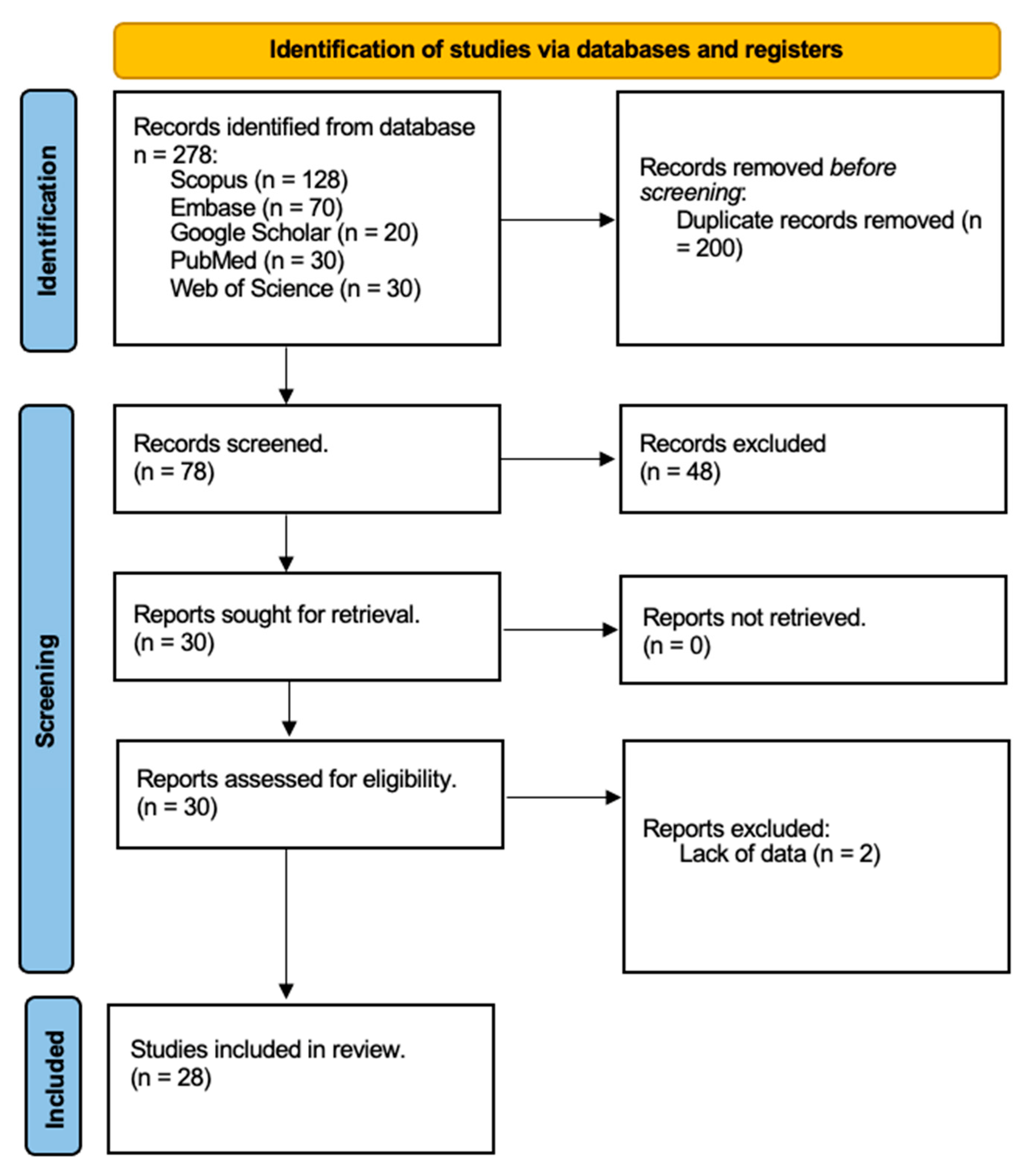
2. Materials and Methods
3. Results
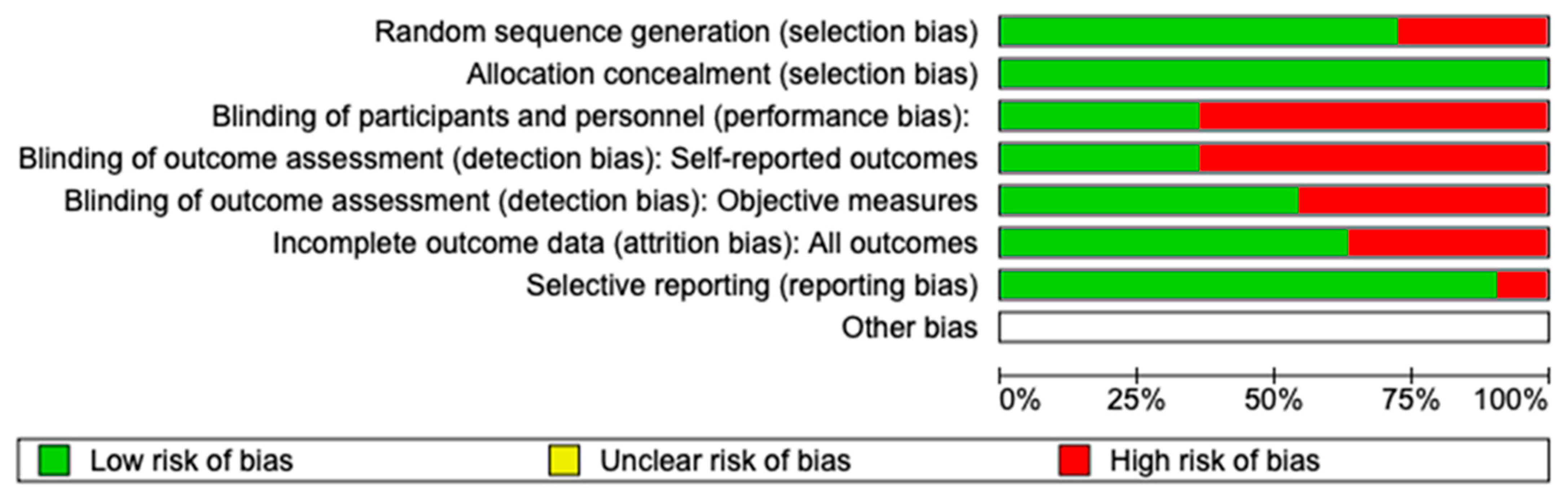
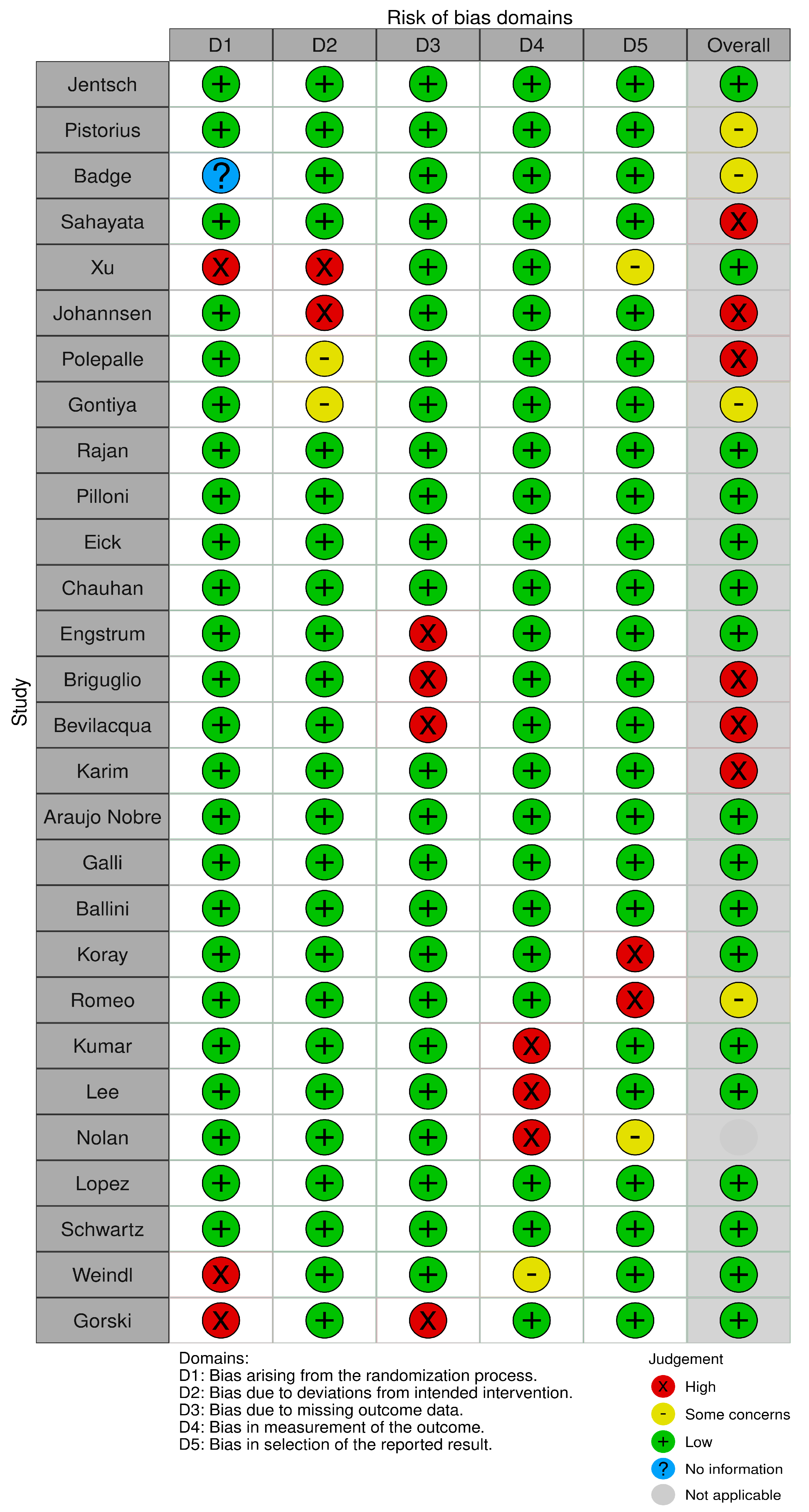
3.1. Quality Assessment and Risk of Bias
3.2. In the Battle Against Gingivitis
3.3. Chronic Periodontitis: A New Frontier
3.4. Surgical Frontiers and Implant Surgery
3.5. The Healing Touch on Oral Ulcers
4. Discussion
5. Conclusions
Author Contributions
Funding
Institutional Review Board Statement
Informed Consent Statement
Data Availability Statement
Conflicts of Interest
References
- Stern, R.; Asari, A.A.; Sugahara, K.N. Hyaluronan fragments: An information-rich system. Eur. J. Cell Biol. 2006, 85, 699–715. [Google Scholar] [CrossRef] [PubMed]
- Jentsch, H.; Pomowski, R.; Kundt, G.; Göcke, R. Treatment of gingivitis with hyaluronan. J. Clin. Periodontol. 2003, 30, 159–164. [Google Scholar] [CrossRef] [PubMed]
- Samiraninezhad, N.; Asadi, K.; Rezazadeh, H.; Gholami, A. Using chitosan, hyaluronic acid, alginate, and gelatin-based smart biological hydrogels for drug delivery in oral mucosal lesions: A review. Int. J. Biol. Macromol. 2023, 252, 126573. [Google Scholar] [CrossRef] [PubMed]
- Ge, W.; Gao, Y.; He, L.; Zeng, Y.; Liu, J.; Yu, Y.; Xie, X.; Xu, R.-A. Combination therapy using multifunctional dissolvable hyaluronic acid microneedles for oral ulcers. Int. J. Biol. Macromol. 2023, 251, 126333. [Google Scholar] [CrossRef]
- Casale, M.; Moffa, A.; Sabatino, L.; Pace, A.; Oliveto, G.; Vitali, M.; Baptista, P.; Salvinelli, F. Hyaluronic Acid: Perspectives in Upper Aero-Digestive Tract. A Systematic Review. PLoS ONE 2015, 10, e0130637. [Google Scholar] [CrossRef]
- Nolan, A.; Baillie, C.; Badminton, J.; Rudralingham, M.; Seymour, R.A. The efficacy of topical hyaluronic acid in the management of recurrent aphthous ulceration. J. Oral Pathol. Med. 2006, 35, 461–465. [Google Scholar] [CrossRef]
- Briguglio, F.; Briguglio, E.; Briguglio, R.; Cafiero, C.; Isola, G. Treatment of infrabony periodontal defects using a resorbable biopolymer of hyaluronic acid: A randomized clinical trial. Quintessence Int. 2013, 44, 231–240. [Google Scholar] [CrossRef]
- Weindl, G.; Schaller, M.; Schäfer-Korting, M.; Korting, H.C. Hyaluronic Acid in the Treatment and Prevention of Skin Diseases: Molecular Biological, Pharmaceutical and Clinical Aspects. Ski. Pharmacol. Physiol. 2004, 17, 207–213. [Google Scholar] [CrossRef]
- Yudaev, P.A.; Chistyakov, E.M. Progress in dental materials: Application of natural ingredients. Russ. Chem. Rev. 2024, 93, RCR5108. [Google Scholar] [CrossRef]
- Zalega, M.; Bociong, K. Antibacterial Agents Used in Modifications of Dental Resin Composites: A Systematic Review. Appl. Sci. 2024, 14, 3710. [Google Scholar] [CrossRef]
- Sasaki, T.; Watanabe, C. Stimulation of osteoinduction in bone wound healing by high-molecular hyaluronic acid. Bone 1995, 16, 9–15. [Google Scholar] [CrossRef] [PubMed]
- Mehta, V.; Kaçani, G.; Moaleem, M.M.A.; Almohammadi, A.A.; Alwafi, M.M.; Mulla, A.K.; Alharbi, S.O.; Aljayyar, A.W.; Qeli, E.; Toti, Ç.; et al. Hyaluronic Acid: A New Approach for the Treatment of Gingival Recession—A Systematic Review. Int. J. Environ. Res. Public Health 2022, 19, 14330. [Google Scholar] [CrossRef] [PubMed]
- Gao, Y.; Wang, R.; Zhang, L.; Fan, Y.; Luan, J.; Liu, Z.; Yuan, C. Oral administration of hyaluronic acid to improve skin conditions via a randomized double-blind clinical test. Ski. Res. Technol. 2023, 29, e13531. [Google Scholar] [CrossRef] [PubMed]
- Polepalle, T.; Srinivas, M.; Swamy, N.; Aluru, S.; Chakrapani, S.; Chowdary, B.A. Local delivery of hyaluronan 0.8% as an adjunct to scaling and root planing in the treatment of chronic periodontitis: A clinical and microbiological study. J. Indian Soc. Periodontol. 2015, 19, 37–42. [Google Scholar] [CrossRef]
- Oldoini, G.; Frabattista, G.R.; Saragoni, M.; Cosola, S.; Giammarinaro, E.; Genovesi, A.M.; Marconcini, S. Ozone Therapy for Oral Palatal Ulcer in a Leukaemic Patient. Eur. J. Case Rep. Intern. Med. 2020, 7, 001406. [Google Scholar] [CrossRef]
- Pistorius, A.; Martin, M.; Willershausen, B.; Rockmann, P. The clinical application of hyaluronic acid in gingivitis therapy. Quintessence Int. 2005, 36, 531–538. [Google Scholar] [PubMed]
- Sahayata, V.N.; Bhavsar, N.V.; Brahmbhatt, N.A. An evaluation of 0.2% hyaluronic acid gel (Gengigel®) in the treatment of gingivitis: A clinical & microbiological study. Oral Health Dent Manag. 2014, 13, 779–785. [Google Scholar]
- Bagde, H.; Pawar, S.K.; Vasisth, D.; Vadvadgi, V.H.; Laddha, R.B.; Wagh, P.P. Comparison of Amnion Membrane and Hyaluronic Acid in Gingival Recession Coverage and Gain in Clinical Attachment Level following Coronally Advanced Flap Procedure—A Clinical Study. J. Pharm. Bioallied Sci. 2023, 15, S1104–S1107. [Google Scholar] [CrossRef]
- Xu, Y.; Höfling, K.; Fimmers, R.; Frentzen, M.; Jervøe-Storm, P.M. Clinical and Microbiological Effects of Topical Subgingival Application of Hyaluronic Acid Gel Adjunctive to Scaling and Root Planing in the Treatment of Chronic Periodontitis. J. Periodontol. 2004, 75, 1114–1118. [Google Scholar] [CrossRef]
- Johannsen, A.; Tellefsen, M.; Wikesjö, U.; Johannsen, G. Local Delivery of Hyaluronan as an Adjunct to Scaling and Root Planing in the Treatment of Chronic Periodontitis. J. Periodontol. 2009, 80, 1493–1497. [Google Scholar] [CrossRef]
- Polepalle, T.; Kumar, R.; Srinivas, M.; Pai, J.; Suragimath, G.; Prasad, K. Efficacy of hyaluronic acid (hyaluronan) in root coverage procedures as an adjunct to coronally advanced flap in Millers Class I recession: A clinical study. J. Indian Soc. Periodontol. 2014, 18, 746–750. [Google Scholar] [CrossRef] [PubMed]
- Gontiya, G.; Galgali, S.R. Effect of hyaluronan on periodontitis: A clinical and histological study. J. Indian Soc. Periodontol. 2012, 16, 184–192. [Google Scholar] [CrossRef] [PubMed]
- Rajan, P. Hyaluronic Acid as an Adjunct to Scaling and Root Planing in Chronic Periodontitis. A Randomized Clinical Trail. J. Clin. Diagn. Res. 2014, 8, ZC11–ZC14. [Google Scholar] [CrossRef] [PubMed]
- Cairo, F.; Cortellini, P.; Pilloni, A.; Nieri, M.; Cincinelli, S.; Amunni, F.; Pagavino, G.; Tonetti, M.S. Clinical efficacy of coronally advanced flap with or without connective tissue graft for the treatment of multiple adjacent gingival recessions in the aesthetic area: A randomized controlled clinical trial. J. Clin. Periodontol. 2016, 43, 849–856. [Google Scholar] [CrossRef]
- Eick, S.; Renatus, A.; Heinicke, M.; Pfister, W.; Stratul, S.; Jentsch, H. Hyaluronic Acid as an Adjunct After Scaling and Root Planing: A Prospective Randomized Clinical Trial. J. Periodontol. 2013, 84, 941–949. [Google Scholar] [CrossRef]
- Bains, V.; Chauhan, A.; Gupta, V.; Singh, G.; Patil, S. Comparative analysis of hyaluronan gel and xanthan-based chlorhexidine gel, as adjunct to scaling and root planing with scaling and root planing alone in the treatment of chronic periodontitis: A preliminary study. Contemp. Clin. Dent. 2013, 4, 54–61. [Google Scholar] [CrossRef]
- Engstrüm, P.; Shi, X.; Tronje, G.; Larsson, A.; Welander, U.; Frithiof, L.; Engstrom, G.N. The Effect of Hyaluronan on Bone and Soft Tissue and Immune Response in Wound Healing. J. Periodontol. 2001, 72, 1192–1200. [Google Scholar] [CrossRef]
- Bevilacqua, L.; Eriani, J.; Serroni, I.; Liani, G.; Borelli, V.; Castronovo, G.; Di Lenarda, R. Effectiveness of adjunctive subgingival administration of amino acids and sodium hyaluronate gel on clinical and immunological parameters in the treatment of chronic periodontitis. Ann. Stomatol. 2012, 3, 75–81. [Google Scholar]
- El-Sayed, K.M.F.; Dahaba, M.A.; Aboul-Ela, S.; Darhous, M.S. Local application of hyaluronan gel in conjunction with periodontal surgery: A randomized controlled trial. Clin. Oral Investig. 2012, 16, 1229–1236. [Google Scholar] [CrossRef]
- Nobre, M.D.A.; Cintra, N.; Maló, P. Peri-implant maintenance of immediate function implants: A pilot study comparing hyaluronic acid and chlorhexidine. Int. J. Dent. Hyg. 2007, 5, 87–94. [Google Scholar] [CrossRef]
- Galli, F.; Zuffetti, F.; Capelli, M.; Fumagalli, L.; Parenti, A.; Testori, T.; Esposito, M. Hyaluronic acid to improve healing of surgical incisions in the oral cavity: A pilot multicentre placebo-controlled randomised clinical trial. Eur. J. Oral Implant. 2008, 1, 199–206. [Google Scholar]
- Ballini, A.; Cantore, S.; Capodiferro, S.; Grassi, F.R. Esterified Hyaluronic Acid and Autologous Bone in the Surgical Correction of the Infra-Bone Defects. Int. J. Med. Sci. 2009, 6, 65–71. [Google Scholar] [CrossRef] [PubMed]
- Koray, M.; Ofluoglu, D.; Onal, E.; Ozgul, M.; Ersev, H.; Yaltirik, M.; Tanyeri, H. Efficacy of hyaluronic acid spray on swelling, pain, and trismus after surgical extraction of impacted mandibular third molars. Int. J. Oral Maxillofac. Surg. 2014, 43, 1399–1403. [Google Scholar] [CrossRef]
- Romeo, U.; Libotte, F.; Palaia, G.; Galanakis, A.; Gaimari, G.; Tenore, G.; Del Vecchio, A.; Polimeni, A. Oral Soft Tissue Wound Healing After Laser Surgery With or Without a Pool of Amino Acids and Sodium Hyaluronate: A Randomized Clinical Study. Photomed. Laser Surg. 2014, 32, 10–16. [Google Scholar] [CrossRef]
- Lee, J.; Jung, J.; Bang, D. The efficacy of topical 0.2% hyaluronic acid gel on recurrent oral ulcers: Comparison between recurrent aphthous ulcers and the oral ulcers of Behçet’s disease. J. Eur. Acad. Dermatol. Venereol. 2008, 22, 590–595. [Google Scholar] [CrossRef]
- Lopez, M.A.; Casale, M.; Candotto, V.; Papalia, R.; Bressi, F.; Carinci, F. The use of hyaluronic acid as a support of two different micronized biomaterials in crestal sinus lift procedures. A report on two case studies with volume comparison. J. Biol. Regul. Homeost. Agents 2017, 31 (Suppl. S2), 129–138. [Google Scholar]
- Schwartz, Z.; Goldstein, M.; Raviv, E.; Hirsch, A.; Ranly, D.M.; Boyan, B.D. Clinical evaluation of demineralized bone allograft in a hyaluronic acid carrier for sinus lift augmentation in humans: A computed tomography and histomorphometric study. Clin. Oral Implant. Res. 2007, 18, 204–211. [Google Scholar] [CrossRef]
- Górski, B.; Skierska, I.; Szerszeń, M.; Mańka-Malara, K. Tunnel technique with cross-linked hyaluronic acid in addition to subepithelial connective tissue graft, compared with connective tissue graft alone, for the treatment of multiple gingival recessions: 6-month outcomes of a randomized clinical trial. Clin. Oral Investig. 2023, 27, 2395–2406. [Google Scholar] [CrossRef]
- Carlstedt, C.A. Mechanical and chemical factors in tendon healing. Effects of indomethacin and surgery in the rabbit. Acta Orthop. 1987, 58, 1–75. [Google Scholar] [CrossRef]
- Galli, C.; Passeri, G.; Macaluso, G.M. FoxOs, Wnts and oxidative stress-induced bone loss: New players in the periodontitis arena? J. Periodontal Res. 2011, 46, 397–406. [Google Scholar] [CrossRef]
- Rosa, A.; Pujia, A.M.; Arcuri, C. Complete Full Arch Supported by Short Implant (<8 mm) in Edentulous Jaw: A Systematic Review. Appl. Sci. 2023, 13, 7162. [Google Scholar] [CrossRef]
- Pardo, A.; Fiorini, V.; Zangani, A.; Faccioni, P.; Signoriello, A.; Albanese, M.; Lombardo, G. Topical Agents in Biofilm Disaggregation: A Systematic Review and Meta-Analysis. J. Clin. Med. 2024, 13, 2179. [Google Scholar] [CrossRef] [PubMed]
- Cankaya, Z.T.; Gurbuz, S.; Bakirarar, B.; Unsal, B.; Kurtis, B. Evaluation of the effect of the application of hyaluronic acid following laser-assisted frenectomy: An examiner-blind, randomized, controlled clinical study. Quintessence Int. 2020, 51, 188–201. [Google Scholar] [CrossRef]
- Ronsivalle, V.M.; Cicciù, M.P.; Fiorillo, L.M. The Effects of a Cool Saline Solution Irrigation on Mandibular Third Molar Extraction Site: A Postoperative Split-Mouth Evaluation. J. Craniofacial Surg. 2024, 35, 1219–1224. [Google Scholar] [CrossRef]
- Franco, R.; Rosa, A.; Lupi, E.; Capogreco, M. The Influence of Dental Implant Roughness on Biofilm Formation: A Comprehensive Strategy. Dent. Hypotheses 2023, 14, 90–92. [Google Scholar] [CrossRef]
- Rosa, A.; Miranda, M.; Franco, R.; Guarino, M.G.; Barlattani, A.; Bollero, P. Experimental protocol of dental procedures In patients with hereditary angioedema: The role of anxiety and the use of nitrogen oxide. Oral Implantol. 2016, 9, 49–53. [Google Scholar] [CrossRef]
- Lorenzi, C.; Leggeri, A.; Cammarota, I.; Carosi, P.; Mazzetti, V.; Arcuri, C. Hyaluronic Acid in Bone Regeneration: Systematic Review and Meta-Analysis. Dent. J. 2024, 12, 263. [Google Scholar] [CrossRef]
- Boccalari, E.; Khijmatgar, S.; Occhipinti, C.; Del Fabbro, M.; Inchingolo, F.; Tartaglia, G.M. Effect of hydrogen peroxide and hyaluronic acid in mouth rinse after third molar extraction: A triple-blind parallel randomized controlled clinical trial. Eur. Rev. Med. Pharmacol. Sci. 2024, 28, 3946–3957. [Google Scholar] [CrossRef]
- Rosa, A.; Pujia, A.M.; Arcuri, C. Investigation of alveolar osteitis and the effectiveness of laser treatment: A unified Meta-analysis and review of the literature. BMC Oral Health 2024, 24, 1–7. [Google Scholar] [CrossRef]
- Rosa, A.; Fiorillo, L.; D’Amico, C.; Pujia, A.; Heboyan, A.; Cervino, G.; Ronsivalle, V.; Claudio, A. Local anesthetic-induced allergic reactions in dentistry: Current perspectives and key considerations. Bull. Stomatol. Maxillofac. Surg. 2024, 20, 93–102. [Google Scholar] [CrossRef]

| Criteria | Inclusion | Exclusion |
|---|---|---|
| Language | Studies published in English | Studies published in other languages |
| Study Design | Human-controlled trials (randomized controlled trials, clinal trails) | Literature reviews, technical notes, letters to editors, instructional courses |
| Population | Human subjects with dental conditions | Animal studies, in vitro research |
| Intervention | Use of hyaluronic acid (HA) and ozone in treating gingivitis, ulcers, wounds, gingival recession | Studies not involving HA or ozone in dental treatment |
| Outcome Measures | Histological or clinical evaluations of the impact of hyaluronic acid in dental disease contexts | Studies without clinical or histological evaluations |
| Publication Type | Primary research articles | Non-primary research (e.g., opinion pieces, conference abstracts) |
| Date Restrictions | No restriction on publication year | None |
| Study Topic | Focus on HA and ozone’s effectiveness in oral health treatments | Focus on unrelated medical conditions or treatments |
| PICO Component | |
|---|---|
| Participants | Healthy participants with no restrictions on age and sex with gingival recession, periodontitis, oral ulcers, surgery wounds |
| Intervention | Application of HA combined with OT in conjunction with surgical procedures |
| Comparison | The same surgical procedures without HA and OT or substitute |
| Outcomes | Pathology reduction |
| Authors | Mean Age | Ha Group | Control Group | Type of Treatment | Parameters Evaluated | Clinical Evidence |
|---|---|---|---|---|---|---|
| Jentsch [2] | 50 male (17 ± 39 y) | 25 with use of HA-OT | 25 with placebo | Gel on gingivitis. | The study evaluated oral health through clinical indices (approximal plaque index, Turesky plaque index, papillary bleeding index) and crevicular fluid markers (peroxidase, lysozyme) initially and after 4, 7, 14, and 21 days. | The test group exhibited notable enhancements in plaque indices from day 4 and in PBI from day 7, outperforming the placebo group. |
| Pistorius [16] | 60 mixed (32 ± 41 y) | 40 with use of HA-OT | 20 with reduced use of HA | Spray on gingivitis. | Clinical measurements including DMF-T index, API, sulcus bleeding index, PBI, and gingival crevicular fluid were recorded at the start, then after 3 and 7 days. | Clinical parameters were assessed initially, and then at 3 and 7 days. The HA group saw decreases in sulcus bleeding index at both time points, with significant drops in PBI values and gingival crevicular fluid. |
| Badge [18] | 21 mixed (22 ± 34 y) | 11 with use of HA-OT | 10 with placebo | Gel in periodontal pocket. | A gingival biopsy for histopathological and immunohistochemical analysis, focusing on Ki-67 expression and inflammatory infiltrate evaluation, was conducted 30 days post-treatment. | Treatment with HA gel notably decreased the proliferation index of gingival epithelium and fibroblast cells. |
| Sahayata [17] | 105 mixed | 50 with use of HA-OT | 50 with reduced use of HA and short follow-up | Gel in periodontal pocket. | Clinical parameters (API, GI, PBI) were assessed at 1, 2, and 4 weeks from baseline; microbiological parameters were checked at 4 weeks. | Significant improvements in GI and PBI were observed in the test group compared to others. At 4 weeks, all treatment groups saw a significant decrease in anaerobic Gram-negative bacilli and an increase in Gram-positive coccoid cells from baseline. |
| Xu [19] | 20 mixed (48 ± 64 y) | 10 with use of HA-OT | 20 with placebo | Gel in periodontal pocket. | SFFR and sulcus bleeding index were measured initially and weekly up to 12 weeks; probing depth and clinical attachment level were checked at the start and at 6 and 12 weeks. Dentists collected subgingival plaque samples to identify specific bacteria at baseline and at 6 and 12 weeks. | This study showed an improvement of all clinical variables in both groups. There are no clinical and microbiological differences between test and control sites. |
| Johannsen [20] | 11 mixed (23 ± 56 y) | 10 with use of HA-OT | 11 with use of placebo | Spray in periodontal pocket. | SFFR and sulcus bleeding index were measured initially and weekly up to 12 weeks; probing depth and clinical attachment level were checked at the start and at 6 and 12 weeks. Dentists collected subgingival plaque samples to identify specific bacteria at baseline and at 6 and 12 weeks. | There are no clinical and microbiological differences between test and control sites. |
| Polepalle [21] | 36 mixed (30 ± 65 y) | 26 with use of HA-OT | 10 with use of reduced HA and short follow-up | Gel in periodontal pocket. | Bleeding on probing (BOP), API, probing pocket depth (PPD), and clinical attachment level (CAL) were assessed at baseline, 1, 4, and 12 weeks. Colony-forming units (CFU) per mL were assessed at baseline, after treatment, and after 2 weeks. | There was a significant reduction in BOP, API, PPD, and CAL in the test sites than control group. In the test sites there was also a significant reduction of CFUs. |
| Gontiya [22] | 26 mixed (25 ± 55 y) | 20 with use of HA-OT | 6 with use of placebo | Gel on gingivitis. | Clinical parameters GI, PBI, PPD, and relative attachment level (RAL) evaluated at baseline (day 0) and weeks 4, 6, and 12. | The test sites showed statistically significant improvement in GI and PBI at 6 and 2 weeks compared to control sites. |
| Rajan [23] | Not specified | 33 with use of HA-OT | Not specified | Gel on gingivitis. | The clinical parameters evaluated: GI, API, BOP, PPD, CAL at three appointments: before SRP, 4 weeks, and 12 weeks after SRP. | The test sites showed statistically significant improvement in GI and PBI at 6 and 2 weeks compared to control sites. |
| Pilloni [24] | 19 mixed (15 ± 41 y) | 15 with use of HA-OT | 4 with use of placebo | Gel and spray in mild chronic periodontitis. | These clinical parameters were evaluated before treatment and repeated at 14 and 21 days: API, BOP, GI, probing attachment level (PAL). | HA gel treatment was more effective, reducing BOP by 92.7% and GI by 96.5%, compared to 75.8% and 79.0% in controls. Periodontitis reduction was significantly greater in the HA-treated area. |
| Eick [25] | 42 mixed (41 ± 72 y) | 17 with use of HA-OT | 17 with use of placebo | Gel and spray in mild chronic periodontitis. | PD and CAL measurements were taken at the start, 3 months, and 6 months, with subgingival plaque and sulcus fluid samples collected for analysis. | The test sites showed statistically significant improvement in GI and PBI at 6 and 2 weeks compared to control sites. |
| Chauhan [26] | 60 mixed (30 ± 65 y) | 30 with use of HA-OT | 30 with use of reduced HA | Gel and spray in mild chronic periodontitis. | PD and CAL measurements were taken at the start, 3 months, and 6 months, with subgingival plaque and sulcus fluid samples collected for analysis. | At 3 months, change in PPD and CAL was greater in the test group than the control group, but the difference was non-significant. |
| Engstrum [27] | 15 mixed (23 ± 54 y) | 8 with use of HA-OT | 7 with use of placebo | Not specified. | PD and CAL measurements were taken at the start, 3 months, and 6 months, with subgingival plaque and sulcus fluid samples collected for analysis. | After 12 months, the test and control groups in surgery showed a bone height difference under 1 mm, visible only in radiographs. Both groups experienced bone height reduction post-scaling. Probing depth decreased as anticipated following surgery and SRP. |
| Briguglio [7] | 15 mixed (23 ± 54 y) | 8 with use of HA-OT | 7 with use of placebo | Not specified. | PD and CAL measurements were taken at the start, 3 months, and 6 months, with subgingival plaque and sulcus fluid samples collected for analysis. | The use of hyaluronic acid in treating infrabony defects provided additional advantages, including improved clinical attachment levels, reduced probing depths, and enhanced predictability, compared to traditional open flap debridement methods. |
| Bevilacqua [28] | 24 mixed (+-51 y) | 11 with use of HA-OT | 13 with use of placebo | Gel in moderate–severe chronic periodontitis. | Clinical variables assessed included API, BOP, CAL, PPD, calprotectin, MPO, and GCF volume on days 45 and 90. Calprotectin, MPO, and GCF quantities were measured at test and control sites on days 7 and 45. | At baseline and 45 days, the HA group showed a significant decrease in probing depth and BOP compared to the control group. Both groups experienced a notable reduction in calprotectin and myeloperoxidase per sample after 1 week, followed by an increase at 45 days. |
| Karim [29] | 14 mixed (23 ± 34 y) | 7 with use of HA-OT | 7 with reduced use of HA | Gel in chronic periodontitis. | BOP, API, PPD, and CAL were assessed at baseline, 1, 4, and 12 weeks. CFU per mL was assessed at baseline, after SRP, and after 2 weeks. | The test sites showed significant improvements in BOP, API, PPD, and CAL compared to the control group, alongside a notable decrease in CFUs. |
| Araujo Nobre [30] | 30 mixed (58.4 ± y) | 15 with use of HA-OT | 15 with use of CHX | Management of the implant platform and healing screw at implant uncovering with gel. | The clinical parameters evaluated: modified plaque index (mPlI), modified bleeding index (mBI), PPD in mL, suppuration (Sup), clinical implant mobility (mob). Both groups were followed up for 6 months, and the clinical observations were performed on day 10 and at 2, 4, and 6 months post-surgery. | HA and CHX effectively supported peri-implant health. The HA group had significantly better modified bleeding index at the second check. At 6 months, CHX showed potentially superior outcomes in modified plaque and bleeding indices. |
| Galli [31] | 8 mixed (36 ± 67 y) | 4 with use of HA-OT | Not specified | Post-implant wound management with gel. | The PPDs, gingival recession, and CAL were evaluated before treatment and after 1 year. | After 1 year the following results were found: PPD reduction, gingival recession increase, and CAL gain. |
| Ballini [32] | 19 mixed (43.8 ± y) | 19 with use of EHA-OT | Not specified | Post-implant wound management with gel. | The PPDs, gingival recession, and CAL were evaluated before treatment and after 1 year. | Clinical results showed a mean gain of CAL (gCAL) of 2.6 mm at the treated sites, confirmed by radiographic evaluation. |
| Koray [33] | 34 mixed (23 ± y) | 34 with use of HA-OT | 34 with use of BnzHCl | Management of bilateral extraction of the lower octaves with HA gel or BnzHCL spray. | Swelling was measured with a tape and trismus by the maximum interincisal opening. Evaluations occurred on the surgery day and 2 and 7 days post-surgery. | The patients with HA gel experienced statistically significant results for the swelling and trismus values compared to those with the BnzHCl spray. |
| Romeo [34] | 49 mixed (45.5 ± y) | 31 with use of HA-OT | 18 with use of placebo | Management of excisional biopsy with HA gel. | The lesion area was measured after surgery (T0) and after 7 days (T1). A percentage healing index (PHI) was calculated, indicating healing extension in 7 days. | Not specified. |
| Kumar [21] | Not specified | 1 with use of HA-OT | Not specified | Gel on gingival recession. | RD, PPD, and CAL were tracked at baseline and then at 1, 3, 6, 12, and 24 weeks post-surgery. | Despite the lack of statistical significance, the experimental group’s root coverage was observed to be more clinically stable than that of the control group at 24 weeks. |
| Lee [35] | 50 mixed (40 y) | 33 with use of HA-OT | 17 with placebo | Gel on oral ulcers in Behçet’s disease. | Subjective assessment: number of ulcers, healing period and VAS; objective assessment: number and maximal size of ulcers. | Ulcer inspection revealed a 57.6% reduction in numbers and a 78.8% decrease in area among patients. Post-treatment, significant improvements were seen in swelling and local heat. |
| Nolan [6] | 106 mixed (37 y) | 60 with use of HA-OT | 56 with use of placebo or reduced level of HA | Gel on oral ulcers. | Average ulcer count, ulcer history over 7 days, patients experiencing ulcers in this period, and treatment assessment scores ranging from very good to not recorded. | Both groups noted quick discomfort relief from ulcers, lasting around 30 min before reverting towards initial levels. Ulcer counts slightly dropped over 7 days, regardless of treatment. By day 5, the HA group reported significantly fewer ulcers compared to the placebo group. Despite new ulcers appearing in both groups during the study, the HA group saw a notably lower incidence of new ulcers by day 4. |
| Lopez [36] | 1 male (32 y) | 1 with use of HA-OT | Not present | Application of HA gel in intracrestal sinus lift. | The filling volume obtained was measured with a comparative software program and using an ellissoid formula. This technique allows the surgery to be performed in a way that is both minimally traumatic and invasive, fully careful of the membrane, and represents a viable alternative to those surgical techniques for crestal sinus lift currently in use. | Not specified. |
| Schwartz [37] | 26 mixed (45 ± y) | 26 with use of HA-OT | Not present | Application of HA gel and bone graft in lateral sinus lift. | All 32 sinus lifts succeeded, with cone beam scans showing bone height increasing from 2.84 mm pre-treatment to 15.2 mm post-treatment. | This study confirmed the hypothesis that new bone formation is graft dependent alone or in combination with other materials. |
| Weindl [8] | 45 mixed (23 ± 45 y) | 25 with use of HA-OT | 20 with use of placebo | Treatment of gingival recession with use of HA gel. | Recession depths in the first, third, and sixth month were 1.82 ± 0.442, 1.31 ± 0.47 mm, and 0.91 ± 0.29, respectively, which showed a significant reduction. | Within the limitations of the present study, the data obtained by periodic assessment of the clinical parameters indicate the use of amnion membrane and hyaluronic acid, and proper technique may thus be the panacea for root coverage procedures. |
| Gorski [38] | 24 mixed (34 ± y) | 24 with use of HA-OT | Not applicable | Use of HA gel in the treatment of multiple gingival recession using the modified coronally advanced tunnel technique (MCAT) combined with subepithelial connective tissue graft (SCTG), with or without cross-linked hyaluronic acid (HA). | No significant improvement in root coverage was observed because of adding HA. After 6 months, mean root coverage (MRC) was 85% for SCTG + HA group and 83% for SCTG group (p = 0.9819). Complete root coverage (CRC) was observed in 91% (test) and 93% (control) of the cases (p = 0.9001). | Both treatments were similarly effective in treating multiple GRs and led to comparable improvements in clinical parameters. However, application of HA improved the appearance of soft tissue texture. |
Disclaimer/Publisher’s Note: The statements, opinions and data contained in all publications are solely those of the individual author(s) and contributor(s) and not of MDPI and/or the editor(s). MDPI and/or the editor(s) disclaim responsibility for any injury to people or property resulting from any ideas, methods, instructions or products referred to in the content. |
© 2024 by the authors. Licensee MDPI, Basel, Switzerland. This article is an open access article distributed under the terms and conditions of the Creative Commons Attribution (CC BY) license (https://creativecommons.org/licenses/by/4.0/).
Share and Cite
Rosa, A.; Pujia, A.M.; Arcuri, C. Hyaluronic Acid Combined with Ozone in Dental Practice. Biomedicines 2024, 12, 2522. https://doi.org/10.3390/biomedicines12112522
Rosa A, Pujia AM, Arcuri C. Hyaluronic Acid Combined with Ozone in Dental Practice. Biomedicines. 2024; 12(11):2522. https://doi.org/10.3390/biomedicines12112522
Chicago/Turabian StyleRosa, Alessio, Alberto Maria Pujia, and Claudio Arcuri. 2024. "Hyaluronic Acid Combined with Ozone in Dental Practice" Biomedicines 12, no. 11: 2522. https://doi.org/10.3390/biomedicines12112522
APA StyleRosa, A., Pujia, A. M., & Arcuri, C. (2024). Hyaluronic Acid Combined with Ozone in Dental Practice. Biomedicines, 12(11), 2522. https://doi.org/10.3390/biomedicines12112522